Next Article in Journal
Utility of Contact Tracing in Reducing the Magnitude of Ebola Disease
Previous Article in Journal
Human Parasitic Protozoa in Drinking Water Sources in Rural Zimbabwe and Their Link to HIV Infection
 
 
GERMS is published by MDPI from Volume 15 Issue 4 (2025). Previous articles were published by another publisher in Open Access under a CC-BY (or CC-BY-NC-ND) licence, and they are hosted by MDPI on mdpi.com as a courtesy and upon agreement with the former publisher Infection Science Forum.
Article

Experimental Approach for Bacteriophage Susceptibility Testing of Planktonic and Sessile Bacterial Populations—Study Protocol

by
Alina Cristina Neguț
1,*,
Oana Săndulescu
2,
Marcela Popa
3,4,
Anca Streinu-Cercel
2,
Zemphira Alavidze
5,
Ioana Berciu
2,
Coralia Bleotu
3,6,
Mircea Ioan Popa
7,
Mariana Carmen Chifiriuc
3,4 and
Adrian Streinu-Cercel
2
1
Carol Davila University of Medicine and Pharmacy, National Institute for Infectious Diseases “Prof. Dr. Matei Balş”, Bucharest, Romania
2
Department of Infectious Diseases, Carol Davila University of Medicine and Pharmacy, National Institute for Infectious Diseases “Prof. Dr. Matei Balş”, Bucharest, Romania
3
Microbiology Department, Faculty of Biology, University of Bucharest, Bucharest, Romania
4
Research Institute of the University of Bucharest, Bucharest, Romania
5
Phage Therapy Center, Tbilisi, Georgia
6
Ștefan S. Nicolau Institute of Virology, Romanian Academy, Bucharest, Romania
7
Department of Microbiology II, Carol Davila University of Medicine and Pharmacy, Bucharest, Romania
*
Author to whom correspondence should be addressed.
GERMS 2014, 4(4), 92-96; https://doi.org/10.11599/germs.2014.1062
Submission received: 6 November 2014 / Revised: 27 November 2014 / Accepted: 28 November 2014 / Published: 1 December 2014

Abstract

Introduction: Antimicrobial resistance is a growing threat for all clinical branches. This phenomenon poses important challenges in controlling infectious diseases. However, multidrug resistance is not the only issue, as bacteria that are otherwise susceptible to common antibiotics express other patterns for evading antibiotherapy, for example they can aggregate within a self-produced matrix to form biofilm. Methods: We intend to perform a prospective laboratory study of the germs isolated from different samples collected from patients admitted with infectious pathology in reference hospitals in Romania. We will perform antibiotic resistance testing as well as phage testing, both on solid and liquid growth medium, for Staphylococcus spp., Enterococcus spp., and Pseudomonas spp. We intend to collect data for 150 patients with different infections with these identified pathogens. Phage susceptibility testing will be performed using 5 types of strain-specific bacteriophage mixtures: PYO, INTESTI, STAPHYLOCOCCAL (Eliava BioPreparations, Tbilisi, Georgia), PHAGYO, PHAGESTI (JSC “Biochimpharm”, Tbilisi, Georgia). For phage-susceptible strains, we will evaluate biofilm formation in the presence of phages, as well as phage effect on already formed biofilm. Expected results: Through this study, we intend to provide the first set of results on bacteriophage-susceptibility of bacteria isolated from patients with hard to treat infections, from reference hospitals in Romania. By evaluating a large number of bacterial strains we aim to predict and project biofilm kinetics, while adding binary phage dilutions at key timepoints during biofilm formation. POSDRU/159/1.5/S/141531; Carol Davila University of Medicine and Pharmacy, Young Researchers Grant no. 28341/2013.

Introduction

In 2011, the World Health Organization (WHO) chose antimicrobial resistance as the central theme for the World Health Day to acknowledge this growing threat for all clinical branches. This phenomenon poses important challenges in controlling infectious diseases, the outcomes being realized with high costs. Antimicrobial resistance has also been associated with an increase in morbidity and mortality. Each year, in the European Union almost 25,000 patients die because of infections caused by multidrug resistant germs and the costs are estimated at about 1.5 billion euros per year [1]. The problem deepens with the lack of new antimicrobial agents entering practice in the past years [2].
However, multidrug resistance is not the only issue that infectious diseases practitioners are faced with. Bacteria that are otherwise susceptible to common antibiotics express different patterns for evading antibiotherapy, for example they can aggregate within a self-produced matrix to form biofilms, and become a metabolically-integrated bacterial population [3] displaying diverse metabolic and resistance patterns within a multi-layered distribution.
Apart from research for identifying and synthetizing new antimicrobials, other modalities of treatment have also become priorities of modern medicine and biotechnology.
Bacteriophages are viruses that can infect bacterial cells. Given the fact that phages have strain or species specificity, they associate less interference with the human microbiome than regular antimicrobials. As bacteriophages are able to reproduce within bacterial cells, an important property is their exponential growth at the site of infection [4].
Phage treatment was used in the pre-antibiotic era, as a way of fighting fire with fire, but it was temporarily abandoned when the first antibiotics were discovered, because of the uncertainty regarding their exact mechanisms of action [5].
A wide range of articles were published between 1960 and 1975 by Romanian researchers from the Cantacuzino Institute, concerning the use of bacteriophages for the treatment of infections with Enterobacteriaceae or S. aureus [6,7,8].
Even during the antibiotic era, phage therapy development continued in countries of the former Soviet Union but starting with 2009, European countries such as France and Poland, and the USA have also initiated experimental therapy with phages.
In the past 15 years, articles and books about the effect of bacteriophages on biofilm have been published internationally, but no specific information is currently available for Romania [9,10,11].
With this background, bacteriophages have a great unexplored potential, in both preclinical and clinical research.

Aims of the study

We aim to optimize an in vitro method for assessing the effects of phages on strains of Staphylococcus spp., Enterococcusspp. and Pseudomonas aeruginosa, in planktonic and metabolically-integrated states. We will also compare the antimicrobial sensitivity profiles with the bacteriophage susceptibility profiles.

Methods

Study design

We intend to perform a prospective laboratory study on bacterial strains isolated from different samples collected from 150 patients admitted with clinically-significant infections in reference hospitals from Bucharest. We will perform antibiotic resistance testing as well as phage susceptibility testing, both on solid and liquid growth medium, for Staphylococcus spp., Enterococcus spp., and Pseudomonas spp. strains.
After signing of the informed consent, the samples will be collected and processed according to the hospital protocol. Data will be collected in a centralized database and each patient will be assigned a study code, for confidentiality and anonymization. We will collect information on age, medical history, diagnosis at admission and discharge, admission duration, complete blood count, inflammatory syndrome tests, microbiology samples collected, information about the germ (identification and antibiogram), treatment received, clinical evolution, follow-up, relapse of infection, etc.
All the strains isolated from the enrolled patients will be stored in soft agar. After strain identification, the procedure for phage susceptibility testing will be performed.
The study uses 5 bacteriophage mixtures: PYO, INTESTI, STAPHYLOCOCCAL (Eliava BioPreparations, Tbilisi, Georgia), PHAGYO, PHAGESTI (JSC “Biochimpharm”, Tbilisi, Georgia). PYO and PHAGYO contain phages specific for Staphylococcus spp., Streptococcus spp., E. coli, Pseudomonas aeruginosa, and Proteus spp. INTESTI and PHAGESTI contain phages for Staphylococcusspp., Enterococcus spp., Shigella spp., Salmonella spp., Proteus spp.,E. coli, Pseudomonas aeruginosa. STAPHYLOCOCCAL contains phages for Staphylococcus spp.

Laboratory procedures

The stored bacteria will be cultured on Columbia agar plus 5% sheep blood medium (BIOMÉRIEUX, France) for 24 h in normal atmosphere at 35±2 °C.
A bacterial inoculum standardized at 0.5 McFarland will be prepared and plated in horizontal lines on Mueller-Hinton solid agar (BIOMÉRIEUX, France) and then 20 μL of each phage mixture will be placed on each bacterial line. The plates will be incubated at 35±2 °C in normal atmosphere for 24 h.
The results will be read the following day, quantifying the aspect of lysis: Positive result: confluent lysis, semi-confluent lysis, ++ (>50 plaques), + (20-50 lysis plaques), ± (<20) or Negative result: absence of lysis.
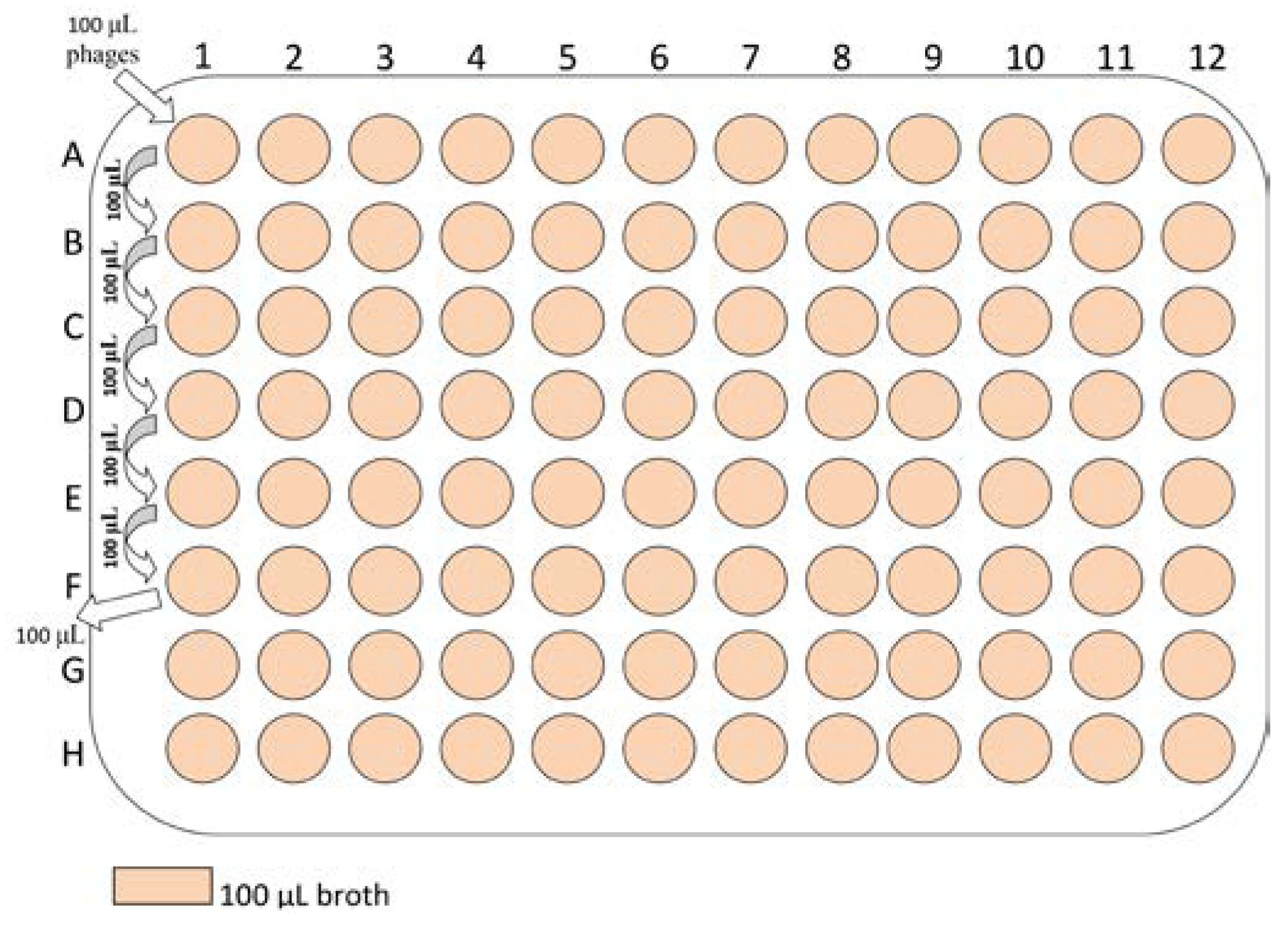
The strains with susceptibility for phages will be tested with binary phage dilutions. The bacterial capacity for adherence and biofilm formation will be evaluated through the microtiter technique. The 96-well plates will be filled with 100 μL of liquid broth per well (Figure 1).
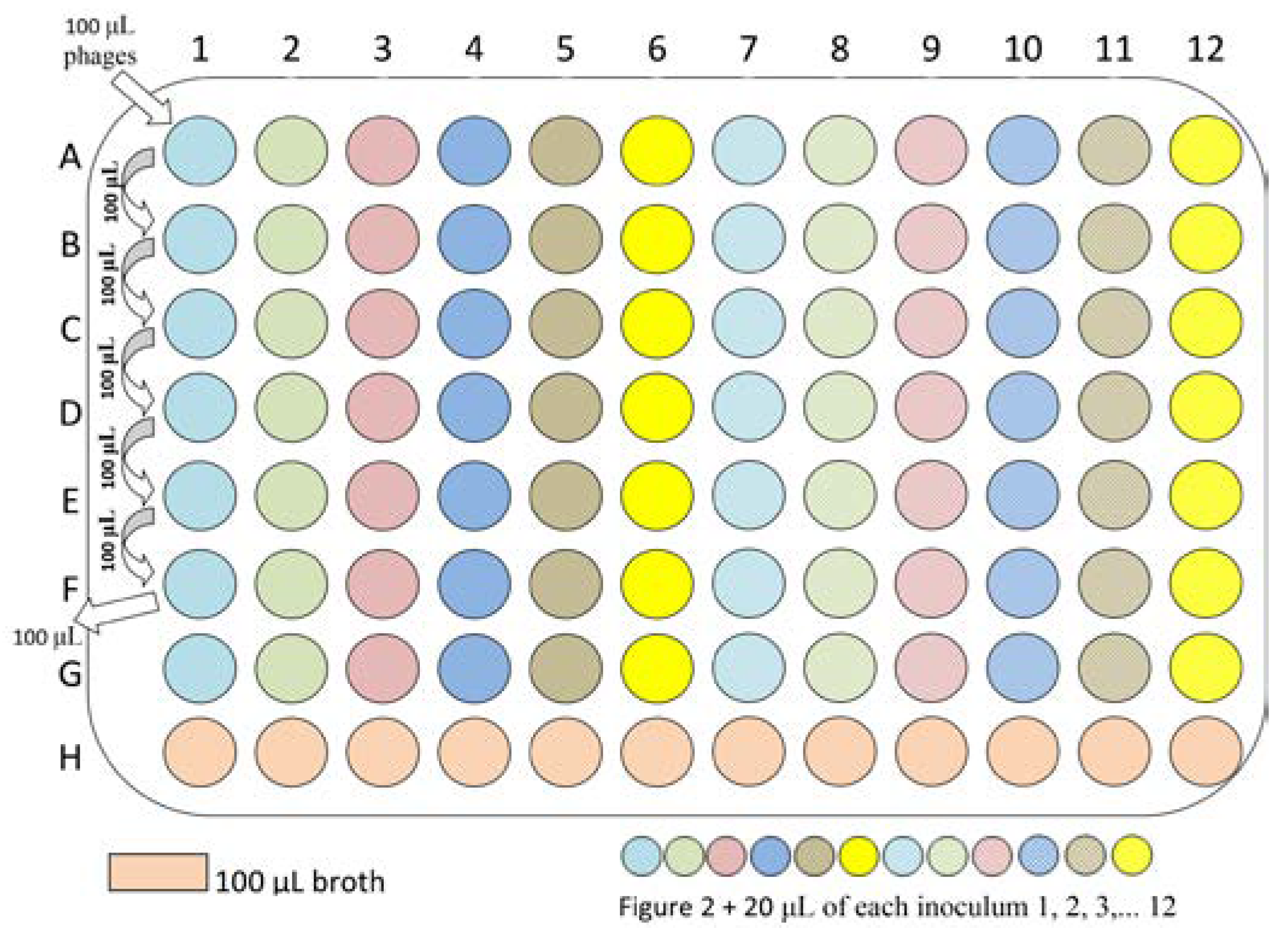
On the first line (A) 100 μL of phage mixture will be added. To obtain binary dilutions, from the A wells, after mixing, 100 μL will be added in the B wells, and the procedure will be continued from A to F and the last 100 μL extracted from F will be thrown out, to ensure equal quantities remain in all wells (Figure 2).
As described, the bacterial suspension (20 μL of 0.5 McFarland inoculum) will be added in all wells except for the H row. In conclusion we will obtain the negative, sterility control (sterile broth) in row H, phages in binary dilution and the microbial culture in rows A to F. In the G row, 100 μL of broth and 20 μL of bacterial inoculum will be added, as culture positive control, while the H row contained the negative control (Figure 3).
After 24 h of incubation, the results will be evaluated qualitatively based on the macroscopic aspect of the liquid medium from the wells. Quantification of bacterial growth will be performed through plating 10 μL from each well, in triplicate, on solid Mueller-Hinton agar followed by 24 h incubation at 35±2 °C in normal atmosphere. The number of colonies will be evaluated.
The liquid left in the wells will be thrown out and the plate will be washed three times with sterile water and fixed with 130 μL cold methanol for 10 min. The plates will be stained with 1% crystal violet. The colored biofilm will be resuspended with 200 μL of 33% acetic acid and the absorbance of the colored suspension will be read through 492 nm spectrophotometry using HumaReader HS (HUMAN Diagnostics Worldwide, Germany). A blank well containing 200 μL of 33% acetic acid will be considered the control.
Through the technique described above, biofilm formation in the presence of phages will be evaluated.
The strains that formed biofilm may be selected and used in the next step of the experiment, for evaluating the effect of phages on already formed biofilm.
Each bacterial strain capable of forming biofilm will be grown in liquid broth in 2 wells (noted a, b) for 24 h—the timespan necessary to ensure biofilm formation. After 24 h any planktonic bacteria left will be washed out; in the “a” well broth will be added, and in the “b” well broth and bacteriophages will be added. These will then be incubated for 24 h. The next day, the “a” and “b” wells will be washed and fixed. In another plate, each strain will be incubated in liquid broth for 24 h; after that the liquid will be washed out and the wells fixed with methanol. Subsequently, all the wells from both plates will be stained with 1% crystal violet and the colored biofilm will be resuspended with 200 μL 33% acetic acid and the same spectrophotometry technique as described above will be used for reading results. A blank well containing 200 μL of 33% acetic acid will be considered the control value.
The values from the wells with the same strain will be compared, evaluating the effect of phages on preformed biofilm.

Expected results

Given the major burden associated with biofilm formation in patients with implanted foreign bodies and recurrent catheter or prosthesis infection, the efficiency of phages should be evaluated not only on planktonic bacteria, but also on biofilm formation. The described method will allow us to evaluate a large number of bacterial strains to predict and project the kinetics of biofilm formation, while adding binary phage dilutions. The effect of phages on preformed biofilm could also be assessed.
Through this study, we intend to provide the first set of results regarding susceptibility to bacteriophages of bacterial strains isolated from patients with hard to treat infections, from reference hospitals in Romania. To further describe the microbiological characteristics of these particular strains, we also aim to assess the correlation between phage susceptibility and antimicrobial sensitivity profiles.
The results derived from this experimental approach will fill a gap in the current knowledge regarding the treatment of patients with problematic recurrent infections with bacterial strains with potentially diminished susceptibility to current antibiotics.
By creating a database for each bacterial species studied, we will collect data on the patient’s profile and through corroborating this information with the laboratory results described for this study, we will be able to better understand the physiologic and pathologic conditions for developing chronic infections or hard to treat infections associated with biofilm formation.
We intend to project and develop further study techniques to assess the potential synergy for co-administration of antibiotics and bacteriophages on both planktonic and sessile bacterial populations.
We intent to use the results derived from this study to generate an algorithm for predicting the clinical outcome and monitoring patients with problematic infections, and to describe the potential role of bacteriophages in clinical practice, alongside antimicrobial therapy, either as synergic therapy providing an extra mechanism of bactericidal action, or as adjuvant therapy potentially disrupting the extracellular polymeric matrix of biofilm, and leaving the bacteria exposed to the direct action of conventional antimicrobials.

Conclusion

To our knowledge, this is the first such study to be performed in Romania, regarding the assessment of susceptibility to bacteriophages of planktonic and aggregated bacterial populations. We consider that this study has the potential to redesign our current understanding of antimicrobial therapy options and to describe the possible role of bacteriophages in clinical practice for recurrent, hard to treat infections.

Author Contributions

All authors had equal contributions.

Funding

This paper is partly supported by the Carol Davila University of Medicine and Pharmacy, Young Researchers Grant, no. 28341/2013.

Acknowledgments

This paper is partly supported by the Sectorial Operational Programme Human Resources Development (SOPHRD), financed by the European Social Fund and the Romanian Government under the contract number POSDRU 141531.

Conflicts of Interest

All authors—none to declare.

References

  1. Leung, E.; Weil, D.E.; Raviglione, M.; Nakatani, H. World Health Organization World Health Day Antimicrobial Resistance Technical Working Group. The WHO policy package to combat antimicrobial resistance. Bull World Health Organ 2011, 89, 390–392. [Google Scholar] [CrossRef] [PubMed]
  2. ECDC/EMEA Joint Working Group. The bacterial challenge: Time to react. A call to narrow the gap between multidrug-resistant bacteria in the EU and development of new antibacterial agents. Stockholm: European Centre for Disease Prevention and Control. 2009.
  3. Parasion, S.; Kwiatek, M.; Gryko, R.; Mizak, L.; Malm, A. Bacteriophages as an alternative strategy for fighting biofilm development. Pol J Microbiol. 2014, 63, 137–145. [Google Scholar] [CrossRef] [PubMed]
  4. Loc-Carrillo, C.; Abedon, S.T. Pros and cons of phage therapy. Bacteriophage 2011, 1, 111–114. [Google Scholar] [CrossRef] [PubMed]
  5. Potera, C. Phage renaissance: New hope against antibiotic resistance. Environ Health Perspect 2013, 121, a48–a53. [Google Scholar] [CrossRef] [PubMed]
  6. Zilișteanu, C.; Mintzer-Morgenstern, L.; Ghyka, G. Influence des bactériophages homologues sur la résistance aux antibiotiques de certaines souches d’Enterobacteriaceae et de staphylocoques. Etude “in vitro”. Arch Roum Pathol Exp Microbiol 1965, 24, 1029–1040. [Google Scholar]
  7. Zilișteanu, C.; Mintzer-Morgenstern, L.; Ionesco, H.; Ionesco-Dorohoi, T. Further data on the association of bacteriophage with antibiotic therapy for the purpose of sterilizing carriers of dysentery bacilli. Arch Roum Pathol Exp Microbiol 1969, 28, 1073–1080. [Google Scholar] [PubMed]
  8. Zilișteanu, C.; Ionescu, H.; Ionescu-Dorohoi, T.; Mintzer, L. Treatment of urinary infections with bacteriophage-autovaccine-antibiotics. Arch Roum Pathol Exp Microbiol 1971, 30, 195–207. [Google Scholar] [PubMed]
  9. Coulter, L.B.; McLean, R.J.; Rohde, R.E.; Aron, G.M. Effect of bacteriophage infection in combination with tobramycin on the emergence of resistance in Escherichia coli and Pseudomonas aeruginosa biofilms. Viruses 2014, 6, 3778–3786. [Google Scholar] [CrossRef] [PubMed]
  10. Kay, M.K.; Erwin, T.C.; McLean, R.J.; Aron, G.M. Bacteriophage ecology in Escherichia coli and Pseudomonas aeruginosa mixed-biofilm communities. Appl Environ Microbiol 2011, 77, 821–829. [Google Scholar] [CrossRef] [PubMed]
  11. Chhibber, S.; Nag, D.; Bansal, S. Inhibiting biofilm formation by Klebsiella pneumoniae B5055 using an iron antagonizing molecule and a bacteriophage. BMC Microbiol 2013, 13, 174. [Google Scholar] [CrossRef] [PubMed]
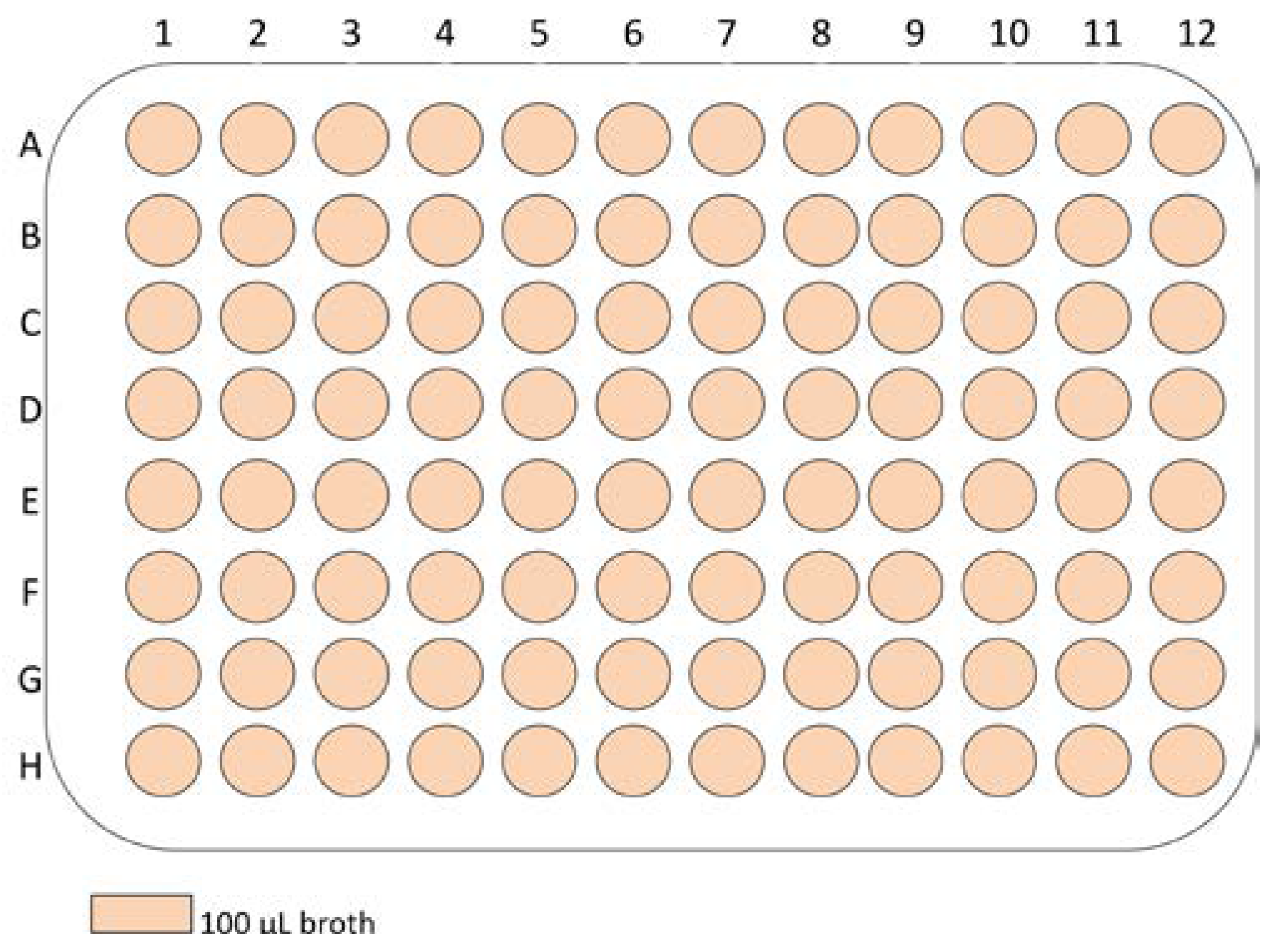
Figure 1. Adding 100 μL broth per well.
Germs 04 00092 g001
Figure 2. Adding the phages and performing the binary dilutions.
Germs 04 00092 g002
Figure 3. Adding the inoculum.
Germs 04 00092 g003