Next Article in Journal
Aggregatibacter actinomycetemcomitans Endocarditis in an Adult Patient with Patent Ductus Arteriosus
Previous Article in Journal
Clinical Spectrum of Extrapulmonary Non-Tuberculous Mycobacterial Disease in Immunocompetent Patients: A Case Series
 
 
Germs is published by MDPI from Volume 15 Issue 4 (2025). Previous articles were published by another publisher in Open Access under a CC-BY (or CC-BY-NC-ND) licence, and they are hosted by MDPI on mdpi.com as a courtesy and upon agreement with the former publisher Infection Science Forum.
Case Report

Rare Fatal Case of Purpura Fulminans Due to Pneumococcal Sepsis in a Child, Associated with Multiorgan Failure

by
Gheorghiţă Jugulete
1,2,
Maria Mădălina Merişescu
1,2,*,
Carmen Pavelescu
1 and
Monica Luminiţa Luminos
1,2
1
Faculty of Dentistry, Carol Davila University of Medicine and Pharmacy, No. 37, Dionisie Lupu Street, 020021 Bucharest, Romania
2
National Institute for Infectious Diseases “Prof. Dr. Matei Balș”, No. 1 Dr. Calistrat Grozovici Street, 021105 Bucharest, Romania
*
Author to whom correspondence should be addressed.
Germs 2024, 14(2), 204-209; https://doi.org/10.18683/germs.2024.1432
Submission received: 6 March 2024 / Revised: 8 April 2024 / Accepted: 9 April 2024 / Published: 30 June 2024

Abstract

Introduction: Streptococcus pneumoniae is one of the associated bacteria that can cause the rare but high mortality hematological pathology known as purpura fulminans (PF) in both adults and children. Pediatric patients with PF can progress quickly to sepsis and multiorgan failure, especially immunocompromised individuals and young children. Due to the thrombotic blockage of blood arteries in PF, there is diffuse intravascular thrombosis and hemorrhagic infarction of the skin, which evolves from ecchymosis to skin necrosis, risk of limb sequelae, sepsis and fatality. Case report: We present a case of a previously healthy 1-year and 9-months old female who was admitted to the Intensive Care Unit of the National Institute of Infectious Diseases "Prof. Dr. Matei Balș"- Bucharest, Romania. On physical examination, she was febrile, hypotensive, tachycardic, and had erythematous patches on her left upper limb and trunk. Initial blood work was significant for creatinine 4.45 mg/dL, aspartate aminotransferase 112 U/L, alanine aminotransferase 130 U/L and fibrinogen 596 mg/dL. Hematological workup showed a white blood cells count of 34 × 109/L, hemoglobin 9.7 g/dL, platelets 23000/L, D-dimers 89000 µg/L, and elevated PT and aPTT. Broad-spectrum antibiotics vancomycin and ceftriaxone were administrated. A lumbar puncture was performed for cerebrospinal fluid (CSF) analysis and culture grew Streptococcus pneumoniae serotype 1A. She required peritoneal dialysis due to acute kidney injury (AKI) and surgeries for affected skin areas. After multiple organ system failures, our patient evolved rapidly to irreversible tissue necrosis and death. Conclusions: We aim to report a rare case of PF associated with pneumococcal meningoencephalitis in an immunocompetent child, to better appreciate the risk of fatal evolution when managing this disease in children.

Introduction

PF is a rapidly progressive fatal syndrome characterized by purpuric lesions and is often associated with ecchymosis and necrosis due to diffuse thrombosis and disseminated intravascular coagulation [1,2,3]. About 90% of the cases that have been described in the pediatric literature have been fatal. Three forms of PF are described as follows: neonatal, idiopathic and acute infection. First is the patient with abnormalities of the protein C or protein S anticoagulant course [3,4]. The second condition is the rarest form of the disease, and the third form is the most common type, and it manifests as hemorrhagic necrosis due to thrombosis of the microvascular system of the skin [2,3,4,5,6]. Streptococcus pneumoniae can cause different kinds of infections, including invasive pneumonia, and can also be associated with a more severe disease course and can cause PF [1,4,5,6]. Pneumococcal infections are dangerous and often require hospitalization, with mortality rates between 8 to 15% [7,8,9,10]. As of clinical presentations, PF first develops cutaneous macules, which become indurated and then evolve into irreversible necrosis of the skin, sepsis, and secondary infections that may occur as gangrene. As necrosis extends to deeper tissues, it is associated with a high incidence of amputations, and skin grafts and decreases in survival rate [11,12,13,14,15]. We herein report a rare case of PF due to acute infection caused by S. pneumoniae, in a healthy and completely vaccinated child [15,16].

Case report

This previously healthy one-year-old nine- month-old girl, pneumococcal vaccine immunized, with a two-day history of fever, nasal obstruction, mucopurulent rhinorrhea, otorrhea, and productive cough developed a high-grade fever (39.8°C) and became lethargic and presented to the Pediatric Intensive Care Unit at the National Institute for Infectious Diseases, „Prof. Dr Matei Balș”, Bucharest, Romania.
From the initial examination in our Pediatric department, she was very ill-appearing, dehydrated, hypotensive (55/30 mmHg), tachycardic (188 beats/min), had delayed capillary refill, and petechial rash on both her arms and trunk (Figure 1). The presumptive diagnosis was purpura fulminans.
On auscultation, there were crepitations at both bases of the lungs. The abdominal examination did not reveal peritoneal signs. Abnormalities in laboratory studies included tests as follows: elevated C reactive protein 97 mg/L, leukocytosis 34.000/µL, anemia (Hb 9.7 g/dL), thrombocytopenia – platelets 23000/µL, impaired renal function (creatinine 4.45 mg/dL), hyponatremia and hyperkalemia. She also presented serum procalcitonin (PCT-Q), hepatic cytolysis syndrome, increasing values of serum urea. Coagulation tests showed disseminated intravascular coagulation (DIC) with prolonged prothrombin time (international normalized ratio>5.59), activated partial thromboplastin time over 160 s, thrombin time over 140 s, and increased D-dimer levels 8.9 mg/dL). Follow-up laboratory tests revealed deterioration of coagulopathy, with increased prothrombin time, (Table 1). The blood cultures were sent for incubation and no growth was detected. Urine culture tests were negative. Nose and throat swabs showed the absence of adenovirus, influenza A, B, and respiratory syncytial virus. Chest radiograph revealed pneumonia and a diagnosis of sepsis due to streptococcal infection was suspected and prompted broad-spectrum antibiotics: vancomycin (60 mg/kg/day) and ceftriaxone (100 mg/kg/day) were administrated. Standardized sepsis therapy was applied according to the surviving sepsis guidelines. On the second day of admission, the lumbar puncture revealed cloudy, purulent cerebrospinal fluid (CSF), latex agglutination positive for Streptococcus pneumoniae serotype 1A. Purpura fulminans was confirmed as a diagnostic when evidence of a severe infection, shock liver, and DIC was associated. The areas of ecchymoses on the left hand lesions became necrotic after 3 days and required surgical debridement, and the patient was transferred to the plastic surgery clinic (Figure 2). After 14 days, the patient developed anuria and required peritoneal dialysis.
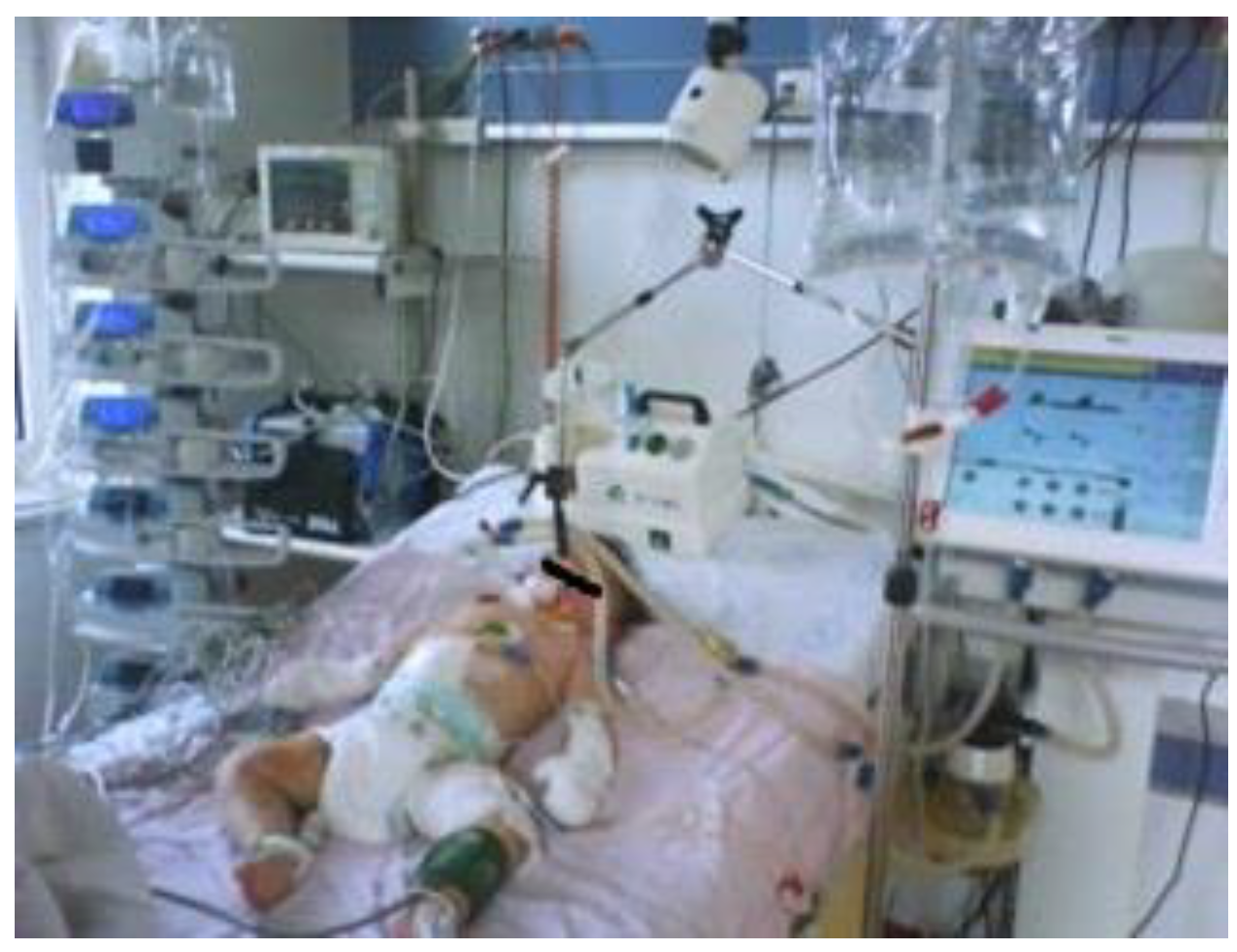
Despite all therapeutic efforts, antibiotic therapy and aggressive supportive care in an intensive care unit, the patient did not recover and multi-organ failure (cardio-respiratory, hematological, hepatic, neurological, renal) developed since day 15 throughout her hospitalization. On the 16th day of hospitalization, the child was intubated, and mechanically ventilated, and complex therapy was administered, supporting vital functions, hydro- electrolytic and acid-base rebalancing, pathogenic methylprednisolone 1 mg/kg/day pulse therapy), antibiotic vancomycin (60 mg/kg/day) and ceftriaxone (100 mg/kg/day), symptomatic therapy, and peritoneal dialysis. (Figure 3). Heparin administration could not be started due to thrombocytopenia. The patient succumbed within 30 days of admission, despite early recognition of purpura fulminans, septic shock and complex therapy administration.

Discussion

Purpura fulminans, also known as purpura gangrenous, is a rare and life-threatening disorder, and it manifests as a widespread purpuric rash that develops hemorrhagic necrosis. It occurs in both adults and children and is accompanied by vascular collapse, fever and DIC [11,12,13,16]. In our case, the clinical symptoms are indicative of microangiopathic thrombosis with hemolysis related to DIC. PF lesions can be distinguished from other purpuric lesions by their distinctive appearance. Rapidly following erythema are black hemorrhagic necrosis centers surrounded by an erythematous border, as our patient developed [16,17,18]. Recent observations include the development of hard eschar and the production of vesicles and bullae, which indicate the progression of hemorrhagic necrosis. Usually, the most seriously affected are the distal extremities. Henoch-Schönlein bullous purpura was first thought to be a differential diagnosis because of the palpable purpura and blisters [13,14,15]. Deep tissue may be compromised by extensive skin necrosis, necessitating surgical debridement and fasciotomy. For this reason, early diagnosis and prompt rehabilitation depend on a multidisciplinary patient follow-up involving plastic surgery healthcare teams. The prognosis for this age category is poor once soft tissue ischemia evolves to necrosis because it is associated with arterial and venous thrombosis leading to serious events and death [19].
We found S. pneumoniae associated with the course of sepsis and PF. The Center for Disease Control (CDC) advises pneumococcal vaccinations for young children (less than 5 years old), as part of regular prophylaxis. Routine vaccination with 13-valent pneumococcal vaccine with 3 doses at ages 2, 4, and 11 months is the indicated active immunization for the prevention of invasive disease caused by Streptococcus pneumoniae, in children aged 6 weeks and older [12,13,14]. Unfortunately, vaccination does not fully eliminate the risk of invasive infection in our case report. Sepsis resulting from pneumococcal infection typically presents so dramatically that immunosuppression is involved. Most examples that have been documented in the literature include people who have some kind of immunodeficiency. This dramatic presentation in a healthy pediatric patient has only been described in a small number of cases. We compared our data with similar articles published on children with PF and the condition was complicated by hemodynamic collapse with skin involvement necrosis of the skin [20,21,22].
Follow-up laboratory tests revealed deterioration and coagulopathy, elongated prothrombin time and dimerized fragment D and fibrinogen. Several differential diagnoses, including drug-related skin reactions like Steven- Johnson syndrome, toxic epidermal necrolysis, cryoglobulinemia as well as autoimmune vasculitis and immunological tests were unspecific in our patient [24]. Other etiologies of infections were found to be negative, and this workup confirmed the cause of PF was Streptococcus pneumoniae serotype 1A. In an intensive-care environment, patients with purpura fulminans and severe bacterial infections should be treated with strong fluid resuscitation, correction of electrolytes, inotropic support if necessary and early delivery of antibacterial medications. Vancomycin is active against strains of S. pneumoniae for the treatment of meningitis and should be administrated with cefotaxime or ceftriaxone to severely sick or immunocompromised children who may have pneumococcal pneumonia, until information on the organism's susceptibilities is available [25,26].
Lerolle and colleagues studied patients with PF, and included patients with severe sepsis. They observed the occurrence of multiple organ failure, with a higher mortality rate [27]. Patients with PF had a low number of platelets, and high levels of D-dimers, but similar levels of fibrinogen. In our situation, the patient status is associated with multiple systems organ failure, but the fibrinogen is increased in the dynamics. It is known that certain infectious diseases are later complicated by PF, after the initial disease has resolved [28].

Conclusions

We think that it's important for clinicians to be knowledgeable of this diagnosis and to have a considerable level of suspicion when disseminated intravascular coagulation and thrombocytopenia are present in conjunction with PF, as this dermatological finding is uncommon among patients in the critical care setting. This clinical case reveals the severity of systemic infection with Streptococcus pneumoniae serotype 1a, in a pediatric patient who developed septic shock, and multiorgan failure. PF can cause irreversible tissue necrosis and, in severe cases, death.

Author Contributions

Conceptualization, GJ, MLL, CP, MMM; Validation, GJ, MLL, CP, MMM; Formal analysis, GJ, CP; Investigation, GJ, CP; Resources, GJ, MLL; Data curation, GJ, MMM; Writing—Original draft preparation, GJ; Writing—Review & Editing, GJ, MLL, CP, MMM; Visualization, GJ, CP; Supervision, GJ; Project administration, GJ, MLL, CP, MMM. All authors read and approved the final version of the manuscript.

Funding

None to declare.

Institutional Review Board Statement

The case study was approved by the institutional review board, C0133/10.06.2023.

Informed Consent Statement

Written informed consent was obtained by the authors from the patient's parents for the publication of this case report and associated images.

Conflicts of Interest

All authors – none to declare.

References

  1. Irfan Kazi, S.G.; Siddiqui, E.; Habib, I.; Tabassum, S.; Afzal, B.; Khan, I.Q. Neonatal purpura fulminans, a rare genetic disorder due to protein C deficiency: a case report. J Pak Med Assoc. 2018, 68, 463–465. [Google Scholar]
  2. Chalmers, E.; Cooper, P.; Forman, K.; et al. Purpura fulminans: recognition, diagnosis and management. Arch Dis Child. 2011, 96, 1066–1071. [Google Scholar] [CrossRef] [PubMed]
  3. Pombar, R.F.; Tellería, R.L.; Bianco, B.; Centeno, M.D.V.; Cervini, A.B. Postinfectious purpura fulminans: a case report. Arch Argent Pediatr. 2024, 122, e202310137. [Google Scholar] [CrossRef]
  4. Li, X.L.; Luan, C.Y.; Fan, Y.J.; et al. A rare case of acute infectious purpura fulminans caused by Klebsiella pneumoniae and human herpesvirus type 5. J Inflamm Res. 2022, 15, 4251–4260. [Google Scholar] [CrossRef]
  5. Findley, T.; Patel, M.; Chapman, J.; Brown, D.; Duncan, A.F. Acquired versus congenital neonatal purpura fulminans: a case report and literature review. J Pediatr Hematol Oncol. 2018, 40, 625–627. [Google Scholar] [CrossRef] [PubMed]
  6. Djurdjevic, N.; Taweesedt, P.T.; Paulson, M.; et al. Septic shock and purpura fulminans due to Streptococcus pneumoniae bacteremia in an unvaccinated immunocompetent adult: case report and review. Am J Case Rep. 2020, 21, e923266. [Google Scholar] [CrossRef] [PubMed]
  7. Dutta, A.K.; Swaminathan Abitbol, V.; Kolhapure, S.; Sathyanarayanan, S. A comprehensive review of meningococcal disease burden in India. Infect Dis Ther. 2020, 9, 537–559. [Google Scholar] [CrossRef]
  8. Merișescu, M.; Jugulete, G.; Bastian, A.E.; Luminos, M. Evaluation of sepsis mortality in children. Rom J Legal Med. 2018, 26, 37–41. [Google Scholar]
  9. Jugulete, G.; Merișescu, M.; Bastian, A.E.; Luminos, M. Clinical aspects and medico-legal implications of purpura fulminans in children. Rom J Legal Med. 2017, 25, 364–368. [Google Scholar]
  10. Lin, C.; Li, D.; Hu, B. Lower limb necrosis secondary to purpura fulminans: a case report. J Burn Care Res. 2023, 44, 477–480. [Google Scholar]
  11. World Health Organization. 2019. Meningococcal meningitis. Available online: https://www.who.int/en/newsroom/fact-sheets/detail/meningococcal-meningitis (accessed on 30 September 2019).
  12. Miron, V.D.; Sandulescu, O.; Visan, C.A.; et al. Pneumococcal colonization and pneumococcal disease in children with influenza. Clinical, laboratory and epidemiological features. Rev Chim. 2018, 69, 2749–2753. [Google Scholar] [CrossRef]
  13. Tekin, R.T.; Dinleyici, E.C.; Ceyhan, M.; et al. The prevalence, serogroup distribution and risk factors of meningococcal carriage in adolescents and young adults in Turkey. Hum Vaccin Immunother. 2017, 13, 1182–1189. [Google Scholar] [CrossRef]
  14. Wang, B.; Santoreneos, R.; Giles, L.; Afzali, H.H.; Marshall, H. Case fatality rates of invasive meningococcal disease by serogroup and age: a systematic review and meta-analysis. Vaccine. 2019, 37, 2768–2782. [Google Scholar] [CrossRef] [PubMed]
  15. Sinclair, D.; Preziosi, M.P.; Jacob John, T.; Greenwood, B. The epidemiology of meningococcal disease in India. Trop Med Int Health. 2010, 15, 1421–1435. [Google Scholar] [CrossRef] [PubMed]
  16. Aye, A.M.M.; Bai, X.; Borrow, R.; et al. Meningococcal disease surveillance in the Asia-Pacific region (2020): the global meningococcal initiative. J Infect. 2020, 81, 698–711. [Google Scholar] [CrossRef]
  17. Lee, J.; Blackburn, J.; Pham-Huy, A. Uncommon clinical presentation of a common bug: Group A Streptococcus meningitis. Paediatr Child Health. 2020, 26, e129–e131. [Google Scholar] [CrossRef]
  18. Sernaqué, C.; Ceresetto, J.; Duboscq, C.; et al. Púrpura fulminans asociada a déficit adquirido de proteína S en una paciente con neumonía por Streptococcus pneumoniae. Hematología. 2021, 24, 71–75. [Google Scholar]
  19. Azzari, C.; Nieddu, F.; Moriondo, M.; et al. Underestimation of invasive meningococcal disease in Italy. Emerg Infect Dis. 2016, 22, 469–475. [Google Scholar] [CrossRef]
  20. Sharip, A.; Sorvillo, F.; Redelings, M.D.; et al. Population- based analysis of meningococcal disease mortality in the United States: 1990-2002. Pediatr Infect Dis J. 2006, 25, 191–194. [Google Scholar] [CrossRef]
  21. Ouldali, N.; Levy, C.; Varon, E.; et al. Incidence of paediatric pneumococcal meningitis and emergence of new serotypes: a time-series analysis of a 16-year French national survey. Lancet Infect Dis. 2018, 18, 983–991. [Google Scholar] [CrossRef]
  22. Green, M.S.; Schwartz, N.; Peer, V. A meta-analytic evaluation of sex differences in meningococcal disease incidence rates in 10 countries. Epidemiol Infect. 2020, 148, e246. [Google Scholar] [CrossRef] [PubMed]
  23. Stein-Zamir, C.; Shoob, H.; Sokolov, I.; Kunbar, A.; Abramson, N.; Zimmerman, D. The clinical features and long-term sequelae of invasive meningococcal disease in children. Pediatr Infect Dise J 2014, 33, 777–779. [Google Scholar] [CrossRef]
  24. Bhatti, U.F.; Williams, A.M.; Raghavendran, K.; Georgoff, P.E. Four-extremity amputation following disseminated intravascular coagulation and purpura fulminans. BMJ Case Rep. 2019, 12, e228028. [Google Scholar] [CrossRef]
  25. Andreasen, T.J.; Green, S.D.; Childers, B.J. Massive infectious soft-tissue injury: diagnosis and management of necrotizing fasciitis and purpura fulminans. Plast Reconstr Surg. 2001, 107, 1025–1034. [Google Scholar] [CrossRef]
  26. Tahmasebi, Z.; Asadollahi, P.; Sadeghifard, N.; Ghafourian, S.; Kalani, B.S.; Kalaei, E.G.P.; Pakzad, I. Microbiological and drug resistance patterns of bronchoalveolar lavage samples taken from hospitalized patients in Iran. Germs. 2022, 12, 333–343. [Google Scholar] [CrossRef]
  27. Chalmers, E.; Cooper, P.; Forman, K.; Grimley, C.; Khair, K.; Minford, A.; et al. Purpura fulminans: recognition, diagnosis and management. Arch Dis Child. 2011, 96, 1066–1071. [Google Scholar] [CrossRef] [PubMed]
  28. Plüß, M.; Zeisberg, M.; Müller, G.A.; Vasko, R.; Korsten, P. Therapeutic response to glucocorticoids, anticoagulation and plasma exchange in a patient with primary antiphospholipid syndrome presenting with purpura fulminans. Lupus. 2018, 27, 2170–2173. [Google Scholar] [CrossRef] [PubMed]
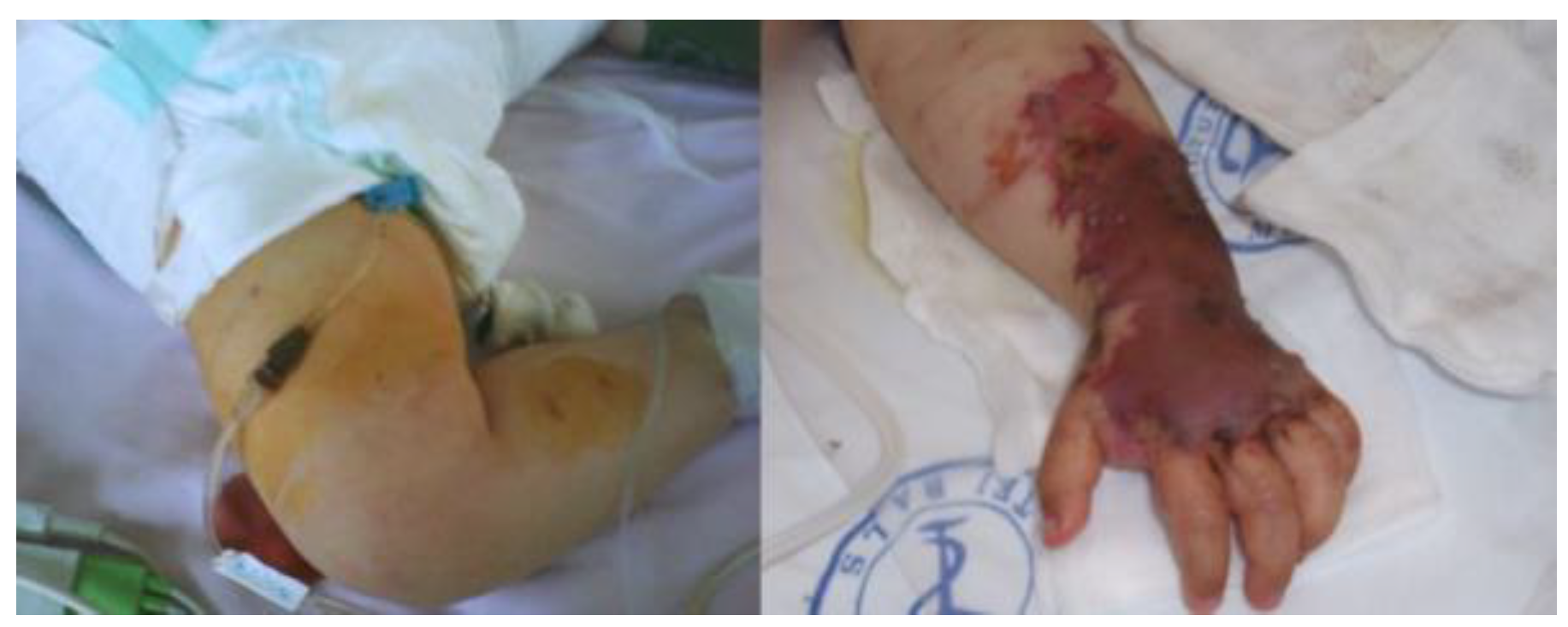
Figure 1. Petechial rash on both arms.
Germs 14 00204 g001
Figure 2. Necrotic lesions on the hand.
Germs 14 00204 g002
Figure 3. Patient was intubated and mechanically ventilated.
Germs 14 00204 g003
Table 1. Laboratory values during hospitalization of the pediatric patient with PF.
Parameter1st week2nd week3rd week4th weekReference range
White blood cells (WBC)
×109/L
3431.523.333.84-11
Platelets ×109/L23282924150-400
Hemoglobin g/dL9.79.610.89.412-15
Fibrinogen mg/dL596697331440<400
Prothrombin time (PT)
seconds
140819013411-15
Aspartate aminotransferase
(AST) U/L
112966410214-50
Alanine aminotransferase
(ALT) U/L
1301221201100-50
Serum creatinine mg/dL4.454.403.002.90<0.7
D-dimers mg/L8.999.3100-0.50