Article

Polymerizable Matrix Metalloproteinases’ Inhibitors with Potential Application for Dental Restorations

1
Centro de investigação interdisciplinar Egas Moniz, Instituto Universitário Egas Moniz, 2829-511 Caparica, Portugal
2
UCIBIO and LAQV, Requimte, Faculdade de Ciências e Tecnologia, Universidade Nova de Lisboa, 2829-516 Caparica, Portugal
3
Unidade de Investigação em Ciências Orais e Biomédicas (UICOB), Faculdade de Medicina Dentária, Universidade de Lisboa, Rua Professora Teresa Ambrósio, 1600-177 Lisboa, Portugal
*
Author to whom correspondence should be addressed.
Biomedicines 2021, 9(4), 366; https://doi.org/10.3390/biomedicines9040366
Submission received: 25 February 2021 / Revised: 19 March 2021 / Accepted: 26 March 2021 / Published: 31 March 2021
(This article belongs to the Section Biomedical Materials and Nanomedicine)

Abstract

:
Collagen cleavage by matrix metalloproteinase (MMP) is considered a major cause of dental resins long term failure. Most MMP inhibitors display significant toxicity and are unsuitable for dental resins’ applications. Here we report a study of a new class of inhibitors that display the unique property of being co-polymerizable with other vinyl compounds present in commercial dental resins, limiting their release and potential toxicity. Computational affinity towards the active site of different MMP-1; -2; -8; -9 and -13 of several compounds showed interesting properties and were synthesized. These free compounds were tested concerning their toxicity upon contact with two different cell types, with no substantial decrease in cell viability at high concentrations. Even so, compound’s safety can be further improved upon copolymerization with commercial dental resins, limiting their release.

1. Introduction

The matrix metalloproteinases (MMPs) belong to the Metzincs superfamily [1,2,3,4,5,6,7,8], and are characterized by a common conserved catalytic domain: VAAHExGHxxGxxH [2,5,6,7,9,10,11,12,13,14]. The MMPs are zinc dependent, functional at neutral pH [13,15] and they are a family of proteolytic enzymes with different substrates, but which share similar structural characteristics [3,5,7,8,10,11,12,13,14,15,16,17,18]. In human tissue there are 23 different types of MMPs [1,3,5,6,7,11,12,15,16] and they can be subdivided according to substrate specificity, sequential similarity and domain organization into collagenases (MMP-1, -8 and -13), gelatinases (MMPs-2 and -9), stromelysins (MMPs-3, -10 and -11), matrilysins (MMP-7 and -26), membrane-type MMPs (MMPs-14, -15, -16, -17, -24 and -25) and other MMPs (MMPs-12, -19, -20, -21, -23, -27 and -28) [1,3,6,7,8,11,12,13,14,15,16,19]. The principal biologic function of MMPs is degradation of extracellular matrix (ECM) proteins and glycoproteins, membrane receptors, cytokines and growth factors [1,2,3,5,6,7,8,11,13,14,16,20,21,22,23,24]. They are involved in several biologic processes [1,2,7,8,10,11,13,15,16,23,25,26] and their deregulation leads to the progression of various pathologies [1,2,8,13,14,19,23].
The MMPs are molecular targets for the development of therapeutics and diagnosis agents [14]. The inhibition of MMP activity can be done at the biomolecular expression or enzyme activity [10,16,17]. The MMPs inhibitors that could affect enzyme activity can be divided into endogenous specific or non-specific inhibitors or synthetic inhibitors [1,3,7,8,12,13,14,16,23,27,28,29,30]. The synthetic MMP inhibitor must have a functional group able to chelate the catalytic zinc ion, at least one group that promote hydrogen bonds and side chains undergoing Van der Waals interactions with the enzyme [11,13,16,31,32,33]. These requirements are crucial to the selectivity of MMP inhibitor, to increasing the efficacy and to preventing the side effects [16]. Several strategies have been suggested to create a specific MMP inhibitor [34], but they have been difficult to develop because MMPs are involved in various pathways. It is necessary to identify the enzymes that are involved in the disease progression, while there are more than 50 human metalloproteinases (23MMPs; 13 ADAM and 19 ADAMTs) [16,17].
There is a wide variety of inhibitors, but few have selectivity and specificity for MMPs [2,27,28,29,30,31,33,35] and most inhibitors have a biphenyl group conjugated to a sulfonamides group [36,37,38,39,40,41]. In this work, a set of several molecules with biphenyl group conjugated with methacrylate or methylacrylamide were studied via computational docking, of which three molecules [42] were selected to be synthesized and tested for cytotoxicity in mouse embryo NIH/3T3 fibroblast cells and human MG-63 osteoblast-like cells.

2. Materials and Methods

2.1. Computational Docking Studies

The 2D structure of the studied compounds was drawn (Figure 1) and the 3D structure of compounds was previously optimized by RHF/3–21G using Chem3D. The crystal structure of the MMPs-1, -2, -8, -9 and -13 was retrieved from the RCSB protein database (Table 1).
In SeeSAR, the binding site of these proteins was defined, containing the conserved sequence VAAHExGHxxGxxH and the S’1 pocket and the number of solutions defined was the TOP10. The results were edited by Discovery Studio and the 2D image were obtained by PoseView.

2.2. Synthesis of A, B and C Compounds

Synthesis was performed according to reference [42]. Briefly a diol moiety was substituted with two additional side chains to yield the final molecule.

2.3. Cytotoxicity Study Using MTT Assay

Two cell lines, fibroblastic cells NIH/3T3 from mouse embryo (93061524) obtained from Health Protection Agency Culture Collections and supplied by Sigma and MG-63 osteoblast-like cells from human osteosarcoma (ATCC® CRL-1427TM) were used for cytotoxicity studies. Defrosting and handling of cell lines was carried out according to supplier information.
The NIH/3T3 cells were grown in Dulbecco’s Modified Eagle Medium (DMEM, Sigma) supplemented with 10% calf bovine serum (Sigma) and 1% Penicillin-Streptomycin solution: 10,000 U/mL penicillin, 10 mg/mL streptomycin, Sigma) and 2.5 mg/L amphotericin B (fungizone, Sigma). MG-63 cells were grown in Eagle’s Minimum Essential Medium (EMEM, Sigma) supplemented with 10% fetal bovine serum and 1% of antibiotics and 2.5 mg/L antimycotics solutions.
Extracts from each compound A, B or C were prepared by spreading 20 μL of a solution with 20 μM, 50 μM or 100 μM of each compound in a glass petri dish, allowed to dry, and incubated at 37 °C under humidified 5% CO2 for 24 h with 10 mL of the corresponding growth medium according to ISO 10993–12: 2012.
The cytotoxicity assay was performed as follows: 1 × 10⁴ cells per well, from passages 8 to 12, were seeded in 96-well plates (eight replicates) and incubated at 37 °C under humidified 5% CO2 for 24 h. After proliferation, the cell medium was replaced by 200 μL of each extract and incubated under the same conditions for 24 h. As positive control cells were grown in their medium containing 7.5% DMSO (Sigma) and as negative control cells proliferated in medium without any toxic corresponding to 100% cell viability. MTT assays were performed as previously described [43]. Briefly, extracts or medium were removed, an MTT solution (0.5 mg/mL prepared in serum-free medium, Sigma) was added to each well and cells were incubated for additional 3 h. After that, a solution containing 0.1% IGEPAL (Merck), 4 mM HCl (Sigma) in isopropanol (Sigma) was used to dissolve formazan. Plates were incubated with agitation for one hour and absorbance was read at 595 nm with a microplate reader (Bio-Rad®, Hercules, California, USA).

2.4. Statistical Analysis

Data analysis was performed using SPSS for MAC, 26 version (Statistic Package for Social Sciences, Inc., New York, NY, USA) and the significant level was set at 5%. Normality distribution of quantitative variables was assessed using Shapiro-Wilk test. A three-way ANOVA was performed to evaluate the effect of cell type, inhibitor compound and inhibitor concentration on cell viability. Due to the existence of interactions between the main factors, the data obtained for each type of cell were analyzed with a two-way ANOVA followed by Tukey HSD post-hoc tests (α = 0.05).

3. Results and Discussion

3.1. Docking Studies

3.1.1. Physicochemical and ADMET Properties of NNGH Commercial Inhibitor, Compounds A, B and C

Knowledge of physicochemical properties and physiological behavior as absorption, distribution, metabolism, excretion and toxicity (ADMET data) is important to predict efficacy and safety and can help drug design [44]. The SeeSAR platform provides a set of physicochemical information (Appendix A), the most relevant of which are shown in Table 2.
The molecular weight (MW) is an important parameter because it can influence several events, such as absorption, bile elimination rate and blood-brain barrier penetration [45,46,47,48,49], while hydrogen bonds acceptors and donors are important parameters for polarity and permeability [50]. Previous studies showed that, for drug development, the number of the hydrogen bonds donors may be more crucial than the number of hydrogen bonds acceptors since compounds with more hydrogen bonds acceptors have more favorable profiles related to bioavailability and membrane permeability [45,48,51,52]. According to these studies, NNGH inhibitor and compounds A and B have better bioavailability and membrane permeability than compound C. Other coefficients as partition, log P (for neutral compounds, such as compounds A, B and C) and distribution Log D (for ionizable compounds, such as NNGH inhibitor) are measures of lipophilicity a rather important characteristic that also impacts certain ADMET parameters and drug potencies. Generally, high lipophilicity compromises water solubility, being more likely to cause toxic effects, though too low lipophilicity could excessively decrease permeability and absorption [53]. Compound B, with the highest lipophilicity value (Log P = 5.089), is the least soluble compound, while the NNGH inhibitor has a low permeability because it has low lipophilicity (Log P = 1.472). These parameters belong to Lipinski’s rule of five, which determine if a compound with biological activity has physicochemical properties that would make it a likely orally active drug. All compounds respect Lipinski’s rule of five, except for compound B that exceeds the threshold Log P. More importantly, as inhibitors to be used in dentistry, will be permanently covalently bound to dental resins, decreasing drastically the eventual free concentration in the organism.
The Topological Polar Surface Area (TPSA) is defined as the surface sum over all polar atoms and is another descriptor related to hydrogen bonding, also important to estimate the permeability and the oral bioavailability. Several models show that these properties decrease with the increase of TPSA and, for Central nervous system (CNS) permeability by passive diffusion, the TPSA must be less than 80 Å2 [45,50,54,55]. Compound A has the highest permeability, while the NNGH inhibitor with the highest TPSA value presents the lowest permeability and is not able to diffuse to CNS (TPSA > 80 Å2).
Drugs directed towards CNS cross the blood-brain barrier (BBB) by passive diffusion, or active transport mechanism [56]. The BBB penetration should be minimal in non-CNS compounds to reduce the possibility of undesired pharmacological events and neurotoxicity [50]. The most common parameter to quantify BBB penetration is the BBB log ([brain]:[blood]) and it determines the total extent of brain exposure, at a steady-state [57]. The P-glycoprotein (P-gp) is an important transporter that belongs to the ATP-binding Cassette superfamily and it can be found in cells throughout the body, including the blood-brain barrier [48,50]. The P-gp plays an important role in the distribution of drugs due to its ability to remove/extract a structurally diverse range of molecules and can reduce drug accumulation in tissues [48,58]. For the compounds A, B and C, none can cross the BBB, since they have a low BBB log value. The NNGH, for having a TPSA greater than 80Å2, is also not able to cross the BBB, despite having a high BBB log. Only compound C can be a substrate for P-gp transporters.

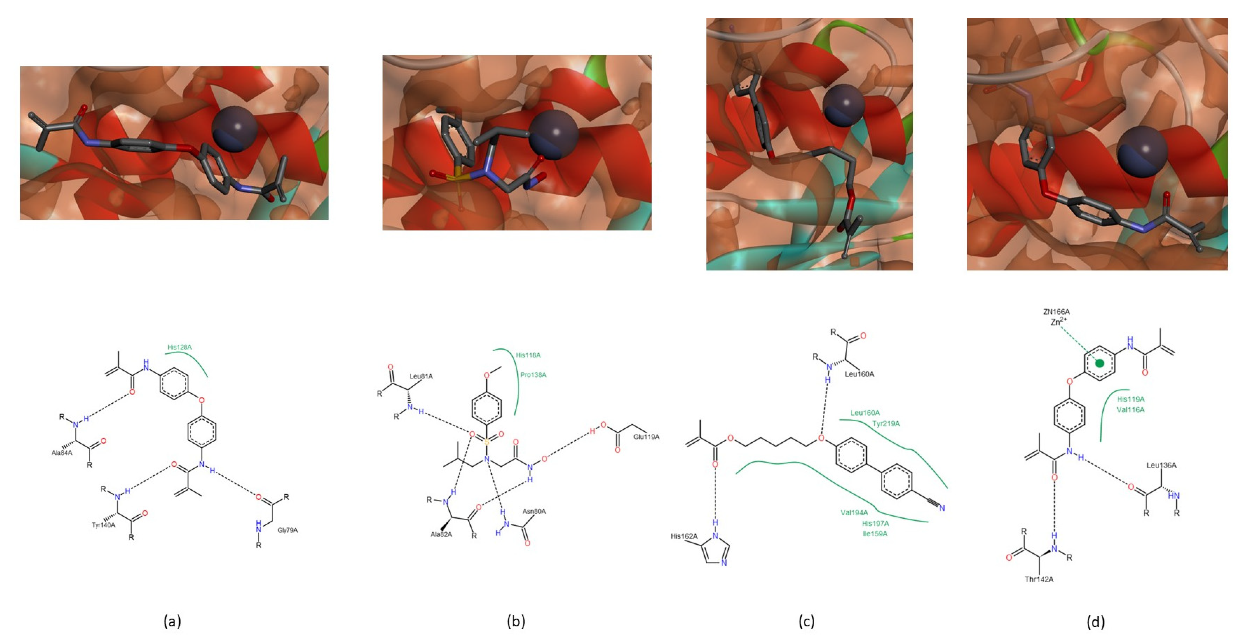
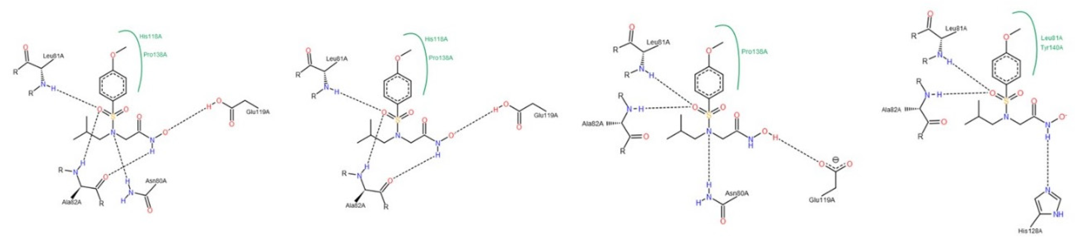
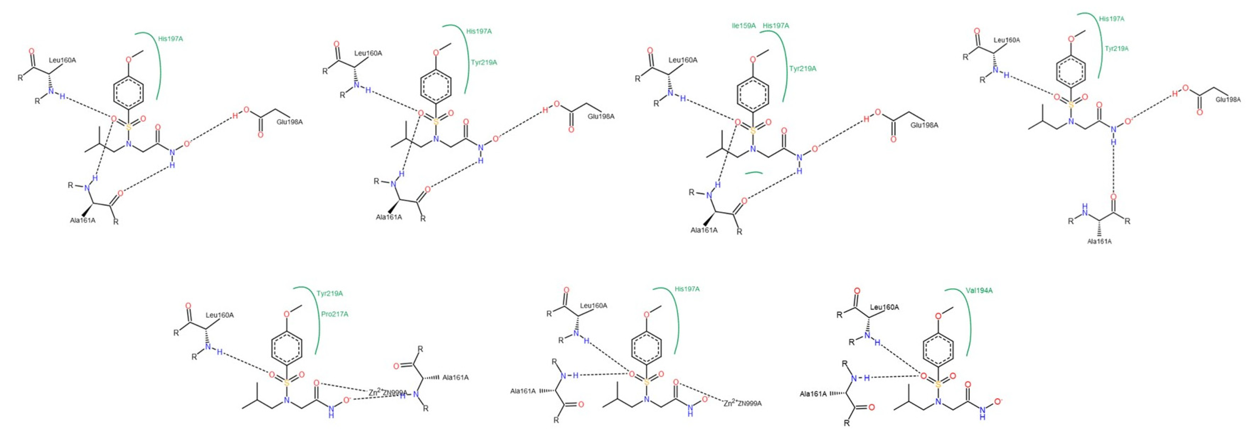
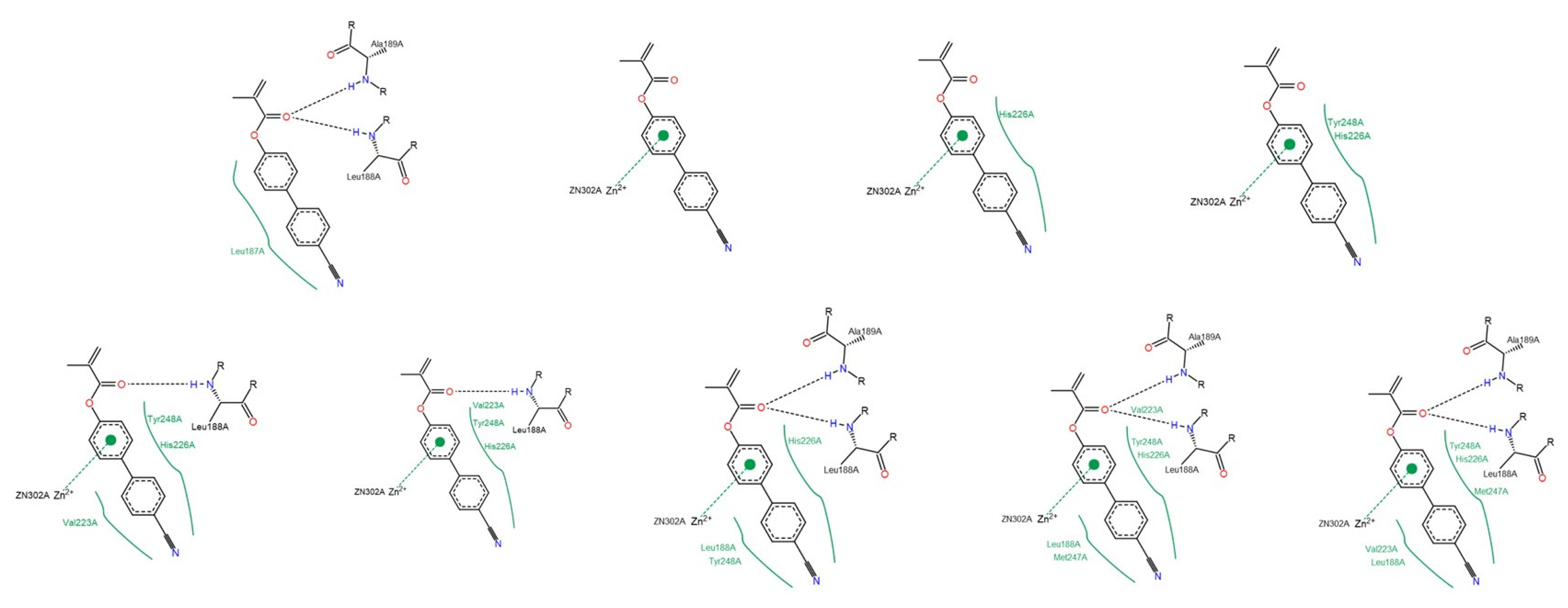
3.1.2. Interactions between Ligand and Protein

A relevant feature of inhibitor specificity is the way it performs as a ligand. The active site of MMPs is a deep cavity and the catalytic domain of different MMPs share a sequential similarity [59]. The catalytic domain is highly conserved, containing three histidine residues responsible for chelates the catalytic zinc ion (VAAHExGHxxGxxH) [1,14,33] and, in the terminal zone, there is the “met-turn” [1,7], that forms the outer wall of pocket S1′ [1,14]. There are six S pockets (S1, S2, S3, S1′, S2′ and S3′) [14] but the S1′ pocket is the most important since it is a determining factor for substrate specificity [1,3,11,16]. The depth of the S11′ pocket can be shallow (MMP-1), intermedium or deep (MMP-2, -8, -9 and -13) cavity (Figure 2) and it is highly hydrophobic [1,7,10,11,12,14,16,60]. The highly conserved sequence (VAAHExGHxxGxxH), the “met-turn” region and the S1′ pocket from collagenases (MMP-1, -8 and -13) and gelatinases (MMP-2 and -9) were considered for docking studies.
The catalytic activity of MMPs requires a catalytic zinc ion, a water molecule flanked by three histidine and one glutamate residues [1,8,16], present in conserved sequence- VAAHExGHxxGxxH. Our hypothesis is that if there is an interaction between the compound and the catalytic zinc ion or any of these residues from the conserved sequence, there will cause enzyme inhibition. The zinc ion demonstrated affinity for all compounds, mainly for the sulfonyl group of NNGH inhibitor and for the aromatic rings of the compounds A (Figure 3a), B (Figure 3b) and C. The three histidine and glutamate residues of the conserved sequence also demonstrated capacity to establish a hydrogen bond with the hydroxamic acid of NNGH inhibitor, in MMPs-1, -2, -8 and -9 and with the methacrylate group of compounds A and B, in all MMPs or methylacrylamide group of compound C, in MMP-1, -2, -8, and -9. Another hypothesis is that the compound can cause enzyme inhibition if blocks the access to S1′ pocket and all compounds besides demonstrated affinity to the active site blocked totally (Figure 3d) or partially (Figure 3c) the S1′.
However, there were additional interactions between the compounds and the enzyme depending on the enzyme class (collagenases and gelatinases) (See Appendix B, Appendix C, Appendix D and Appendix E). Preliminary biochemical tests (not shown) gave encouraging indications on activity reduction achieved for most MMPs by compound A, B and C, even though their low solubility and polymerization tendency.

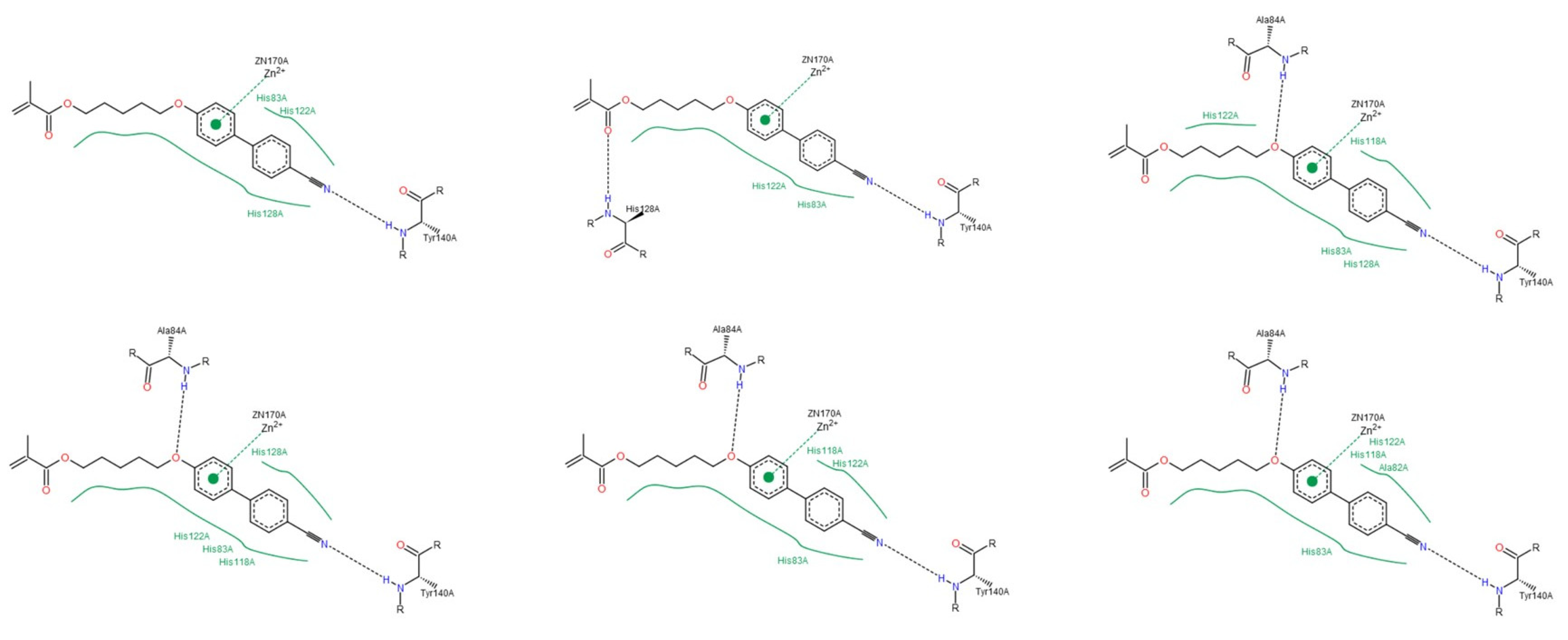
Collagenases (MMP-1, -8 and -13)

The residues Leu81, Ala82 and Ala84 of the MMP-1, the residues Leu160, Ala161 and Ala160 of the MMP-8, and the residues Leu82, Ala83 and Ala85 of the MMP-13 are located at the same distance from the conserved sequence in the three enzymes and their neighboring peptide bond groups can establish hydrogen bonds with the compounds studied (Table 3), through the sulfonyl group or hydroxamic acid of the NNGH inhibitor, both oxygen atoms of the methacrylate group from compound A, CN group of the compounds A and B, the oxygen atom adjacent to the biphenyl group of the compound B and the methylacylamide group of the compound C.
In MMP-1, there are three more spots on the peptide that can interact with the studied compounds. The peptide bond group adjacent to Gly79 can establish a hydrogen bond with the NH group of methylacrylamide group of compound C (Figure 4a). The residue Asn80 interacts with the tertiary amine of NNGH inhibitor (Figure 4b) and with the oxygen atom adjacent to the biphenyl group of compound A (Figure 3a). The Tyr140 can interact with the CN group of compound B (Figure 3b) or with the oxygen atom of the methylacrylamide group of compound C (Figure 4a). In MMP-8, the residue His162 establishes a hydrogen bond with the oxygen atom of the carbonyl group of methacrylate function of compound B (Figure 4c). In MMP-13, the residue Thr142 establishes a hydrogen bond with compound C, through the oxygen atom of the methylacrylamide group (Figure 4d) and residue Thr144 can establish an interaction with the oxygen atom of the methacrylate group of compound B (Figure 3d).

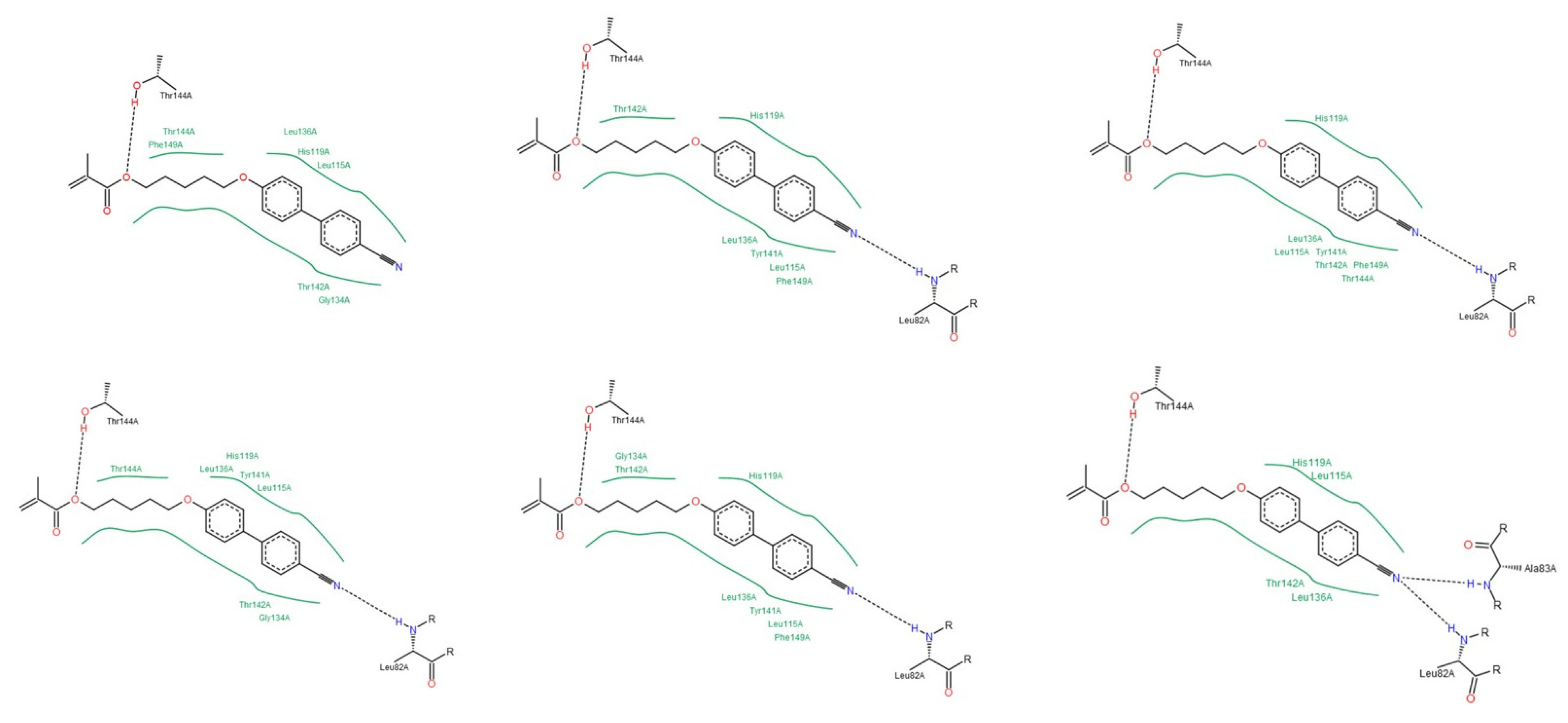
Gelatinases (MMP-2 and -9)

In MMP-2, the peptide bond adjacent to Leu83 and Ala84 can establish hydrogen bonds with all of the compounds. In the case of the NNGH inhibitor, the oxygen atom of the sulfonyl group and the NH group of the hydroxamic acid interact with both residues. The oxygen atom of the carbonyl group of methacrylate in compound A can establish a hydrogen bond with Leu83 and Ala84 (Figure 5a). The CN group of compound A and B can interact with Leu83 or Ala84, in the case of compound B (Figure 5b). The oxygen atom adjacent to the biphenyl group in compound B can establish a hydrogen bond with Leu83 (Figure 5c). The methylacrylamide group of compound C interacts with Leu83 and Ala84 (Figure 5d). The residue His85 can establish a hydrogen bond with the oxygen atom of the sulfonyl group in the NNGH inhibitor and with the methacrylate group in compound B (Figure 5c).
The residues Leu137, Tyr142 and Thr143 belong to the “met-turn” region and interact only with the compounds A, B and C. The residue Leu137 establishes a hydrogen bond only with the NH group of the methylacrylamide of the compound C (Figure 5d). The residue Tyr142 can interact with one or both aromatic rings of the biphenyl group of the compounds A and B (Figure 5b). The residue Thr143 establishes a hydrogen bond with the oxygen atom of the methacrylate group, in compounds A and B or methylacrylamide group in compound C (Figure 5b,d).
In MMP-9, the residues Leu188, Ala189 and His190 interact with the NNGH inhibitor and the compounds A, B and C. The sulfonyl group and the NH group of the hydroxamic acid of the NNGH inhibitor (Figure 6a) and the methacrylate group of compound A (Figure 6b) can interact with Leu188 and Ala189. In compound B, the methacrylate group establishes a hydrogen bond with His190 and the oxygen atom adjacent to the biphenyl group interacts with Leu188 and Ala189 (Figure 6c). The methylacrylamide group of compound C can establish a hydrogen bond with all amino acids (Figure 3c and Figure 6d). This group of compound C can also establish a hydrogen bond with the residue Asp235 in MMP-9, a variable residue of the conserved sequence-VAAHExGHxxGxxH (Figure 6d).

3.2. Cytotoxicity Study

In general, MG-63 cells showed significantly (p < 0.001) more resistance to the presence of MMP inhibitors than NIH/3T3 cells. The compound interaction with cellular membrane depend on their physical and chemicals characteristics [61]. In fact, cellular differentiation can be the answer why osteoblasts (MG-63) are more resistant than fibroblasts (NIH/3T3); it is known that growing factors and gene expression are the responsible ones for this differentiation [61,62].
Compounds A, B and C decrease cell viability (Figure 7 and Table 4) and can even be considered toxic for NIH/3T3 cells at 50 and 100 µM concentration since the number of viable cells decreased more than 30% (ISO 10993–5: 2009). The viability of NIH/3T3 cell was significantly (p < 0.05) higher for compound A than for compound B and C; and significantly (p < 0.001) higher at 20 µM than the other two concentrations tested.
A decrease in cell viability was also observed in human MG-63 cells, but not statistical significantly differences were found between compounds (p = 0.307) and concentrations (p = 0.406). However, according to ISO standards, only compound B at 50/100 μM and compound C at 100 μM were toxic. Nevertheless, none of the three compounds highlighted serious toxicity.
Although compounds A, B and C present some toxicity towards the two cell types, it may be much less as compounds are to be used copolymerization with dental resins, which reduce their free concentration. Preliminary results by FTIR spectroscopy using commercial resins showed a decrease in the vinyl bands of the free compounds upon light polymerization (results not shown).

4. Conclusions

According to the computational docking simulations, all compounds (NNGH inhibitor and the compounds A, B and C) have the potential to be good inhibitors of MMPs. These compounds were chosen among a pool of previously synthesized compounds in the laboratory and a few hundred other proposals as the ones with better affinity score towards the MMPs active sites. The compounds showed an affinity for the active center of MMPs, which may block access to the S1′ pocket, and they can interact with the catalytic zinc ion. The residues histidine and glutamate, belong to the conserved sequence, and some residues of the “met-turn” can also establish hydrogen bonds with the compounds. Altogether available data lead us to conclude that the compounds interact with the crucial components for the catalytic activity of MMPs. Furthermore, cell viability does not decrease much upon direct contact with the free non-polymerized compounds and toxicity is expected to further decrease upon covalent binding to the dental resin matrix, promising improvements in dental resins mechanical strength.

Author Contributions

Conceptualization, J.C.; I.B.; M.S.-O.; methodology, H.L.; I.C.; software, H.L.; validation, I.C. writing—review and editing, J.P.; A.A.; M.P.; J.C.; M.S.-O.; K.T.P.; funding acquisition, J.C. All authors have read and agreed to the published version of the manuscript.

Funding

This research was funded by A Molecular View of Dental Restoration, grant number PTDC/SAU-BMA/122444/2010 and by Molecular Design for Dental Restauration, grant number SAICT-POL/24288/2016, suported by FEDER thru Programas Operacionais financiadores do projeto (PO Lisboa e PO Norte). The work was supported by the Associate Laboratory for Green Chemistry- LAQV FCT/MCTES (UIDB/50006/2020) and Applied Molecular Biosciences Unit- UCIBIO FCT/MCTES (UID/Multi/04378/2019), which are financed by national funds. This work is financed by national funds through the FCT—Foundation for Science and Technology, I.P, under the project UIDB/04585/2020.

Institutional Review Board Statement

Not applicable.

Informed Consent Statement

Not applicable.

Conflicts of Interest

The authors declare no conflict of interest.

Appendix A

Table A1. Physicochemical and ADMET properties, by SeeSAR.
ParameterNNGHCompound ACompound BCompound C
MW316.375263.295349.428336.389
H-bonds acceptors6342
H-bonds donors2002
Aromatic rings1222
Stereo centers0000
Rotatable bonds5060
Total charge0 (OH)/ −1 (O−)000
Log P1.4723.8285.0892.943
Log D−0.0753.8285.0892.943
TPSA95.9450.0959.3267.43
BBB category++
BBB log ([brain]:[blood])0.703−0.158−0.150−0.492
P-gp categoryNoNoNoYes
2C9 pki4.3774.9315.0405.125
2D6 affinityLowMediumHighMedium
HIA category++++
PPB90 categoryLowHighHighHigh
hERG pIC504.3195.0735.7494.894
Log S (pH = 7.4)3.2640.653-0.2172.336

Appendix B

Figure A1. Interactions between NNGH inhibitor and MMP-1, by PoseView calculations.
Biomedicines 09 00366 g0a1
Figure A2. Interactions between NNGH inhibitor and MMP-2, by PoseView calculations.
Biomedicines 09 00366 g0a2
Figure A3. Interactions between NNGH inhibitor and MMP-8, by PoseView calculations.
Biomedicines 09 00366 g0a3
Figure A4. Interactions between NNGH inhibitor and MMP-9, by PoseView calculations.
Biomedicines 09 00366 g0a4
Figure A5. Interactions between NNGH inhibitor and MMP-13, by PoseView calculations.
Biomedicines 09 00366 g0a5

Appendix C

Figure A6. Interactions between compound A and MMP-1, by PoseView calculations.
Biomedicines 09 00366 g0a6
Figure A7. Interactions between compound A and MMP-2, by PoseView calculations.
Biomedicines 09 00366 g0a7
Figure A8. Interactions between compound A and MMP-8, by PoseView calculations.
Biomedicines 09 00366 g0a8
Figure A9. Interactions between compound A and MMP-9, by PoseView calculations.
Biomedicines 09 00366 g0a9
Figure A10. Interactions between compound A and MMP-13, by PoseView calculations.
Biomedicines 09 00366 g0a10

Appendix D

Figure A11. Interactions between compound B and MMP-1, by PoseView calculations.
Biomedicines 09 00366 g0a11
Figure A12. Interactions between compound B and MMP-2, by PoseView calculations.
Biomedicines 09 00366 g0a12
Figure A13. Interactions between compound B and MMP-8, by PoseView calculations.
Biomedicines 09 00366 g0a13
Figure A14. Interactions between compound B and MMP-9, by PoseView calculations.
Biomedicines 09 00366 g0a14
Figure A15. Interactions between compound B and MMPb-13, by PoseView calculations.
Biomedicines 09 00366 g0a15

Appendix E

Figure A16. Interactions between compound C and MMP-1, by PoseView calculations.
Biomedicines 09 00366 g0a16
Figure A17. Interactions between compound C and MMP-2, by PoseView calculations.
Biomedicines 09 00366 g0a17
Figure A18. Interactions between compound C and MMP-8, by PoseView calculations.
Biomedicines 09 00366 g0a18
Figure A19. Interactions between compound C and MMP-9, by PoseView calculations.
Biomedicines 09 00366 g0a19
Figure A20. Interactions between compound C and MMP-13, by PoseView calculations.
Biomedicines 09 00366 g0a20

References

  1. Cui, N.; Hu, M.; Khalil, R.A. Biochemical and Biological Attributes of Matrix Metalloproteinases. Prog. Mol. Biol. Transl. Sci. 2017, 147, 1–73. [Google Scholar] [CrossRef] [PubMed] [Green Version]
  2. Fischer, T.; Senn, N.; Riedl, R. Design and Structural Evolution of Matrix Metalloproteinase Inhibitors. Chemistry 2019, 25, 7960–7980. [Google Scholar] [CrossRef] [PubMed]
  3. Nagase, H.; Visse, R.; Murphy, G. Structure and function of matrix metalloproteinases and TIMPs. Cardiovasc. Res. 2006, 69, 562–573. [Google Scholar] [CrossRef] [PubMed] [Green Version]
  4. Goodwin, L. A Closer Look At Metalloproteinases; Nova Science Publishers, Inc.: New York, NY, USA, 2019; p. 310. [Google Scholar]
  5. Klein, T.; Bischoff, R. Physiology and pathophysiology of matrix metalloproteases. Amino Acids 2011, 41, 271–290. [Google Scholar] [CrossRef] [Green Version]
  6. Visse, R.; Nagase, H. Matrix metalloproteinases and tissue inhibitors of metalloproteinases: Structure, function, and biochemistry. Circ. Res. 2003, 92, 827–839. [Google Scholar] [CrossRef] [Green Version]
  7. Maskos, K. Crystal structures of MMPs in complex with physiological and pharmacological inhibitors. Biochimie 2005, 87, 249–263. [Google Scholar] [CrossRef]
  8. Tallant, C.; Marrero, A.; Gomis-Rüth, F.X. Matrix metalloproteinases: Fold and function of their catalytic domains. Biochim. Biophys. Acta 2010, 1803, 20–28. [Google Scholar] [CrossRef]
  9. Oberholzer, A.E.; Bumann, M.; Hege, T.; Russo, S.; Baumann, U. Metzincin’s canonical methionine is responsible for the structural integrity of the zinc-binding site. Biol. Chem. 2009, 390, 875–881. [Google Scholar] [CrossRef] [PubMed] [Green Version]
  10. Jacobsen, J.A.; Major Jourden, J.L.; Miller, M.T.; Cohen, S.M. To bind zinc or not to bind zinc: An examination of innovative approaches to improved metalloproteinase inhibition. Biochim. Biophys. Acta 2010, 1803, 72–94. [Google Scholar] [CrossRef] [Green Version]
  11. Verma, R.P.; Hansch, C. Matrix metalloproteinases (MMPs): Chemical-biological functions and (Q)SARs. Bioorg. Med. Chem. 2007, 15, 2223–2268. [Google Scholar] [CrossRef]
  12. Murphy, G.; Nagase, H. Progress in matrix metalloproteinase research. Mol. Aspects Med. 2008, 29, 290–308. [Google Scholar] [CrossRef] [PubMed] [Green Version]
  13. Mannello, F.; Medda, V. Nuclear localization of matrix metalloproteinases. Prog. Histochem. Cytochem. 2012, 47, 27–58. [Google Scholar] [CrossRef] [PubMed]
  14. Rangasamy, L.; Geronimo, B.D.; Ortín, I.; Coderch, C.; Zapico, J.M.; Ramos, A.; de Pascual-Teresa, B. Molecular Imaging Probes Based on Matrix Metalloproteinase Inhibitors (MMPIs). Molecules 2019, 24, 2982. [Google Scholar] [CrossRef] [PubMed] [Green Version]
  15. Amălinei, C.; Căruntu, I.D.; Bălan, R.A. Biology of metalloproteinases. Rom. J. Morphol. Embryol. 2007, 48, 323–334. [Google Scholar]
  16. Liu, J.; Khalil, R.A. Matrix Metalloproteinase Inhibitors as Investigational and Therapeutic Tools in Unrestrained Tissue Remodeling and Pathological Disorders. Prog. Mol. Biol. Transl. Sci. 2017, 148, 355–420. [Google Scholar] [CrossRef] [Green Version]
  17. Cerofolini, L.; Fragai, M.; Luchinat, C. Mechanism and Inhibition of Matrix Metalloproteinases. Curr. Med. Chem. 2019, 26, 2609–2633. [Google Scholar] [CrossRef]
  18. Laronha, H.; Caldeira, J. Structure and Function of Human Matrix Metalloproteinases. Cells 2020, 9, 1076. [Google Scholar] [CrossRef]
  19. Vandenbroucke, R.E.; Dejonckheere, E.; Libert, C. A therapeutic role for matrix metalloproteinase inhibitors in lung diseases? Eur. Respir. J. 2011, 38, 1200–1214. [Google Scholar] [CrossRef]
  20. Sekhon, B.S. Matrix metalloproteinases—An overview. Dovepress 2010, 1, 1–20. [Google Scholar]
  21. Henn, S.; De Carvalho, R.; Ogliari, F.; De Souza, A.; Line, S.; Da Silva, A.; Demarco, F.; Etges, A.; Piva, E. Addition of zinc methacrylate in dental polymers: MMP-2 inhibition and ultimate tensile strength evaluation. Clin. Oral Investig. 2012, 16, 531–536. [Google Scholar] [CrossRef] [PubMed]
  22. Hidalgo, M.; Eckhardt, S.G. Development of matrix metalloproteinase inhibitors in cancer therapy. J. Natl. Cancer Inst. 2001, 93, 178–193. [Google Scholar] [CrossRef] [PubMed] [Green Version]
  23. Tokuhara, C.K.; Santesso, M.R.; Oliveira, G.S.N.; Ventura, T.M.D.S.; Doyama, J.T.; Zambuzzi, W.F.; Oliveira, R.C. Updating the role of matrix metalloproteinases in mineralized tissue and related diseases. J. Appl. Oral Sci. 2019, 27, e20180596. [Google Scholar] [CrossRef]
  24. Young, D.; Das, N.; Anowai, A.; Dufour, A. Matrix Metalloproteases as Influencers of the Cells’ Social Media. Int. J. Mol. Sci. 2019, 20, 3847. [Google Scholar] [CrossRef] [PubMed] [Green Version]
  25. Hannas, A.R.; Pereira, J.C.; Granjeiro, J.M.; Tjäderhane, L. The role of matrix metalloproteinases in the oral environment. Acta Odontol. Scand. 2007, 65, 1–13. [Google Scholar] [CrossRef]
  26. Lührs, A.K.; De Munck, J.; Geurtsen, W.; Van Meerbeek, B. Does inhibition of proteolytic activity improve adhesive luting? Eur. J. Oral Sci. 2013, 121, 121–131. [Google Scholar] [CrossRef] [PubMed]
  27. Nuti, E.; Tuccinardi, T.; Rossello, A. Matrix metalloproteinase inhibitors: New challenges in the era of post broad-spectrum inhibitors. Curr. Pharm. Des. 2007, 13, 2087–2100. [Google Scholar] [CrossRef]
  28. Li, K.; Tay, F.R.; Yiu, C.K.Y. The past, present and future perspectives of matrix metalloproteinase inhibitors. Pharmacol. Ther. 2020, 207, 107465. [Google Scholar] [CrossRef] [PubMed]
  29. Hu, J.; Van den Steen, P.E.; Sang, Q.X.; Opdenakker, G. Matrix metalloproteinase inhibitors as therapy for inflammatory and vascular diseases. Nat. Rev. Drug Discov. 2007, 6, 480–498. [Google Scholar] [CrossRef] [PubMed]
  30. Laronha, H.; Carpinteiro, I.; Portugal, J.; Azul, A.; Polido, M.; Petrova, K.T.; Salema-Oom, M.; Caldeira, J. Challenges in Matrix Metalloproteinases Inhibition. Biomolecules 2020, 10, 717. [Google Scholar] [CrossRef] [PubMed]
  31. Georgiadis, D.; Yiotakis, A. Specific targeting of metzincin family members with small-molecules inhibitors: Progress toward a multifarious challenge. Bioorg. Med. Chem. 2008, 8781–8794. [Google Scholar] [CrossRef]
  32. Whittaker, M.; Floyd, C.D.; Brown, P.; Gearing, A.J. Design and therapeutic application of matrix metalloproteinase inhibitors. Chem. Rev. 1999, 99, 2735–2776. [Google Scholar] [CrossRef] [PubMed]
  33. Cuniasse, P.; Devel, L.; Makaritis, A.; Beau, F.; Georgiadis, D.; Matziari, M.; Yiotakis, A.; Dive, V. Future challenges facing the development of specific active-site-directed synthetic inhibitors of MMPs. Biochimie 2005, 87, 393–402. [Google Scholar] [CrossRef]
  34. Fields, G.B. The Rebirth of Matrix Metalloproteinase Inhibitors: Moving Beyond the Dogma. Cells 2019, 8, 984. [Google Scholar] [CrossRef] [Green Version]
  35. Folgueras, A.R.; Pendás, A.M.; Sánchez, L.M.; López-Otín, C. Matrix metalloproteinases in cancer: From new functions to improved inhibition strategies. Int. J. Dev. Biol. 2004, 48, 411–424. [Google Scholar] [CrossRef] [PubMed] [Green Version]
  36. Rossello, A.; Nuti, E.; Orlandini, E.; Carelli, P.; Rapposelli, S.; Macchia, M.; Minutolo, F.; Carbonaro, L.; Albini, A.; Benelli, R.; et al. New N-arylsulfonyl-N-alkoxyaminoacetohydroxamic acids as selective inhibitors of gelatinase A (MMP-2). Bioorg. Med. Chem. 2004, 12, 2441–2450. [Google Scholar] [CrossRef]
  37. Tamura, Y.; Watanabe, F.; Nakatani, T.; Yasui, K.; Fuji, M.; Komurasaki, T.; Tsuzuki, H.; Maekawa, R.; Yoshioka, T.; Kawada, K.; et al. Highly selective and orally active inhibitors of type IV collagenase (MMP-9 and MMP-2): N-sulfonylamino acid derivatives. J. Med. Chem. 1998, 41, 640–649. [Google Scholar] [CrossRef] [PubMed]
  38. Terp, G.E.; Cruciani, G.; Christensen, I.T.; Jørgensen, F.S. Structural differences of matrix metalloproteinases with potential implications for inhibitor selectivity examined by the GRID/CPCA approach. J. Med. Chem. 2002, 45, 2675–2684. [Google Scholar] [CrossRef] [PubMed]
  39. Tuccinardi, T.; Martinelli, A.; Nuti, E.; Carelli, P.; Balzano, F.; Uccello-Barretta, G.; Murphy, G.; Rossello, A. Amber force field implementation, molecular modelling study, synthesis and MMP-1/MMP-2 inhibition profile of (R)- and (S)-N-hydroxy-2-(N-isopropoxybiphenyl-4-ylsulfonamido)-3-methylbutanamides. Bioorg. Med. Chem. 2006, 14, 4260–4276. [Google Scholar] [CrossRef] [PubMed]
  40. O’Brien, P.M.; Ortwine, D.F.; Pavlovsky, A.G.; Picard, J.A.; Sliskovic, D.R.; Roth, B.D.; Dyer, R.D.; Johnson, L.L.; Man, C.F.; Hallak, H. Structure-activity relationships and pharmacokinetic analysis for a series of potent, systemically available biphenylsulfonamide matrix metalloproteinase inhibitors. J. Med. Chem. 2000, 43, 156–166. [Google Scholar] [CrossRef]
  41. Gooyit, M.; Suckow, M.A.; Schroeder, V.A.; Wolter, W.R.; Mobashery, S.; Chang, M. Selective gelatinase inhibitor neuroprotective agents cross the blood-brain barrier. ACS Chem. Neurosci. 2012, 3, 730–736. [Google Scholar] [CrossRef] [PubMed] [Green Version]
  42. Barros, M.T.; Mouquinho, A.I.; Petrova, K.T.; Saavedra, M.D.; Sotomayor, J.C. Fast synthesis employing a microwave assisted neat protocol of new monomers potentially useful for the preparation of PDLC films. Cent. Eur. J. Chem. 2011, 9. [Google Scholar] [CrossRef]
  43. Bandarra, S.; Mascarenhas, P.; Luís, A.R.; Catrau, M.; Bekman, E.; Ribeiro, A.C.; Félix, S.; Caldeira, J.; Barahona, I. In vitro and in silico evaluations of resin-based dental restorative material toxicity. Clin. Oral Investig. 2020, 24, 2691–2700. [Google Scholar] [CrossRef] [PubMed]
  44. Morphy, R.; Rankovic, Z. The physicochemical challenges of designing multiple ligands. J. Med. Chem. 2006, 49, 4961–4970. [Google Scholar] [CrossRef]
  45. Leeson, P.D. Molecular inflation, attrition and the rule of five. Adv. Drug Deliv. Rev. 2016, 101, 22–33. [Google Scholar] [CrossRef] [PubMed]
  46. Meanwell, N.A. Improving drug candidates by design: A focus on physicochemical properties as a means of improving compound disposition and safety. Chem. Res. Toxicol. 2011, 24, 1420–1456. [Google Scholar] [CrossRef] [PubMed]
  47. Gleeson, M.P.; Hersey, A.; Hannongbua, S. In-silico ADME models: A general assessment of their utility in drug discovery applications. Curr. Top. Med. Chem. 2011, 11, 358–381. [Google Scholar] [CrossRef] [PubMed]
  48. Gleeson, M.P. Generation of a set of simple, interpretable ADMET rules of thumb. J. Med. Chem. 2008, 51, 817–834. [Google Scholar] [CrossRef]
  49. Gleeson, M.P.; Hersey, A.; Montanari, D.; Overington, J. Probing the links between in vitro potency, ADMET and physicochemical parameters. Nat. Rev. Drug Discov. 2011, 10, 197–208. [Google Scholar] [CrossRef]
  50. Lagorce, D.; Douguet, D.; Miteva, M.A.; Villoutreix, B.O. Computational analysis of calculated physicochemical and ADMET properties of protein-protein interaction inhibitors. Sci. Rep. 2017, 7, 46277. [Google Scholar] [CrossRef] [PubMed]
  51. Leeson, P.D.; Davis, A.M. Time-related differences in the physical property profiles of oral drugs. J. Med. Chem. 2004, 47, 6338–6348. [Google Scholar] [CrossRef]
  52. Baell, J.; Congreve, M.; Leeson, P.; Abad-Zapatero, C. Ask the experts: Past, present and future of the rule of five. Future Med. Chem. 2013, 5, 745–752. [Google Scholar] [CrossRef] [PubMed]
  53. Waring, M.J. Lipophilicity in drug discovery. Expert Opin. Drug Discov. 2010, 5, 235–248. [Google Scholar] [CrossRef]
  54. Veber, D.F.; Johnson, S.R.; Cheng, H.Y.; Smith, B.R.; Ward, K.W.; Kopple, K.D. Molecular properties that influence the oral bioavailability of drug candidates. J. Med. Chem. 2002, 45, 2615–2623. [Google Scholar] [CrossRef]
  55. Palm, K.; Stenberg, P.; Luthman, K.; Artursson, P. Polar molecular surface properties predict the intestinal absorption of drugs in humans. Pharm. Res. 1997, 14, 568–571. [Google Scholar] [CrossRef] [PubMed]
  56. Jeffrey, P.; Summerfield, S. Assessment of the blood-brain barrier in CNS drug discovery. Neurobiol. Dis. 2010, 37, 33–37. [Google Scholar] [CrossRef]
  57. Carpenter, T.S.; Kirshner, D.A.; Lau, E.Y.; Wong, S.E.; Nilmeier, J.P.; Lightstone, F.C. A method to predict blood-brain barrier permeability of drug-like compounds using molecular dynamics simulations. Biophys. J. 2014, 107, 630–641. [Google Scholar] [CrossRef] [PubMed] [Green Version]
  58. Chen, L.; Li, Y.; Yu, H.; Zhang, L.; Hou, T. Computational models for predicting substrates or inhibitors of P-glycoprotein. Drug Discov. Today 2012, 17, 343–351. [Google Scholar] [CrossRef]
  59. Lovejoy, B.; Cleasby, A.; Hassell, A.M.; Longley, K.; Luther, M.A.; Weigl, D.; McGeehan, G.; McElroy, A.B.; Drewry, D.; Lambert, M.H. Structure of the catalytic domain of fibroblast collagenase complexed with an inhibitor. Science 1994, 263, 375–377. [Google Scholar] [CrossRef]
  60. Gimeno, A.; Beltrán-Debón, R.; Mulero, M.; Pujadas, G.; Garcia-Vallvé, S. Understanding the variability of the S1’ pocket to improve matrix metalloproteinase inhibitor selectivity profiles. Drug Discov. Today 2019. [Google Scholar] [CrossRef]
  61. Xia, T.; Li, N.; Nel, A.E. Potential health impact of nanoparticles. Annu. Rev. Public Health 2009, 30, 137–150. [Google Scholar] [CrossRef] [PubMed] [Green Version]
  62. Singer, M. Advances in the management of macular degeneration. F1000Prime Rep. 2014, 6, 29. [Google Scholar] [CrossRef] [PubMed]
Figure 1. Compounds selected from docking studies. (a) N-Isobutyl-N-(4-methoxyphenylsulfonyl)glycyl hydroxamic acid- NNGH, inhibitor of commercial MMP enzymatic test kit (b) 4′-cyano-[1,1′-bihenyl]-4-yl methacrylate- Compound A [42] (c) 5-((4′-cyano-[1,1′-biphenyl]-4-yl)oxy)pentyl methacrylate- Compound B [42] (d) N,N′-(oxybis(4,1-phenylene))bis(2-methylacrylamide)- Compound C [42].
Biomedicines 09 00366 g001
Figure 2. (a) Active site of MMP-1. (b) Active site of MMP-13. The S1′ pocket of MMP-1 is shallow and the S1′ pocket of MMP-13 is deep.
Biomedicines 09 00366 g002
Figure 3. Location of compounds A, B and C in the active center of MMPs. (a) Compound A in the active site of MMP-1. The catalytic zinc ion can establish two interactions with the two aromatic rings, simultaneously. (b) Compound B in the active site of MMP-1. The catalytic zinc ion establishes an interaction with an aromatic ring. (c) Compound C in the active site of MMP-9. Compound C partially through the S1′ pocket. (d) Compound B in the active site of MMP-13 and the molecule totally through the S1′ pocket.
Biomedicines 09 00366 g003
Figure 4. Interactions between MMP-1, -8 and -13 and the NNGH inhibitor and the compounds B and C. (a) The residue Gly79 and Tyr140, of MMP-1, establish a hydrogen bond with the NH group and the oxygen atom, respectively, of methylacrylamide function of compound C. (b) The Asn80 of MMP-1 establishes a hydrogen bond with the tertiary amine of NNGH inhibitor. (c) The His162 of MMP-8 interacts with the oxygen atom of the carbonyl group of methacrylate of the compound B. (d) In MMP-13, the residue Thr142 establishes a hydrogen bond with the oxygen atom of methylacrylamide of compound C.
Biomedicines 09 00366 g004
Figure 5. Interactions between compounds A, B and C and the MMP-2. (a) Compound A. The oxygen atom of the carbonyl group of methacrylate establishes hydrogen bonds with Leu83 and Ala84, simultaneously. (b) Compound B. The CN group establishes hydrogen bonds with Leu83 and Ala84, simultaneously. The residue Tyr142 interacts with the oxygen atom of the carbonyl group of methacrylate and the residue Thr143 interacts with the biphenyl group. (c) Compound B. The Leu83 and His85 establish a hydrogen bond with the oxygen atom of the methacrylate group and with the oxygen atom adjacent to the biphenyl group. (d) Compound C. The Ala84, Leu137 and Thr143 establish a hydrogen bond with the methylacrylamide group.
Biomedicines 09 00366 g005
Figure 6. Interactions between the NNGH inhibitor and the compounds A, B and C and the MMP-9. (a) NNGH inhibitor. Leu188 and Ala189 interact with the oxygen atom of the sulfonyl group and the NH group of hydroxamic acid. (b) Compound A. Leu188 and Ala189 establish a hydrogen bond with the oxygen atom of the carbonyl group, simultaneously. (c) Compound B. Interactions between Leu188, Ala189 and His190 and the oxygen atom of methacrylate and adjacent to the biphenyl group, respectively. (d) Compound C. The methylacrylamide can establish a hydrogen bond with Leu188, Ala189 and the residue Asp235.
Biomedicines 09 00366 g006
Figure 7. NIH/3T3 (a) and MG-63 (b) cell viability upon exposure to compounds A (4′-cyano-[1,1′-bihenyl]-4-yl methacrylate), B (5-((4′-cyano-[1,1′-biphenyl]-4-yl)oxy)pentyl methacrylate) and C (N,N′-(oxybis(4,1-phenylene))bis(2-methylacrylamide).
Biomedicines 09 00366 g007
Table 1. MMP information obtained from the Protein Data Bank (PDB).
MMPPDB IDTotal Structure Weight (kDa)MethodResolution (Å)R-Factor (%)
12tcl19.63X-ray diffraction2.2016.2
21HOV19.28Solution NMR
81BZS19.22X-ray diffraction1.7019.2
94XCT18.97X-ray diffraction1.3017.0
131FM119.21Solution NMR
Table 2. Physicochemical and ADMET properties of the NNGH inhibitor and the compounds A, B and C.
PropertyNNGHCompound ACompound BCompound C
Molecular weight (MW)316.375263.295349.428336.389
H-bonds acceptor6342
H-bonds donor2002
Log P1.4723.8285.0892.943
Log D−0.0753.8285.0892.943
Topological polar surface area (TPSA)95.9450.0959.3267.43
BBB category++
BBB log[(brain): (blood)]0.703−0.158−0.15−0.492
P-gp categoryNoNoNoYes
Table 3. Interactions between the residues Leu, Ala and Ala of the MMPs-1, -8 and -13 and the NNGH inhibitor and the compounds A, B and C.
ResidueNNGHCompound ACompound BCompound C
Leu81 (MMP-1);
Leu160 (MMP-8);
Leu82 (MMP-13)
All
collagenases
All collagenasesMMP-8
MMP-13
MMP-8
Ala82 (MMP-1);
Ala161 (MMP-8);
Ala83 (MMP-13)
All collagenasesMMP-1MMP-8
MMP-13
MMP-8
MMP-13
Ala84 (MMP-1);
Ala163 (MMP-8);
Ala85 (MMP-13)
No interactMMP-13MMP-1All collagenases
Table 4. Percentage of mean (M) and standard variation (SD) of cell viability for the two type of cells with the several inhibitor compounds in different concentrations.
GroupsCell Viability (%)
Inhibitor
Compound
Inhibitor
Concentration
NIH/3T3MG-63
MSDMSD
A20 μM85.10.7182.62.28
50 µM54.24.4773.60.78
100 µM45.55.3073.05.40
B20 µM78.61.4178.11.41
50 µM50.54.4270.32.03
100 µM40.51.6065.90.82
C20 µM83.71.4185.00.36
50 µM46.44.2265.82.94
100 µM42.48.2869.29.24
Publisher’s Note: MDPI stays neutral with regard to jurisdictional claims in published maps and institutional affiliations.

