Article

Targeting CDK4/6 in Cancer: Molecular Docking and Cytotoxic Evaluation of Thottea siliquosa Root Extract

by
Maruthamuthu Rathinam Elakkiya
1,
Mohandas Krishnasreya
1,
Sureshkumar Tharani
2,
Muthukrishnan Arun
3,
L. Vijayalakshmi
4,*,
Jiseok Lim
4,
Ayman A. Ghfar
5 and
Balasundaramsaraswathy Chithradevi
1,*
1
Department of Botany, PSGR Krishnammal College for Women, Coimbatore 641004, Tamil Nadu, India
2
GRG BIRAC EYUVA Centre, PSGR Krishnammal College for Women, Coimbatore 641004, Tamil Nadu, India
3
Department of Biotechnology, Bharathiar University, Coimbatore 641046, Tamil Nadu, India
4
Department of Mechanical Engineering, Yeungnam University, Gyeongsan-si 38541, Republic of Korea
5
Department of Chemistry, College of Science, King Saud University, Riyadh 11451, Saudi Arabia
*
Authors to whom correspondence should be addressed.
Biomedicines 2025, 13(7), 1658; https://doi.org/10.3390/biomedicines13071658
Submission received: 23 April 2025 / Revised: 1 July 2025 / Accepted: 2 July 2025 / Published: 7 July 2025
(This article belongs to the Special Issue Progress in Cytotoxicity of Biomaterials)

Abstract

Background: Cyclin-dependent kinases 4 and 6 (CDK4/6) are pivotal regulators of the cell cycle, whose dysregulation is closely linked to cancer progression. While synthetic CDK4/6 inhibitors such as Palbociclib and Ribociclib are clinically effective, their use is limited by significant adverse effects. Methods: In this study, the aqueous root extract of Thottea siliquosa, a traditionally used medicinal plant, was evaluated for its potential as a natural CDK4/6 inhibitor. Phytochemical profiling using GC-MS identified bioactive compounds, which were subsequently subjected to molecular docking, ADME prediction, and in vitro cell-based assays using HCT116 and L929 cells. Results: The docking results revealed that Isocorydine (−7.4 kcal/mol for CDK4 and −7.2 kcal/mol for CDK6) and Thunbergol (−6.5 kcal/mol for CDK4 and −7.0 kcal/mol for CDK6) exhibited promising binding affinities comparable to standard CDK inhibitors, Palbociclib (−7.2, −8.3 kcal/mol) and Ribociclib (−7.1, −8.1 kcal/mol). Among the other tested natural compounds, Squalene (−7.1 kcal/mol for CDK4) and 2-palmitoylglycerol (−5.2 kcal/mol for CDK4, −4.9 kcal/mol for CDK6) demonstrated moderate binding affinities. ADME analysis confirmed favorable drug-like properties with minimal toxicity alerts. The extract displayed dose-dependent cytotoxicity with an IC50 of 140 μg/mL and reduced cell migration in HCT116 cells, indicating potential anti-proliferative effects. These findings suggest that T. siliquosa root extract, through synergistic phytochemical interactions, holds promise as a multi-targeted, plant-based therapeutic candidate for CDK4/6-associated cancers, warranting further in vitro and in vivo validation.

1. Introduction

Cyclin-dependent kinases (CDKs) are essential regulatory molecules that govern cell cycle progression by modulating cellular growth and proliferation. Among them, CDK4 and CDK6 play critical roles in driving the transition from the G1 to S phase of the cell cycle [1,2,3]. During the process of malignant formation, the proto-oncogene cyclin D1 binds with CDK4 and CDK6, forming active complexes that promote uncontrolled cell proliferation, an underlying hallmark of cancer development [4,5]. Consequently, targeting CDK4 and CDK6 has emerged as a promising strategy in anticancer drug development.
Despite advances in chemotherapy and radiotherapy, these conventional treatments are often limited by high costs and severe side effects. In recent years, medicinal plants have gained increasing attention as potential alternatives in cancer therapy, offering diverse bioactive compounds with therapeutic potential. Notably, approximately 50% of commercially available antineoplastic drugs are plant-derived, including well-known compounds such as leucovorin, vincristine, taxol, camptothecin, and podophyllotoxin [6,7]. The adverse effects and resistance associated with synthetic chemotherapeutic agents further emphasize the need for safer, targeted therapies. In this regard, medicinal plants, with their broad spectrum of pharmacologically active compounds, present valuable candidates for drug discovery, often exhibiting fewer side effects and enhancing the safety margin [8].
Thottea siliquosa, a medicinal shrub belonging to the family Aristolochiaceae, native to India and Sri Lanka, holds significant value in Ayurvedic medicine and is traditionally used by several tribal communities to manage various ailments. The roots of T. siliquosa are known for managing headaches, coughs, and chest pain, and are even used as an antidote for poisonous bites. Pharmacological studies have further reported a wide range of biological activities, including anticancer, antimicrobial, antioxidant, antigenotoxic, anti-inflammatory, and cytotoxic activities [9,10]. Given the therapeutic potential of T. siliquosa, exploring its phytochemical constituents for their ability to modulate molecular targets such as CDK4 and CDK6 offers a promising direction for anticancer research.
Molecular docking provides an efficient computational strategy to predict and visualize interactions between bioactive compounds and target proteins, enabling the identification of potential lead molecules [11]. In this study, T. siliquosa root extract was subjected to phytochemical profiling using GC-MS, followed by molecular docking, ADME prediction, cytotoxicity evaluation, cell cycle analysis, and cell migration assays to comprehensively assess its potential as a natural source of CDK-4 and CDK-6 inhibitors with possible therapeutic applications in cancer.

2. Materials and Methods

2.1. Collection and Authentication of Plant Material

T. siliquosa saplings were collected from Vithura town, Nedumangad, located in Thiruvananthapuram district, Kerala, India, in May 2023 and maintained in a greenhouse. The specimen was duly authenticated at the Botanical Survey of India, Southern Regional Centre, Coimbatore-03. The same specimen was preserved as a herbarium for future reference.

2.2. Phytochemical Extraction
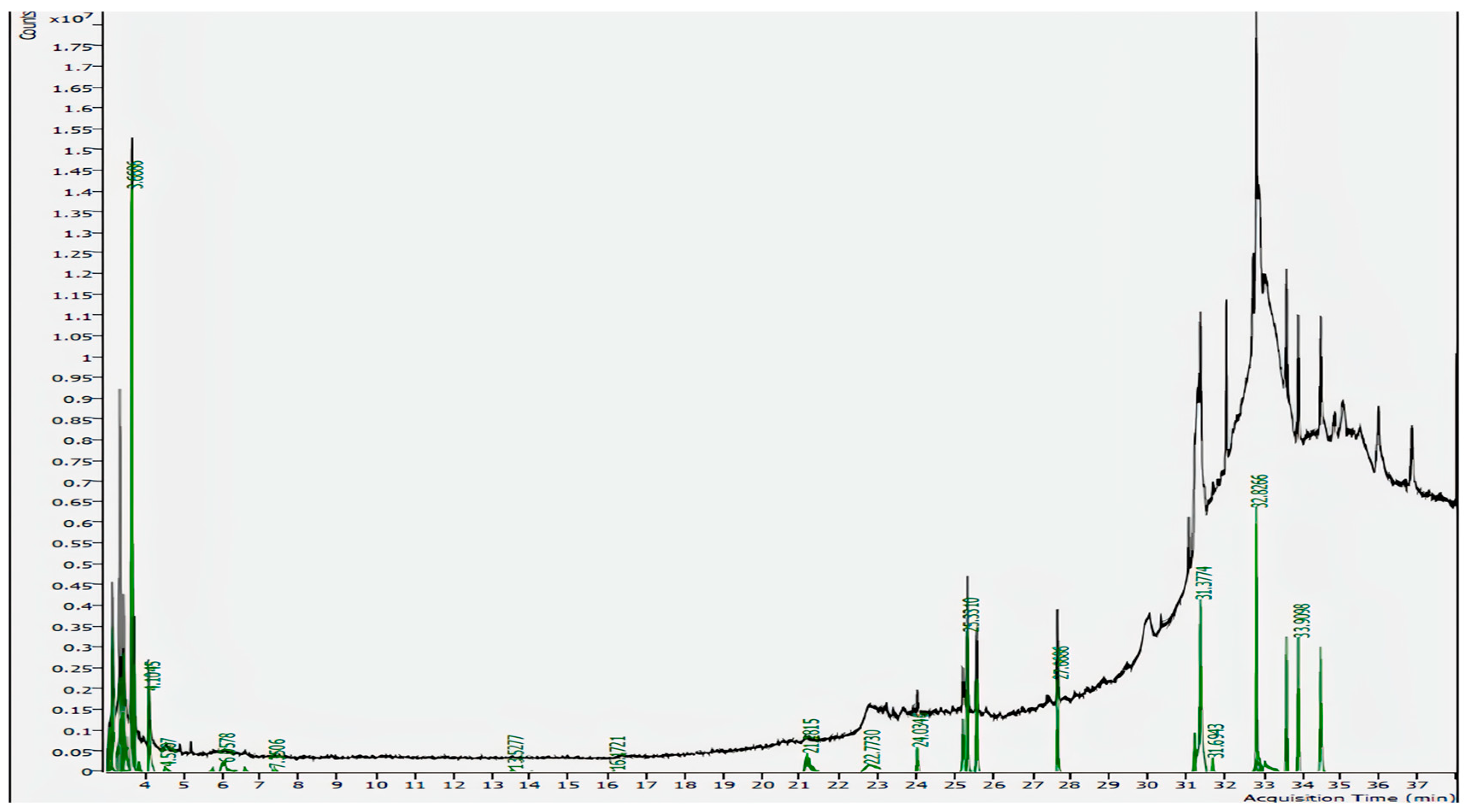
Fresh roots were collected in January 2024, washed, shade dried, and powdered. Twenty grams of the powdered sample was decocted in distilled water for 20 min, filtered, and dried in a hot air oven. It was then stored in vials for further analysis. Aqueous extraction mimics the traditional preparation methods of T. siliquosa, which minimizes the use of organic solvents and avoids toxic residues [12].

2.3. GC-MS Analysis

GC-MS analysis was conducted using a gas chromatography system (Model: CH-GCMSMS02, 8890) integrated with a 7000 GC/TQ mass spectrometer. A capillary column (30 m × 250 mm × 0.5 μm) was used with helium as the carrier gas. Methanol was used as the solvent. The thermal program involved gradual heating to 280 °C over a total run time of 38 min. The mass range was set to 30–900 m/z, and compounds were identified via NIST library comparison.

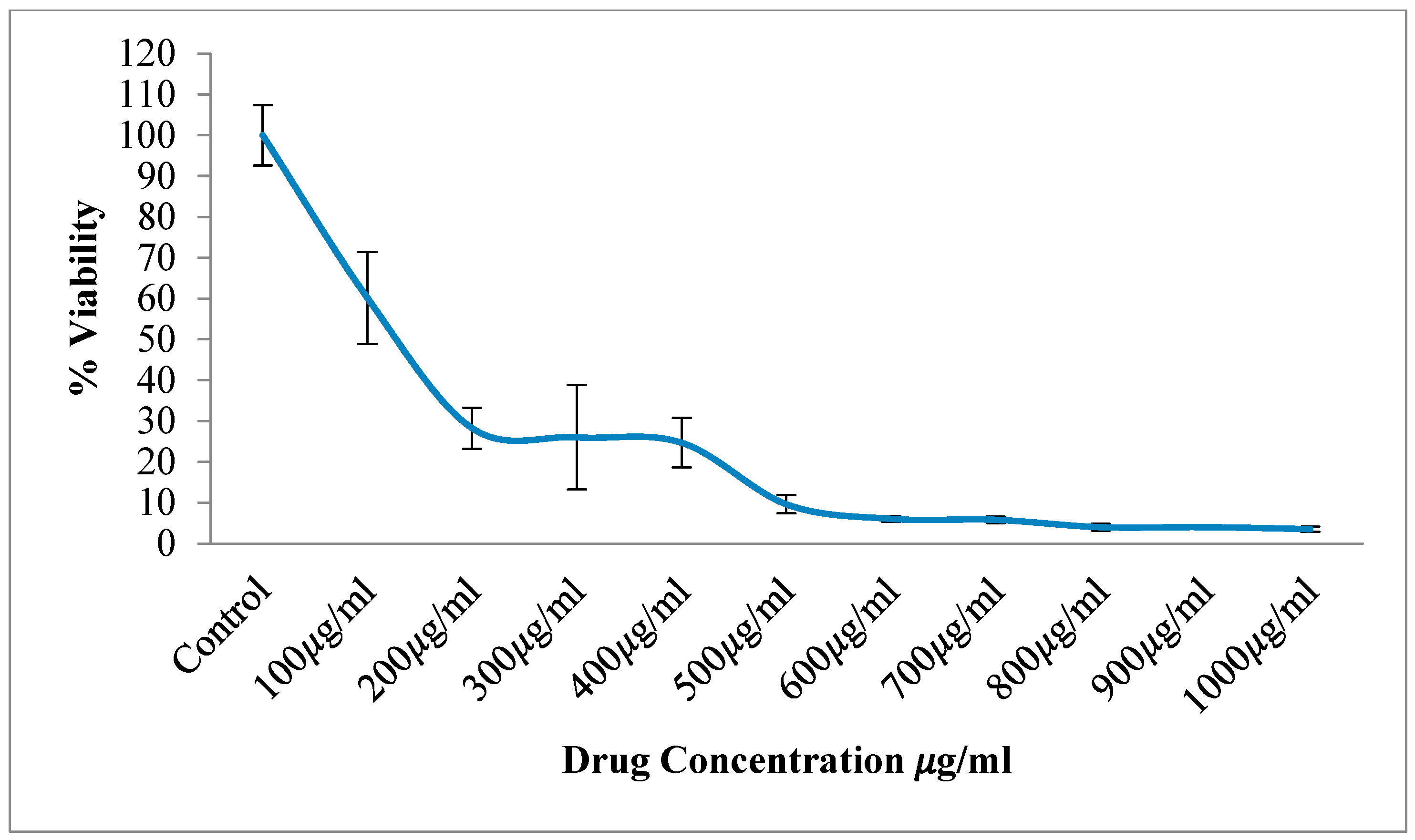
2.4. Cell Culture and Cytotoxicity (MTT Assay)

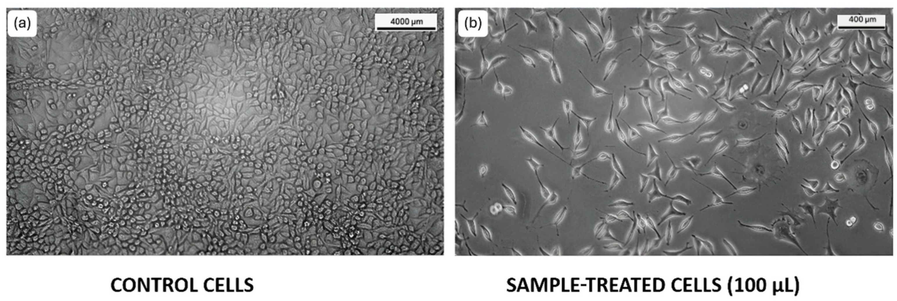
2.4.1. HCT116 Cell Lines

The human colorectal carcinoma cell line (HCT116) was selected for this study due to its well-characterized cell cycle regulation and high CDK activity, making it a suitable model for evaluating the effects of CDK inhibitors [13]. The cells were obtained from NCCS, Pune, India, and were cultured in Dulbecco’s Modified Eagle’s Medium (DMEM, HiMedia Laboratories Pvt. Ltd., Mumbai, India), which was supplemented with 10% Fetal Bovine Serum (Gibco, Thermo Fisher Scientific, Waltham, MA, USA), 1% Sodium Bicarbonate, and 1% Sodium pyruvate (HiMedia Laboratories Pvt. Ltd., Mumbai, India). The cells were maintained in a humidified 5% CO2 atmosphere at 37 °C for further experiments. MTT (tetrazolium salt; HiMedia Laboratories Pvt. Ltd., Mumbai, India) was used to analyze the effective cytotoxic concentration of the root extract. HCT116 cells were seeded (1 × 10⁴ cells per well) in a 96-well plate. The cells were then incubated for 24 h. Cells were treated with the extract (10 concentrations from 100 μg/mL to 1000 μg/mL) and incubated for another 24 h. Then, 100 μL of complete media containing MTT (0.5 mg/mL) was added to each well, the cells were incubated for 5 h, the media was carefully removed, and the formazan crystals were dissolved in 100 μL DMSO for 30 min. The absorbance was then obtained on a microplate reader at 570 nm and 650 nm, and the IC50 value was calculated [14].

2.4.2. L929 Cell Lines

L-929 fibroblast cell lines, obtained from NCCS, Pune, India, were cultured in DMEM medium supplemented with 10% FBS, penicillin, and streptomycin, and maintained at 37 °C with 5% CO2. Cells were seeded in 96-well plates and treated with varying concentrations (5–100 μg/mL) of the extract. The MTT assay was performed to evaluate cytotoxicity following incubation with MTT reagent, solubilization with acidified isopropanol, and absorbance measurement at 650 nm.

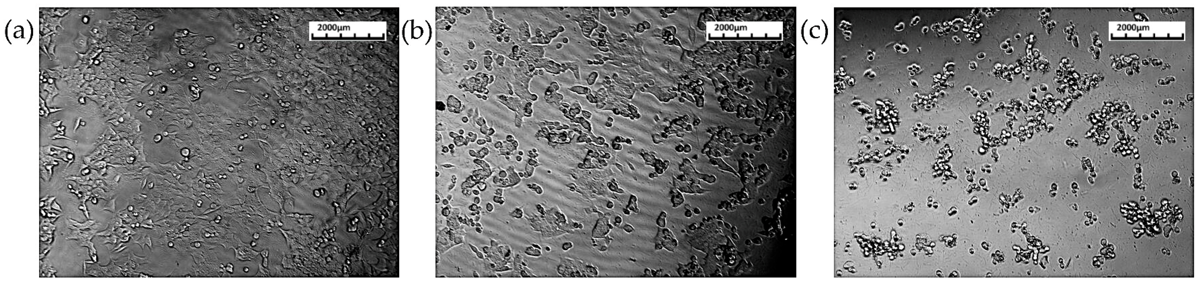
2.5. Cell Migration Assay

2.5.1. HCT116 Cell Lines

To assess the anti-migratory effect of the extract, a wound healing assay was performed. HCT116 cells were seeded (2 × 10⁵ cells per well) in a 24-well plate and then incubated for 24 h; a scratch was made in the middle by a 200 μL pipette tip after 24 h of incubation of cells to reach 90% confluency. The cells were washed with PBS to remove any floating cells after wound creation. Only 50% of the IC50 concentration was administered to reduce cytotoxic impact, considering that the full IC50 concentration is cytotoxic to 50% of cells. The cells were then incubated for another 24 h, while the untreated cells served as a control. The potential of the cells to move from the edge to the wounded area is observed for three time periods (0 h, 24 h, and 36 h).

2.5.2. L929 Cell Lines

L-929 cells were seeded in 6-well plates to confluence, and a scratch was made using a sterile pipette tip. The cell surface was then washed with serum-free culture medium three times to remove dislodged cells. Wound closure was monitored by collecting digitized images at 0 and 24 h after the scratch was made.

2.6. Cell Cycle Analysis

The cell cycle arrest was evaluated in the HCT116 cell line treated with IC50 concentrations of the extract. 1.2 × 10⁵ HCT116 cells were seeded in each well of the 24-well plate and grown with DMEM media for 24 h under standard cell culture conditions till they reached 80–90% confluency. The cells were treated with the extract and incubated for 24 h, while untreated cells served as controls. Then, the trypsinized cells were washed twice with PBS, and the cells were suspended in PI staining solution (50 μg/mL propidium iodide, 200 μg/mL DNase-free RNase, 4 mM sodium citrate, 0.1% Triton X-100) and incubated in the dark. The fluorescently labeled cells were then analyzed using flow cytometry (DxFLEX flow cytometer, Beckman Coulter, Brea, CA, USA).

2.7. Selection and Preparation of Ligand Molecule

The bioactive compounds identified through GC-MS and FDA-approved CDK inhibitors (Palbociclib and Ribociclib) were retrieved from the PubChem database (http://pubchem.ncbi.nlm.nih.gov/) (accessed on 28 March 2025) and saved in a structure data file format (*.sdf file). These were then converted into protein data bank file format (*.pdb file) using OpenBabel 2.4.1 software and prepared for docking [15].

2.8. Target Retrieval

The crystal structures of CDK4 (PDB Code: 2W9F) and CDK6 (PDB Code: 1JOW) were obtained from the RCSB Protein Data Bank (https://www.rcsb.org) (accessed on 28 March 2025).

2.9. Molecular Docking

Molecular docking was performed using Autodock 4.2 and Autodock Vina. The target proteins were prepared by removing water molecules from the target protein. In due course, polar hydrogens, Kollman charges, and Gasteiger charges were added, and AD4-type atoms were assigned. The torsion tree was set up for the ligand. These prepared proteins and ligands were saved in PDBQT format (*.pdbqt file). Blind docking was carried out, considering the entire protein molecule as the binding site. The results were analyzed based on binding energies [16,17].

2.10. ADME Profiling

SwissADME (http://www.swissadme.ch/) (accessed on 11 February 2025) was used to predict ADME properties, evaluating absorption, distribution, metabolism, and excretion potential [18].

2.11. Statistical Analysis

The mean ± standard deviation (SD) was calculated for all experiments conducted in triplicate. IC50 values were estimated from dose–response curves, and all plots were prepared using Microsoft Excel.

3. Results

3.1. GC-MS Analysis

GC-MS analysis identified seven phytochemicals in the T. siliquosa aqueous root extract, including 2-palmitoylglycerol, Hentriacontane, Isocorydine, Squalene, Methyl Palmitate, Thunbergol, and Methyl Stearate. The compounds were analyzed for their retention time (RT), molecular formula, molecular mass, and peak area percentage (Figure 1, Table 1). Notably, 2-palmitoylglycerol exhibited the highest peak area (19.23%), followed by Hentriacontane (15.97%) and Isocorydine (8.73%).

3.2. Cell Viability and Cytotoxicity (MTT Assay)

3.2.1. HCT116 Cell Lines

The cytotoxic effect of the aqueous root extract of T. siliquosa was assessed using the MTT assay on HCT116 cell lines (Figure 2). The results demonstrated a dose-dependent cytotoxicity during the 24 h treatment with increasing concentrations of the extract (100 to 1000 μg/mL). The extract exhibited potent cytotoxic effects, reducing cell viability to below 10% at higher concentrations. The calculated IC50 value was 140 μg/mL, indicating a moderate to strong cytotoxic potential of the crude extract against HCT116 cells (Figure 3, Table 2).

3.2.2. L929 Cell Lines

The cytotoxic effect of the Thottea siliquosa root extract on L-929 fibroblast cells was evaluated using the MTT assay. The results revealed a dose-dependent reduction in cell viability. At a low concentration of 5 μg/mL, cell viability remained high (90.66%), indicating minimal impact on cell metabolic activity. As the extract concentration increased, a gradual decline in viability was observed, with the lowest value of 45.50% recorded at 100 μg/mL (Figure 4). However, the extract did not achieve 50% inhibition (IC50) within the tested concentration range. It is important to note that the observed decrease in OD values may partly reflect reduced metabolic activity due to serum starvation or other stress conditions rather than direct cytotoxic effects. These findings suggest that the extract exhibits low to moderate toxicity towards normal fibroblast cells and appears relatively safe at lower concentrations.

3.3. Cell Migration Assay

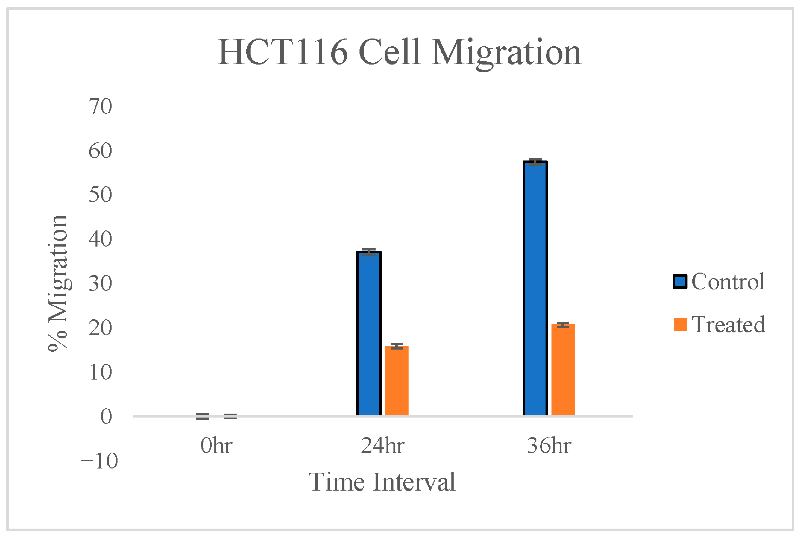
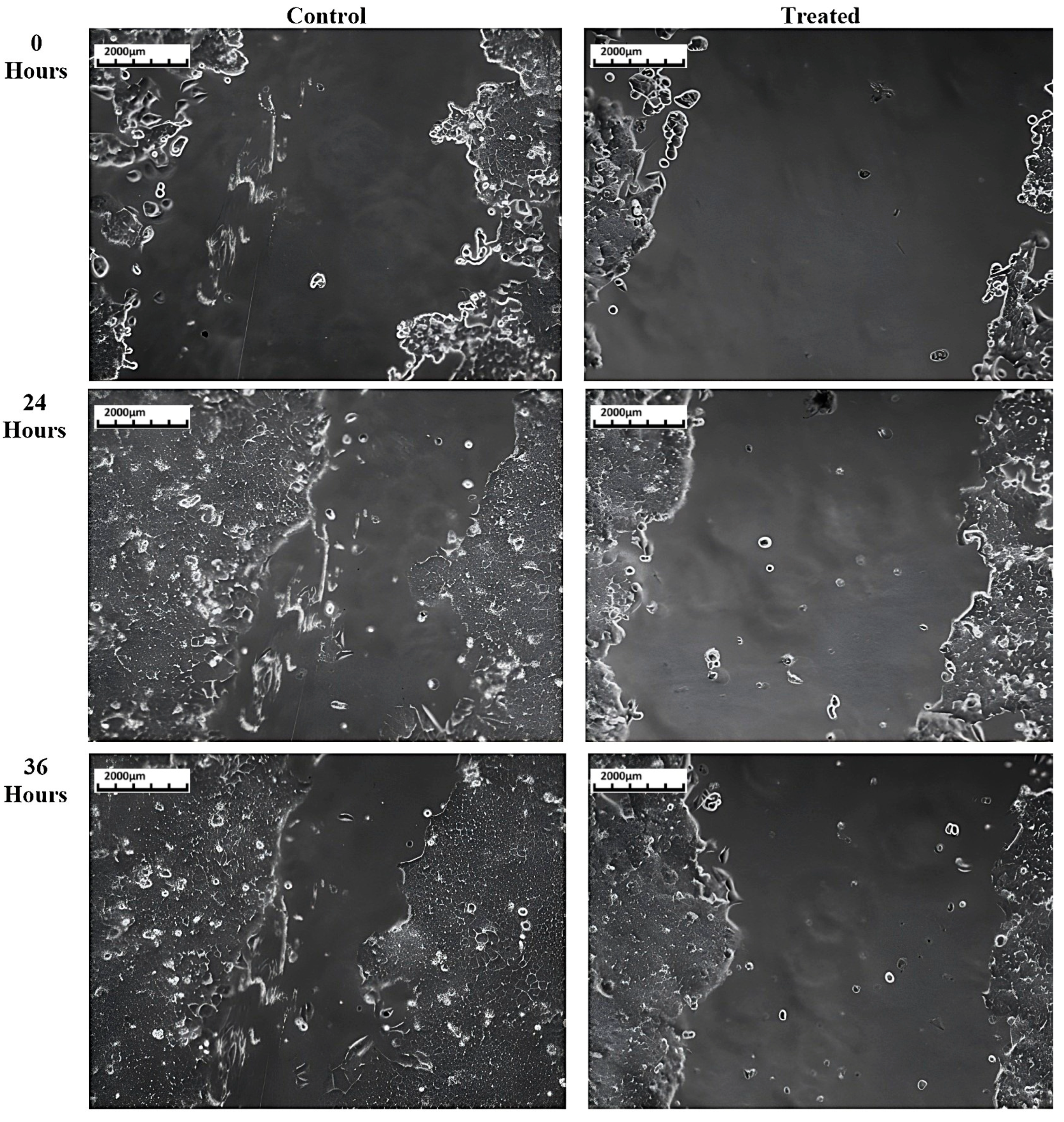
3.3.1. HCT116 Cell Lines

The anti-migratory potential of T. siliquosa root extract was evaluated through a cell migration (scratch) assay on HCT116 cells. The cells were treated only with 50% of the IC50 concentration, which is 70 μg/mL. This minimizes the cytotoxic effect on cells, as the IC50 concentration (140 μg/mL) can be cytotoxic to 50% of the cell population. A reference line was drawn in the middle of 24-well plates to ensure images were taken at the same field for the three time periods where the wound was created. The wound made in the human colorectal carcinoma cells (HCT116) is observed under a phase contrast microscope at three time intervals: 0 h, 24 h, and 36 h (Figure 5). Images were analyzed with ImageJ software version 1.54p to find the percentage of migration between the control and treated cells (Figure 6). The results reveal that the extract is capable of significantly inhibiting the migration of HCT116 cells (Table 3).

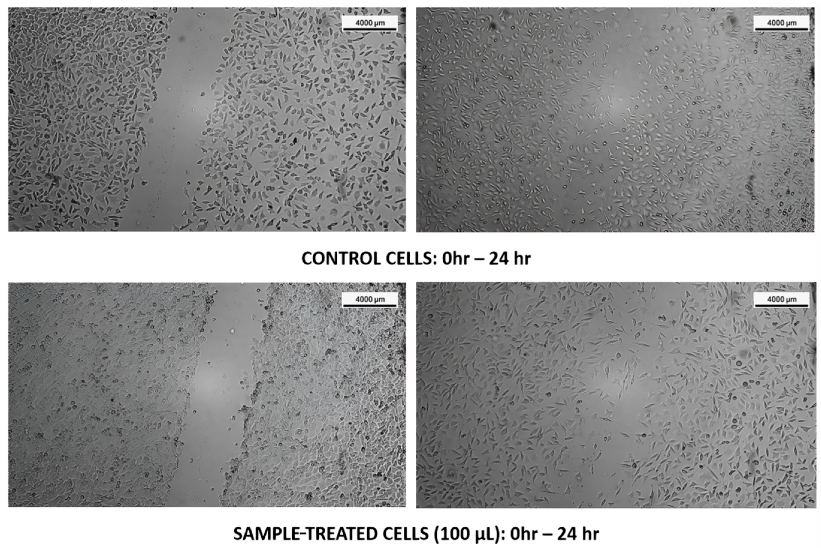
3.3.2. L929 Cell Lines

The cell migration potential of T. siliquosa root extract (100 μg/mL) was evaluated using the wound scratch assay on L-929 fibroblast cells. The assay revealed that after 24 h, untreated control cells exhibited significant migration into the scratch area, consistent with normal proliferative and migratory behavior. Similarly, cells treated with the root extract also demonstrated marked migration into the wound region, indicating that the extract did not inhibit the migratory capacity of these normal fibroblasts. This observation suggests that the extract is non-toxic to normal cells and selectively inhibits malignant cell proliferation, aligning with its proposed role as a natural CDK4/6 inhibitor (Figure 7).

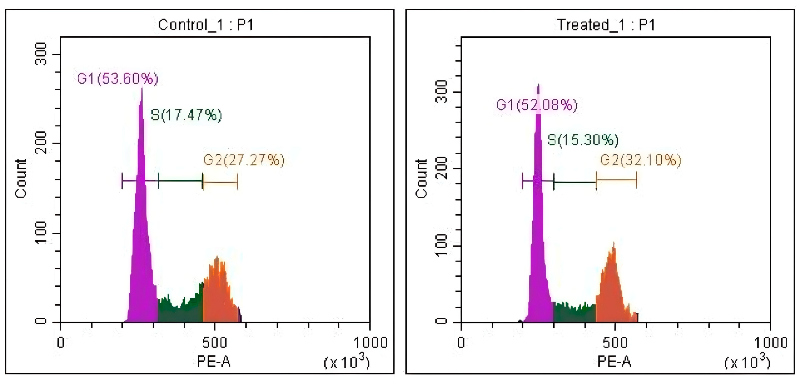
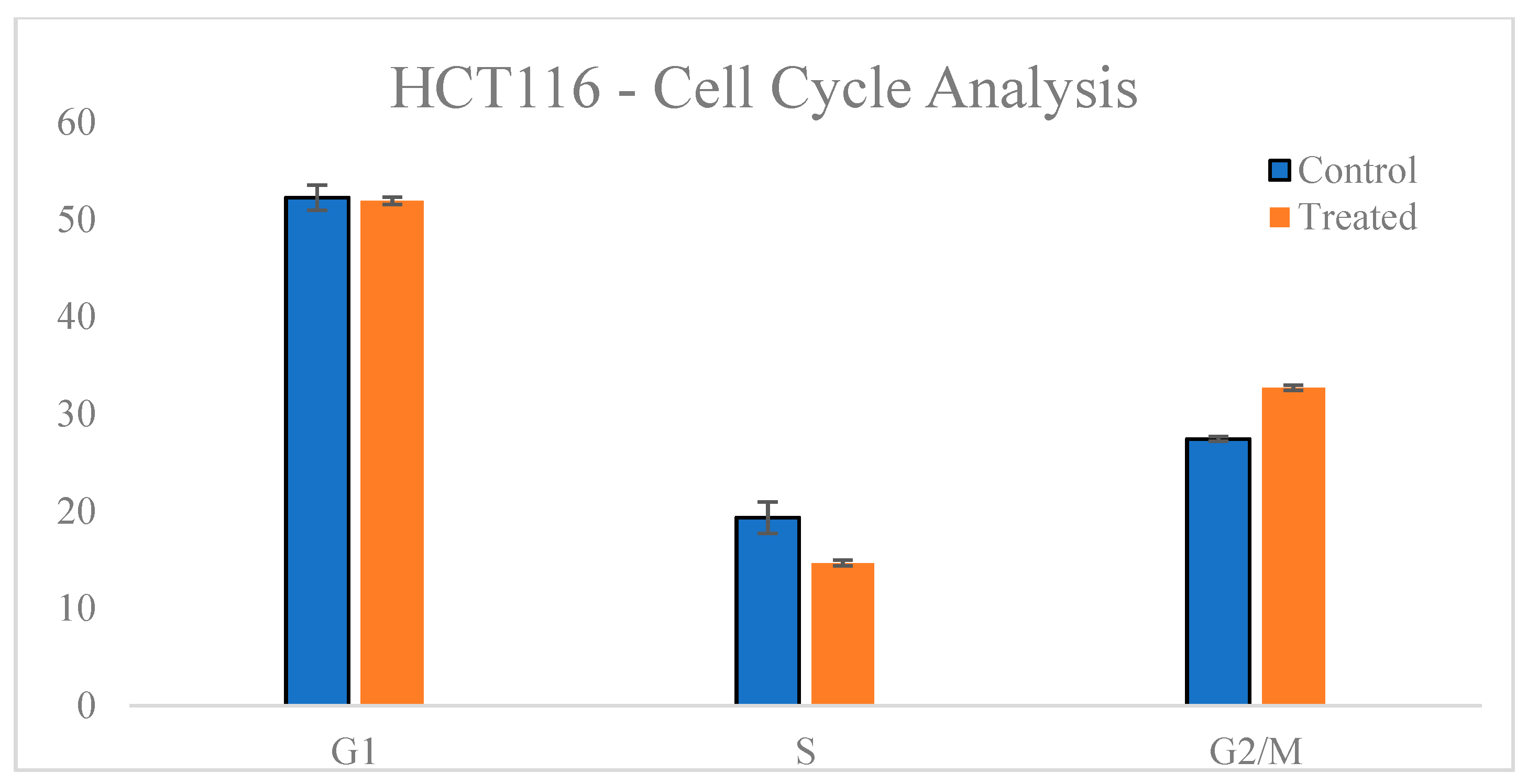
3.4. Cell Cycle Analysis

To further investigate the mechanism underlying the cytotoxic effects of T. siliquosa root extract, cell cycle analysis was performed on HCT116 cells treated with the IC50 concentration (140 μg/mL) for 24 h. Flow cytometry following propidium iodide (PI) staining revealed a notable accumulation of cells in the G2/M phase of the cell cycle compared to the control. In untreated cells, the average distribution was 52.25% (G1 phase), 19.34% (S phase), and 27.44% (G2/M checkpoint). Upon treatment, the G2/M population increased to 32.7%, accompanied by a decrease in the S phase population to 14.67% (Table 4, Figure 8 and Figure 9). This shift indicates that the extract induces cell cycle arrest at the G2/M checkpoint, thereby suppressing cell division. Although CDK4/6 is primarily involved in the G1-S transition, the minimal change in G1 phase suggests partial inhibition, but not complete suppression, of CDK4/6 activity. The prominent G2/M arrest indicates that the extract likely affects additional regulatory kinases such as CDK1 or Cyclin B1, supporting a potential multi-targeted mechanism of anticancer action.

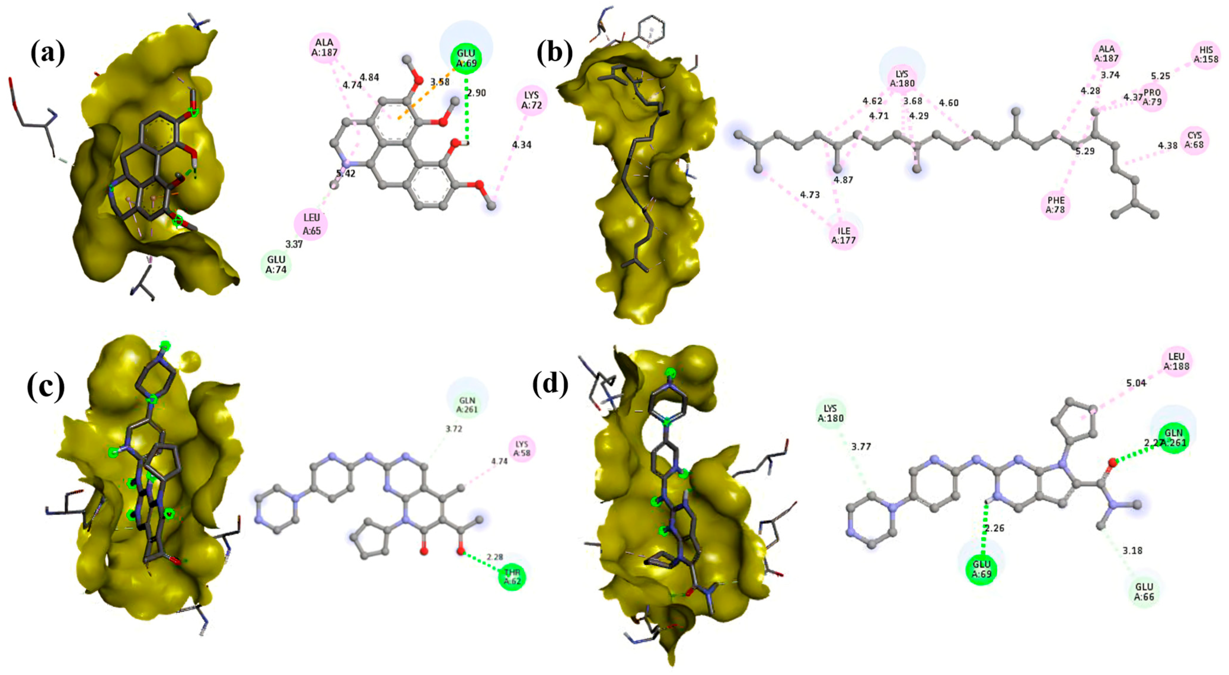
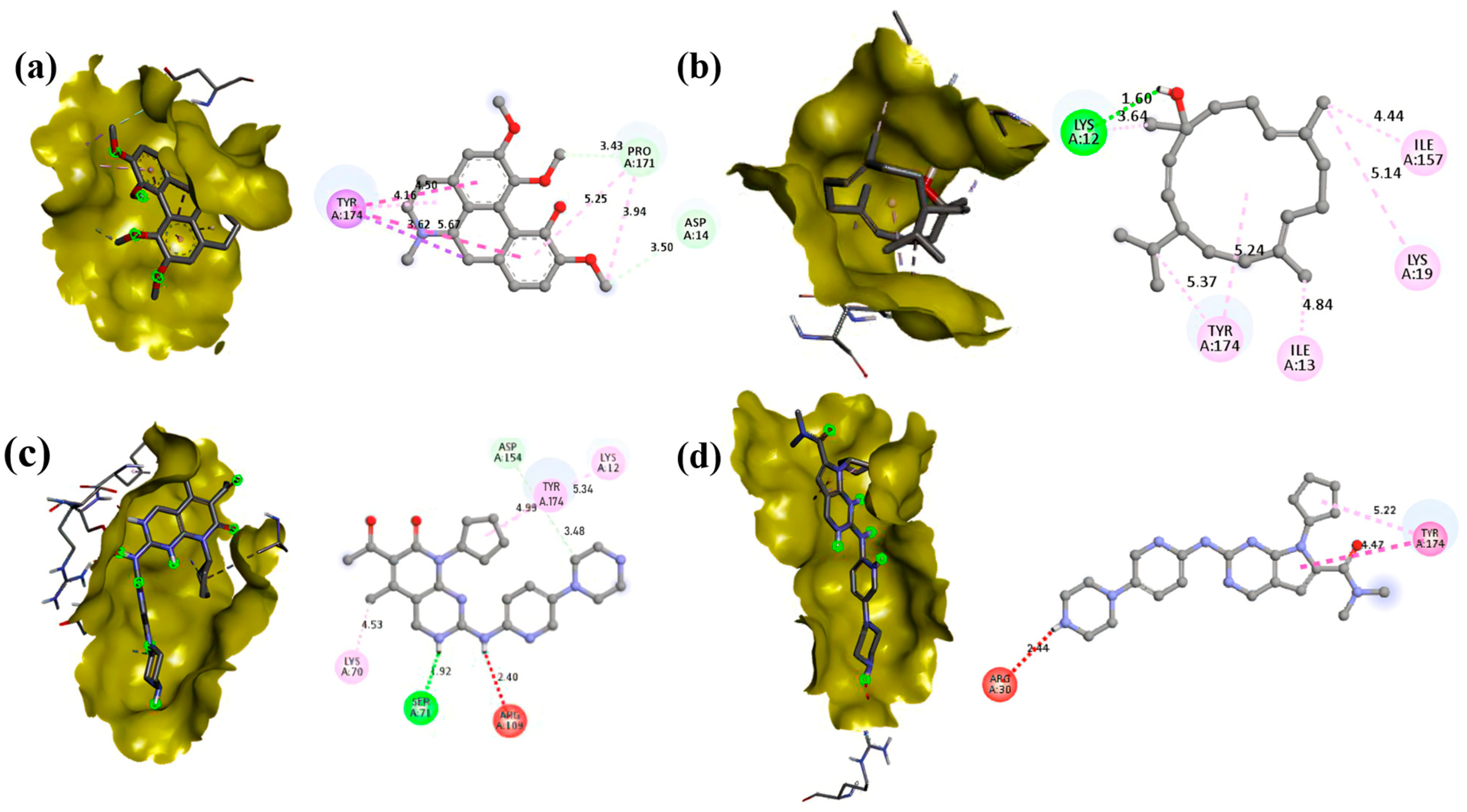
3.5. Molecular Docking

Molecular docking studies revealed significant interactions between phytochemicals from T. siliquosa and the target proteins CDK4 (2W9F) and CDK6 (1JOW). Among the tested compounds, Isocorydine exhibited the highest docking scores, recording binding energies of −7.4 kcal/mol with CDK4 and −7.2 kcal/mol with CDK6. Thunbergol showed strong interaction with CDK6 (−7 kcal/mol) and moderate interaction with CDK4 (−6.5 kcal/mol). Squalene demonstrated notable docking scores as well, with binding energies of −7.1 kcal/mol against CDK4 and −4.9 kcal/mol against CDK6. Other phytochemicals, such as Methyl Palmitate, Methyl Stearate, 2-palmitoylglycerol, and Hentriacontane, displayed weaker interaction (Table 5).
In comparison, the FDA-approved CDK4/6 inhibitors Palbociclib exhibited −7.2 kcal/mol (CDK4) and −8.3 kcal/mol (CDK6) binding energies, and Ribociclib displayed −7.1 kcal/mol (CDK4) and −8.1 kcal/mol (CDK6) binding energies, respectively. Additionally, the specific amino acid residues interacting with the phytochemicals and the control drugs are detailed in Table 6 and Table 7. Notably, the docking scores of key phytochemicals, such as Isocorydine, Thunbergol, and Squalene, were comparable to those of the control drugs. This suggests that these phytochemicals might act synergistically with existing CDK4/6 inhibitors, offering enhanced therapeutic potential. The strong and stable interactions of Isocorydine, Thunbergol, and Squalene with the active sites of CDK4/6 highlight their promise as natural drug candidates for further development and exploration (Figure 10 and Figure 11).

3.6. ADME Profiling

In silico ADME (Absorption, Distribution, Metabolism, and Excretion) profiling, conducted using the SwissADME tool, evaluated the pharmacokinetic properties of the phytochemicals from T. siliquosa in comparison to the control drugs Palbociclib and Ribociclib. The analysis highlighted that the phytochemicals, such as Isocorydine, Methyl Palmitate, and Thunbergol, exhibit pharmacokinetic properties comparable to the control drugs, underscoring their potential as drug-like candidates. The phytochemicals complied with Lipinski’s Rule of Five (Ro5), demonstrating parameters like molecular weight, hydrogen bond donors/acceptors, lipophilicity, and topological polar surface area (TPSA) within favorable ranges, similar to that of the control drugs. Notably, Isocorydine and Methyl Palmitate exhibited high gastrointestinal (GI) absorption, mirroring the oral bioavailability characteristics of Palbociclib and Ribociclib. Furthermore, minimal blood–brain barrier (BBB) permeation was observed for Squalene and Hentriacontane, suggesting a reduced likelihood of central nervous system side effects, similar to the safety profile of the control drugs.
The absence of significant PAINS (Pan-assay Interference Compounds) alerts among the tested phytochemicals indicates a lower risk of non-specific interactions in bioassays, further reinforcing their drug-like potential (Table 8). Collectively, these attributes align the phytochemicals closely with established pharmacokinetic profiles of Palbociclib and Ribociclib, suggesting their potential not only as standalone therapeutic agents but also as complementary molecules that might enhance or synergize with existing CDK4/6 inhibitors.

4. Discussion

The present study provides scientific evidence supporting the potential of T. siliquosa root extract as a natural source of bioactive compounds with inhibitory effects on cyclin-dependent kinases CDK4 and CDK6, key regulators of cell cycle progression commonly implicated in various cancers [19]. GC-MS analysis of the aqueous root extract revealed seven major phytochemical compounds, each known for diverse pharmacological properties, including anticancer, antioxidant, anti-inflammatory, and antitumor effects (Table 9).
This phytochemical diversity highlights the therapeutic potential of T. siliquosa as a natural resource for drug discovery. Importantly, this study supports the concept of synergistic action, where the combined effects of multiple phytochemicals within the crude extract may enhance biological activity and mitigate potential side effects compared to isolated compounds [34,35].
Following the identification of bioactive compounds through GC-MS analysis, the crude extract was evaluated for biological activity. The cytotoxicity assay demonstrated a dose-dependent cytotoxic effect of the crude extract on HCT116 cancer cells, with an IC50 of 140 μg/mL. This dose-dependent profile indicated a potential therapeutic window where selective cytotoxicity can be harnessed against target cells while sparing normal cells [36]. The cell migration assay further indicated that the extract significantly inhibited cancer cell migration, as demonstrated by reduced wound closure in treated cells compared to the control [37]. This anti-migratory effect suggests potential anti-proliferative and anti-metastatic properties, aligning with the proposed mechanism of CDK4/6 inhibition, which is known to arrest the cell cycle and suppress cancer cell progression. Supporting this, the extract did not exhibit cytotoxic or anti-migratory effects on L929 murine fibroblast cells, which continued to proliferate and migrate normally in the scratch assay. These results suggest that the extract is non-toxic to normal cells, reinforcing its selective action against cancer cells.
Furthermore, flow cytometric analysis revealed that treatment with the extract demonstrated a marked accumulation of cells at the G2/M checkpoint alongside minimal change in G1 phase (53.60% to 52.08%). These findings suggest that while CDK4/6 inhibition was proposed based on molecular docking, the extract’s in vitro effect may not be limited to CDK4/6 alone. The observed G2/M arrest implies potential interference with other key regulators of cell cycle progression, such as CDK1, Cyclin B1, or PLK1, which are essential for G2/M transition. Given the complex nature of crude plant extracts, it is plausible that multiple cell cycle regulators are concurrently affected. Therefore, while partial inhibition of CDK4/6 cannot be excluded, the overall effect likely involves a multi-targeted mechanism. Further studies employing protein-level validation, like Western blotting, RT-qPCR, or kinase activity assays, are warranted to confirm the specific contribution of CDK4/6 inhibition to the extract’s anticancer activity [38,39].
Subsequent molecular docking studies revealed significant interactions between phytochemicals identified from the crude extract and the target proteins CDK-4 and CDK-6. Isocorydine exhibited the highest binding affinity, followed closely by Thunbergol and Squalene. These targets are crucial regulators of the cell cycle and have been implicated in various cancers, including breast cancer, glioblastoma, and melanoma [40,41,42]. Notably, their binding energies were comparable to the FDA-approved CDK4/6 inhibitors Palbociclib and Ribociclib, reinforcing the potential of T. siliquosa phytochemicals as natural CDK inhibitors.
Additionally, compounds with the most negative binding energies and lowest inhibition constants were considered the most effective binders, validating the potential of these phytochemicals as promising therapeutic agents [43,44]. The amino acid residues involved in the docking interactions, detailed in Table 4 and Table 5, provide further evidence of the stability and effectiveness of these interactions. Given the central role of CDK4/6 in promoting uncontrolled cell proliferation in cancer, these results are significant and suggest that the extract could serve as a multi-targeted therapeutic agent. The weaker interactions observed for Methyl Palmitate, Methyl Stearate, 2-palmitoylglycerol, and Hentriacontane may still contribute to the overall pharmacological profile when acting in synergy with the more potent compounds.
Further in silico ADME profiling strengthened the potential of the extract as a drug-like candidate. Most phytochemicals satisfied Lipinski’s Rule of Five, exhibited good gastrointestinal absorption, and showed low blood–brain barrier penetration, reducing the likelihood of central nervous system toxicity. Minimal PAINS alerts also suggest a low risk of non-specific interactions, which is favorable for drug development [45].
While Palbociclib and Ribociclib have demonstrated clinical success as selective CDK4/6 inhibitors, their usage is often associated with notable side effects. Each drug presents a distinct profile of frequently reported adverse effects, including anemia; neutropenia; fatigue; gastrointestinal disturbances like nausea, diarrhea, etc.; and increased risk of infections. In particular, Palbociclib is known for causing stomatitis, fatigue, epistaxis, alopecia, and hot flushes, with neutropenia, leukopenia, infections, and gastrointestinal disturbances. Ribociclib has been associated with anemia, polyneuropathy, thrombocytopenia, QT interval prolongation, and spinal pain, along with hematological abnormalities and elevated liver enzymes [46,47,48,49,50]. These limitations highlight the need for alternative therapeutic agents with reduced toxicity profiles.
Overall, the combined results from cytotoxicity, cell migration assays, cell cycle arrest, molecular docking, and ADME profiling emphasize the potential of T. siliquosa root extract as a natural, multi-compound therapeutic candidate. Further in vitro and in vivo studies are warranted to validate these findings, explore the mechanistic pathways involved, and assess the extract’s safety and efficacy in CDK4/6-associated cancers.

5. Conclusions

This study highlights the therapeutic potential of T. siliquosa root extract as a natural and multi-compound agent capable of targeting cyclin-dependent kinases CDK4 and CDK6, which are key drivers of cell cycle progression in various cancers. Through a combination of GC-MS analysis, biological assays, molecular docking, and ADME profiling, this study has demonstrated that the extract exhibits diverse pharmacological properties, including cytotoxicity, anti-migratory effects, cell cycle arrest in cancer cells, and favorable drug-likeness. Molecular docking revealed strong interactions between the phytochemicals and cancer-associated proteins, with binding affinities comparable to FDA-approved inhibitors, supporting its viability as a natural CDK inhibitor. Additionally, ADME profiling confirmed the extract’s drug-like attributes, such as favorable gastrointestinal absorption and minimal risks of central nervous system toxicity, which are essential for safe therapeutic application. Given the limitations and side effects of existing CDK4/6 inhibitors, T. siliquosa root extract offers a promising alternative with potential advantages in toxicity and multi-targeted action. While these findings provide a solid foundation, further in vitro and in vivo studies are crucial to validate the efficacy, elucidate the mechanistic pathways involved, and ensure its safety in clinical settings. These findings pave the way for further research involving protein-level validation, which is essential to delineate the precise molecular targets and mechanisms to confirm the extract’s effectiveness and safety in treating CDK4/6-related cancers.

Supplementary Materials

The following supporting information can be downloaded at: https://www.mdpi.com/article/10.3390/biomedicines13071658/s1.

Author Contributions

Conceptualization, L.V. and B.C.; Data curation, L.V. and B.C.; Funding acquisition, J.L. and A.A.G.; Investigation, M.R.E. and M.K.; Methodology, M.R.E. and M.K.; Project administration, J.L. and A.A.G.; Resources, S.T., M.A. and A.A.G.; Supervision, L.V. and B.C.; Validation, M.R.E., M.K., S.T., L.V., and B.C.; Writing—original draft, M.R.E., M.K., and B.C.; Writing—review and editing, L.V. and J.L. All authors have read and agreed to the published version of the manuscript.

Funding

This research was supported by the Basic Science Research Program through the National Research Foundation of Korea (NRF), funded by the Ministry of Education (Grant Nos. NRF-2021R1A6A1A03039493 and NRF-2022R1A2C1009389). The authors are also grateful to the Researchers Supporting Project number (RSP2025R407), King Saud University, Riyadh, Saudi Arabia, for the financial assistance. The authors acknowledge the Department of Science and Technology—FIST (DST-FIST), New Delhi, for providing essential infrastructure support.

Institutional Review Board Statement

Not applicable.

Informed Consent Statement

Not applicable.

Data Availability Statement

The original contributions presented in this study are included in the article/Supplementary Materials. Further inquiries can be directed to the corresponding authors.

Conflicts of Interest

The authors declare no conflicts of interest.

Abbreviations

The following abbreviations are used in this manuscript:
CDKCyclin-Dependent Kinase
ADMEAbsorption, Distribution, Metabolism, and Excretion
GC-MSGas Chromatography-Mass Spectrometry
HCT116Human Colonic Tumor—116 Cancer Cell Line
MTT3-(4,5-dimethylthiazol-2yl)-2,5-diphenyltetrazolium bromide
IC50Half-Maximal Inhibitory Concentration
DMEMDulbecco’s Modified Eagle’s Medium
DMSODimethyl Sulfoxide
PBSPhosphate-Buffered Saline
PIPropidium Iodide
RCSBResearch Collaboratory for Structural Bioinformatics
AD4AutoDock4
FDAFood and Drug Administration
G1 phaseGap1
S phaseSynthesis Phase
G2 phaseGap2
M phaseMitosis

References

  1. Pellarin, I.; Dall’Acqua, A.; Favero, A.; Segatto, I.; Rossi, V.; Crestan, N.; Karimbayli, J.; Belletti, B.; Baldassarre, G. Cyclin-Dependent Protein Kinases and Cell Cycle Regulation in Biology and Disease. Signal Transduct. Target. Ther. 2025, 10, 11. [Google Scholar] [CrossRef] [PubMed]
  2. Longworth, S.; Damania, B. Modulation of Cell Cycle Kinases by Kaposi’s Sarcoma-Associated Herpesvirus. J. Med. Virol. 2025, 97, e70157. [Google Scholar] [CrossRef] [PubMed]
  3. Oner, M.; Cheng, Y.-C.; Soong, S.-W.; Cheng, P.-T.; Wang, Y.-H.; Yang, S.-F.; Tsai, S.C.-S.; Lin, H. Dinaciclib Interrupts Cell Cycle and Induces Apoptosis in Oral Squamous Cell Carcinoma: Mechanistic Insights and Therapeutic Potential. Int. J. Mol. Sci. 2025, 26, 2197. [Google Scholar] [CrossRef]
  4. Priyadharshini, G.; Sukumaran, G.; Dilipan, E.; Ramani, P. Targeting Oral Cancer: In Silico Docking Studies of Phytochemicals on Oncogenic Molecular Markers. Asian Pac. J. Cancer Prev. 2024, 25, 2069–2075. [Google Scholar] [CrossRef]
  5. Zhang, W.; Hong, W. Upregulation of miR-519d-3p Inhibits Viability, Proliferation, and G1/S Cell Cycle Transition of Oral Squamous Cell Carcinoma Cells Through Targeting CCND1. Cancer Biother. Radiopharm. 2024, 39, 153–163. [Google Scholar] [CrossRef]
  6. Dehelean, C.A.; Marcovici, I.; Soica, C.; Mioc, M.; Coricovac, D.; Iurciuc, S.; Cretu, O.M.; Pinzaru, I. Plant-Derived Anticancer Compounds as New Perspectives in Drug Discovery and Alternative Therapy. Molecules 2021, 26, 1109. [Google Scholar] [CrossRef] [PubMed]
  7. Jayaraman, L.; Shivaji, S.; Anandakumar, S. Phytochemical screening, cytotoxic activity and molecular docking studies of Eclipta alba leaves extract against oral cancer. Rasayan J. Chem. 2022, 15, 676–685. [Google Scholar] [CrossRef]
  8. Vasan, N.; Baselga, J.; Hyman, D.M. A View on Drug Resistance in Cancer. Nature 2019, 575, 299–309. [Google Scholar] [CrossRef]
  9. Nusaiba, S.A.W.; Murugan, K. In Vitro Analysis on Bactericidal Screening and Antioxidant Potentiality of Leaf and Root Extracts of Thottea siliquosa (Lam.) Ding Hou. An Ethnobotanical Plant. Asian Pac. J. Trop. Biomed. 2013, 3, 859–865. [Google Scholar] [CrossRef]
  10. Vrundha, C.P.K.; Thomas, T.D. Control of Media Browning during Micropropagation and Assessment of Biochemical and Clonal Fidelity of in Vitro-Derived and Mother Plants in Thottea siliquosa (Lamk.) Ding Hou., an Important Ethnomedicinal Shrub. J. Genet. Eng. Biotechnol. 2023, 21, 70. [Google Scholar] [CrossRef]
  11. de Angelo, R.M.; de Sousa, D.S.; da Silva, A.P.; Chiari, L.P.A.; da Silva, A.B.F.; Honorio, K.M. Molecular Docking: State-of-the-Art Scoring Functions and Search Algorithms. In Computer-Aided and Machine Learning-Driven Drug Design: From Theory to Applications; Maltarollo, V.G., Ed.; Springer Nature: Cham, Switzerland, 2024; pp. 163–198. ISBN 978-3-031-76718-0. [Google Scholar]
  12. Abubakar, A.R.; Haque, M. Preparation of Medicinal Plants: Basic Extraction and Fractionation Procedures for Experimental Purposes. J. Pharm. Bioallied Sci. 2020, 12, 1–10. [Google Scholar] [CrossRef] [PubMed]
  13. Schneider, J.S.; Khaled, N.B.; Ye, L.; Wimmer, R.; Hammann, L.; Weich, A.; Suppan, C.; Mahajan, U.M.; Jung, A.; Kumbrink, J.; et al. Efficacy of CDK4/6 Inhibition in Colorectal Cancer and the Role of P16 Expression in Predicting Drug Resistance. Cell Oncol. 2025, 1–13. [Google Scholar] [CrossRef]
  14. Ravi, S.; Gaddamedi, S.; Gopal Agrawal, H.; Soni, A.; Kumar Mishra, A.; Narayan Rath, S. Design, Synthesis, Biological Evaluation, and Molecular Docking Studies of N3-Functionalized TZD-Indole Conjugates as Potential Anticancer Agents. ChemistrySelect 2024, 9, e202400123. [Google Scholar] [CrossRef]
  15. O’Boyle, N.M.; Banck, M.; James, C.A.; Morley, C.; Vandermeersch, T.; Hutchison, G.R. Open Babel: An Open Chemical Toolbox. J. Cheminform. 2011, 3, 33. [Google Scholar] [CrossRef]
  16. Morris, G.M.; Huey, R.; Lindstrom, W.; Sanner, M.F.; Belew, R.K.; Goodsell, D.S.; Olson, A.J. AutoDock4 and AutoDockTools4: Automated Docking with Selective Receptor Flexibility. J. Comput. Chem. 2009, 30, 2785–2791. [Google Scholar] [CrossRef] [PubMed]
  17. Trott, O.; Olson, A.J. AutoDock Vina: Improving the Speed and Accuracy of Docking with a New Scoring Function, Efficient Optimization and Multithreading. J. Comput. Chem. 2010, 31, 455–461. [Google Scholar] [CrossRef] [PubMed]
  18. Chikowe, I.; Bwaila, K.D.; Ugbaja, S.C.; Abouzied, A.S. GC–MS Analysis, Molecular Docking, and Pharmacokinetic Studies of Multidentia Crassa Extracts’ Compounds for Analgesic and Anti-Inflammatory Activities in Dentistry. Sci. Rep. 2024, 14, 1876. [Google Scholar] [CrossRef]
  19. Goel, S.; Bergholz, J.S.; Zhao, J.J. Targeting CDK4 and CDK6 in Cancer. Nat. Rev. Cancer. 2022, 22, 356–372. [Google Scholar] [CrossRef]
  20. Wu, F.; Tian, M.; Sun, Y.; Wu, C.; Liu, X. Efficacy, Chemical Composition, and Pharmacological Effects of Herbal Drugs Derived from Fritillaria Cirrhosa D. Don and Fritillaria Thunbergii Miq. Front. Pharmacol. 2022, 13, 985935. [Google Scholar] [CrossRef]
  21. Kocanci, F.; Aslim, B. Chemical Composition and Neurotherapeutic Potential of Glaucium Corniculatum Extracts. Pharmacogn. Mag. 2021, 17, 67. [Google Scholar] [CrossRef]
  22. Madriwala, B.; Suma, B.V.; Jays, J. Molecular Docking Study Of Hentriacontane For Anticancer And Antitubercular Activity. Int. J. Chem. Res. 2022, 6, 1–4. [Google Scholar] [CrossRef]
  23. Dong, J.-W.; Li, X.-J.; Xu, X.-X.; Gu, S.-F.; Zhao, H.; Wang, X.-X. Solid-State Fermentation of Aspergillus Sydowii G12, an Approach to Produce Isocorydine. Process Biochem. 2024, 147, 147–151. [Google Scholar] [CrossRef]
  24. Luo, J.; Wang, N.; Hua, L.; Deng, F.; Liu, D.; Zhou, J.; Yuan, Y.; Ouyang, F.; Chen, X.; Long, S.; et al. The Anti-Sepsis Effect of Isocorydine Screened from Guizhou Ethnic Medicine Is Closely Related to Upregulation of Vitamin D Receptor Expression and Inhibition of NFκB P65 Translocation into the Nucleus. J. Inflamm. Res. 2022, 15, 5649–5664. [Google Scholar] [CrossRef]
  25. Song, L.; Zhao, F.; Liu, Y.; Guo, X.; Wu, C.; Liu, J. Effects of 8-Amino-Isocorydine, a Derivative of Isocorydine, on Gastric Carcinoma Cell Proliferation and Apoptosis. Curr. Ther. Res. 2021, 94, 100624. [Google Scholar] [CrossRef] [PubMed]
  26. Huang, Z.-R.; Lin, Y.-K.; Fang, J.-Y. Biological and Pharmacological Activities of Squalene and Related Compounds: Potential Uses in Cosmetic Dermatology. Molecules 2009, 14, 540–554. [Google Scholar] [CrossRef]
  27. Micera, M.; Botto, A.; Geddo, F.; Antoniotti, S.; Bertea, C.M.; Levi, R.; Gallo, M.P.; Querio, G. Squalene: More than a Step toward Sterols. Antioxidants 2020, 9, 688. [Google Scholar] [CrossRef] [PubMed]
  28. Muzalevskaya, E.N.; Miroshnichenko, L.A.; Nikolaevskii, V.A.; Ushakov, I.B.; Chernov, Y.N.; Alabovskii, V.V.; Batishcheva, G.A.; Buzlama, A.V. Squalene: Physiological and pharmacological properties. Eksperimental Naia I Klin. Farmakol. 2015, 78, 30–36. [Google Scholar]
  29. Yuefan, L.I.; Yuanyuan, W.; Gaiqin, M.A.; Qi, L.I.; Xiuzhu, Y.U. Research progress on sources, extraction and functional properties of squalene. J. Henan Univ. Technol. 2022, 43, 19–29. [Google Scholar] [CrossRef]
  30. Jokar, E.; Sateei, A.; Ebadi, M.; Ahmadi Golsefidi, M. Changes in the medicinal and antimicrobial compounds, methyl palmitate, methyl stearate, and bis (2-ethylhexyl) phthalate in American agave (Agave Americana L. cv Marginata) under urea treatment. Iran. J. Plant Physiol. 2023, 4, 4637. [Google Scholar] [CrossRef]
  31. Olivia, N.U.; Goodness, U.C.; Obinna, O.M. Phytochemical Profiling and GC-MS Analysis of Aqueous Methanol Fraction of Hibiscus Asper Leaves. Future J. Pharm. Sci. 2021, 7, 59. [Google Scholar] [CrossRef]
  32. Chellappan, B.M.; Venkatachalam, G.; Reddy, C.U.; Kumar, M.; Venketeswarawarlu, B.S. GC-MS Profiling and in Vitro Assessment of Antioxidant and Neuroprotective Properties of Ethanolic and Acetone Extracts of Oldenlandia corymbosa L. Asian J. Life Sci. 2022, 11, 188–192. [Google Scholar] [CrossRef]
  33. Xu, C.; Zhao, S.; Li, M.; Dai, Y.; Tan, L.; Liu, Y. Chemical Composition, Antimicrobial and Antioxidant Activities of Essential Oil from Flue-Cured Tobacco Flower Bud. Biotechnol. Biotechnol. Equip. 2016, 30, 1026–1030. [Google Scholar] [CrossRef]
  34. Boța, M.; Vlaia, L.; Jîjie, A.-R.; Marcovici, I.; Crişan, F.; Oancea, C.; Dehelean, C.A.; Mateescu, T.; Moacă, E.-A. Exploring Synergistic Interactions between Natural Compounds and Conventional Chemotherapeutic Drugs in Preclinical Models of Lung Cancer. Pharmaceuticals 2024, 17, 598. [Google Scholar] [CrossRef]
  35. Chaachouay, N. Synergy, Additive Effects, and Antagonism of Drugs with Plant Bioactive Compounds. Drugs Drug Candidates 2025, 4, 4. [Google Scholar] [CrossRef]
  36. Childs, R.W.; Carlsten, M. Therapeutic Approaches to Enhance Natural Killer Cell Cytotoxicity against Cancer: The Force Awakens. Nat. Rev. Drug. Discov. 2015, 14, 487–498. [Google Scholar] [CrossRef] [PubMed]
  37. Ghadimi, P.; Ghorbian, S. Effects of Melatonin on the Expression of Invasion-Related Markers (MMP2 and MMP9) in Breast Cancer Cells. J. Cell. Biochem. 2025, 126, e70010. [Google Scholar] [CrossRef]
  38. Nabi, N.; Nisa, M.U.; Khan, M.S.; Gillani, S.Q. Role of Signaling Pathways Regulating Cell Cycle Progression in Cancer. In Cell Signaling Pathways and Their Therapeutic Implication in Cancers; Rehman, M.U., Khan, M.S., Eds.; Springer Nature: Singapore, 2025; pp. 357–393. ISBN 978-981-9627-63-9. [Google Scholar]
  39. Yang, L.; Zhu, Y.; Ding, X.; Sun, J.; Cao, X.; Zhang, Z.; Li, L.; Lin, J. M7: A Novel in Vitro CDK1 Inhibitor Suppressing Colorectal Cancer Cell Proliferation, Migration and Promoting Apoptosis. Eur. J. Med. Chem. Rep. 2025, 14, 100271. [Google Scholar] [CrossRef]
  40. Guo, L.; Qi, J.; Wang, H.; Jiang, X.; Liu, Y. Getting under the Skin: The Role of CDK4/6 in Melanomas. Eur. J. Med. Chem. 2020, 204, 112531. [Google Scholar] [CrossRef]
  41. Nardone, V.; Barbarino, M.; Angrisani, A.; Correale, P.; Pastina, P.; Cappabianca, S.; Reginelli, A.; Mutti, L.; Miracco, C.; Giannicola, R.; et al. CDK4, CDK6/Cyclin-D1 Complex Inhibition and Radiotherapy for Cancer Control: A Role for Autophagy. Int. J. Mol. Sci. 2021, 22, 8391. [Google Scholar] [CrossRef]
  42. Tadesse, S.; Yu, M.; Kumarasiri, M.; Le, B.T.; Wang, S. Targeting CDK6 in Cancer: State of the Art and New Insights. Cell Cycle 2015, 14, 3220–3230. [Google Scholar] [CrossRef]
  43. Azad, I. Molecular Docking in the Study of Ligand-Protein Recognition: An Overview. In Molecular Docking—Recent Advances; IntechOpen: London, UK, 2023; ISBN 978-1-80356-468-5. [Google Scholar]
  44. Spassov, D.S. Binding Affinity Determination in Drug Design: Insights from Lock and Key, Induced Fit, Conformational Selection, and Inhibitor Trapping Models. Int. J. Mol. Sci. 2024, 25, 7124. [Google Scholar] [CrossRef] [PubMed]
  45. Ahsan, R.; Paul, S.; Alam, M.S.; Rahman, A.F.M.M. Synthesis, Biological Properties, In Silico ADME, Molecular Docking Studies, and FMO Analysis of Chalcone Derivatives as Promising Antioxidant and Antimicrobial Agents. ACS Omega 2025, 10, 4367–4387. [Google Scholar] [CrossRef]
  46. Lin, W.; Zeng, Y.; Weng, L.; Yang, J.; Zhuang, W. Comparative Analysis of Adverse Events Associated with CDK4/6 Inhibitors Based on FDA’s Adverse Event Reporting System: A Case Control Pharmacovigilance Study. BMC Pharmacol. Toxicol. 2024, 25, 47. [Google Scholar] [CrossRef]
  47. Peng, Y.; Zhou, Y.; Zhou, X.; Jia, X.; Zhong, Y. A Disproportionality Analysis of CDK4/6 Inhibitors in the FDA Adverse Event Reporting System (FAERS). Expert Opin. Drug Saf. 2025, 24, 25–33. [Google Scholar] [CrossRef] [PubMed]
  48. Shen, J.; Luo, P.; Xu, J. Adverse Event Profiles of CDK4/6 Inhibitors: Data Mining and Disproportionality Analysis of the FDA Adverse Event Reporting System. Ther. Adv. Drug Saf. 2024, 15, 20420986241278498. [Google Scholar] [CrossRef] [PubMed]
  49. Stanciu, I.-M.; Parosanu, A.I.; Nitipir, C. An Overview of the Safety Profile and Clinical Impact of CDK4/6 Inhibitors in Breast Cancer—A Systematic Review of Randomized Phase II and III Clinical Trials. Biomolecules 2023, 13, 1422. [Google Scholar] [CrossRef]
  50. Yang, L.; Xue, J.; Yang, Z.; Wang, M.; Yang, P.; Dong, Y.; He, X.; Bao, G.; Peng, S. Side Effects of CDK4/6 Inhibitors in the Treatment of HR+/HER2− Advanced Breast Cancer: A Systematic Review and Meta-Analysis of Randomized Controlled Trials. Ann. Palliat. Med. 2021, 10, 5590599–5595599. [Google Scholar] [CrossRef]
Figure 1. GC-MS spectrum of aqueous root extract of Thottea siliquosa showing peaks of bioactive compounds.
Biomedicines 13 01658 g001
Figure 2. Cytotoxicity assay showing morphological changes in HCT116 cells. Subfigures (ac) depict cell death after 24 h in control, 100 μg/mL-treated, and 200 μg/mL-treated groups, respectively.
Biomedicines 13 01658 g002
Figure 3. Dose–response curve showing % viability of HCT116 cells treated with T. siliquosa root extract.
Biomedicines 13 01658 g003
Figure 4. Cytotoxicity assay showing morphological changes in L929 cells. Subfigures (a,b) depict control and 100 μg/mL-treated group after 24 h incubation.
Biomedicines 13 01658 g004
Figure 5. % Cell migration in HCT116 cells induced by T. siliquosa root extract.
Biomedicines 13 01658 g005
Figure 6. Cell migration assay images at 0 h, 24 h, and 36 h post-treatment with T. siliquosa extract (70 μg/mL).
Biomedicines 13 01658 g006
Figure 7. Cell migration assay images at 0 h and 24 h post-treatment with T. siliquosa extract (100 μg/mL).
Biomedicines 13 01658 g007
Figure 8. Differential cell cycle distribution in HCT116 cells under control cells exhibited a typical distribution with the majority of cells in G1 phase. Treated cells showed a marked increase in the G2/M population, indicating cell cycle arrest.
Biomedicines 13 01658 g008
Figure 9. Quantitative bar graphs represent the percentage of cells in each phase (G1, S, and G2/M). The minimal change in G1 population suggests partial CDK4/6 inhibition, while the increase in G2/M points to involvement of other regulatory proteins such as CDK1 and Cyclin B1.
Biomedicines 13 01658 g009
Figure 10. Docking complexes and interactions between the CDK4 protein and the ligands with the best binding energies. Subfigures (a,b) show the interactions of Isocorydine (−7.4) and Squalene (−7.1), respectively, whereas (c,d) depict the interactions of the standard drugs Palbociclib (−7.2) and Ribociclib (−7.1).
Biomedicines 13 01658 g010
Figure 11. Docking complexes and interactions between the CDK6 protein and the ligands with the best binding energies. Subfigures (a,b) show the interactions of Isocorydine (−7.2) and Thunbergol (−7), respectively, whereas (c,d) depict the interactions of the standard drugs Palbociclib (−8.3) and Ribociclib (−8.1).
Biomedicines 13 01658 g011
Table 1. Phytochemical compounds identified from T.siliquosa using GC-MS analysis.
RT (Min)Molecular FormulaMolecular Mass (g/mol)Compound NamePeak Area%Match Factor
31.3774C19H38O4330.502-palmitoylglycerol19.23%81.4
32.8266C31H64436.85Hentriacontane15.97%90.1
34.4893C20H23NO4341.4Isocorydine8.73%79.6
33.9098C30H50410.73Squalene7.69%85.5
25.3310C17H34O2270.45Methyl Palmitate7.63%94.8
25.5736C20H34O290.5Thunbergol5.77%77.9
27.6686C19H38O2298.50Methyl Stearate5.04%93.0
Table 2. % of cell viability and cytotoxicity of control and sample-treated cells.
Concentrations
(µg/mL)
% of Viability ± SD
Control cells 100 ± 6.04
Sample-treated cells100 60.18 ± 9.21
20028.24 ± 4.12
30026.02 ± 10.43
40024.70 ± 4.93
5009.64 ± 1.82
6006.06 ± 0.58
7005.82 ± 0.63
8003.99 ± 0.67
9004.02 ± 0.43
10003.53 ± 0.50
Table 3. % of cell migration in control and sample-treated cells.
% Migration
Control CellsTreated Cells
0 h00
24 h34.9327.01
36 h57.2630.78
Table 4. % distribution of HCT116 cells in different phases of cell cycle.
GroupG1 Phase (%)S Phase (%)G2 Phase (%)
Control52.25 ± 0.6619.34 ± 1.2227.44 ± 0.24
Treated51.95 ± 0.3614.67 ± 0.6032.70 ± 0.45
Table 5. Binding energy of selected ligands against CDK4/6.
CompoundPubChem IDBinding Energy (kcal/mol)
CDK4 (2W9F)CDK6 (1JOW)
Isocorydine10143 −7.4−7.2
Thunbergol5363523−6.5−7
Squalene638072−7.1−4.9
2-Palmitoylglycerol123409 −5.2−4.9
Methyl palmitate8181−4.5−5.5
Methyl stearate8201 −4.4−4.7
Hentriacontane12410 −4.8−3.9
Palbociclib5330286−7.2−8.3
Ribociclib44631912−7.1−8.1
Table 6. Interacting amino acid residues of the CDK4 (2W9F) with the phytochemicals and standards.
10143536352363807212340981818201124105330286
(Standard)
4431912
(Standard)
-------Lys58-
----Gln183Gln183
(Pi-Sigma)
---
Glu69
(Pi-Alkyl)
-------Glu69
(Pi-Sigma)
--Phe78-Phe78
(Pi-Alkyl)
----
-------Thr62
(Pi-Alkyl)
-
Ala187Ala187Ala187
(Pi-Alkyl)
Ala187Ala187Ala187Ala187--
---Ser258-----
-----Cys73
(Pi-Alkyl)
---
--------Glu66
(Alkyl)
--Ile177------
--Pro79-Pro79
(Pi-Sigma)
----
-Leu188----Leu188
(Pi-Sigma)
-Leu188
Glu74
(Pi-Alkyl)
--------
--Cys68
(Pi-Sigma)
Cys68
(Pi-Alkyl)
Cys68-Cys68--
-Gln261
(Pi-Alkyl)
-Gln261---Gln261Gln261
Leu65Leu65-Leu65--Leu65--
--Lys180-Lys180
(Pi-Alkyl)
Lys180Lys180-Lys180
(Pi-Sigma)
Lys72-------
--His158
(Pi-Sigma)
------
Table 7. Interacting amino acid residues of the CDK6 (1JOW) with the phytochemicals and standards.
10143536352363807212340981818201124105330286
(Standard)
4431912
(Standard)
----Leu185
(Pi-Alkyl)
----
Tyr174Tyr174-Tyr174-Tyr174-Tyr174
(Pi-Sigma)
Tyr174
----Leu32----
--Trp58---Trp58
(Pi-Sigma)
--
-Ile13-------
--------Arg30
Asp14--------
---Pro158
(Pi-Alkyl)
-Pro158
(Pi-Sigma)
---
---Asp154-Asp154-Asp154-
--Ile54---Ile54--
----Leu34
(Pi-Sigma)
----
-Lys12-----Lys12-
--Leu254---Leu254--
-------Lys70
(Pi-Alkyl)
-
-Lys19-Lys19-----
--Leu55
(Pi-Alkyl)
---Leu55--
-Ile157-Ile157-Ile157---
-------Arg109-
----Phe37----
--Lys91---Lys91--
----Pro195----
Pro171--Pro171-Pro171---
-------Ser71
(Pi-Alkyl)
-
----Ile198----
--Leu94
(Pi-Alkyl)
---Leu94--
Table 8. ADME properties of ligands.
LigandsTPSA (Å2)Water SolubilityGI AbsorptionBBB PermeationDrug LikelinessLipinski’s Rule of FivePAINS Alert
Isocorydine 51.16SolubleHighYesYes0 violation0
Thunbergol20.23ModerateHighNoYes1 violation
(LogP = 5.64)
0
Squalene0.00PoorLowNoYes1 violation
(LogP = 10.74)
0
2-palmitoylglycerol66.76ModerateHighYesYes0 violation0
Methyl palmitate26.30ModerateHighYesYes1 violation
(LogP = 6.34)
0
Methyl stearate26.30ModerateHighNoYes1 violation
(LogP = 7.15)
0
Hentriacontane0.00InsolubleLowNoYes1 violation
(LogP = 12.29)
0
Palbociclib105.04SolubleHighNoYes0 violation0
Ribociclib91.21SolubleHighNoYes0 violation0
Table 9. Pharmacological activities of bioactive compounds from T.siliquosa.
Compound NamePharmacological ActivitiesReferences
2-palmitoylglycerolAnti-inflammatory, Prevents apoptosis[20,21]
HentriacontaneAnticancer, Antioxidant, Anti-inflammatory, Anti-tubercular, Immunomodulator, Hepatoprotective, Antimicrobial[22]
IsocorydineAnticancer, Antioxidant, Anti-inflammatory, Antisepsis, Anti-arrhythmia, Vasodilation, Antitumor, [23,24,25]
SqualeneAnticancer, Antioxidant, Anti-inflammatory, Cardioprotective, Hepatoprotective, Promotes skin health[26,27,28,29]
Methyl PalmitateAnticancer, Antioxidant, Anti-inflammatory, Antimicrobial, Hypocholesterolemic, Hemolytic[30,31]
ThunbergolAntioxidant, Antimicrobial, Neuro-protective[32,33]
Methyl StearateAnticancer, Anti-inflammatory, Antimicrobial[30]
Disclaimer/Publisher’s Note: The statements, opinions and data contained in all publications are solely those of the individual author(s) and contributor(s) and not of MDPI and/or the editor(s). MDPI and/or the editor(s) disclaim responsibility for any injury to people or property resulting from any ideas, methods, instructions or products referred to in the content.

