Review

Mechanisms for Orofacial Pain: Roles of Immunomodulation, Metabolic Reprogramming, Oxidative Stress and Epigenetic Regulation

Department of Biomedical Sciences, Texas A&M University College of Dentistry, 3302 Gaston Ave., Dallas, TX 75246, USA
*
Author to whom correspondence should be addressed.
Biomedicines 2025, 13(2), 434; https://doi.org/10.3390/biomedicines13020434
Submission received: 31 December 2024 / Revised: 8 February 2025 / Accepted: 10 February 2025 / Published: 11 February 2025
(This article belongs to the Special Issue Recent Advances in Craniofacial Pain and Headaches—Second Edition)

Abstract

:
Background and Objectives: Orofacial pain corresponds to pain sensitization originating from the facial and oral regions, often accompanied by diagnostic complexity due to a multitude of contributory factors, leading to significant patient distress and impairment. Here, we have reviewed current mechanistic pathways and biochemical aspects of complex orofacial pain pathology, highlighting recent advancements in understanding its multifactorial regulation and signaling and thus providing a holistic approach to challenging it. Materials and Methods: Studies were identified from an online search of the PubMed database without any search time range. Results: We have discussed neuron–glia interactions and glial cell activation in terms of immunomodulatory effects, metabolism reprogramming effects and epigenetic modulatory effects, in response to orofacial pain sensitization comprising different originating factors. We have highlighted the fundamental role of oxidative stress affecting significant cellular pathways as well as cellular machinery, which renders pain pathology intricate and multidimensional. Emerging research on the epigenetic modulation of pain regulatory genes in response to molecular and cellular environmental factors is also discussed, alongside updates on novel diagnostic and treatment approaches. Conclusions: This review deliberates the integrative perspectives and implications of modulation in the immune system, glucose metabolism, lipid metabolism and redox homeostasis accompanied by mitochondrial dysfunction as well as epigenetic regulation accommodating the effect of dysregulated non-coding RNAs for an interdisciplinary understanding of pain pathology at the molecular level, aiming to improve patient outcomes with precise diagnosis offering improved pain management and treatment.

1. Introduction

Orofacial pain is a clinical condition encompassing painful sensations originating from oral and craniofacial regions, and it represents one of the most rampant pain conditions [1]. Classically, it is characterized as odontogenic, comprising pathologies like dental caries, tooth decay, pulpitis and dentine hypersensitivity; however, any inflammatory, nociceptive, stress or tissue injury stimuli in oral and craniofacial regions contribute to its pathogenesis [2].
Owing to the complexity of the facial region, orofacial pain can be attributed to many sources, rendering its management complicated [3], and also subjecting its classification to being challenged with a high risk of misdiagnosis and mismanagement. The International Classification of Orofacial Pain (ICOP) provides the most accepted and widely recognized classification of orofacial pain. Broadly, it comprises six chapters detailing pain in dentoalveolar and anatomically related tissues, muscle pain, temporomandibular joint (TMJ) pain, neuropathic pain affecting cranial nerves, pain resembling primary headaches, and idiopathic pain [4].
Clinical conditions in the TMJ are defined as TMJ disorders (TMDs). TMDs are more frequent in women compared to men and are related to rampant cases of orofacial pain, comprising jaw ache, earache, toothache, facial pain, headache or facial pressure. These pain conditions are subcategorized into primary and secondary. Classically, the pain conditions worsen during jaw movement or palpation. Also, any myofascial pain sensation referring to the dentialveolar region is categorized under TMDs [4]. TMD cases are identified using magnetic resonance imaging, employed as a standard diagnostic tool [2]. Technically, TMDs are manifestations of impaired TMJ biomechanics resulting in disc displacement, internal disarrangement or osteoarthritic changes. Their treatment usually involves the use of analgesics, nonsteroidal anti-inflammatory drugs (NSAIDs), local anesthetics, oral and injectable corticosteroids, sodium hyaluronate injections, muscle relaxants, botulinum toxin injections and antidepressants [3].
Pain perception and transmission involve the stimulation of neurons as well as immune cells. An understanding of orofacial pain pathophysiology, i.e., origin, persistence, enhancement or alleviation, can support a better diagnosis and prognosis of orofacial pain leading to an appropriate pharmacological treatment as well as management plans. An orofacial pain signal is transmitted from the primary afferent neurons or pain-sensing neurons (nociceptors) to the central nervous system (CNS) via trigeminal ganglia (TG). During this, the associated non-neuronal immune cells are activated and supplement the pain signaling by contributing several immunomodulatory factors such as cytokines and neurotrophic and tumor necrosis factors. In non-nervous tissues, non-neuronal immune cells (e.g., macrophages) are involved in peripheral sensitization, while in the central nervous system, glial cells (e.g., microglia and astrocytes) contribute to central sensitization [5]. It is at this stage that these neuronal cells undergo several physiological changes in terms of altered cellular metabolism directing mitochondrial dysfunction [6,7,8]. In addition to this, a genome-wide association study (GWAS) on pain-specific genes has demonstrated that many pain-related genes exhibit altered expression at both the mRNA and protein levels, influencing the pathogenesis and persistence of chronic pain concurrently [9].
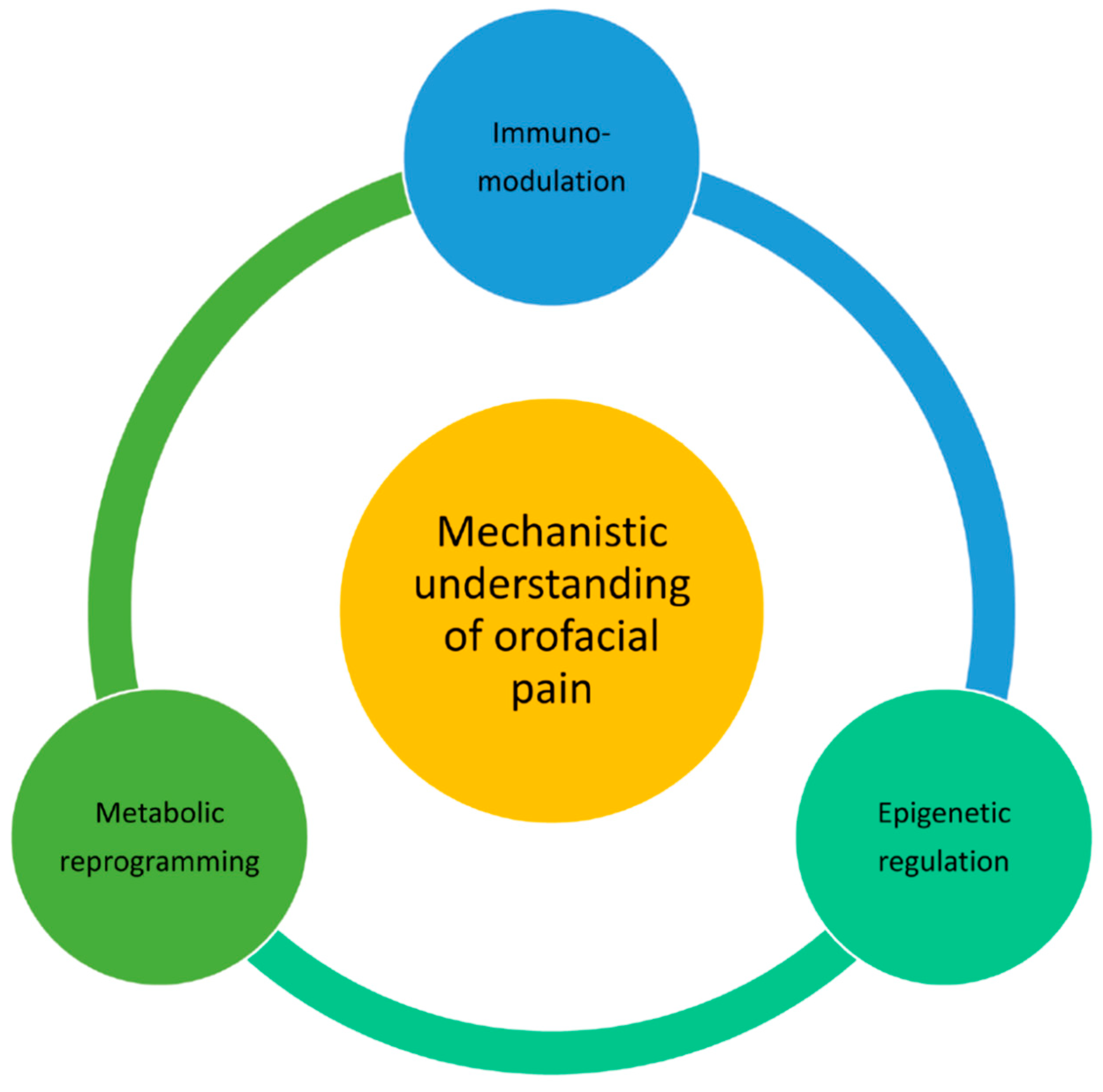
With the advancement in understanding pain pathways at a molecular and cellular level, it is now evident that pain signaling involves an interplay of immunomodulation, metabolic reprogramming and epigenetic modifications in the physiological systems of nerve and immune cells [1,3,5,7,9]. Here, in this review, we present a thorough insight into the disturbed homeostasis of neural cells and have signified the importance of understanding of pathophysiology of orofacial pain at the molecular level. Such comprehensive understanding can pave the way for developing novel strategies and treatment approaches resulting in better pain management.
We conducted comprehensive searches of the literature focusing on orofacial pain and its mechanisms in PubMed and Google Scholar. The following keywords were used for selecting the cited studies: immunoregulation of orofacial pain, oxidative stress in orofacial pain, redox homeostasis in orofacial pain, reactive radicals in orofacial pain, mitochondrial dysfunction in orofacial pain, epigenetic regulation of orofacial pain, non-coding RNAs in orofacial pain, and clinical studies of orofacial pain. The publications not available in English were excluded. We prioritized those studies published within the last ten years.

2. Glial Cell Activation and Orofacial Pain Chronicity

Immune cells impart a homeostatic environment in the brain as they continuously patrol the brain parenchyma, acting as sensors for any physiological alteration [10,11]. They execute a profound role in pain signaling as they interact with neurons while directly influencing their functional capability of excitability [12,13,14,15]. Generally, macrophages as well as glial cells exhibit functional and structural heterogeneity, potentiating pain signaling through various mediators such as chemokines, neurotrophins, neuropeptides, prostaglandins and cytokines which bind to their respective receptors expressed on nociceptive primary afferent neurons. Factors like C-C motif chemokine ligand 2 (CCL2), C-X3-C motif chemokine ligand 1 (CX3CL1), brain-derived growth factor (BDFN), tumor necrosis factor alpha (TNFα), nerve growth factor (NGF), leukemia inhibitory factor (LIF), interleukin 1 beta (IL-1β) and interleukin 6 (IL-6) all have been characterized with distinct roles in pain pathophysiology [12,15,16,17]. Glial cells like astrocytes, microglia, oligodendrocytes, satellite glial cells (SGCs) and Schwann cells contribute to immunomodulation of orofacial pain [18], including both the development and chronic progression of orofacial pain. It has been reported that SGCs derive neuron–glia interactions as well as signaling in TG generating peripheral sensitization; on the other hand, microglia and astrocytes in the CNS contribute to central sensitization in spinal trigeminal nucleus caudalis (Sp5C) [19]. Figure 1 shows a brief outline of chief contributors in immunomodulatory system of orofacial pain pathophysiology.
The increase in orofacial pain in patients receiving cancer therapy has also been attributed to the vasodilation and vasoconstriction effect of astrocytes that release mediators into the vascular system [20]. The role of activated astrocytes and microglia in craniofacial cancers and tongue cancer has also been documented [21,22,23]. The pain-augmenting role of activated Schwann cells in oral carcinogenesis is mediated by the release of TNFα. The Schwann cells reciprocate with cancer cells and increase adenosine production with enhanced secretion of IL-6, TNFα and NGF, which ultimately have an augmenting effect on cancer proliferation, migration and invasion [24,25].
It has been reported that migraine-associated genes carry signal transduction and protein modification in astrocytes and oligodendrocytes [26]. In temporomandibular joint (TMJ) inflammation, activation of SGCs has been reported in the TG [27], and the activation of microglia as well as astrocytes has been reported in the Sp5C [18]. The activated SGCs are facilitated by the upregulation of purinergic G-protein-coupled nucleotide P2Y receptors (P2Y1 and P2Y2) [28]. They exhibit increased intercoupling and formation of gap junctions which ultimately aid in signal modulation [29,30]. They also release several pro-inflammatory cytokines and prostaglandins such as IL1β and PGE2 facilitating neuronal hyperexcitability through overexpression of voltage-dependent sodium channel Nav1.7 in TG neurons [31]. In a TMJ pain mouse model, we demonstrate the crucial role of trigeminal TNFα, which is regulated by a DNA methylation-mediated epigenetic mechanism [32]. New findings have widened the role of oligodendrocytes in neuropathic pain, in which oligodendroglia exhibit an enhancement in the production of cytokines, such as interleukin 33 (IL-33), leading to pain exacerbation [33]. Thus, all these studies project a strong foothold of immunomodulatory action of different glial cells at various stages of pain sensitization, leading to substantial alterations in pain pathophysiology.

3. Metabolic Reprogramming and Orofacial Pain

The severity and maintenance of orofacial pain have been related to the observed metabolic changes in neurons and glial cells. The shift in cellular energy metabolism is central to pain physiology and is widely being investigated for a profound understanding of pain pathology [34]. Glucose is a carbohydrate biomolecule that serves as a primary energy provider of the nervous system. It is taken up by the brain cells, crossing the blood–brain barrier via facilitated diffusion through sodium-independent transporters known as GLUT1-6 and GLUT8, as well as being co-transported along with sodium ion via SGLT1 [35]. It is sourced either through dietary supplementation or through glycogenolysis as well as gluconeogenesis. The glucose molecule starts the energy pathway by undergoing glycolysis followed by the tricarboxylic acid (TCA) cycle and oxidative phosphorylation ultimately producing ATP, the energy currency of cells. The ATP requirement of nerve cells depends upon the condition of the cells. For more neuronal activity or immune cell activation, more ATP is required [36]. In addition to ATP generation, glucose contributes to the synthesis of various neurotransmitters/neuromodulators, transmembrane ionic concentration gradients, neuronal plasticity, redox status, learning and memory [37].
Under normal conditions, pyruvate is produced as an end product of glycolysis that enters the TCA cycle, generating 32 ATP via oxidative phosphorylation; however, in hypoxic conditions, pyruvates are converted to lactate via anaerobic respiration, generating 2 ATP molecules only [38]. The anaerobic route yields less ATP at a higher rate as compared to oxidative phosphorylation which is more efficient but has a slower rate. Immune cells derive their ATP primarily from oxidative phosphorylation at the resting state; however, upon activation, their ATP supply shifts to the anaerobic route to facilitate faster energy supply. This leads to glycometabolism reprogramming characterized by impaired oxidative phosphorylation and an enhanced glycolytic rate. It has been established that upon nerve injury, the neurons and supporting immune cells undergo glycometabolism reprogramming affecting pain sensitization, development and transmission via altered synaptic plasticity and neuronal excitability [8]. Glucose metabolites have a direct role in pain mechanisms as they can alter the expression of pain-related genes via substrate-level gene modification. A perturbed oxidative phosphorylation results in an imbalanced NAD+/NADH ratio which directly affects the acetylation–deacetylation cycle of histones, thereby triggering epigenetic regulation of pain-specific genes [8,39].
Altered levels of several metabolic intermediates such as succinate, lactate, citrate, α-ketoglutarate (α-KG), 2-hydroxyglutarate (2-HG), S-adenosylmethionine (SAM) and fructose-1,6-bisphosphate (FBP) can influence pain sensitization and provide mechanistic insights into their respective roles at the molecular level [8]. Succinate plays a crucial role in inflammatory kinetics. Its cellular and extracellular accumulation leads to hypoxia-inducible factor-1α stabilization, thereby enhancing inflammation by inhibiting prolyl hydroxylases, and it can also signal and modulate immune cell activity using succinate receptor 1 on nerve cells [40]. Extensive studies have shown that lactate acts as an excellent sensor and signaling molecule imparting its effect on various physio-pathological processes of brain metabolism as well as synaptic plasticity [41,42]. Its anti-inflammatory effect projects it as an emerging therapeutic target for inflammatory orofacial pain. It regulates the secretion of IL-1β, IL-10, NF-κB and IL-8 through various pathways [43,44,45]. In the brain, lactate serves as the primary fuel for neurons or glial cells. The excitatory synaptic activity runs primarily on lactate transporting neuron–glia neurometabolic coupling known as the astrocyte-to-neuron lactate shuttle (ANLS) [46]. Thus, fluctuations in lactate levels can directly affect the functioning of neurons involved in pain sensation. Recent findings suggest the lactate-mediated neuro-glia crosstalk as an indicator of pain sensitization. A reduction in lactate transport through this shuttle can attenuate nociceptive, inflammatory and pathological pain. It has been reported recently that a reduction in lactate shuttling via ANLS attenuates opioid-induced hyperalgesia as well as central sensitization [47]. On the other hand, the transfer of lactate via ANLS can persistently sensitize neurons, resulting in chronic neuropathic pain [48,49]. The modulating effects of citric acid, α-KG, 2-HG and SAM on macrophage activity and T-cell proliferation have been well documented, implicating their respective roles in pain sensitization [8]. Fructose 1,6-bisphosphate not only holds anti-inflammatory potential by inhibiting cytokine and adenosine production but also has an antinociceptive effect on hyperalgesia [50].
Generally, cellular lipids along with their metabolites act as endogenous ligands in several pathways, and their association with pain signaling can produce pain-augmenting effects. Both glial cells and neurons can be the source of these molecules in response to either inflammation or oxidative stress. Oxides and epoxides of essential fatty acids like linoleic acid and arachidonic acid, hydroxy and dihydroxy octadecadienoic acid, lysophospholipids and sphingolipids can sensitize nociceptor neurons to alter pain sensitization either directly by activating nociceptors, ion channels and G-protein-coupled receptors or indirectly through inflammatory mediators and intracellular protein kinases [51,52,53]. The pain aggravation effect of arachidonic acid-derived prostaglandins is well documented, and these factors either act as intracellular pro-inflammatory mediators or directly incite peripheral sensitization [54].
Taken together, various cellular metabolites modulate vital biological pathways and contribute to inflammation, pain sensitization and synaptic plasticity through intricate and diverse mechanisms, highlighting a dynamic interplay between metabolic pathways and immune responses deriving cellular machinery in an altered functional way.

4. Oxidative Stress and Mitochondrial Dysfunction in the Pathogenesis of Orofacial Pain

The involvement of perturbed redox homeostasis owing to oxidative stress generated by reactive oxygen species (ROS) such as hydroxyl radical (OH), hydrogen peroxide (H2O2) and superoxide anions (O2) as well as reactive nitrogen species (RNS) such as nitric oxide (NO) and its derivatives has been observed in orofacial pain. It can significantly impact the etiology and modulation of orofacial pain, and with the availability of enough literature, we can comprehend a mutually reciprocating rapport between pain and cellular redox status. It is established that ROS perturbations in sensory neurons influence pain intensity or hyperalgesia as well as neuronal plasticity by regulating the excitability of respective neurons [55].
Previous studies indicate that oxidative stress in the trigeminal nucleus is involved in orofacial pain in a rat model [55,56]. Clinical studies have established a direct correlation between oxidative stress and pain intensity in patients with TMJ disorders [57,58]. The evaluation of oxidative stress biomarkers such as 8-Hydroxydeoxyguanosine (8-OHdG), protein carbonylation (PC), malondialdehyde (MDA) or nuclear factor erythroid 2-related factor 2 (Nrf2) is also being reserved as a diagnostic tool for TMJ disorders [59,60,61]. There are reports suggesting immunomodulatory effects of oxidative stress generating differential pain phenotypes. ROS increment can lead to a pro-inflammatory response in our nervous system through factors such as IL-1β, COX-2, the mitogen-activated protein kinase (MAPK) pathway, toll-like receptors and TNF-α, thereby aggravating pain conditions [55].
As for the role of ROS in neurodegenerative diseases, ROS are involved in almost every pathway of neuropathic pain, which indicates their contribution to pain pathophysiology. On the other hand, it is also established that the redox status of the cell plays a decisive role in regulating mitochondrial function and remodeling, a phenomenon characterized by the differential distribution of mitochondria on the basis of their shape and size [62]. However, studies have not explored whether mitochondrial dysfunction has a role in orofacial pain. It has been reported that people with heritable mitochondrial diseases experience chronic pain, suggesting a direct correlation between chronic pain and mitochondrial disturbance [63]. Electron transport chain inhibitors affecting respiration and ATP production, antioxidants scavenging accumulated ROS or mitochondrial Ca+2 uptake regulation has shown inhibitory effects on nociceptive behavior in different pain models [64]. Persistent mitochondrial disturbance in the soma of sensory neurons further triggers pain signaling and causes a transition to chronic pain [7]. Accumulated ROS as well as mitochondrial dysfunction can also result in dysregulated autophagy, thereby aggravating pain conditions [6].

5. Epigenetic Regulation of Orofacial Pain

Epigenetics deals with DNA methylation and histone modification of our genetic system at the chromatin level to regulate gene expression without any change in the primary structure of DNA. These alterations are heritable and can elicit a distinct phenotype. It primarily navigates the accessibility of genetic sequence to be expressed in an altered way by modulating the binding of transcription factors or by chromatin remodeling affecting proximity to the DNA strand [65]. Altered pain behavior can be manifested by DNA methylation, histone modification such as acetylation, methylation, phosphorylation, ADP-ribosylation, and miRNA expression [66]. A genome-wide assessment of DNA methylation further validates the role of epigenetics in chronic pain [67]. Additionally, perturbed cellular oxidative machinery is accompanied by an imbalanced NAD+/NADH ratio that impacts the histone acetylation or deacetylation process, thereby sparking epigenetic modifications in pain signaling [8,39].
Histone modification, DNA methylation, and non-coding RNA modification are critical for the modulation of neuropathic pain [68]. There have been at least 358 genes, including voltage-gated sodium channel-type 1-alpha subunit (SCN1A), angiotensin I-converting enzyme 2 (ACE2), prostaglandin-endoperoxide synthase 1 (PTGS1) and amyloid-beta precursor protein (APP), identified to be associated with TMD pain [69]. In a rat TMJ osteoarthritis model, DNA methylation has been shown to contribute to inflammation, immune response, angiogenesis and ossification, directing the remodeling of respective joint structures [70]. BDNF is an important neurotrophin involved in neuronal plasticity and nociceptive excitability [71], and its gene can be modified at both the DNA and histone levels. DNA comprising exon III of the gene has been found to be methylated, whereas there is an increment in histone dimethylation of k4 and k9 and a decrement in k29 acetylation [72].
Owing to the profound impact of histone deacetylation catalyzed by histone deacetylases (HDACs) on craniofacial pain intensity, HDAC inhibitors, such as HDAC5 inhibitor LMK253, have demonstrated promising therapeutic potential for such pain [73]. It has also been reported that persistent pain accompanies the epigenetic suppression of the Gad2 gene, which encodes glutamic acid decarboxylase 65, which impairs GABAergic synaptic inhibition via HDAC-mediated histone hypoacetylation [74]. These observations highlight the impact of epigenetic regulation on pain mechanisms. Figure 2 depicts all the factors that contribute to pain pathophysiology.
In addition, the role of non-coding RNAs in pain signaling is also being investigated [75]. Non-coding RNAs comprise part of transcribed RNAs that do not code for proteins [76], but they exhibit regulatory effects on various signaling pathways, gene transcription and chromatin remodeling and can act as oncogenic and tumor-suppressive factors [77,78,79]. Also, their regulatory effects on the wiring and remodeling of neural circuits have been very well established [80]. Thus, many disease conditions have now been identified with dysregulated non-coding RNAs [81], indicating that they can serve as potential biomarkers for various clinical conditions including cancer [82,83]. Various studies have also shown that these non-coding RNAs play a crucial role in oral inflammation and lesion formation, suggesting their foothold in the signaling of oral pain pathways [75,84]. Clinically, microRNAs, among other non-coding RNAs, exhibit significant regulatory effects on several oral pathologies like pulpitis, periodontitis, oral lichen planus and gingival epithelium. Studies on various temporomandibular complications have also validated the role of dysregulated microRNAs in inflammation and inflammatory pain [75]. A study on injured lingual tissues has shown that the dysregulation of several microRNAs like miR-138, miR-138 and miR-29a can cause augmented pain behavior [85], whereas another study on saliva samples from burning mouth syndrome observed the upregulation of miR-1273h-5p/1273a/1304-3p/4449/1285-3p/6802-5p/1268a/1273d/1273f/423-5p and downregulation of miR-27b-3p/16-5p/186-5p/142-3p/141-3p/150-5p/374a-5p/93-5p/29c-3p/29a-3p/148a-3p/22-3p/27a-3p/424-5p/19b-3p/99a-5p/548d-3p/19a-3p, suggesting a role of these microRNAs in the development of neuropathic orofacial pain [86]. Additionally, in serum samples from patients with trigeminal neuralgia, dysregulation in the levels of microRNAs such as miR-132-3p, miR-146b-5p, miR-155-5p and miR-384 contributes to the development of pain [87]. The role of MRAK009713 in regulating the expression and function of the P2X3 receptor suggests its involvement in neuropathic pain [88]. It has been reported that the interactions among non-coding RNAs and mRNAs can cause pain augmentation through the regulation of pain-related genes and nociceptive signaling [89].

6. Primary Experimental or Clinical Data in Orofacial Pain Research

An experimental mechanistic insight into pain pathology provides significant primary data, which could serve as a basis for human clinical studies to pave the way for developing advanced therapeutics. Presently, clinical trials for the management of TMDs and trigeminal neuralgia involve a range of pharmacological treatments targeting various physiological pathways. A study on 60 patients experiencing sleep bruxism and muscle-related TMDs demonstrates the pain-alleviating effect of cannabidiol formulations [90]. These formulations target the endocannabinoid system, involving the regulation of lipid messengers on specific cannabinoid receptors both at peripheral and central levels, thus ameliorating inflammation and neuropathic orofacial pain [91]. Also, several clinical trials have studied formulations like gabapentin and botulinum toxins to explore their pain-alleviating potential [92,93,94,95,96]. In addition to drug-based treatment, current clinical paradigms have incorporated other therapies like physical therapy, cognitive behavior therapy, laser acupuncture therapy, transcranial magnetic stimulation and stem cell therapies to examine their advances in pain management [97,98,99,100,101,102,103,104,105,106,107]. Thus, strategic and evidential collaborations of the scientific community including scientific researchers and clinicians worldwide can benefit from the above explained multidimensional approaches inclusive of all immunomodulating, metabolic and epigenetic factors.

7. Roles of Psychosocial Factors and Systemic Health Conditions in Orofacial Pain

As the diagnosis of the origin of orofacial pain involves a thorough investigation into etiological factors along with odontogenic factors, it is now very well established that patients’ psychological status and systemic health conditions play conspicuous roles in the development and exacerbation of orofacial pain. Several studies have shown a positive correlation between pain associated with TMDs and depression, anxiety, stress or emotional distress. This correlation can be attributed to the altered neuroplasticity and disturbed neurobiological homeostasis involving neuroendocrine dysfunction and chronic inflammation during the development of orofacial pain [108,109,110,111,112,113,114,115,116]. A case study on patients with orofacial pain and early-onset diabetes has shown that the experience of nociceptive pain is exacerbated by diabetes, thereby validating a link between orofacial pain and systemic health conditions [117]. Another case study has demonstrated that trigeminal nociception, intraoral sensitization and structural nerve change are related to diabetic peripheral neuropathy [118]. Moreover, some investigations have indicated the fact that the risk of periodontitis is increased by type 1 and 2 diabetes [119,120,121,122]. In addition, coronary artery diseases or ischemic heart diseases can cause orofacial pain, which can be used as a diagnostic symptom for further cardiac malfunction [123,124,125].

8. Conclusions

A profound understanding of pain pathogenesis and mechanisms requires a comprehensive approach that encompasses the co-interaction of various immunomodulatory, metabolic and epigenetic factors (Figure 3). It is imperative to create a holistic treatment strategy targeting multimodal mechanisms behind the multitude of observed systemic and localized pain perceptions culminating in differential pain behaviors. The pathophysiology of orofacial pain involves an interplay of the immune and nervous systems under the direction of fluctuating oxidative metabolism and genetic regulation. Glucose and lipid metabolites modulate several critical physiological pathways with perturbed redox homeostasis. The generated ROS interfere with cellular machinery accompanied by further molecular perturbations along with epigenetic modulation. The link between oxidative stress and epigenetic modulation emphasizes the critical role of the metabolic factor in chronic orofacial pain. This comprehensive review bridges our understanding of orofacial pain pathology from the molecular level to the gene level; therefore, it provides a window for various researchers and clinicians to understand the mechanisms of orofacial pain.

Author Contributions

Conceptualization, S.K. and F.T.; Methodology, S.K.; Resources, S.K. and F.T.; Data Curation, S.K.; Writing—Original Draft Preparation, S.K.; Writing—Editing, F.T.; Supervision, F.T.; Funding Acquisition, F.T. All authors have read and agreed to the published version of the manuscript.

Funding

This work was supported by the National Institutes of Health Grants R01 DE031255 and R01 DE032061.

Acknowledgments

The figures presented were created with Office 365 MS Powerpoint and Edraw Mindmap software (https://www.edrawsoft.com/).

Conflicts of Interest

The authors have no conflicts of interest to declare.

References

  1. Chichorro, J.G.; Porreca, F.; Sessle, B. Mechanisms of craniofacial pain. Cephalalgia 2017, 37, 613–626. [Google Scholar] [CrossRef] [PubMed]
  2. Labanca, M.; Gianò, M.; Franco, C.; Rezzani, R. Orofacial Pain and Dentistry Management: Guidelines for a More Comprehensive Evidence-Based Approach. Diagnostics 2023, 13, 2854. [Google Scholar] [CrossRef] [PubMed]
  3. Romero-Reyes, M.; Uyanik, J.M. Orofacial pain management: Current perspectives. J. Pain Res. 2014, 7, 99–115. [Google Scholar] [CrossRef]
  4. Pigg, M.; Nixdorf, D.R.; Law, A.S.; Renton, T.; Sharav, Y.; Baad-Hansen, L.; List, T. New International Classification of Orofacial Pain: What Is in It For Endodontists? J. Endod. 2021, 47, 345–357. [Google Scholar] [CrossRef] [PubMed]
  5. Shinoda, M.; Imamura, Y.; Hayashi, Y.; Noma, N.; Okada-Ogawa, A.; Hitomi, S.; Iwata, K. Orofacial Neuropathic Pain-Basic Research and Their Clinical Relevancies. Front. Mol. Neurosci. 2021, 14, 691396. [Google Scholar] [CrossRef] [PubMed]
  6. Shrivastava, M.; Ye, L. A Review on Autophagy in Orofacial Neuropathic Pain. Cells 2022, 11, 3842. [Google Scholar] [CrossRef]
  7. Willemen, H.L.D.M.; Santos Ribeiro, P.S.; Broeks, M.; Meijer, N.; Versteeg, S.; Tiggeler, A.; De Boer, T.P.; Małecki, J.M.; Falnes, P.Ø.; Jans, J.; et al. Inflammation-induced mitochondrial and metabolic disturbances in sensory neurons control the switch from acute to chronic pain. Cell Rep. Med. 2023, 4, 101265. [Google Scholar] [CrossRef]
  8. Kong, E.; Li, Y.; Deng, M.; Hua, T.; Yang, M.; Li, J.; Feng, X.; Yuan, H. Glycometabolism Reprogramming of Glial Cells in Central Nervous System: Novel Target for Neuropathic Pain. Front. Immunol. 2022, 13, 861290. [Google Scholar] [CrossRef]
  9. Bai, G.; Ren, K.; Dubner, R. Epigenetic regulation of persistent pain. Transl. Res. 2015, 165, 177–199. [Google Scholar] [CrossRef]
  10. Norris, G.T.; Kipnis, J. Immune cells and CNS physiology: Microglia and beyond. J. Exp. Med. 2019, 216, 60–70. [Google Scholar] [CrossRef]
  11. D’Alessandro, G.; Marrocco, F.; Limatola, C. Microglial cells: Sensors for neuronal activity and microbiota-derived molecules. Front. Immunol. 2022, 13, 1011129. [Google Scholar] [CrossRef] [PubMed]
  12. Ji, R.-R.; Chamessian, A.; Zhang, Y.-Q. Pain regulation by non-neuronal cells and inflammation. Science 2016, 354, 572–577. [Google Scholar] [CrossRef] [PubMed]
  13. Talbot, S.; Foster, S.L.; Woolf, C.J. Neuroimmunity: Physiology and Pathology. Annu. Rev. Immunol. 2016, 34, 421–447. [Google Scholar] [CrossRef]
  14. Ji, R.-R.; Xu, Z.-Z.; Gao, Y.-J. Emerging targets in neuroinflammation-driven chronic pain. Nat. Rev. Drug Discov. 2014, 13, 533–548. [Google Scholar] [CrossRef] [PubMed]
  15. Shinoda, M.; Kubo, A.; Hayashi, Y.; Iwata, K. Peripheral and Central Mechanisms of Persistent Orofacial Pain. Front. Neurosci. 2019, 13, 1227. [Google Scholar] [CrossRef]
  16. Lee, S.; Zhang, J. Heterogeneity of macrophages in injured trigeminal nerves: Cytokine/chemokine expressing vs. phagocytic macrophages. Brain Behav. Immun. 2012, 26, 891–903. [Google Scholar] [CrossRef]
  17. Batbold, D.; Shinoda, M.; Honda, K.; Furukawa, A.; Koizumi, M.; Akasaka, R.; Yamaguchi, S.; Iwata, K. Macrophages in trigeminal ganglion contribute to ectopic mechanical hypersensitivity following inferior alveolar nerve injury in rats. J. Neuroinflamm. 2017, 14, 249. [Google Scholar] [CrossRef]
  18. Ye, Y.; Salvo, E.; Romero-Reyes, M.; Akerman, S.; Shimizu, E.; Kobayashi, Y.; Michot, B.; Gibbs, J. Glia and Orofacial Pain: Progress and Future Directions. Int. J. Mol. Sci. 2021, 22, 5345. [Google Scholar] [CrossRef]
  19. Zhang, S.; Azubuine, J.; Schmeer, C. A systematic literature review on the role of glial cells in the pathomechanisms of migraine. Front. Mol. Neurosci. 2023, 16, 1219574. [Google Scholar] [CrossRef]
  20. Epstein, J.B.; Hong, C.; Logan, R.M.; Barasch, A.; Gordon, S.M.; Oberlee-Edwards, L.; McGuire, D.; Napenas, J.J.; Elting, L.S.; Spijkervet, F.K.L.; et al. A systematic review of orofacial pain in patients receiving cancer therapy. Support. Care Cancer 2010, 18, 1023–1031. [Google Scholar] [CrossRef]
  21. Hidaka, K.; Ono, K.; Harano, N.; Sago, T.; Nunomaki, M.; Shiiba, S.; Nakanishi, O.; Fukushima, H.; Inenaga, K. Central glial activation mediates cancer-induced pain in a rat facial cancer model. Neuroscience 2011, 180, 334–343. [Google Scholar] [CrossRef] [PubMed]
  22. Sago, T.; Ono, K.; Harano, N.; Furuta-Hidaka, K.; Hitomi, S.; Nunomaki, M.; Yoshida, M.; Shiiba, S.; Nakanishi, O.; Matsuo, K.; et al. Distinct time courses of microglial and astrocytic hyperactivation and the glial contribution to pain hypersensitivity in a facial cancer model. Brain Res. 2012, 1457, 70–80. [Google Scholar] [CrossRef] [PubMed]
  23. Tamagawa, T.; Shinoda, M.; Honda, K.; Furukawa, A.; Kaji, K.; Nagashima, H.; Akasaka, R.; Chen, J.; Sessle, B.J.; Yonehara, Y.; et al. Involvement of Microglial P2Y12 Signaling in Tongue Cancer Pain. J. Dent. Res. 2016, 95, 1176–1182. [Google Scholar] [CrossRef] [PubMed]
  24. Salvo, E.; Saraithong, P.; Curtin, J.G.; Janal, M.N.; Ye, Y. Reciprocal interactions between cancer and Schwann cells contribute to oral cancer progression and pain. Heliyon 2019, 5, e01223. [Google Scholar] [CrossRef]
  25. Salvo, E.; Tu, N.H.; Scheff, N.N.; Dubeykovskaya, Z.A.; Chavan, S.A.; Aouizerat, B.E.; Ye, Y. TNFα promotes oral cancer growth, pain, and Schwann cell activation. Sci. Rep. 2021, 11, 1840. [Google Scholar] [CrossRef]
  26. Eising, E.; De Leeuw, C.; Min, J.L.; Anttila, V.; Verheijen, M.H.; Terwindt, G.M.; Dichgans, M.; Freilinger, T.; Kubisch, C.; Ferrari, M.D.; et al. Involvement of astrocyte and oligodendrocyte gene sets in migraine. Cephalalgia 2016, 36, 640–647. [Google Scholar] [CrossRef]
  27. Villa, G.; Ceruti, S.; Zanardelli, M.; Magni, G.; Jasmin, L.; Ohara, P.T.; Abbracchio, M.P. Temporomandibular Joint Inflammation Activates Glial and Immune Cells in Both the Trigeminal Ganglia and in the Spinal Trigeminal Nucleus. Mol. Pain 2010, 6, 1744–8069. [Google Scholar] [CrossRef]
  28. Magni, G.; Merli, D.; Verderio, C.; Abbracchio, M.P.; Ceruti, S. P2Y2 receptor antagonists as anti-allodynic agents in acute and sub-chronic trigeminal sensitization: Role of satellite glial cells. Glia 2015, 63, 1256–1269. [Google Scholar] [CrossRef]
  29. Spray, D.C.; Hanani, M. Gap junctions, pannexins and pain. Neurosci. Lett. 2019, 695, 46–52. [Google Scholar] [CrossRef]
  30. Ahmed, F.; Rahman, M.; Thompson, R.; Bereiter, D.A. Role of Connexin 43 in an Inflammatory Model for TMJ Hyperalgesia. Front. Pain Res. 2021, 2, 715871. [Google Scholar] [CrossRef]
  31. Zhang, P.; Gan, Y.-H. Prostaglandin E2 Upregulated Trigeminal Ganglionic Sodium Channel 1.7 Involving Temporomandibular Joint Inflammatory Pain in Rats. Inflammation 2017, 40, 1102–1109. [Google Scholar] [CrossRef] [PubMed]
  32. Bai, Q.; Liu, S.; Shu, H.; Tang, Y.; George, S.; Dong, T.; Schmidt, B.L.; Tao, F. TNFα in the Trigeminal Nociceptive System Is Critical for Temporomandibular Joint Pain. Mol. Neurobiol. 2019, 56, 278–291. [Google Scholar] [CrossRef]
  33. Kim, W.; Angulo, M.C. Unraveling the role of oligodendrocytes and myelin in pain. J. Neurochem. 2024, 169, e16206. [Google Scholar] [CrossRef]
  34. Jha, M.K.; Song, G.J.; Lee, M.G.; Jeoung, N.H.; Go, Y.; Harris, R.A.; Park, D.H.; Kook, H.; Lee, I.-K.; Suk, K. Metabolic Connection of Inflammatory Pain: Pivotal Role of a Pyruvate Dehydrogenase Kinase-Pyruvate Dehydrogenase-Lactic Acid Axis. J. Neurosci. 2015, 35, 14353–14369. [Google Scholar] [CrossRef] [PubMed]
  35. Koepsell, H. Glucose transporters in brain in health and disease. Pflügers Arch.—Eur. J. Physiol. 2020, 472, 1299–1343. [Google Scholar] [CrossRef] [PubMed]
  36. Rumpf, S.; Sanal, N.; Marzano, M. Energy metabolic pathways in neuronal development and function. Oxf. Open Neurosci. 2023, 2, kvad004. [Google Scholar] [CrossRef] [PubMed]
  37. Nimgampalle, M.; Chakravarthy, H.; Devanathan, V. Glucose metabolism in the brain: An update. In Recent Developments in Applied Microbiology and Biochemistry; Elsevier: Amsterdam, The Netherlands, 2021; pp. 77–88. [Google Scholar]
  38. Fadaka, A.; Ajiboye, B.; Ojo, O.; Adewale, O.; Olayide, I.; Emuowhochere, R. Biology of glucose metabolization in cancer cells. J. Oncol. Sci. 2017, 3, 45–51. [Google Scholar] [CrossRef]
  39. Zhang, W.; Jiao, B.; Yu, S.; Zhang, C.; Zhang, K.; Liu, B.; Zhang, X. Histone deacetylase as emerging pharmacological therapeutic target for neuropathic pain: From epigenetic to selective drugs. CNS Neurosci. Ther. 2024, 30, e14745. [Google Scholar] [CrossRef]
  40. Huang, H.; Li, G.; He, Y.; Chen, J.; Yan, J.; Zhang, Q.; Li, L.; Cai, X. Cellular succinate metabolism and signaling in inflammation: Implications for therapeutic intervention. Front. Immunol. 2024, 15, 1404441. [Google Scholar] [CrossRef]
  41. Certo, M.; Llibre, A.; Lee, W.; Mauro, C. Understanding lactate sensing and signalling. Trends Endocrinol. Metab. 2022, 33, 722–735. [Google Scholar] [CrossRef]
  42. Wang, Q.; Hu, Y.; Wan, J.; Dong, B.; Sun, J. Lactate: A Novel Signaling Molecule in Synaptic Plasticity and Drug Addiction. BioEssays 2019, 41, 1900008. [Google Scholar] [CrossRef] [PubMed]
  43. Errea, A.; Cayet, D.; Marchetti, P.; Tang, C.; Kluza, J.; Offermanns, S.; Sirard, J.-C.; Rumbo, M. Lactate Inhibits the Pro-Inflammatory Response and Metabolic Reprogramming in Murine Macrophages in a GPR81-Independent Manner. PLoS ONE 2016, 11, e0163694. [Google Scholar] [CrossRef] [PubMed]
  44. Nasi, A.; Fekete, T.; Krishnamurthy, A.; Snowden, S.; Rajnavölgyi, E.; Catrina, A.I.; Wheelock, C.E.; Vivar, N.; Rethi, B. Dendritic Cell Reprogramming by Endogenously Produced Lactic Acid. J. Immunol. 2013, 191, 3090–3099. [Google Scholar] [CrossRef]
  45. Cai, M.; Wan, J.; Cai, K.; Song, H.; Wang, Y.; Sun, W.; Hu, J. Understanding the Contribution of Lactate Metabolism in Cancer Progress: A Perspective from Isomers. Cancers 2022, 15, 87. [Google Scholar] [CrossRef] [PubMed]
  46. Benarroch, E. What Is the Role of Lactate in Brain Metabolism, Plasticity, and Neurodegeneration? Neurology 2024, 102, e209378. [Google Scholar] [CrossRef]
  47. Ma, X.; Qi, Q.; Wang, W.; Huang, M.; Wang, H.; Luo, L.; Xu, X.; Yuan, T.; Shi, H.; Jiang, W.; et al. Astrocytic pyruvate dehydrogenase kinase-lactic acid axis involvement in glia-neuron crosstalk contributes to morphine-induced hyperalgesia in mice. Fundam. Res. 2024, 4, 820–828. [Google Scholar] [CrossRef]
  48. Wang, Y.; Peng, Y.; Zhang, C.; Zhou, X. Astrocyte-neuron lactate transport in the ACC contributes to the occurrence of long-lasting inflammatory pain in male mice. Neurosci. Lett. 2021, 764, 136205. [Google Scholar] [CrossRef]
  49. Miyamoto, K.; Ishikura, K.I.; Kume, K.; Ohsawa, M. Astrocyte-neuron lactate shuttle sensitizes nociceptive transmission in the spinal cord. Glia 2019, 67, 27–36. [Google Scholar] [CrossRef]
  50. Valério, D.; Ferreira, F.; Cunha, T.; Alves-Filho, J.; Lima, F.; De Oliveira, R., Jr.; Ferreira, S.; Cunha, F.; Queiroz, R.; Verri, W., Jr. Fructose-1,6-bisphosphate reduces inflammatory pain-like behaviour in mice: Role of adenosine acting on A1 receptors. Br. J. Pharmacol. 2009, 158, 558–568. [Google Scholar] [CrossRef]
  51. Osthues, T.; Sisignano, M. Oxidized Lipids in Persistent Pain States. Front. Pharmacol. 2019, 10, 1147. [Google Scholar] [CrossRef]
  52. Hargreaves, K.M.; Ruparel, S. Role of Oxidized Lipids and TRP Channels in Orofacial Pain and Inflammation. J. Dent. Res. 2016, 95, 1117–1123. [Google Scholar] [CrossRef] [PubMed]
  53. Shapiro, H.; Singer, P.; Ariel, A. Beyond the classic eicosanoids: Peripherally-acting oxygenated metabolites of polyunsaturated fatty acids mediate pain associated with tissue injury and inflammation. Prostaglandins Leukot. Essent. Fat. Acids 2016, 111, 45–61. [Google Scholar] [CrossRef] [PubMed]
  54. Jang, Y.; Kim, M.; Hwang, S.W. Molecular mechanisms underlying the actions of arachidonic acid-derived prostaglandins on peripheral nociception. J. Neuroinflamm. 2020, 17, 30. [Google Scholar] [CrossRef] [PubMed]
  55. Gianò, M.; Franco, C.; Castrezzati, S.; Rezzani, R. Involvement of Oxidative Stress and Nutrition in the Anatomy of Orofacial Pain. Int. J. Mol. Sci. 2023, 24, 13128. [Google Scholar] [CrossRef]
  56. Viggiano, E.; Monda, M.; Viggiano, A.; Viggiano, A.; Aurilio, C.; De Luca, B. Persistent facial pain increases superoxide anion production in the spinal trigeminal nucleus. Mol. Cell. Biochem. 2010, 339, 149–154. [Google Scholar] [CrossRef]
  57. Rodríguez De Sotillo, D.; Velly, A.M.; Hadley, M.; Fricton, J.R. Evidence of oxidative stress in temporomandibular disorders: A pilot study. J. Oral. Rehabil. 2011, 38, 722–728. [Google Scholar] [CrossRef]
  58. Eslami, H.; Katebi, K.; Ghaffaripour Saleh, S.; Mirizadeh, L.; Hashemi, M. The relationship between oxidative stress markers and temporomandibular disorders: A systematic review and meta-analysis. J. Res. Med. Sci. 2024, 29, 33. [Google Scholar] [CrossRef]
  59. Nguyen, T.T.; Ngo, L.Q.; Promsudthi, A.; Surarit, R. Salivary oxidative stress biomarkers in chronic periodontitis and acute coronary syndrome. Clin. Oral. Investig. 2017, 21, 2345–2353. [Google Scholar] [CrossRef]
  60. Goriuc, A.; Cojocaru, K.-A.; Luchian, I.; Ursu, R.-G.; Butnaru, O.; Foia, L. Using 8-Hydroxy-2′-Deoxiguanosine (8-OHdG) as a Reliable Biomarker for Assessing Periodontal Disease Associated with Diabetes. Int. J. Mol. Sci. 2024, 25, 1425. [Google Scholar] [CrossRef]
  61. Xu, M.; Fang, L.; Xue, Q.; Zhang, X.; He, Y. The Nrf2 Pathway Alleviates Overloading Force-Induced TMJ Degeneration by Downregulating Oxidative Stress Reactions. J. Inflamm. Res. 2023, 16, 5601–5612. [Google Scholar] [CrossRef]
  62. Tilokani, L.; Nagashima, S.; Paupe, V.; Prudent, J. Mitochondrial dynamics: Overview of molecular mechanisms. Essays Biochem. 2018, 62, 341–360. [Google Scholar] [PubMed]
  63. Van Den Ameele, J.; Fuge, J.; Pitceathly, R.D.S.; Berry, S.; McIntyre, Z.; Hanna, M.G.; Lee, M.; Chinnery, P.F. Chronic pain is common in mitochondrial disease. Neuromuscul. Disord. 2020, 30, 413–419. [Google Scholar] [CrossRef] [PubMed]
  64. Guo, B.-L.; Sui, B.-D.; Wang, X.-Y.; Wei, Y.-Y.; Huang, J.; Chen, J.; Wu, S.-X.; Li, Y.-Q.; Wang, Y.-Y.; Yang, Y.-L. Significant changes in mitochondrial distribution in different pain models of mice. Mitochondrion 2013, 13, 292–297. [Google Scholar] [CrossRef] [PubMed]
  65. Fox, S.A.; Tiwari, L.; Farah, C.S. Epigenetics and oral disease. In Translational Systems Medicine and Oral Disease; Elsevier: Amsterdam, The Netherlands, 2020; pp. 163–206. [Google Scholar]
  66. Nirvanie-Persaud, L.; Millis, R.M. Epigenetics and Pain: New Insights to an Old Problem. Cureus 2022, 14, e29353. [Google Scholar] [CrossRef]
  67. Massart, R.; Dymov, S.; Millecamps, M.; Suderman, M.; Gregoire, S.; Koenigs, K.; Alvarado, S.; Tajerian, M.; Stone, L.S.; Szyf, M. Overlapping signatures of chronic pain in the DNA methylation landscape of prefrontal cortex and peripheral T cells. Sci. Rep. 2016, 6, 19615. [Google Scholar] [CrossRef]
  68. Luo, D.; Li, X.; Tang, S.; Song, F.; Li, W.; Xie, G.; Liang, J.; Zhou, J. Epigenetic modifications in neuropathic pain. Mol. Pain 2021, 17, 174480692110567. [Google Scholar] [CrossRef]
  69. Smith, S.B.; Mir, E.; Bair, E.; Slade, G.D.; Dubner, R.; Fillingim, R.B.; Greenspan, J.D.; Ohrbach, R.; Knott, C.; Weir, B.; et al. Genetic Variants Associated With Development of TMD and Its Intermediate Phenotypes: The Genetic Architecture of TMD in the OPPERA Prospective Cohort Study. J. Pain 2013, 14, T91–T101.e3. [Google Scholar] [CrossRef]
  70. Xiao, J.-L.; Meng, J.-H.; Gan, Y.-H.; Li, Y.-L.; Zhou, C.-Y.; Ma, X.-C. DNA methylation profiling in different phases of temporomandibular joint osteoarthritis in rats. Arch. Oral Biol. 2016, 68, 105–115. [Google Scholar] [CrossRef]
  71. Bonomini, F.; Favero, G.; Castrezzati, S.; Borsani, E. Role of Neurotrophins in Orofacial Pain Modulation: A Review of the Latest Discoveries. Int. J. Mol. Sci. 2023, 24, 12438. [Google Scholar] [CrossRef]
  72. Nakae, A.; Nakai, K.; Tanaka, T.; Hosokawa, K.; Mashimo, T.; Oro-facial Study Group. Epigenetic regulation of BDNF genes in rat orofacial neuropathic pain model: 14AP3-1. Eur. J. Anaesthesiol. 2013, 30, 210–211. [Google Scholar] [CrossRef]
  73. Westlund, K.N.; Montera, M.; Goins, A.E.; Shilling, M.W.; Afaghpour-Becklund, M.; Alles, S.R.A.; Elise Hui, S. Epigenetic HDAC5 Inhibitor Reverses Craniofacial Neuropathic Pain in Mice. J. Pain 2024, 25, 428–450. [Google Scholar] [CrossRef] [PubMed]
  74. Zhang, Z.; Cai, Y.-Q.; Zou, F.; Bie, B.; Pan, Z.Z. Epigenetic suppression of GAD65 expression mediates persistent pain. Nat. Med. 2011, 17, 1448–1455. [Google Scholar] [CrossRef] [PubMed]
  75. Roganović, J.; Petrović, N. Clinical Perspectives of Non-Coding RNA in Oral Inflammatory Diseases and Neuropathic Pain: A Narrative Review. Int. J. Mol. Sci. 2022, 23, 8278. [Google Scholar] [CrossRef] [PubMed]
  76. Mattick, J.S.; Makunin, I.V. Non-Coding RNA. Hum. Mol. Genet. 2006, 15, R17–R29. [Google Scholar] [CrossRef]
  77. Fu, X.D. Non-Coding RNA: A New Frontier in Regulatory Biology. Natl. Sci. Rev. 2014, 1, 190–204. [Google Scholar] [CrossRef]
  78. Slack, F.J.; Chinnaiyan, A.M. The Role of Non-Coding RNAs in Oncology. Cell 2019, 179, 1033–1055. [Google Scholar] [CrossRef]
  79. Nemeth, K.; Bayraktar, R.; Ferracin, M.; Calin, G.A. Non-Coding RNAs in Disease: From Mechanisms to Therapeutics. Nat. Rev. Genet. 2023, 25, 211–232. [Google Scholar] [CrossRef]
  80. Soutschek, M.; Schratt, G. Non-Coding RNA in the Wiring and Remodeling of Neural Circuits. Neuron 2023, 111, 2140–2154. [Google Scholar] [CrossRef]
  81. El-Gewely, M.R. Dysregulation of Regulatory NcRNAs and Diseases. Int. J. Mol. Sci. 2023, 25, 24. [Google Scholar] [CrossRef]
  82. Loganathan, T.; Doss C, G.P. Non-Coding RNAs in Human Health and Disease: Potential Function as Biomarkers and Therapeutic Targets. Funct. Integr. Genom. 2023, 23, 33. [Google Scholar] [CrossRef]
  83. Malgundkar, S.H.; Tamimi, Y. The Pivotal Role of Long Non-Coding RNAs as Potential Biomarkers and Modulators of Chemoresistance in Ovarian Cancer (OC). Hum. Genet. 2024, 143, 107–124. [Google Scholar] [CrossRef] [PubMed]
  84. Zhang, K.; Qiu, W.; Wu, B.; Fang, F. Long Non-Coding RNAs Are Novel Players in Oral Inflammatory Disorders, Potentially Premalignant Oral Epithelial Lesions and Oral Squamous Cell Carcinoma (Review). Int. J. Mol. Med. 2020, 46, 535. [Google Scholar] [CrossRef] [PubMed]
  85. Tavares-Ferreira, D.; Lawless, N.; Bird, E.V.; Atkins, S.; Collier, D.; Sher, E.; Malki, K.; Lambert, D.W.; Boissonade, F.M. Correlation of MiRNA Expression with Intensity of Neuropathic Pain in Man. Mol. Pain 2019, 15, 1744806919860323. [Google Scholar] [CrossRef]
  86. Kim, K.-Y.; Byun, J.-S.; Jung, J.-K.; Choi, J.-K. Profiling of Salivary Exosomal Micro RNAs in Burning Mouth Syndrome Patients. J. Oral Med. Pain 2019, 44, 25–30. [Google Scholar]
  87. Li, X.; Wang, D.; Zhou, J.; Yan, Y.; Chen, L. Evaluation of Circulating MicroRNA Expression in Patients with Trigeminal Neuralgia: An Observational Study. Medicine 2020, 99, e22972. [Google Scholar] [CrossRef]
  88. Li, G.; Jiang, H.; Zheng, C.; Zhu, G.; Xu, Y.; Sheng, X.; Wu, B.; Guo, J.; Zhu, S.; Zhan, Y.; et al. Long Noncoding RNA MRAK009713 Is a Novel Regulator of Neuropathic Pain in Rats. Pain 2017, 158, 2042–2052. [Google Scholar] [CrossRef]
  89. Fang, Z.H.; Liao, H.L.; Tang, Q.F.; Liu, Y.J.; Zhang, Y.Y.; Lin, J.; Yu, H.P.; Zhou, C.; Li, C.J.; Liu, F.; et al. Interactions Among Non-Coding RNAs and MRNAs in the Trigeminal Ganglion Associated with Neuropathic Pain. J. Pain Res. 2022, 15, 2967–2988. [Google Scholar] [CrossRef]
  90. Walczyńska-Dragon, K.; Kurek-Górecka, A.; Niemczyk, W.; Nowak, Z.; Baron, S.; Olczyk, P.; Nitecka-Buchta, A.; Kempa, W.M. Cannabidiol Intervention for Muscular Tension, Pain, and Sleep Bruxism Intensity—A Randomized, Double-Blind Clinical Trial. J. Clin. Med. 2024, 13, 1417. [Google Scholar] [CrossRef]
  91. Crescente, G.; Minervini, G.; Spagnuolo, C.; Moccia, S. Cannabis Bioactive Compound-Based Formulations: New Perspectives for the Management of Orofacial Pain. Molecules 2022, 28, 106. [Google Scholar] [CrossRef]
  92. Sitnikova, V.; Kämppi, A.; Kämppi, L.; Alvesalo, E.; Burakova, M.; Kemppainen, P.; Teronen, O. Clinical Benefit of Botulinum Toxin for Treatment of Persistent TMD-Related Myofascial Pain: A Randomized, Placebo-Controlled, Cross-over Trial. Pain Pract. 2024, 24, 1014–1023. [Google Scholar] [CrossRef]
  93. Ma, S.J.; Iovoli, A.J.; Wang, K.; Neimanis, D.; Smith, K.A.; Attwood, K.; Farrugia, M.; Hermann, G.; Singh, A.K. Efficacy of Prophylactic High-Dose Gabapentin and Venlafaxine on Reducing Oral Mucositis Pain Among Patients Treated With Chemoradiation for Head and Neck Cancer: A Single-Institution, Phase 2, Randomized Clinical Trial. Int. J. Radiat. Oncol. Biol. Phys. 2023, 116, 797–806. [Google Scholar] [CrossRef] [PubMed]
  94. Sulukdjian, A.; L’Homme, R.; Chanlon, A.; Moreau, N. Gabapentinoid Prescription in Oral Medicine and Oral Surgery Practice. Part I—Experience from a French Orofacial Pain Clinic. J. Oral Med. Oral Surg. 2020, 26, 13. [Google Scholar] [CrossRef]
  95. De la Torre Canales, G.; Poluha, R.L.; Bonjardim, L.R.; Ernberg, M.; Conti, P.C.R. Botulinum Toxin-A Effects on Pain, Somatosensory and Psychosocial Features of Patients with Refractory Masticatory Myofascial Pain: A Randomized Double-Blind Clinical Trial. Sci. Rep. 2024, 14, 4201. [Google Scholar] [CrossRef]
  96. Cook, A.; Modh, A.; Ali, H.; Sheqwara, J.; Chang, S.; Ghanem, T.; Momin, S.; Wu, V.; Tam, S.; Money, S.; et al. Randomized Phase 3, Double-Blind, Placebo-Controlled Study of Prophylactic Gabapentin for the Reduction of Oral Mucositis Pain During the Treatment of Oropharyngeal Squamous Cell Carcinoma. Int. J. Radiat. Oncol. Biol. Phys. 2022, 112, 926–937. [Google Scholar] [CrossRef]
  97. Yamada, Y.; Nakamura-Yamada, S.; Konoki, R.; Baba, S. Promising Advances in Clinical Trials of Dental Tissue-Derived Cell-Based Regenerative Medicine. Stem Cell Res. Ther. 2020, 11, 175. [Google Scholar] [CrossRef]
  98. Hanif, M.; Sheikh, M.A. Efficacy of Platelet Rich Plasma (PRP) on Mouth Opening and Pain after Surgical Extraction of Mandibular Third Molars. J. Oral Med. Oral Surg. 2021, 27, 9. [Google Scholar] [CrossRef]
  99. Li, F.; Wu, C.; Sun, H.; Zhou, Q. Effect of Platelet-Rich Plasma Injections on Pain Reduction in Patients with Temporomandibular Joint Osteoarthrosis: A Meta-Analysis of Randomized Controlled Trials. J. Oral Facial Pain Headache 2020, 34, 149–156. [Google Scholar] [CrossRef]
  100. Clinical Trial of the Safety and Efficacy of Peripheral Nerve Stimulation in the Treatment of Postherpetic Neuralgia. Clinical Research Trial Listing. Available online: https://www.centerwatch.com/clinical-trials/listings/NCT06290661/clinical-trial-of-the-safety-and-efficacy-of-peripheral-nerve-stimulation-in-the-treatment-of-postherpetic-neuralgia (accessed on 22 January 2025).
  101. UCSF Pain Trial → Transcranial Magnetic Stimulation for Chronic Neuropathic Pain. Available online: https://clinicaltrials.ucsf.edu/trial/NCT05593237 (accessed on 22 January 2025).
  102. Liao, M.; Diao, Y.; Pan, J.; Wong, L.S.; Subramaniam, G.; Vasanthi, R.K.; Liao, L. Efficacy and Safety of Repetitive Transcranial Magnetic Stimulation with Different Frequencies on Neuropathic Orofacial Pain: A Systematic Literature Review and Meta-Analysis. J. Oral Facial Pain Headache 2024, 38, 48–67. [Google Scholar] [CrossRef]
  103. Di Francesco, F.; Minervini, G.; Siurkel, Y.; Cicciù, M.; Lanza, A. Efficacy of Acupuncture and Laser Acupuncture in Temporomandibular Disorders: A Systematic Review and Meta-Analysis of Randomized Controlled Trials. BMC Oral Health 2024, 24, 174. [Google Scholar] [CrossRef]
  104. Ferrando, M.; Galdón, M.J.; Durá, E.; Andreu, Y.; Jiménez, Y.; Poveda, R. Enhancing the Efficacy of Treatment for Temporomandibular Patients with Muscular Diagnosis through Cognitive-Behavioral Intervention, Including Hypnosis: A Randomized Study. Oral Surg. Oral Med. Oral Pathol. Oral Radiol. 2012, 113, 81–89. [Google Scholar] [CrossRef]
  105. Patil, D.J.; Dheer, D.S. Psychological Assessment and Cognitive Behavioral Therapy in Temporomandibular Joint Disorders: A Randomized Controlled Study. J. Indian Acad. Oral Med. Radiol. 2023, 35, 326–330. [Google Scholar] [CrossRef]
  106. Reviewing the Evidence: Can Cognitive Behavioral Therapy Improve Outcomes for Patients with Chronic Orofacial Pain? Available online: https://www.jofph.com/articles/10.11607/jofph.24.2.04 (accessed on 22 January 2025).
  107. Shi, Y.; Wu, W. Multimodal Non-Invasive Non-Pharmacological Therapies for Chronic Pain: Mechanisms and Progress. BMC Med. 2023, 21, 1–29. [Google Scholar] [CrossRef] [PubMed]
  108. Aboushaar, N.; Serrano, N. The Mutually Reinforcing Dynamics between Pain and Stress: Mechanisms, Impacts and Management Strategies. Front. Pain Res. 2024, 5, 1445280. [Google Scholar] [CrossRef]
  109. De Ridder, D.; Adhia, D.; Vanneste, S. The Anatomy of Pain and Suffering in the Brain and Its Clinical Implications. Neurosci. Biobehav. Rev. 2021, 130, 125–146. [Google Scholar] [CrossRef] [PubMed]
  110. Sheng, J.; Liu, S.; Wang, Y.; Cui, R.; Zhang, X. The Link between Depression and Chronic Pain: Neural Mechanisms in the Brain. Neural Plast. 2017, 2017, 9724371. [Google Scholar] [CrossRef]
  111. Assiri, K. Relationships between Personality Factors and DC/TMD Axis II Scores of Psychosocial Impairment among Patients with Pain Related Temporomandibular Disorders. Sci. Rep. 2024, 14, 26869. [Google Scholar] [CrossRef]
  112. Hekmati, A.; Mortazavi, N.; Ozouni-Davaji, R.B.; Vakili, M. Personality Traits and Anxiety in Patients with Temporomandibular Disorders. BMC Psychol. 2022, 10, 86. [Google Scholar] [CrossRef]
  113. Xiang, Y.; Song, J.; Liang, Y.; Sun, J.; Zheng, Z. Causal Relationship between Psychiatric Traits and Temporomandibular Disorders: A Bidirectional Two-Sample Mendelian Randomization Study. Clin. Oral Investig. 2023, 27, 7513–7521. [Google Scholar] [CrossRef]
  114. Ye, C.; Pu, D.; Zhang, J.; Jia, M.; Zhang, Y.; Du, S.; Wang, J.; Xiong, X. Unlocking the Link between Temporomandibular Disorders and Suicide Ideation in Pre-Orthodontic Patients: A Moderated Mediation Model of Depression and Anxiety. J. Affect. Disord. 2024, 349, 486–493. [Google Scholar] [CrossRef]
  115. Lee, Y.H.; Auh, Q.S. Clinical Factors Affecting Depression in Patients with Painful Temporomandibular Disorders during the COVID-19 Pandemic. Sci. Rep. 2022, 12, 14667. [Google Scholar] [CrossRef]
  116. Calixtre, L.B.; Da Grüninger, B.L.S.; Chaves, T.C.; De Oliveira, A.B. Is There an Association between Anxiety/Depression and Temporomandibular Disorders in College Students? J. Appl. Oral Sci. 2014, 22, 15–21. [Google Scholar] [CrossRef] [PubMed]
  117. Xu, Z.; Zhang, P.; Long, L.; He, H.; Zhang, J.; Sun, S. Diabetes Mellitus in Classical Trigeminal Neuralgia: A Predisposing Factor for Its Development. Clin. Neurol. Neurosurg. 2016, 151, 70–72. [Google Scholar] [CrossRef] [PubMed]
  118. Costa, Y.M.; Karlsson, P.; Bonjardim, L.R.; Conti, P.C.R.; Tankisi, H.; Jensen, T.S.; Nyengaard, J.R.; Svensson, P.; Baad-Hansen, L. Trigeminal Nociceptive Function and Oral Somatosensory Functional and Structural Assessment in Patients with Diabetic Peripheral Neuropathy. Sci. Rep. 2019, 9, 169. [Google Scholar] [CrossRef] [PubMed]
  119. Deshpande, K.; Jain, A.; Sharma, R.; Prashar, S.; Jain, R. Diabetes and Periodontitis. J. Indian Soc. Periodontol. 2010, 14, 207–212. [Google Scholar] [CrossRef]
  120. Salvi, G.E.; Carollo-Bittel, B.; Lang, N.P. Effects of Diabetes Mellitus on Periodontal and Peri-Implant Conditions: Update on Associations and Risks. J. Clin. Periodontol. 2008, 35, 398–409. [Google Scholar] [CrossRef]
  121. Gurav, A.; Jadhav, V. Periodontitis and Risk of Diabetes Mellitus. J. Diabetes 2011, 3, 21–28. [Google Scholar] [CrossRef]
  122. Pontes Andersen, C.C.; Flyvbjerg, A.; Buschard, K.; Holmstrup, P. Relationship between Periodontitis and Diabetes: Lessons from Rodent Studies. J. Periodontol. 2007, 78, 1264–1275. [Google Scholar] [CrossRef]
  123. López-López, J.; Garcia-Vicente, L.; Jané-Salas, E.; Estrugo-Devesa, A.; Chimenos-Küstner, E.; Roca-Elias, J. Orofacial Pain of Cardiac Origin: Review Literature and Clinical Cases. Med. Oral Patol. Oral Cir. Bucal 2012, 17, e538. [Google Scholar] [CrossRef]
  124. Kreiner, M.; Okeson, J.; Tanco, V.; Waldenström, A.; Isberg, A. Orofacial Pain and Toothache as the Sole Symptom of an Acute Myocardial Infarction Entails a Major Risk of Misdiagnosis and Death. J. Oral Facial Pain Headache 2020, 34, 53–60. [Google Scholar] [CrossRef]
  125. Choi, E.; Lee, Y.H.; Park, H.K. Orofacial Pain with Cardiac Origin of Coronary Artery Disease: A Case Report and Literature Review. Case Rep. Dent. 2023, 2023, 6304637. [Google Scholar] [CrossRef]
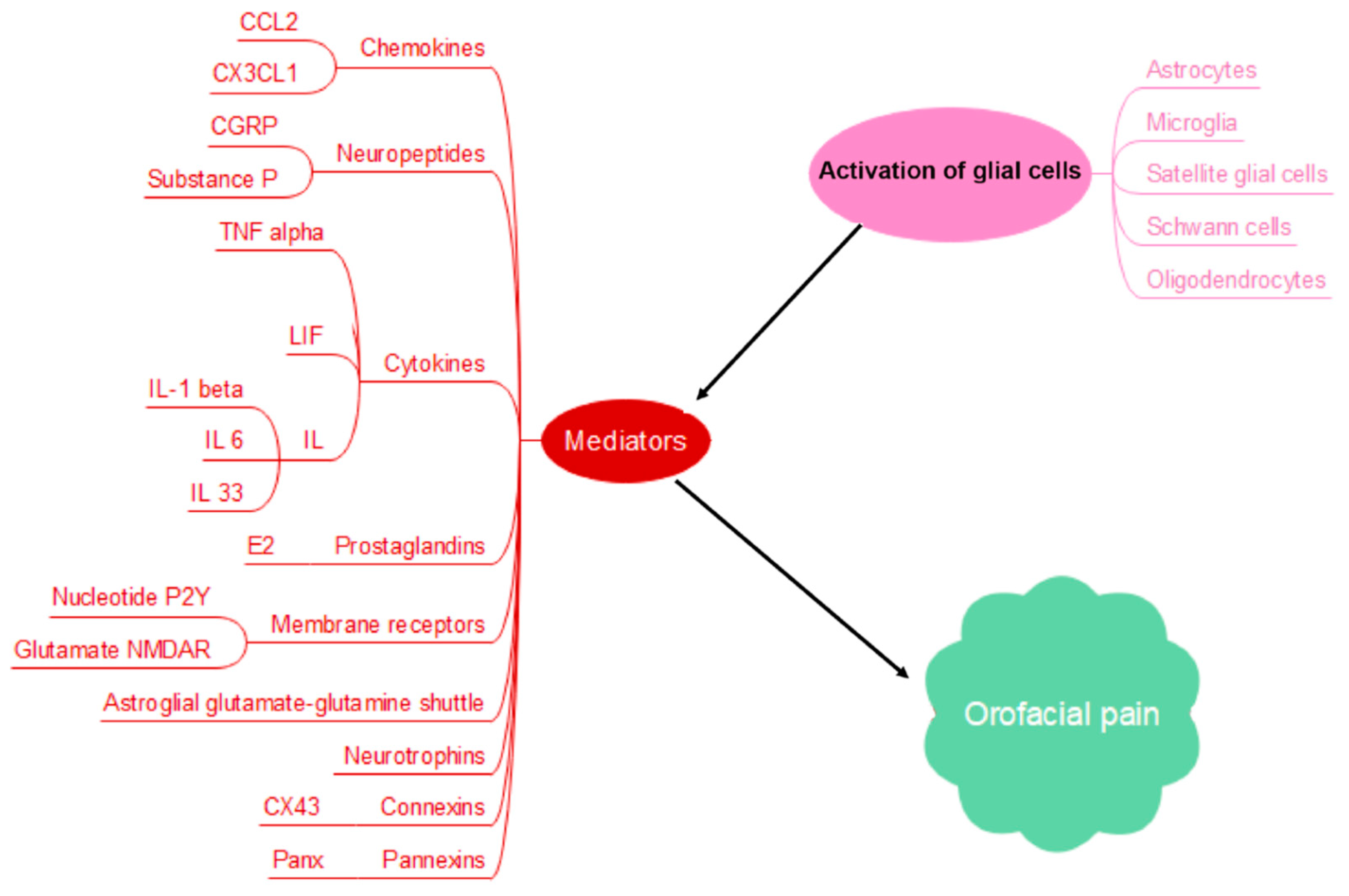
Figure 1. Glial cell activation and immunomodulation of orofacial pain through various mediators, including chemokines like CCL2 and CX3CL1, neuropeptides like calcitonin gene-related peptide (CGRP) and Substance P, cytokines like TNFα, neurotrophins like BDNF and NGF, leukemia inhibitory factor (LIF), interleukins (IL-1β, IL-6, IL33), receptors like nucleotide P2Y receptors and glutamate N-methyl-d-aspartate receptor (NMDAR), prostaglandins like E2, astroglial glutamate–glutamine shuttle, gap junctions connexins like CX43, and pannexins like Panx.
Biomedicines 13 00434 g001
Figure 2. Orofacial pain pathophysiology is controlled by a multitude of factors encompassing metabolic and epigenetic elements. The metabolic factors include glucose and lipid metabolites along with reactive species regulating mitochondrial function, and the epigenetic mechanism is mainly composed of DNA methylation and histone modification.
Biomedicines 13 00434 g002
Figure 3. A holistic approach integrating immunomodulatory, metabolic and epigenetic regulation of pain pathways to fully elucidate mechanisms underlying pain pathophysiology.
Biomedicines 13 00434 g003
Disclaimer/Publisher’s Note: The statements, opinions and data contained in all publications are solely those of the individual author(s) and contributor(s) and not of MDPI and/or the editor(s). MDPI and/or the editor(s) disclaim responsibility for any injury to people or property resulting from any ideas, methods, instructions or products referred to in the content.