
Investigation of UCHL3 and HNMT Gene Polymorphisms in Greek Patients with Type 2 Diabetes Mellitus and Diabetic Retinopathy

 ,
,  , and
, and
Abstract
1. Introduction
2. Materials and Methods
2.1. Study Design and Population
2.2. Genotyping
2.3. Statistical Analysis
3. Results
4. Discussion
5. Conclusions
Author Contributions
Funding
Institutional Review Board Statement
Informed Consent Statement
Data Availability Statement
Conflicts of Interest
Abbreviations
| DR | diabetic retinopathy |
| SNP | single-nucleotide polymorphism |
| T2DM | type 2 diabetes mellitus |
| NDR | non-diabetic retinopathy |
| HbA1c | hemoglobin A1c |
| DM | diabetes mellitus |
| T1DM | type 1 diabetes mellitus |
| ETDRS | Early Treatment Diabetic Retinopathy Study |
| NPDR | Non-Proliferative Diabetic Retinopathy |
| IRMAs | intraretinal microvascular abnormalities |
| PDR | Proliferative Diabetic Retinopathy |
| DME | Diabetic Macular Edema |
| BMI | Body Mass Index |
| SBP | systolic blood pressure |
| DBP | diastolic blood pressure |
| GFR | glomerular filtration rate |
| OCT | Optical Coherence Tomography |
| OCT-A | Optical Coherence Tomography–Angiography |
| IQRs | interquartile ranges |
| OR | odds ratio |
| CI | confidence interval |
| AMD | age-related macular degeneration |
| RR | risk ratio |
| INSR | insulin receptor |
References
- Ulbig, M.W.; Kollias, A.N. Diabetische Retinopathie: Frühzeitige Diagnostik Und Effiziente Therapie. Dtsch. Arztebl. 2010, 107, 75–84. [Google Scholar] [CrossRef]
- Cheung, N.; Mitchell, P.; Wong, T.Y. Diabetic Retinopathy. Lancet 2010, 376, 124–136. [Google Scholar] [CrossRef]
- Hou, Y.; Cai, Y.; Jia, Z.; Shi, S. Risk Factors and Prevalence of Diabetic Retinopathy: A Protocol for Meta-Analysis. Medicine 2020, 99, E22695. [Google Scholar] [CrossRef]
- Heng, L.Z.; Comyn, O.; Peto, T.; Tadros, C.; Ng, E.; Sivaprasad, S.; Hykin, P.G. Diabetic Retinopathy: Pathogenesis, Clinical Grading, Management and Future Developments. Diabet. Med. 2013, 30, 640–650. [Google Scholar] [CrossRef] [PubMed]
- Leley, S.P.; Ciulla, T.A.; Bhatwadekar, A.D. Diabetic Retinopathy in the Aging Population: A Perspective of Pathogenesis and Treatment. Clin. Interv. Aging 2021, 16, 1367–1378. [Google Scholar] [CrossRef] [PubMed]
- Fung, T.H.; Patel, B.; Wilmot, E.G.; Amoaku, W.M. Diabetic Retinopathy for the Non-Ophthalmologist. Clin. Med. 2022, 22, 112–116. [Google Scholar] [CrossRef] [PubMed]
- Li, H.; Liu, X.; Zhong, H.; Fang, J.; Li, X.; Shi, R.; Yu, Q. Research Progress on the Pathogenesis of Diabetic Retinopathy. BMC Ophthalmol. 2023, 23, 372. [Google Scholar] [CrossRef]
- Simó-Servat, O.; Hernández, C.; Simó, R. Diabetic Retinopathy in the Context of Patients with Diabetes. Ophthalmic Res. 2019, 62, 211–217. [Google Scholar] [CrossRef]
- Lin, K.-Y.; Hsih, W.-H.; Lin, Y.-B.; Wen, C.-Y.; Chang, T.-J. Update in the Epidemiology, Risk Factors, Screening, and Treatment of Diabetic Retinopathy. J. Diabetes Investig. 2021, 12, 1322–1325. [Google Scholar] [CrossRef] [PubMed]
- Yue, T.; Shi, Y.; Luo, S.; Weng, J.; Wu, Y.; Zheng, X. The Role of Inflammation in Immune System of Diabetic Retinopathy: Molecular Mechanisms, Pathogenetic Role and Therapeutic Implications. Front. Immunol. 2022, 13, 1055087. [Google Scholar] [CrossRef]
- Sharma, A.; Valle, M.L.; Beveridge, C.; Liu, Y.; Sharma, S. Unraveling the Role of Genetics in the Pathogenesis of Diabetic Retinopathy. Eye 2019, 33, 534–541. [Google Scholar] [CrossRef] [PubMed]
- Sharma, A.; Liu, H.; Tobar-Tosse, F.; Dakal, T.C.; Ludwig, M.; Holz, F.G.; Loeffler, K.U.; Wüllner, U.; Herwig-Carl, M.C. Ubiquitin Carboxyl-Terminal Hydrolases (Uchs): Potential Mediators for Cancer and Neurodegeneration. Int. J. Mol. Sci. 2020, 21, 3910. [Google Scholar] [CrossRef]
- Fang, Y.; Fu, D.; Shen, X.Z. The Potential Role of Ubiquitin C-Terminal Hydrolases in Oncogenesis. Biochim. Biophys. Acta-Rev. Cancer 2010, 1806, 1–6. [Google Scholar] [CrossRef] [PubMed]
- Fang, Y.; Shen, X. Ubiquitin Carboxyl-Terminal Hydrolases: Involvement in Cancer Progression and Clinical Implications. Cancer Metastasis Rev. 2017, 36, 669–682. [Google Scholar] [CrossRef]
- Suzuki, M.; Setsuie, R.; Wada, K. Ubiquitin Carboxyl-Terminal Hydrolase L3 Promotes Insulin Signaling and Adipogenesis. Endocrinology 2009, 150, 5230–5239. [Google Scholar] [CrossRef]
- Sheu, W.H.H.; Kuo, J.Z.; Lee, I.T.; Hung, Y.J.; Lee, W.J.; Tsai, H.Y.; Wang, J.S.; Goodarzi, M.O.; Klein, R.; Klein, B.E.K.; et al. Genome-Wide Association Study in a Chinese Population with Diabetic Retinopathy. Hum. Mol. Genet. 2013, 22, 3165–3173. [Google Scholar] [CrossRef]
- Yoshikawa, T.; Nakamura, T.; Yanai, K. Histamine N-Methyltransferase in the Brain. Int. J. Mol. Sci. 2019, 20, 737. [Google Scholar] [CrossRef] [PubMed]
- Raftogianis, B.B.; Aksoy, S.; Weinshilboum, R.M. Human Histamine N-Methyltransferase Gene: Structural Characterization and Chromosomal Localization. J. Investig. Med. 1996, 44, 548–554. [Google Scholar]
- Jiménez-Jiménez, F.J.; Alonso-Navarro, H.; García-Martín, E.; Agúndez, J.A.G. Thr105Ile (Rs11558538) Polymorphism in the Histamine N -Methyltransferase (HNMT) Gene and Risk for Parkinson Disease. Medicine 2016, 95, e4147. [Google Scholar] [CrossRef] [PubMed]
- American Diabetes Association. Standards of Medical Care in Diabetes—2014. Diabetes Care 2014, 37 (Suppl. S1), S14–S80. [Google Scholar] [CrossRef]
- Gouliopoulos, N.; Siasos, G.; Bouratzis, N.; Oikonomou, E.; Kollia, C.; Konsola, T.; Oikonomou, D.; Rouvas, A.; Kassi, E.; Tousoulis, D.; et al. Polymorphism Analysis of ADIPOQ Gene in Greek Patients with Diabetic Retinopathy. Ophthalmic Genet. 2022, 43, 326–331. [Google Scholar] [CrossRef] [PubMed]
- Dai, C.; Zhang, Y.; Zhan, X.; Tian, M.; Pang, H. Association Analyses of SNAP25, HNMT, FCHSD1, and DBH Single-Nucleotide Polymorphisms with Parkinson’ s Disease in a Northern Chinese Population. Neuropsychiatr. Dis. Treat. 2021, 17, 1689–1695. [Google Scholar] [CrossRef] [PubMed]
- Dulull, N.; Kwa, F.; Osman, N.; Rai, U.; Shaikh, B.; Thrimawithana, T.R. Recent Advances in the Management of Diabetic Retinopathy. Drug Discov. Today 2019, 24, 1499–1509. [Google Scholar] [CrossRef] [PubMed]
- Gale, M.J.; Scruggs, B.A.; Flaxel, C.J. Diabetic Eye Disease: A Review of Screening and Management Recommendations. Clin. Exp. Ophthalmol. 2021, 49, 128–145. [Google Scholar] [CrossRef] [PubMed]
- Torm, M.E.W.; Dorweiler, T.F.; Fickweiler, W.; Levine, S.R.; Fort, P.E.; Sun, J.K.; Gardner, T.W. Frontiers in Diabetic Retinal Disease. J. Diabetes Complicat. 2023, 37, 108386. [Google Scholar] [CrossRef]
- Le, H.G.; Shakoor, A. Diabetic and Retinal Vascular Eye Disease. Med. Clin. N. Am. 2021, 105, 455–472. [Google Scholar] [CrossRef] [PubMed]
- Sun, W.J.; An, X.D.; Zhang, Y.H.; Zhao, X.F.; Sun, Y.T.; Yang, C.Q.; Kang, X.M.; Jiang, L.L.; Ji, H.Y.; Lian, F.M. The Ideal Treatment Timing for Diabetic Retinopathy: The Molecular Pathological Mechanisms Underlying Early-Stage Diabetic Retinopathy Are a Matter of Concern. Front. Endocrinol. 2023, 14, 1270145. [Google Scholar] [CrossRef] [PubMed]
- Whitehead, M.; Wickremasinghe, S.; Osborne, A.; Van Wijngaarden, P.; Martin, K.R. Diabetic Retinopathy: A Complex Pathophysiology Requiring Novel Therapeutic Strategies. Expert Opin. Biol. Ther. 2018, 18, 1257–1270. [Google Scholar] [CrossRef] [PubMed]
- Forrester, J.V.; Kuffova, L.; Delibegovic, M. The Role of Inflammation in Diabetic Retinopathy. Front. Immunol. 2020, 11, 10319–10329. [Google Scholar] [CrossRef] [PubMed]


| Variable | NDR n = 71 (45.5) | DR n = 85 (54.5) | Total n = 156 (100) | p-Value |
|---|---|---|---|---|
| Female | 28 (39.4) | 26 (30.6) | 54 (34.6) | 0.25 |
| Age (years) | 74 [67–80] | 69 [65–74] | 72 [65.3–77] | 0.01 |
| BMI (kg/m2) | 27.7 [25.7–30.4] | 27.8 [25.9–30.3] | 27.7 [25.7–30.3] | 0.95 |
| DM duration (years) | 14 [6–18] | 21 [13.5–29] | 17 [10–26.75] | <0.01 |
| HbA1c (%) | 6.5 [6.1–7] | 6.9 [6.3–7.2] | 6.7 [6.2–7.1] | 0.01 |
| NPDR | - | 63 (74.1) | - | |
| Duration DR (years) | - | 12 [7–18] | - | |
| Macular Diabetic Edema | - | 61 (71.8) | - | |
| Argon Laser Photocoagulation | 0 (0) | 19 (22.4) | 19 (12.2) | <0.01 |
| Intravitreal injections | 9 (12.7) | 65 (76.5) | 74 (47.4) | <0.01 |
| Vitrectomy | 8 (11.3) | 19 (22.4) | 27 (17.3) | 0.07 |
| Cataract surgery | 29 (40.8) | 40 (47.1) | 69 (44.2) | 0.44 |
| Glaucoma | 11 (15.5) | 19 (22.4) | 30 (19.2) | 0.28 |
| Dry AMD | 23 (32.4) | 5 (5.9) | 28 (17.9) | <0.01 |
| Wet AMD | 17 (23.9) | 0 (0) | 17 (10.9) | <0.01 |
| Tablets | 70 (98.6) | 65 (76.5) | 135 (86.5) | <0.01 |
| Insulin | 7 (9.9) | 35 (41.2) | 42 (26.9) | <0.01 |
| Hypertension | 57 (80.3) | 67 (78.8) | 124 (79.5) | 0.82 |
| Smoker | 47 (66.2) | 56 (65.9) | 103 (66) | 0.97 |
| Smoking (pack-years) | 31 [0–65] | 41 [0–64.5] | 36 [0–65] | 0.59 |
| Alcohol | 14 (19.7) | 14 (16.5) | 28 (17.9) | 0.60 |
| Cardiovascular disease | 27 (38) | 40 (47.1) | 67 (42.9) | 0.26 |
| Stroke | 4 (5.6) | 6 (7.1) | 10 (6.4) | 0.72 |
| Dyslipidemia | 49 (69) | 70 (82.4) | 119 (76.3) | 0.05 |
| Anemia | 0 (0) | 1 (1.2) | 1 (0.6) | 0.36 |
| Nephropathy | 4 (5.6) | 7 (8.2) | 11 (7.1) | 0.53 |
| Ca | 7 (9.9) | 1 (1.2) | 8 (5.1) | 0.01 |
| Benign prostate hyperplasia | 5 (7) | 6 (7.1) | 11 (7.1) | 1 |
| Hypothyroidism | 7 (9.9) | 5 (5.9) | 12 (7.7) | 0.35 |
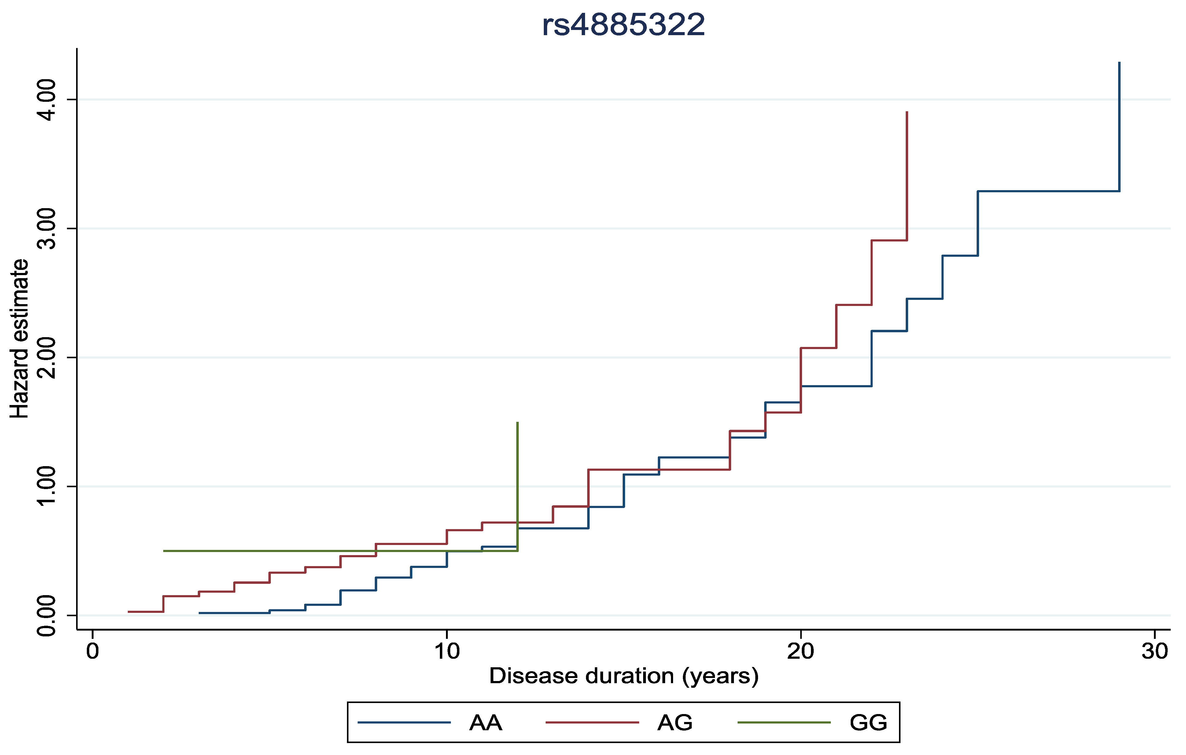
| rs4885322 | ||||
| AA | 55 (77.5) | 49 (57.6) | 104 (66.7) | |
| AG | 15 (21.1) | 34 (40) | 49 (31.4) | |
| GG | 1 (1.4) | 2 (2.4) | 3 (1.9) | 0.03 |
| rs11558538 | ||||
| CC | 64 (90.1) | 64 (75.3) | 128 (82.1) | |
| CT | 7 (9.9) | 20 (23.5) | 27 (17.3) | |
| TT | 0 (0) | 1 (1.2) | 1 (0.6) | 0.05 |
| Main | Excluding Extreme BMI (n = 3) | |||
|---|---|---|---|---|
| OR (95% CI) | p-Value | OR (95% CI) | p-Value | |
| rs4885322 | ||||
| per allele | 2.04 (1.03, 4.04) | 0.04 | 2.02 (1.01, 4.04) | 0.05 |
| GG vs. AA+AG | 1.35 (0.10, 18.00) | 0.82 | 1.31 (0.10, 16.97) | 0.84 |
| AG+GG vs. AA | 2.23 (1.07, 4.64) | 0.03 | 2.22 (1.06, 4.67) | 0.04 |
| G vs. A | 1.87 (0.98, 3.55) | 0.06 | 1.86 (0.97, 3.58) | 0.06 |
| rs11558538 | ||||
| per allele | 3.27 (1.27, 8.39) | 0.01 | 3.59 (1.34, 9.62) | 0.01 |
| TT vs. CC+CT | - | - | - | - |
| CT+TT vs. CC | 3.31 (1.27, 8.61) | 0.01 | 3.64 (1.34, 9.89) | 0.01 |
| T vs. C | 3.05 (1.24, 7.52) | 0.02 | 3.36 (1.30, 8.67) | 0.01 |
| RR (95% CI) | p-Value | |
|---|---|---|
| rs4885322 | ||
| per allele | 0.53 (0.19, 1.48) | 0.225 |
| AG+GG vs. AA | 0.55 (0.18, 1.64) | 0.283 |
| G vs. A | 0.60 (0.24, 1.51) | 0.279 |
| rs11558538 | ||
| per allele | 0.56 (0.17, 1.86) | 0.345 |
| CT+TT vs. CC | 0.57 (0.16, 2.00) | 0.382 |
| T vs. C | 0.58 (0.18, 1.85) | 0.360 |
Disclaimer/Publisher’s Note: The statements, opinions and data contained in all publications are solely those of the individual author(s) and contributor(s) and not of MDPI and/or the editor(s). MDPI and/or the editor(s) disclaim responsibility for any injury to people or property resulting from any ideas, methods, instructions or products referred to in the content. |
© 2025 by the authors. Licensee MDPI, Basel, Switzerland. This article is an open access article distributed under the terms and conditions of the Creative Commons Attribution (CC BY) license (https://creativecommons.org/licenses/by/4.0/).
Share and Cite
Flindris, K.; Lagkada, V.; Christodoulou, A.; Gazouli, M.; Moschos, M.; Markozannes, G.; Kitsos, G. Investigation of UCHL3 and HNMT Gene Polymorphisms in Greek Patients with Type 2 Diabetes Mellitus and Diabetic Retinopathy. Biomedicines 2025, 13, 341. https://doi.org/10.3390/biomedicines13020341
Flindris K, Lagkada V, Christodoulou A, Gazouli M, Moschos M, Markozannes G, Kitsos G. Investigation of UCHL3 and HNMT Gene Polymorphisms in Greek Patients with Type 2 Diabetes Mellitus and Diabetic Retinopathy. Biomedicines. 2025; 13(2):341. https://doi.org/10.3390/biomedicines13020341
Chicago/Turabian StyleFlindris, Konstantinos, Vivian Lagkada, Aikaterini Christodoulou, Maria Gazouli, Marilita Moschos, Georgios Markozannes, and George Kitsos. 2025. "Investigation of UCHL3 and HNMT Gene Polymorphisms in Greek Patients with Type 2 Diabetes Mellitus and Diabetic Retinopathy" Biomedicines 13, no. 2: 341. https://doi.org/10.3390/biomedicines13020341
APA StyleFlindris, K., Lagkada, V., Christodoulou, A., Gazouli, M., Moschos, M., Markozannes, G., & Kitsos, G. (2025). Investigation of UCHL3 and HNMT Gene Polymorphisms in Greek Patients with Type 2 Diabetes Mellitus and Diabetic Retinopathy. Biomedicines, 13(2), 341. https://doi.org/10.3390/biomedicines13020341