Review

Clinical and Molecular Insights of Arterial and Venous Thrombosis in Myeloproliferative Diseases—Case-Based Narrative Review

by
Anca Drăgan
1,*,
Mădălina Găvănescu
2,
Adrian Ştefan Drăgan
3,*,
Alexandru Bardaş
4,
Monica Dobrovie
5 and
Anca Doina Mateescu
3,6
1
Department of Cardiovascular Anaesthesiology and Intensive Care, “Prof. Dr. C.C. Iliescu” Emergency Institute for Cardiovascular Diseases, 258 Fundeni Road, 022328 Bucharest, Romania
2
Department of Vascular Surgery, “Prof. Dr. C.C. Iliescu” Emergency Institute for Cardiovascular Diseases, 258 Fundeni Road, 022328 Bucharest, Romania
3
Faculty of General Medicine, Carol Davila University of Medicine and Pharmacy, 8 Eroii Sanitari Boulevard, 050474 Bucharest, Romania
4
Department of Hematology and Bone Marrow Transplant, Fundeni Clinical Institute, 258 Fundeni Road, 022328 Bucharest, Romania
5
Department of Radiology, Fundeni Clinical Institute, 258 Fundeni Road, 022328 Bucharest, Romania
6
Department of Cardiology, “Prof. Dr. C.C. Iliescu” Emergency Institute for Cardiovascular Diseases, 258 Fundeni Road, 022328 Bucharest, Romania
*
Authors to whom correspondence should be addressed.
Biomedicines 2025, 13(10), 2543; https://doi.org/10.3390/biomedicines13102543 (registering DOI)
Submission received: 7 September 2025 / Revised: 14 October 2025 / Accepted: 16 October 2025 / Published: 18 October 2025
(This article belongs to the Section Molecular and Translational Medicine)

Abstract

The myeloproliferative neoplasms (MPN), a heterogeneous group of disorders characterized by specific genetic mutations, have the development of arterial and venous thrombosis as their main complication. Almost 40–50% of MPN patients encountered arterial or venous thrombosis during the course of their disease. Moreover, arterial thrombosis is linked to significant mortality, progression to myelofibrosis, and an increased risk of developing second cancers. Despite significant advancements in medical research, there are still unmet needs in this field. Our narrative review provides clinical and genetic insights into thrombosis associated with myeloproliferative neoplasms. We focus on the underlying pathophysiological processes, assessment methods, and risk stratification related to thrombotic events. This information aims to assist clinicians in accurately assessing the risks associated with MPN thrombosis, enabling a more personalized and effective approach to patient care. We based our review on a rare case of MPN-associated thrombosis, whose clinical presentation was marked by acute ischemia in both lower limbs. The thrombosis affected the distal aortic arch, thoracic and abdominal aorta, celiac trunk, common and proper hepatic arteries, proximal left renal artery, several segmental arteries in the right kidney, and the portal vein thrombosis. Our review presents various therapeutic options for these conditions. In the presented case, the multiple thrombi were treated medically, except for the popliteal artery thromboses, which required surgical management. This case may serve as a valuable reference for choosing treatment options for aortic and portal vein thrombosis, highlighting the multidisciplinary approach.

1. Introduction

BCR-ABL1-negative myeloproliferative neoplasms (MPN) represent a heterogeneous group of disorders characterized by specific genetic mutations that result in the abnormal clonal expansion of the hematopoietic cell proliferation, leading to the overproduction of leukocytes, erythrocytes, or platelets. The main MPN types include polycythemia vera (PV), essential thrombocythemia (ET), primary myelofibrosis (PMF), and unclassifiable MPN (MPN-U) [1,2].
The MPN complications are related to the development of bone marrow fibrosis, leukemic transformation, and arterial and venous thrombosis [3]. Zhang et al. reported that 40–50% of patients with MPN encountered arterial or venous thrombosis during the course of their disease [4]. Notably, arterial thrombosis is linked to significant mortality [5,6,7,8,9], progression to myelofibrosis [5,6,7], and an increased risk of developing second cancers [5,10,11,12]. Thrombosis may arise prior to, at the time of, or subsequent to the diagnosis of MPN. Hultcrantz et al. demonstrated that the rate of thrombosis was significantly higher in MPN patients compared to the general population, with the highest hazard ratio observed shortly after the MPN diagnosis [13]. It is well known that thrombosis in MPN occurs in the medium-sized arteries and veins, and also in the microvessels [14]. Unusual sites of venous thrombosis include upper extremity veins, splanchnic veins, cerebral veins, and retinal veins, and unusual sites of arterial thrombosis include renal, adrenal, splenic and mesenteric arteries, and intracardiac and aortal locations [15]. Despite remarkable advancements in medical research, the persistent prevalence of arterial and venous thrombosis remains a serious challenge in the healthcare landscape. Current efforts to assess and stratify thrombotic risk have progressed from conventional methods to sophisticated risk scores and machine learning algorithms, all designed to tailor patient management more effectively. Moreover, gaining a deeper understanding of the pathophysiology of thrombotic events in the MPN context is crucial; this knowledge not only enhances risk assessment but also paves the way for targeted and more effective treatments. Furthermore, it is imperative to raise awareness of these serious manifestations and embrace a multidisciplinary approach to their management. By doing so, we can improve outcomes and enhance the quality of care for patients facing these conditions.
Our narrative review offers clinical and genetic insights into thrombosis associated with myeloproliferative neoplasms. We focus on pathophysiological processes, assessment methods, and risk stratification related to thrombotic events. This information aims to assist clinicians in accurately assessing the risks associated with MPN thrombosis, enabling a more personalized and effective approach to patient care. To illustrate the complexities of this condition, we present a rare case of MPN-U associated thrombosis affecting the distal aortic arch, thoracic and abdominal aorta, celiac trunk, common and proper hepatic arteries, proximal left renal artery, as well as several segmental arteries in the right kidney. Additionally, we report portal vein thrombosis (PVT) in this case. The clinical presentation was marked by acute ischemia in both lower limbs, highlighting the need for a multidisciplinary approach to management. Previously reported cases of MPN thrombosis in large vessels, such as the aorta and iliac artery, have typically required surgical intervention [16]. However, urgent surgical treatment of aortic thrombi is associated with a high mortality rate. There are only a few documented instances of medical management for an MPN-associated aortic thrombus [17]. Similarly, PVT is rare and can sometimes be the first clinical indication of an underlying MPN [18]. Our review presents various therapeutic options for these conditions. In our case, multiple thrombi were treated medically, except for the popliteal artery thromboses, which required different management. This case may serve as a valuable reference for choosing treatment options for aortic and portal vein thrombosis.

2. Case Presentation

A 64-year-old woman was referred to our hospital with acute bilateral lower limb ischemia. Our patient complained of sudden coldness, paresthesia, and pain in the lower limbs that began 8 h before the presentation to the emergency department. Apparently, the patient was in good health, without a significant medical history, except for a diagnosis of uninvestigated thrombocytosis and leukocytosis for almost one year. Moreover, she described diffuse abdominal pain for the past three months, which she had ignored. The patient denied fever, cough, headache, melena, and tarry stool. She had no chest pain, syncope, or palpitations. There was no family history of coagulation disorders. She denied a personal or family history of cerebrovascular attack and ischemic heart disease. The patient had no associated cardiovascular risk factors and was not taking any medications at home.
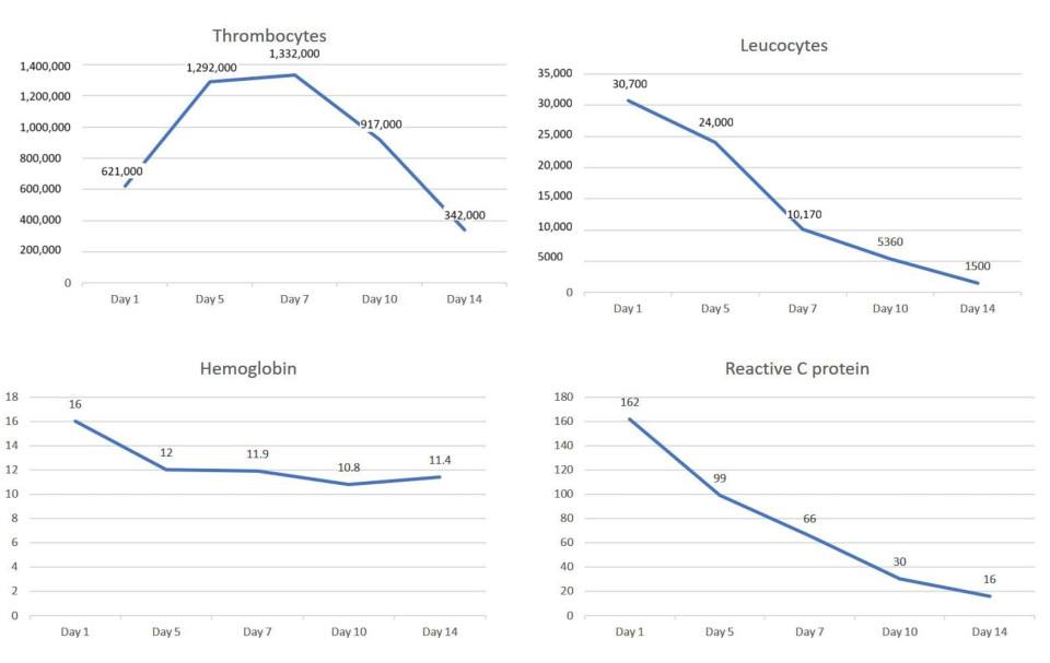
Examination in the emergency room showed the abolition of both the right and left pedal and posterior tibial arterial pulses. The other peripheral pulses were present. Both lower limbs were pale and cold, with mild loss of sensibility limited to the left foot. Normal motor function was evidenced. Otherwise, her physical examination revealed no other abnormal clinical signs or symptoms, except for an enlarged spleen. Vital signs were within normal range: a blood pressure of 140/64 mmHg, a regular pulse rate of 76/min, and a respiratory rate of 16 per/minute. Her body temperature was 36.7 °C. At admission, our patient had a leukocytosisof 31 × 103/μL with 93% neutrophils and 3% lymphocytes. a hemoglobin level of 16 g/dL, and platelet count of 621 × 103/μL. A mild increase in liver enzymes was also present (AST/ALT of 167/204 UI/L). No other abnormalities were detected in other standard laboratory results. No proteinuria nor hematuria was found. Doppler ultrasound examination confirmed the occlusion of the arterial trunks in both lower limbs. Cardiological evaluation in the emergency department revealed a normal electrocardiogram with no cardiac abnormalities at transthoracic echocardiography.
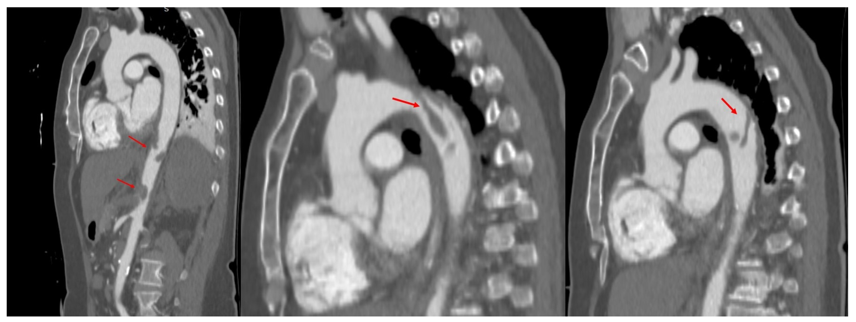
Emergency surgery was necessary with a diagnosis of acute bilateral lower limb ischemia. Under general anesthesia, bilateral popliteal and tibial arterial axis dethrombosis with restoration of arterial flow was performed via the distal popliteal route with removal of multiple thrombi. However, the etiology of the acute lower limb ischemia remained uncertain in a patient in sinus rhythm. Moreover, the transesophageal echocardiography (TOE) performed in the operating room revealed multiple thrombi of various forms, highly mobile, attached to the aortic arch and the wall of the entire descending thoracic aorta. (Figure 1). Doppler ultrasound of the extracranial carotid arteries did not reveal any abnormalities.
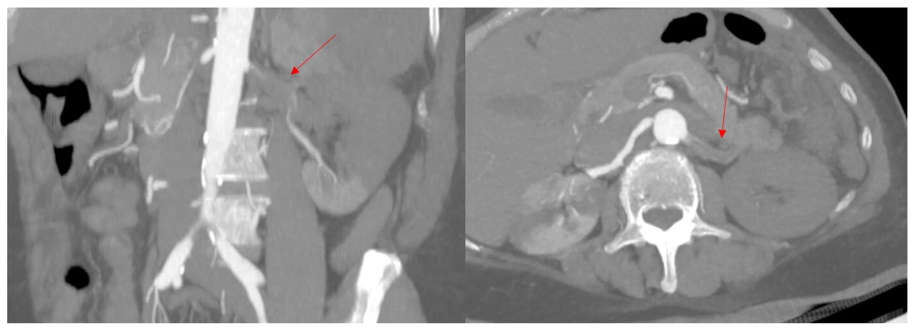
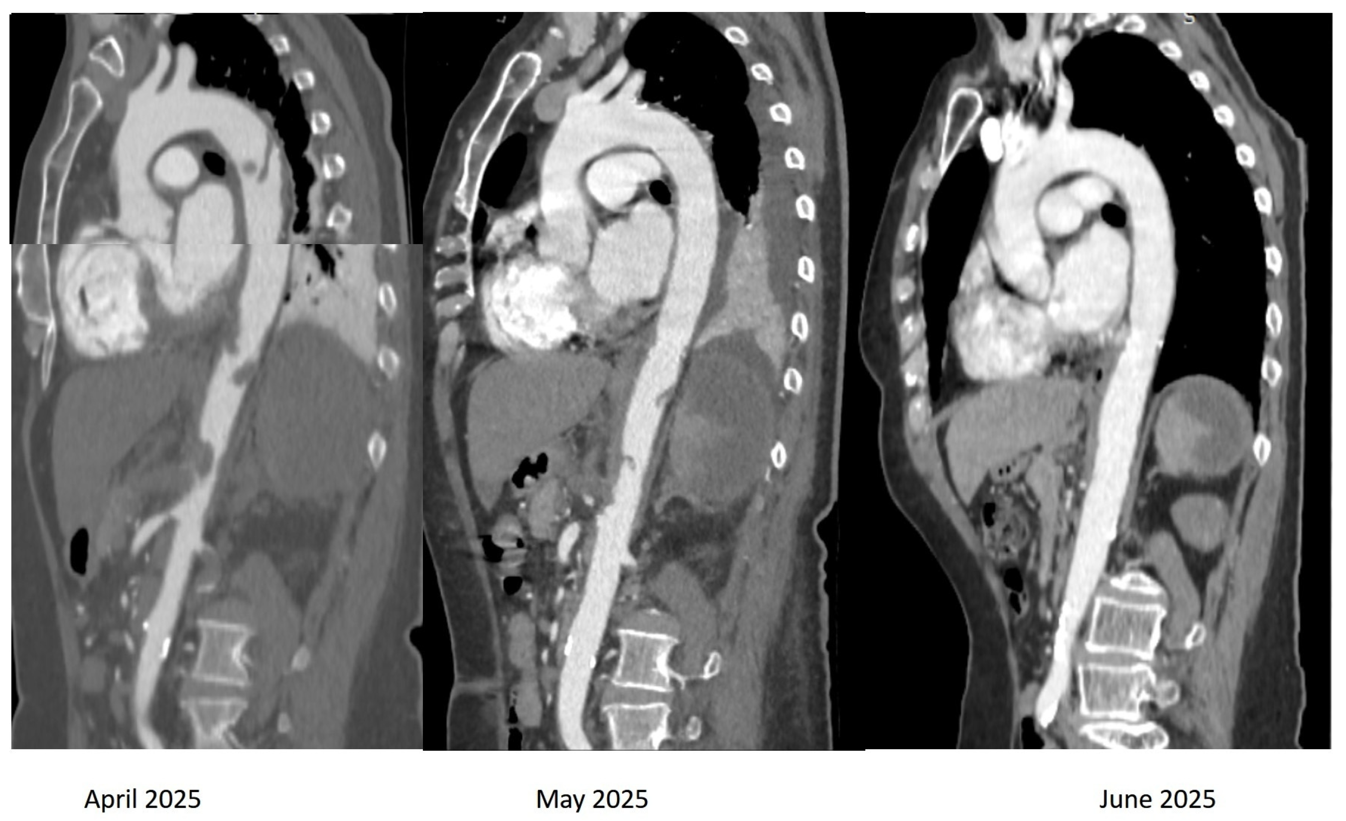
Due to TOE findings, we performed, early postoperatively, a computed tomography (CT) scan of her thorax and abdomen, which showed the presence of multiple thrombi in the distal aortic arch, thoracic, and abdominal aorta, celiac trunk, common and proper hepatic arteries, proximal left renal artery, and some segmental arteries at the level of the right kidney. Arteriosclerosis was not recognized (Figure 2 and Figure 3).
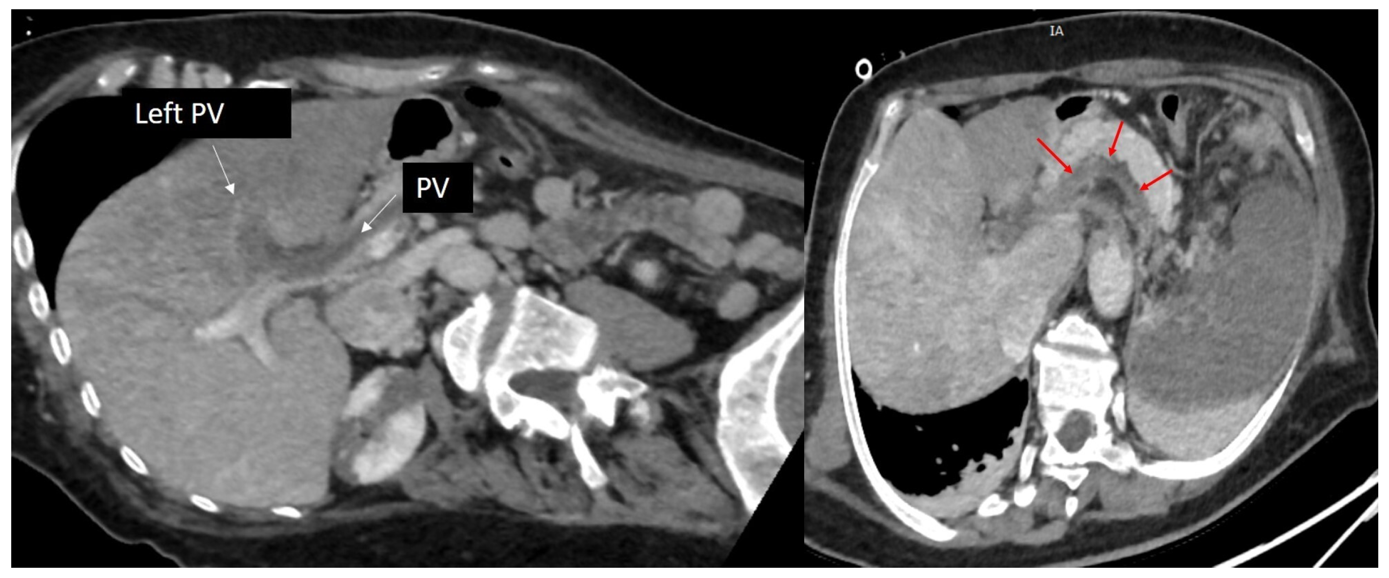
Moreover, there was a completely obstructive thrombosis of the left portal vein, extended to the portal vein trunk and the splenic vein, while the superior mesenteric vein and the iliac veins remained permeable (Figure 4).
Vascular-ischemic changes were noted in the spleen (almost total damage), left kidney (extensive ischemic changes in the upper pole, in the middle third, and partial changes in the lower pole), right kidney (partial ischemic changes in the upper pole and in one-third), and the left hepatic lobe. There was a fluid accumulation adjacent to the pericardium on the left side, measuring 97/57 mm, most likely due to left incised pleurisy. The liver exhibited ischemic changes in the left lobe, accompanied by perfusion disorders in the pancreas and adrenal glands. No signs of oncological disease were detected at the CT scan.
At admission to the intensive care unit (ICU), the parenteral anticoagulant therapy with unfractioned heparin, along with aspirin as antiplatelet treatment, was started. The postoperative course was favorable and uneventful, with the patient being fast extubated in the ICU. Hydroxyurea was initiated on the second day of her admission and uptitrated to a maximum dose of 3g/day (Figure 5).
On the 11th day of admission, the dose of hydroxyurea was reduced to 2 g/day. The numbness in both of her lower extremities disappeared within two days. Additionally, the number and dimensions of the aortic thrombi decreased at the repeated TOE and CT-scan on the 7th day of her hospital stay (Figure 6).
The hematological evaluation raised the suspicion of a myeloproliferative disorder. The bone marrow aspirate revealed a hypercellular marrow and panmyelosis, an appearance compatible with a chronic MPN-U. Moreover, the JAK2 V617F mutation burden was 13%. The hematologist diagnosed this case as MPN-U JAK2 V617F positive, and the aortic thrombus was thought to be associated with MPN. An MPN-U is a hematological neoplasm characterized by clonal proliferation of myeloid precursors in the bone marrow, blood, and other tissues (spleen, liver); however, it does not meet the specific diagnostic criteria for a distinct type of MPN. It is a diagnosis of exclusion used when clinical, morphological, and molecular features of an MPN are present, but insufficient for a definitive classification into categories like PV or essential thrombocythemia. On one hand, it is characterized by dysplasia and ineffective hematopoiesis, and on the other hand, it is characterized by an increase in the number of more than one type of myeloid lineage. Symptoms can be non-specific and variable, including fatigue, weight loss, and splenomegaly and hepatomegaly, particularly in later stages.
The patient was transferred to the Hematological Department, where she continued medical treatment with enoxaparin 1 mg/kg twice-daily initially, aspirin 75 mg/day, and hydroxyurea 1.5 g/day. At discharge, she received novel anticoagulant therapy with apixaban 5 mg bi-daily along with aspirin 75 mg/day and hydroxyurea 1 g/day for 5 days/week and 1.5g/day for 2 days/week. At the 3-month CT follow-up (Figure 6), we noted the disappearance of the aortic thrombus. However, CT revealed persistent stenosis at the origin of the celiac trunk, persistent left PVT with small adjacent collaterals, and post-thrombotic occlusion of the splenic vein with collateral formation and necrosis of about 80% of the splenic volume. The patient presented no symptoms at 6-month follow-up.

3. Insights into Pathophysiological Mechanisms of Thrombosis in Myeloproliferative Neoplasms

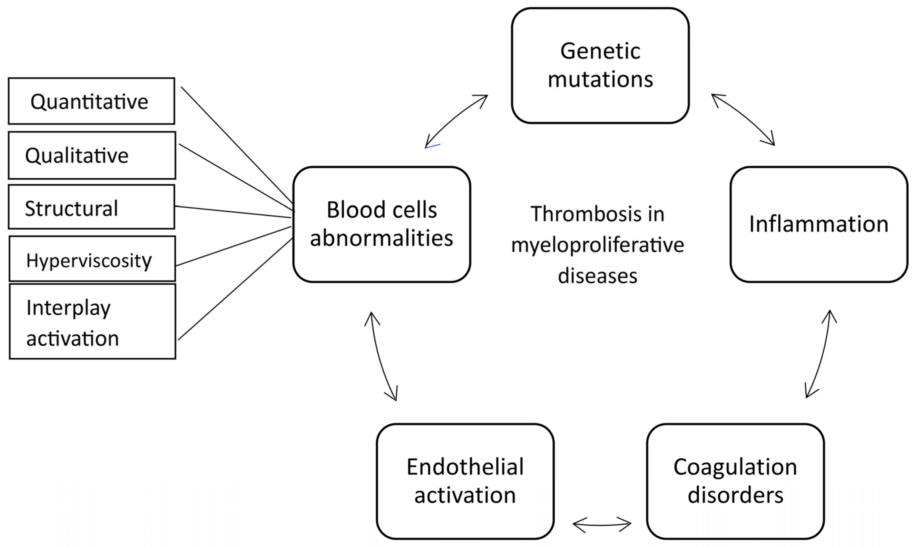
The mechanism of thrombosis in MPN is complex and multifactorial. Various components, including the quantity and activation patterns of blood cells, endothelial involvement, coagulation disorders, and thrombo-inflammatory processes, all play significant roles in this phenomenon (Figure 7).

3.1. Genetic Mutations

Notably, genetic mutations exert a substantial influence on the overall environment, thereby impacting thrombosis development. JAK2/CALR/MPL gene mutations are the driver mutations in MPN [19,20]. One of the most common mutations in the Janus kinase 2 (JAK2) gene is the V617F, which is a somatic gain-of-function mutation. This mutation alters the 1849th coding nucleotide, changing it from guanine (G) to thymine (T), resulting in the replacement of the amino acid valine with phenylalanine [20]. JAK2 encodes a non-receptor tyrosine kinase involved in cell growth, differentiation, development, or histone modification. JAK2 facilitates the phosphorylation of STAT proteins, which, upon phosphorylation, dimerize in the cytoplasm and are transported into the nucleus to activate specific genes. Among these mutations, JAK2V617F was the most prevalent driver mutation in MPN associated with thrombotic complications. Moreover, thrombosis may be exacerbated by co-occurring mutations such as TET2, DNMT3A, and ASXL1 [19,20].
Research suggested that even in populations lacking a formal MPN diagnosis, individuals with a JAK2V617F allele burden above 1% faced a heightened risk of venous thromboembolism compared to those without the mutation [21]. Additionally, Cordua et al. reported a JAK2 V617F prevalence of 3.1% in their study of the Danish general population [21]. In MPN diseases, Vignoli et al. demonstrated that patients with more than 50% JAK2V617F allele burden had a significantly higher platelet adhesion capacity compared to subjects with less than 50% JAK2V617F allele burden, thus promoting thrombosis [22].
A single mutation in the JAK2 gene cannot fully explain the diverse MPN landscape. While no link has been established between MPL mutations and thrombosis, a significant correlation has been reported between the JAK2 V617F mutation and ASXL1 gene mutations in relation to thrombotic events [20]. However, the underlying mechanism related to the ASXL1 mutation remains incompletely understood [20]. Furthermore, mutations in the TET2 gene have been identified in approximately 15% of MPN patients [23]. TET2 plays various roles during hematopoiesis, including stem cell self-renewal, lineage commitment, and the terminal differentiation of monocytes. Additionally, TET2 facilitates JAK2 or STAT5 signal transduction and modulates the epigenetic composition of genomic DNA, influencing both DNA and histone methylation and acetylation [23]. The order in which mutations occur influences the proliferative response to JAK2 V617F and affects the ability of double-mutant hematopoietic and progenitor cells to form colonies. In patients where TET2 mutations occurred first, the hematopoietic stem and progenitor cell compartment was primarily made up of TET2 single-mutant cells. In contrast, in patients where JAK2 mutations occurred first, the compartment was dominated by JAK2-TET2 double-mutant cells. Prior mutation of TET2 altered the transcriptional effects of JAK2 V617F in a cell-intrinsic manner, preventing JAK2 V617F from upregulating genes associated with proliferation [24]. DNMT3A mutations, an independent risk factor for arterial thrombosis in MPN, leads to aggressive inflammatory transcriptome, enhance macrophage activity and stimulate proinflammatory T-cell polarization [25]. Morath et al. suggested a similar relationship for DNMT3A with JAK2 V617F as TET2 in thrombotic events [25]. MPN thrombosis is also related to epigenetic dysregulations, that refers to the mutations in genes that encode proteins which regulate the chromatin structure (ASXL1, TET2, IDH1/2, EZH2, IKZF1, JAK2 V617F, and PRMT5), and to the methylation status of promoter sites of genes that coordinate cell growth, differentiation, and survival [20].

3.2. The Coagulation Disorders and Platelet Involvement

Virchow’s triad described the conditions that lead to thrombosis endothelial injury/dysfunction, stasis of blood flow, and hypercoagulability. Studies have shown that MPN patients exhibit a continuous hypercoagulable state, even in the absence of overt thrombosis [26]. Levels of proteins C and S were significantly lower in MPN patients compared to control subjects [26]. Whole blood rotational thromboelastometry (ROTEM) revealed a hypercoagulable profile in MPN patients [27]. Giaccherini et al. showed that the ROTEM parameters were significantly affected by platelet count. After adjusting for platelet count, the maximum clot firmness (MCF) values indicated reduced platelet reactivity in MPN patients, supporting the hypothesis that platelet function becomes exhausted during clotting activation [27]. Higher mean MCF values were also found in the ET and PV settings by Şahin et al. [28]. Additionally, the PDW and (mean platelet volume) MPV values suggested the presence of large-sized platelets, with a higher variation in size, especially in ET patients [26]. Moreover, the increase in thrombin generation may be attributed to platelets that arise from the clonal process, resulting in ongoing activation of the TGF-beta/ALK5 and PAR-2/PAR-1 signaling pathways [29].
Recently, Ross et al. demonstrated that there may be a thrombotic risk associated with increased mitochondrial mass in platelets from MPN patients, although no correlation between the variant allele frequency (VAF) of JAK2 V617F (measured in leukocyte DNA) and mitochondrial mass in platelets was found [30]. They found that the mitochondrial membrane potential is altered in circulating platelets before stimulation, and that MPN platelets are more sensitive to mitochondrial membrane depolarization, leading to the formation of balloon-shaped platelets. Additionally, mitochondria in MPN platelets may have a greater tendency to be released as microparticles [30].

3.3. The Leukocytes Involvement

Platelet activation has also the capacity to stimulate neutrophils [19]. Vignoli et al. reported that platelet P-selectin binding to PSGL-1 activates neutrophils, leading to increased surface CD11b expression, cathepsin-G release from neutrophil granules, and the generation of procoagulant microparticles from both platelets and leukocytes [22].
Nayak et al. highlighted the role of activated neutrophils in both arterial and venous thrombosis, identifying the transcription factor Krüppel-like factor 2 (KLF2) as a key regulator of this process [31]. Activated neutrophils cluster P-selectin glycoprotein ligand 1 (PSGL-1) through cortical actin remodeling, increasing their adhesion at thrombosis sites. This pathway may be effectively targeted using immunoregulatory nanoparticles [31]. Zhang et al. reported that neutrophil extracellular traps (NETs) was significantly enriched in the neutrophils of PV patients, leading to thrombosis through the PRKCD-mediated NETs pathway [32].
The expression of multiple genes associated with hypoxia-inducible factor (HIF) might increase due to the JAK2-V617F mutation in leukocytes. HIF activates pathways associated with inflammation and thrombosis by modulating the production of KLF2 and vascular endothelial growth factor (VEGF) [19]. Additionally, the activated leukocytes had elevated phosphatidylserine expression enhancing the formation of tenase and prothrombinase complexes. This process subsequently increases the production of FXa, thrombin, and fibrin, thereby shortening coagulation time [19]. Moreover, the neutrophil-derived microparticles expose active CD11b/CD18 integrin molecules that lead to platelet activation and P-selectin expression [33].

3.4. The Thrombo-Inflammatory Process

Leukocytes are crucial in the thrombo-inflammatory MPN process. They release various proinflammatory and prothrombotic factors and interact with other cells, megakaryocytes, and ECs, contributing to the amplification of the clotting cascade and subsequent thrombosis [19]. Elevated platelet-leukocyte aggregates were reported to be an independent risk factor for MPN thrombosis, indicating that platelets could be a mediator between thrombosis and inflammation [34,35]. He et al. identified the PI3K/AKT/mTOR signaling pathway as a significant contributor to platelet dysregulation, and reported that metabolic intervention via α-KG supplementation suppressed platelet activation by inhibiting this pathway, thereby reducing hyperactivation associated with MPN [35].
Platelets also exhibit immune cell-like behavior, leading to the release of various inflammatory mediators, thereby promoting inflammation-based thrombosis [19]. Thrombo-inflammation is the phenomenon involving activation of Toll-like receptor 2 on platelets, which leads to inflammation and promotes thrombosis. Later, this was defined as a pathological response occurring in the vasculature following blood vessel injury [19]. The inflammatory state leads to the production of microparticles that damage vascular endothelial cells (ECs). High levels of these circulating microparticles, which contain prothrombotic mediators, alter the inflammatory response, activate blood cells, and disrupt the coagulation system [33].

3.5. The Endothelial Involvement

JAK2V617F mutation can alter endothelial function. The JAK-STAT pathway leads to vascular activation, while the JAK-STAT inhibitors reduce critical proadhesive and prothrombotic markers, and prevent increased leukocyte-endothelial adhesion [36]. ECs JAK2V617F-mutated promote thrombosis through induction of endothelial P-selectin expression [36,37], and von Willebrand factor (VWF) [36]. Furthermore, Diz-Küçükkaya et al. reported for the first time that the JAK2V617F mutation was identified somatically in both peripheral blood cells and ECs from patients with atherosclerosis [38]. The authors suggested that this finding may support the idea that ECs and hematopoietic cells originate from the same clone or that somatic mutations could be transmitted to ECs through other mechanisms that require further investigation [38]. Previously, Hekimoğlu et al. were the first to demonstrate the significance of endothelial microparticles released by ECs carrying the JAK2V617F mutation [39]. These microparticles play a crucial role in cellular communication, triggering inflammation and thrombosis. Their production should be viewed as a result of cellular activation initiated by the JAK2V617F mutation, rather than merely a sign of endothelial cell dysfunction [39].
Šefer et al. conducted a prospective analysis to examine the impact of activated blood cells and ECs on thrombosis in MPN patients. They investigated the correlation between circulating leukocyte-platelet aggregates and soluble selectins. Their results revealed that monocyte-platelet aggregates are an independent risk factor for thrombosis in this patient population [34].

3.6. Red Blood Cells Involvement

An elevated red blood cell (RBC) count influences the thrombotic process by increasing blood viscosity and slowing blood flow, thereby serving as a strong prothrombotic factor. Even a minor reduction in RBC deformability can significantly increase microvascular flow resistance and blood viscosity, potentially resulting in thrombotic complications [40,41]. Blood viscosity is a recognized risk factor for thrombosis and increases nonlinearly with hematocrit [39,40]. RBCs promote arterial thrombosis by increasing thrombus growth through a platelet-dependent mechanism that does not rely on thrombin generation [41]. Therefore, hematocrit has a significantly greater impact on platelet adhesion than the platelet count [22]. An increased hematocrit promotes platelet margination, permitting shear-induced platelet aggregation through αIIbβIII-mediated adhesion at supraphysiological shear rates [42].
Recent studies have identified erythrocyte-derived microvesicles that are associated with a dose- and time-dependent increase in thrombin generation and a reduction in clotting time, indicating an enhancement of hypercoagulability [43]. RBCs in MPN patients exhibit greater adhesion to the endothelium as a result of elevated levels of Lutheran/BCAM protein (Lu) on their surface [44,45]. The overexpression of Lu/BCAM facilitates binding to laminin α5, which is expressed on ECs. Researchers proposed that variability in the expression of adhesion molecules throughout the vascular system, as well as differences in hemodynamic conditions, may account for the site-specific occurrence of thrombosis [44].

4. Thrombosis Risk Assessment and Stratification

4.1. Clinical Assessment

Age has been identified as a risk factor for arterial thrombosis, but not for venous thrombosis in MPN patients [46,47,48]. Barbui et al. included for the first-time patients aged more than 60 years old and those with a history of thrombosis in their assessment of high-risk patients regarding thrombotic events in MPN [49]. Individuals aged 60 years and older have been integrated into the newly established risk scores designed to predict thrombosis. In the European Collaboration on Low-Dose Aspirin in Polycythemia Vera (ECLAP) prospective multicenter project, age older than 65 years and prior thrombotic events were identified as risk factors for cardiovascular events among PV patients [50,51]. The older age, the use of JAK inhibitors or low-molecular-weight heparin, previous arterial or venous events, were identified as independent risk factors for new arterial or venous events in a recently published analysis of 1079 MF patients [52]. Advanced age and a prior history of thrombotic events have been shown to be significantly associated with the occurrence of thrombosis in adult PV patients according to the findings of the REVEAL study [53]. Ripamonti et al. demonstrated that age above 60 years old predicted independently only the venous thrombosis, not the arterial events [54]. However, a study by Shimano et al. observed a 12% rate of thrombosis even in a pediatric cohort. In this group, there was a significantly higher incidence of thrombosis in patients with JAK2V617F mutations and those diagnosed with PV [55]. Wille et al. showed that more than half of the thromboembolic complications (53.89%) occurred before or at the time of MPN diagnosis [56].
Researchers reported sex differences regarding thrombosis associated with MPN. Women show unique patterns of thrombosis and conflicting bleeding tendencies [57]. Notably, they have a higher prevalence of unusual thromboses, such as cerebral and splanchnic vein thrombosis. Additionally, women are at an increased risk of hemorrhage due to anti-thrombotic medication, as well as conditions like acquired von Willebrand syndrome and platelet dysfunction [57]. However, female sex was associated with more favorable clinical outcomes, including lower rates of progression to myelofibrosis or leukemia, as well as a reduced incidence of secondary cancers [58]. Venous thrombosis was correlated with female sex, regardless of age, phenotype at diagnosis, or MPN-specific mutations [53,59]. In PV, females with palpable splenomegaly and a history of major hemorrhage were more likely to exhibit a venous thrombotic event [60]. Specifically, Karantanos et al. reported a venous thrombosis incidence of 14.7% in women compared to 7.2% in men [59]. The REVEAL study also indicated that male sex was significantly linked to a reduction in thrombosis events, while female sex was risk factor [53]. In contrast, studies reported no difference in the incidence of arterial ischemic events between sexes [53,59]. Furthermore, Shimano et al. found no difference in thrombosis incidence between sexes, studying a pediatric cohort [55].
An important issue to consider in MPN arterial thrombosis is the increased risk of secondary cancer (SSC). Hindilerden et al. recently reported a bidirectional link between arterial thrombosis in MPN patients and SSC in a retrospective multicenter study [12]. They also emphasized the protective effect of interferon-based therapy against SSC and the need for SCC search in MPN patients with arterial thrombosis. Carcinoma emerged as the most prevalent form of SSC among MPN patients, with older age at the time of MPN diagnosis and male sex identified as significant risk factors [12].

4.2. Cardiovascular Risk Factors

The cardiovascular risk factors have been shown to influence the incidence of thrombosis in MPN patients. Gu et al. identified independent risk factors for thrombosis in PV patients, including age 60 or older, cardiovascular risk factors, at least one high-risk mutation (DNMT3A, ASXL1, or BCOR/BCORL1), and a history of previous thrombosis [61]. Obesity, dyslipidemia, diabetes, hypertension, and smoking predicted the thrombotic complications after MPN diagnosis in the same cohort (HR 4.22; CI 95%: 2.00–8.92) [61]. Aswad et al. showed that a history of thrombosis (HR 2.23; p 0.019), age over 60 years at diagnosis (HR 1.56, p 0.037), the presence of JAK2V617F mutation (HR 3.01, p 0.002), and tobacco smoking (HR 1.75, p 0.01) were independent risk factors of the thrombotic complications during the follow-up [62]. Notably, arterial hypertension has been identified as a significant risk factor for both thrombosis and kidney dysfunction in the MPN population in Gecht et al. study [63]. On the contrary, Ripamonti et al. demonstrated in a ET cohort that diabetes mellitus (DM) and hypertension predicted only the arterial thrombotic events [54]. Recently, How et al. conducted the largest retrospective analysis assessing the impact of cardiovascular (CV) risk factors in MPN patients within a real-world population. Their findings indicated that the risk of arterial thrombosis was particularly pronounced in PV patients. Specifically, having one or more CV risk factors was associated with nearly a six-fold increase in the likelihood of subsequent arterial thromboses [64]. Additionally, MF patients were more likely to have a CV risk factor compared to those with ET and PV, especially concerning hypertension and hyperlipidemia. Furthermore, DM2 emerged as an independent risk factor for both arterial and venous thrombosis in PV (HR 3.60; CI 95%: 1.11–11.68) and MF (HR 4.09; CI 95%: 1.20–13.95), but not in ET (HR 0.90; CI 95%: 0.19–4.34) [64]. Previously, Cerquozzi et al. found diabetes to be associated with arterial thrombosis, along with older age (≥60 years), hypertension, diabetes, hyperlipidemia, and normal karyotype [60]. Recently, Krecak et al. established a prognostic cutoff for HbA1c at ≥7.2% to assess the thrombotic risk in MPN patients [65]. Their research found no correlations between HbA1c levels and various blood cell count components at the time of diagnosis. However, they identified significant interactions between high HbA1c levels, older age, and different mature myeloid cell lineages, indicating that uncontrolled HbA1c may act synergistically with the MPN clone, contributing to cardiovascular complications in MPN patients [63]. Hyperlipidemia (HR 1.8; CI 95%: 1.1–2.9) and prior arterial events (HR 2.7; CI 95%: 1.7–4.5) emerged as predictors of subsequent arterial events in the same PV cohort [58]. In a different study, Furuya et al. discovered that hypertriglyceridemia (HR 3.364; CI 95%: 1.541–7.346) acted as an independent risk factor for thrombosis in a Japanese population diagnosed with ET [66]. They also noted that patients with serum triglyceride levels of ≥1.2 mmol/L or those with two or more cardiovascular risk factors tended to have poorer thrombosis-free survival [66]. Additionally, Zhang et al. reported that dyslipidemia was more prevalent among MPN individuals who experienced thrombotic events compared to those who remained thrombosis-free (62.1% vs. 37.9%; p < 0.0001) [67]. The Lai et al.’ study demonstrated for the first time that inflammation and abnormal lipid metabolism were closely related to the high risk of thrombosis in PV patients [68]. Lipid abnormalities contribute to the development of atherosclerosis and interact with oxidative stress, inflammation, genetics, and various other factors to increase the risk of thrombosis in MPNs [69]. Recently, Enblom-Larsson et al. found that previous ischemic heart disease and hypertension were significantly associated with an increased risk of a new arterial event in both PV and ET [52]. COVID-19 was also reported in MPN patients as a risk factor for arterial thrombosis but not VTE, bleeding, and death compared with non-MPN patients [70].
In conclusion, it is imperative for clinicians to recognize that cardiovascular risk factors, which predominantly align with those associated with atherosclerosis, must be effectively managed in MPN patients to mitigate the risk of thrombosis events.

4.3. Blood Cells Assessment

Researchers investigated blood cells in relation to the risk of thrombosis in MPN patients. In their prospective, interventional MAJIC-PV study, Harrison et al. discovered that achieving normalization of all blood counts results in significantly longer thrombosis event-free survival compared to patients who did not reach this goal [71]. In PV setting, maintaining hematocrit levels below 45% was recommended to reduce the thrombotic risk [72,73]. Furthermore, the Cytoreductive Therapy in Polycythemia Vera (CYTO-PV) large-scale, multicenter, prospective, randomized clinical trial reported that maintaining a hematocrit target of 45% to 50% through conventional treatments (including phlebotomy, hydroxyurea, or a combination of both) resulted in a fourfold increase in the rate of death from cardiovascular causes or major thrombosis, compared to maintaining a hematocrit target of less than 45% [74]. Chojecki et al. showed that ruxolitinib was effective in controlling hematocrit levels after three and six months of treatment in PV patients, leading to a lower thrombotic risk [75]. In contrast, Shimoda et al. found in a Japanese cohort that leukocyte counts exceeding 15 × 109/L and platelet counts above 1000 × 109/L were independent predictors of thrombosis in PV patients, rather than hematocrit levels above 45% [76].
A recent analysis from the REVEAL study provided valuable insights into the relationship between hematocrit levels, leukocyte counts, platelet counts, and absolute neutrophil counts and the risk of thrombosis in PV patients. This research is the largest prospective multicenter observational study in this setting. A hematocrit level higher than 45%, a leukocyte count exceeding 11 × 109/L, a platelet count above 400 × 109/L, and an absolute neutrophil count greater than 7 × 10⁹/L are all associated with the occurrence of thrombosis in adult PV patients [53]. Specifically, a hematocrit level above 45%, a platelet count over 400 × 109/L, and older age significantly correlated with an increased risk of arterial thrombosis [53]. Interestingly, a leukocyte count greater than 11 × 109/L was significantly linked to both venous and arterial thrombosis. Even when hematocrit levels were below 45%, a leukocyte count exceeding 11 × 109/L was linked to an approximately twofold increase in thrombotic risk compared to patients with leukocyte counts below this threshold [53]. These findings emphasized the importance of monitoring leukocyte levels in PV patients to evaluate and manage their thrombosis risk effectively. Cerquozzi et al. previously found that the presence of major hemorrhage at diagnosis, a leukocyte count above 11 × 109/L, and history of prior venous events predicted subsequent venous thrombotic events, not arterial ones, in patients with PV [60].
Carobbio et al. demonstrated that the influence of leukocytosis was more pronounced in ET (RR 1.65; CI 95%: 1.43–1.91) compared with PV patients (RR 1.34; CI 95%: 1.08–1.66). This effect appears to be exclusively associated with arterial events (RR 1.45; CI 95%: 1.13–1.86) because no effect was found in venous setting (RR 1.14; CI 95%: 0.65–1.98) [77]. In a previous analysis by Carobbio et al., several independent risk factors for arterial thrombotic events in ET patients were identified, including age over 60 years, a history of thrombosis, the presence of cardiovascular risk factors (such as tobacco use, hypertension, and DM), a leukocyte count higher than 11 × 109/L, and the presence of the JAK2V617F mutation [46].
Regarding thrombocytosis, Galvez et al. reported that many retrospective and prospective trials argued against associations between thrombocytosis and risk of thrombosis in patients with ET and PV. Furthermore, most studies indicate that extreme thrombocytosis was linked to a higher risk of hemorrhagic events, a paradoxical situation with significant clinical implications [78]. In the JSH-MPN-R18 study, thrombocytosis was identified as a risk factor for hemorrhagic events and prognosis but not for thrombotic events [79]. Conversely, the Reveal trial found that a platelet count exceeding 400 × 109/L was associated with the occurrence of thrombosis in adult patients with PV [53]. Shimoda et al. also reported that platelet counts above 1000 × 109/L one of the independent predictors of thrombosis in PV patients [76]. In a study by Lucijanic et al., the platelet count also exhibited significant results in correlation with a shorter time to thrombosis in prefibrotic patients (values greater than 752 × 109/L) and in overt fibrotic patients (values lower than 385 × 109/L) [80]. The thrombotic risk in MPN patients was not directly correlated with platelet count, but rather with the factors that mediate aberrant platelet activation in this setting [35].
Other indexes could help risk assessment in MPN thrombosis. The SHIP-study described red blood cell distribution width (RDW) as an independent predictive parameter for thromboembolic events in the general population [81]. Their research demonstrated a significant association between an early high RDW and the diagnosis of acute unprovoked venous thrombosis. Specifically, a RDW of 14% or greater was identified as an independent predictor of unprovoked venous thrombosis in adult patients [82]. Higher RDW means decreased RBC deformability leading to erythrocyte aggregation, increased blood viscosity, and thrombotic susceptibility [43]. Furthermore, several studies have shown that RDW can predict thrombosis in patients with PV [43,83] and ET [43,80]. The absolute neutrophil count has been identified as an independent risk factor for thrombotic events in PV patients before or at the time of diagnosis, according to Zhang et al. [32]. In a study by Lucijanic at al., the absolute neutrophil count higher than 8.33 × 10⁹/L, and, respectively, 8.8 × 10⁹/L was also significantly associated with a shorter time to thrombosis in prefibrotic patients and overt fibrotic patients [80]. Absolute lymphocytes count also exhibited significant results in correlation with a shorter time to thrombosis in prefibrotic patients (values greater than 2.58 × 10⁹/L) and in overt fibrotic patients (values lower than 1.43 × 10⁹/L) [80]. In Carobbio et al.’s research, the history of previous venous events (HR = 5.48, p ≤ 0.001), and the neutrophil-to lymphocyte ratio (NLR) greater than 5 (HR = 2.13, p = 0.001) were strongly associated with the risk of venous thrombosis in PV patients [84]. Furthermore, Lai et al. found the neutrophil/HDL ratio (NHR) was the most effective predictor of thrombosis in PV patients, outperforming the NLR in this setting (AUC = 0.791 compared to 0.658) [68]. Regarding ET, Ripamonti et al. demonstrated that an NLR higher than 4 predicted arterial thrombotic events, along with several cardiovascular risk factors (DM and hypertension) [54]. Additionally, they found that individuals over 60 years old and an NLR greater than 5 independently predicted venous thrombotic events [54].
In conclusion, clinicians should be cautious to maintain hematocrit levels below 45% and avoid leukocytosis to prevent thrombosis events. Additionally, monitoring platelet count and function is essential, as these factors may predict both thrombosis and hemorrhage.

4.4. Genetic Risk Factors

The primary genetic mutations identified in MPN patients are JAK2, CALR, and MPL. A strong correlation was reported between thrombotic events and mutations in the JAK2 and ASXL1 genes. In contrast, MPL mutations have not been associated with thrombosis [20]. Additionally, mutations in epigenetic modifier genes such as TET2, ASXL1, DNMT3A, and EZH2 occur frequently in MPN cases, with reported frequencies ranging from 1% to 30% [20].
In a study by Ha et al., the allele JAK2V617F burden was found to be highest in PV (66.0% ± 24.9%) compared with ET (40.5% ± 25.2%) and PMF patients (31.5% ± 37.0%) [85]. In contrast, a study by Chatambudza et al. involving an African cohort reported a significantly lower allele burden in ET patients (24.9%), but higher allele burden in PV (71.1%) and PMF (55.8%) patients [86]. In PV patients, a VAF higher than 50% has been associated with an increased risk of venous thrombosis, even after adjusting for prior events, white blood cell count, and age [87,88]. There was no significant association between a JAK2V617F allele burden and arterial thrombosis [89]. Moreover, Nikolova did not find a statistically significant difference between JAK2 V617F mutation and the frequency of thrombotic events [90].
Recently, Pasquer et al. included the presence of the JAK2V617F mutation in the Venous Risk Score [91]. According to a study by Guglielmelli, the risk of venous thrombosis at any time was significantly higher in patients with the JAK2V617F mutation compared to those with CALR mutations [88]. Conversely, Zhang et al., in their study of 1537 patients with JAK2V617F, found that reducing the JAK2V617F allele burden could help prevent thrombosis, as well as control hematocrit levels and mitigate cardiovascular risk factors [67].
CALR-mutated patients had a significantly lower arterial or venous thrombotic risk than patients with a JAK2 mutation (HR: 0.346; 95% CI: 0.172, 0.699) in Wille et al. study [56]. Additionally, a study by Narlı et al. involving Turkish patients with essential thrombocythemia found that patients with mutant CALR were younger at diagnosis, had higher platelet counts, and lower hemoglobin levels than those with the mutant JAK2V617F [92]. No differences were observed in the incidence of thrombotic events between patients with type 1 and type 2 CALR mutations [92]. On the other hand, Pérez Encinas et al.’ multicentric study concluded that CALR mutation type has prognostic value for the stratification of thrombotic risk in ET patients finding that CALR-type 2 mutation was a statistically significant protective factor for thrombosis [93].
Recently, Morath et al. reported the results of a study involving MF patients. They found JAK2-V617F was identified in 55% patients, CALR in 32% patients, and 5% patients with an MPL variant [25]. JAK2-V617F mutation was associated with an increased risk of venous thrombosis (OR 2.6, 95% CI 1.01–7.16). In contrast, the DNMT3A mutation served an independent predictor of arterial thrombotic events (OR 5.40, 95% CI 1.30–22.42) [25]. Interestingly, a high JAK2-V617F variant allele frequency of greater than 50% did not correlate with an increased risk of thrombosis in their MF cohort [25].
The relationship between JAK2 and TET2 mutations in MPN patients and their association with thrombosis has been investigated. Wang et al. concluded that TET2 mutation may be more useful for predicting thrombosis in ET patients compared to PV [94]. Furthermore, ET patients with TET2 mutations were older and exhibited different patterns in coagulation markers compared to those without the mutation [94]. Significantly higher D-Dimer, lower antithrombin III), and a higher percentage of D-Dimer and fibrin degradation products were found in ET patients with TET2 mutations [94]. Ortmann et al. found that PV patients with an initial JAK2 mutation had a higher risk of thrombosis, while those with an initial TET2 mutation had a more indolent disease course [24].
Mroczkowska-Bękarciak et al. recently introduced NGS panel testing, a tool that can simultaneously detect mutations in multiple genes and quantify mutation burden, which serves as a basis for personalized prognosis prediction. However, there are limitations associated with this method, such as the requirement for highly trained staff and robust bioinformatics support [95].
The JAK2V617F mutation is the primary genetic alteration associated with thrombosis in MPN patients. A comprehensive understanding of the entire mutational landscape is crucial for accurately assessing thrombotic risk, tailoring therapeutic strategies, and implementing effective monitoring protocols.

4.5. Risk Scores

Based on the identified risk factors, several risk scores have been developed to stratify thrombotic risk in MPN patients (Table 1).
Initially, the conventional approach considered only age over 60 years and a history of thrombosis [49]. Subsequent risk models incorporated additional variables, such as genetic data. The IPSET score included leukocyte count greater than 11 × 109/L [96], the IPSET-thrombosis model considered the presence of the JAK2V617F mutation and of the cardiovascular risk factors [97], while the revised IPSET-thrombosis model included only the presence of the JAK2V617F mutation [99]. All these risk scores were reported in ET setting. Haider et al. performed the external validation of revised IPSET-thrombosis in a ET cohort [103]. Guglielmelli et al. studied IPSET score in a cohort of pre-PMF patients, and demonstrated that IPSET score was superior to both the conventional and the revised IPSET in predicting thrombosis [98].
Mancuso et al. reported that the high-risk patients were the most represented (70.8%, 70.3%, and 60.9%, respectively) either using the traditional system, the IPSET thrombosis, or the revised IPSET thrombosis score for the assessment of the thrombotic risk [104]. Interestingly, Saelue et al. found that the conventional model had optimal calibration and a good discrimination (C-index, 0.67; 95%CI: 0.55–0.79), compared to the IPSET thrombosis (C-index 0.33; 95%CI: 0.20–0.49) and revised IPSET thrombosis (C-index 0.31; 95%CI: 0.18–0.44) in a Thai ET cohort [105].
Alvarez-Larrán et al. performed the first evaluation of IPSET-thrombosis and revised IPSET-thrombosis scores as thrombosis predictors in a ET large cohort with prospective follow-up. Both risk scales identified patients at increased risk of arterial thrombosis but failed to predict venous thrombosis [106].
Pasquer et al. recently proposed different risk scores for arterial and venous thrombotic events risk assessment in ET, PV, MF, and unclassified cases. The Arterial Risk Score included data on the cardiovascular risk factors (including male sex, tobacco, hypertension, diabetes, or hypercholesterolemia),TET2 or DNMT3A mutation, age at diagnosis of more than 60 years, and arterial thrombosis prior to or at diagnosis, while the Venous Risk Score included only venous thrombosis history prior to or at diagnosis, and JAK2 V617F mutation [91].
Elsayed et al. highlighted the limitations and utility of artificial intelligence (AI) and machine learning (ML) technologies in MPN. These models may review a large volume of data, identifying patterns and trends more easily. However, they proposed these tools as complements to the standard diagnostic criteria and clinical judgment [107].
Verstovsek et al. proposed a ML model (PV-AIM) for ET patients treated with hydroxyurea and validated it using an independent dataset. They included demographic, clinical, and laboratory data, highlighting the importance of the lymphocyte percentage, neutrophil percentage, and RDW as key markers of TE risk [102].
After discovering the independent risk factors of thrombosis, Gu et al., assigned coefficient-weighted scores to each risk factor, and developed a multiple factor-based prognostic score system of thrombosis (MFPS-PV). Age greater than 60 years, the presence of the cardiovascular risk factors, mutation for thrombosis (DNMT3A, ASXL1, or BCOR/BCORL1), and previous thrombosis made part of this score [61]. The authors suggested that MFPS-PV can identify high-risk patients ignored by the conventional model and non–high-risk patients over-evaluated by the conventional model [61].
Abu-Zeinah et al. developed an ML algorithm-based clinical decision support system that predicts the incidence of thrombosis in the following 3 months in PV patients, starting from the most significant 60 clinicopathologic variables. They used a random survival forest (RSF) model, a point beeing allocated for the presence of each of the five variables: age, old thrombosis, recent thrombosis, leukocytosis, and recent diagnosis [100].
Krichevsky et al. used ML to review high-volume clinical data to predict near-term thrombosis risk in PV. The prospective validation demonstrated the model’s generalizability. They used intrinsic factors (age, blood type), disease events (time since diagnostics/thrombosis), and short-term changes (body mass index) to tailor risk mitigation strategies [101].
Khosla et al. reported a ML algorithm to predict thrombosis in ICU patients, diagnosed with MPN. The top ten variables included in the deep learning model showed that the top ten variables associated with venous thrombosis were oxygen saturation, length of stay, blood urea nitrogen, other insurance type, anion gap, respiratory rate, bicarbonate, pressure of oxygen, pH value, and race other than white [108].

5. Treatment Insights

5.1. Prevention of the Thrombotic Complications in Myeloproliferative Disorders

Much of the current treatment recommendations in MPN setting focus on preventing thrombotic complications.
Careful risk stratification in these cases is essential to establish the appropriate management in these settings. In low-risk patients, low-dose aspirin and phlebotomy are recommended to maintain a hematocrit level lower than 45%, while in high-risk patients, cytoreduction is usually started in addition to aspirin and phlebotomy [108,109]. According to the ECLAP study, aspirin is considered a successful treatment for thrombosis prevention in PV disease [110]. There are also some reports recommending aspirin twice a day for patients at high risk of arterial thrombosis [111,112]. However, this approach is not currently endorsed by guidelines, and the competing risk of increased bleeding must be considered and cautiously balanced. In the presence of extreme thrombocytosis, the use of aspirin may exacerbate bleeding.
A selective JAK inhibitor, Ruxolitinib, has shown significant effectiveness in controlling hematocrit and symptoms in patients with myeloproliferative disorders who are intolerant or have developed resistance to hydroxyurea [113,114]. On the other hand, a recent meta-analysis of clinical trials using Ruxolitinib failed to demonstrate that its use reduces thrombosis in polycythemia vera patients compared to the previously available therapy [115]. Interferon-alpha (IFNα), an immune-modulating cytoreductive agent, has opened a new perspective for treating patients with myeloproliferative disorders [116,117]. Interferon α-2 has been evaluated successfully in the treatment of these patients [118]. However, the exact mechanism of its selective suppression of the myeloproliferative clone is still unknown [119].
Studies demonstrated that MPN arterial thrombosis was associated with increased SSC risk [5,10,11,12]. Screening for SSC in MPN patients with arterial thrombosis may be valuable in this clinical context [H].

5.2. Treatment of Acute Thrombotic Events in Myeloproliferative Disorders

Traditionally, the treatment of acute thrombotic events of large arterial trunks is based on anticoagulants in the acute phase, followed by aspirin for secondary prevention. However, in MPN, the efficacy of anticoagulants in the treatment of thrombotic events in large arteries is still under debate.
Baysal et al. recently conducted the largest analysis of MNP patients to date, comparing the efficacy and safety of direct oral anticoagulants (DOACs) with vitamin K antagonists (VKAs). They suggest that DOACs are promising alternatives to VKAs for managing thrombotic risk in MPNs, with an acceptable level of toxicity [120]. The thrombotic and bleeding risks were similar between the two groups, with p-values of 0.708 and 0.158, respectively. The incidence of thrombosis was 1.1% in the VKA group and 1.9% in the DOAC group. Additionally, the incidence of major bleeding was 0.6% for the VKA group and 1.6% for the DOAC group [120]. Similarly, Lee et al. reported that DOACs are both effective and safe in the MPN context [121]. Previously, Barbui et al. found that DOACs and VKAs exhibit a substantially similar risk–benefit profile for the prophylaxis of venous thromboembolism in MPN patients [122].
Considering that the aortic thrombi described in patients with these hematological disorders are ‘white thrombi’, consisting of primarily aggregated platelets, it is intuitive to treat patients with aspirin, which is a cyclooxygenase (COX) inhibitor [17]. Aspirin disrupts thromboxan A2 production by inhibiting COX-1 of the arachidonic acid cascade, and it exhibits antiplatelet action [17]. Indeed, the efficacy of aspirin in the prevention and treatment of thrombotic events in the microcirculation is well demonstrated in myeloproliferative neoplasms; however, its effectiveness in the primary prevention or treatment of large artery thrombosis is still under debate [123]. Platelet aggregation inhibitor combined with cytoreductive treatment appears to be sufficient [123]. Indeed, cytoreductive treatment is essential to reduce the platelet count below 500,000/mm3, relying mainly on hydroxyurea, the only agent with proven efficacy in preventing thrombotic events, but with a possible leukemogenic risk. Anagrelide has no leukemogenic risk, but is considered a second-line treatment when hydroxyurea becomes ineffective or poorly tolerated [123].
Although rare, medical treatment of extensive artery thrombosis has previously been reported. Fang et al. presented a case in which a large aortic thrombus associated with ET was medically treated with aspirin 325 mg daily, combined with cytoreductive therapy with hydroxyurea [17]. They adopted this management strategy due to the ET diagnosis and the histology of the aortic thrombus, which was a white thrombus consisting primarily of aggregated platelets with a minimal fibrin network and almost no entrapped erythrocytes. This case was the first reported of an extensive intra-aortic thrombosis associated with ET that has been successfully managed with medical therapy alone. In this case, symptoms disappeared within two days (similar to our case), and the disappearance of the thrombus was confirmed three weeks later by CT scan. In our case, we observed the reduction in the number and dimensions of the aortic thrombi at the repeated TOE and CT-scan on the 7th day of medical treatment. However, our patient received anticoagulant therapy with unfractionated heparin alongside aspirin and cytoreduction therapy. Our patient’s favorable and uneventful evolution demonstrates that pharmacological treatment is effective and can prevent necrosis in the organ. This implies that medical therapy can be used as the primary treatment for this condition in the future.
On the other hand, surgical treatment with thrombus removal prevents necrosis in the organ due to a potential embolism in the peripheral arteries. Although the surgical removal of large vessel thrombus was performed exceptionally, there are reported cases of successful aortic thrombectomy in ET patients and intra-aortic thrombus [14,16]. The removed thrombus consisted of platelets, and it was termed ‘white clot’ due to the characteristic thrombocythaemia [14]. However, aortic thrombectomy is a high-risk procedure. Ehrenfeld et al. reported the case of a patient who died of shock after an aortic thrombectomy for thrombocytosis [124]. In our case, surgical thrombectomy was not feasible, considering the multiple thrombi located in both large arteries and veins. Finally, emergency endovascular intervention does not seem appropriate, with a mortality rate of up to 52%, due to acute renal failure, hemorrhagic syndrome or ischemic colitis [125].

6. Conclusions and Future Directions

There remain critical unresolved issues concerning the pathophysiology of thrombosis within the MPN context of myeloproliferative neoplasms (MPNs), which significantly impact intervention strategies. Advancements in next-generation sequencing (NGS) panel testing are emerging as vital components of the solution [95]. However, the requirement for highly trained personnel and robust bioinformatics support poses substantial challenges in clinical practice. By investigating the molecular and functional changes that the JAK2V617F mutation induces in ECs, we may develop new treatment strategies applicable not only in MPN but also in conditions such as atherosclerosis. This is especially important because ECs and hematopoietic cells originate from the same precursor cells, and somatic mutations can be transmitted to ECs through various pathways.
Nonetheless, effective risk assessment and stratification are essential, as preventing thrombotic events is a crucial focus in MPN management. Currently, the absence of a definitive risk scale hampers progress in this area. However, leveraging clinical and genetic variables can significantly enhance the information available to clinicians. The PROSPERO study aims to identify and validate new variables that will help in developing precise and integrated prediction models, although it focuses solely on high-risk PV patients 72]. The integration of machine learning algorithms has the potential to revolutionize our approach to addressing complex challenges in atherogenesis and thrombogenesis.
Another challenge is related to the bidirectional relationship between MPN thrombosis and SSC. Further research is essential to determine whether MPN patients are predisposed to SSC, regardless of their use of cytoreductive therapy. Additionally, it is important to explore whether the duration and cumulative dose of cytoreductive therapy affect the incidence of SSC, confirm the potential protective IFN role, and identify other factors that may contribute to the development of SSC.
Balancing the risks of thrombosis and bleeding in MPN patients remains a significant challenge in treatment. Direct oral anticoagulants (DOACs) appear to offer a potential solution [120,121,122]; however, additional prospective studies are necessary to confirm their effectiveness. Another promising area of research is the potential of α-ketoglutarate (α-KG) supplementation, which may help reduce platelet hyperreactivity in MPN patients [35]. More studies are urgently needed to investigate this possibility. Additionally, effectively managing modifiable risk factors such as diabetes, obesity, hyperlipidemia, and hypertension is essential but demands comprehensive strategies that go beyond specific MPN treatments. Future therapies should focus on targeting the complex mechanisms involved in both atherogenesis and thrombogenesis, including new cytoreductive drugs targeting the somatic mutations, such as interferon and Jak2 inhibitors, and anti-inflammatory drugs for primary and secondary prevention of thrombosis.

Author Contributions

Conceptualization, A.D., A.Ş.D., M.G., A.B., M.D. and A.D.M.; methodology, A.D., A.Ş.D., M.G., A.B., M.D. and A.D.M.; software, A.D., A.Ş.D., M.G., A.B., M.D. and A.D.M.; validation, A.D., A.Ş.D., M.G., A.B., M.D. and A.D.M.; investigation, A.D., A.Ş.D., M.G., A.B., M.D. and A.D.M.; resources, A.D., A.Ş.D. and A.D.M.; data curation, A.D., A.Ş.D., M.G., A.B., M.D. and A.D.M.; writing—original draft preparation, A.D., A.Ş.D., M.G., A.B., M.D. and A.D.M.; writing—review and editing, A.D., A.Ş.D., M.G., A.B., M.D. and A.D.M.; visualization, A.D., A.Ş.D., M.G., A.B., M.D. and A.D.M.; supervision, A.D., A.Ş.D., M.G., A.B., M.D. and A.D.M.; project administration, A.D., A.Ş.D. and A.D.M.; funding acquisition, A.D., A.Ş.D. and A.D.M. All authors have read and agreed to the published version of the manuscript.

Funding

This research received no external funding.

Institutional Review Board Statement

Not applicable.

Informed Consent Statement

Written informed consent has been obtained from the patient to publish this paper.

Data Availability Statement

No new data were created or analyzed in this study. Data sharing is not applicable to this article.

Conflicts of Interest

The authors declare no conflicts of interest.

References

  1. Khoury, J.D.; Solary, E.; Abla, O.; Akkari, Y.; Alaggio, R.; Apperley, J.F.; Bejar, R.; Berti, E.; Busque, L.; Chan, J.K.C.; et al. The 5th edition of the World Health Organization Classification of Haematolymphoid Tumours: Myeloid and Histiocytic/Dendritic Neoplasms. Leukemia 2022, 36, 1703–1719. [Google Scholar] [CrossRef] [PubMed]
  2. Arber, D.A.; Hasserjian, R.P.; Orazi, A.; Mathews, V.; Roberts, A.W.; Schiffer, C.A.; Roug, A.S.; Cazzola, M.; Döhner, H.; Tefferi, A. Classification of myeloid neoplasms/acute leukemia: Global perspectives and the international consensus classification approach. Am. J. Hematol. 2022, 97, 514–518. [Google Scholar] [CrossRef]
  3. Moliterno, A.R.; Ratchford, E.V. Shedding New Light on the Magnitude of Thrombosis Risk in Patients with Myeloproliferative Neoplasms. Ann. Intern. Med. 2018, 168, 363–364. [Google Scholar] [CrossRef]
  4. Zhang, H.; Kafeiti, N.; Masarik, K.; Lee, S.; Yang, X.; Zheng, H.; Zhan, H. Decoding Endothelial MPL and JAK2V617F Mutation: Insight Into Cardiovascular Dysfunction in Myeloproliferative Neoplasms. Arterioscler. Thromb. Vasc. Biol. 2024, 44, 1960–1974. [Google Scholar] [CrossRef]
  5. Barbui, T.; Ghirardi, A.; Carobbio, A.; De Stefano, V.; Rambaldi, A.; Tefferi, A.; Vannucchi, A.M. Thrombosis in myeloproliferative neoplasms: A viewpoint on its impact on myelofibrosis, mortality, and solid tumors. Blood Cancer J. 2024, 14, 188. [Google Scholar] [CrossRef]
  6. Carobbio, A.; Vannucchi, A.M.; Rumi, E.; De Stefano, V.; Rambaldi, A.; Carli, G.; Randi, M.L.; Gisslinger, H.; Passamonti, F.; Thiele, J.; et al. Survival expectation after thrombosis and overt-myelofibrosis in essential thrombocythemia and prefibrotic myelofibrosis: A multistate model approach. Blood Cancer J. 2023, 13, 115. [Google Scholar] [CrossRef]
  7. Barbui, T.; Carobbio, A.; Thiele, J.; Gangat, N.; Rumi, E.; Rambaldi, A.; Vannucchi, A.M.; Tefferi, A.; IWG-MRT group; Jeryczynski, G.; et al. The impact of thrombosis on probabilities of death and disease progression in polycythemia vera: A multistate transition analysis of 1545 patients. Blood Cancer J. 2023, 13, 187. [Google Scholar] [CrossRef] [PubMed]
  8. Pemmaraju, N.; Gerds, A.T.; Yu, J.; Parasuraman, S.; Shah, A.; Xi, A.; Kumar, S.; Scherber, R.M.; Verstovsek, S. Thrombotic events and mortality risk in patients with newly diagnosed polycythemia vera or essential thrombocythemia. Leuk. Res. 2022, 115, e106809. [Google Scholar] [CrossRef] [PubMed]
  9. Barbui, T.; Carobbio, A. Prediction models for essential thrombocythemia from two longitudinal studies involving 2000 patients. Blood Cancer J. 2024, 14, 17. [Google Scholar] [CrossRef]
  10. De Stefano, V.; Ghirardi, A.; Masciulli, A.; Carobbio, A.; Palandri, F.; Vianelli, N.; Rossi, E.; Betti, S.; Di Veroli, A.; Iurlo, A.; et al. Arterial thrombosis in philadelphia-negative myeloproliferative neoplasms predicts second cancer: A case-control study. Blood 2020, 135, 381–386. [Google Scholar] [CrossRef]
  11. Ghirardi, A.; Carobbio, A.; Guglielmelli, P.; Rambaldi, A.; De Stefano, V.; Vannucchi, A.M.; Tetfferi, A.l.; Barbui, T. Age-stratified analysis reveals arterial thrombosis as a predictor for gender-related second cancers in myeloproliferative neoplasms: A case-control study. Blood Cancer J. 2024, 14, 68. [Google Scholar] [CrossRef]
  12. Hindilerden, F.; Akay, Ö.N.; Aksoy, E.; Dağlar-Aday, A.; Gültürk, E.; Nalçacı, M.; Yönal-Hindilerden, İ. Secondary Solid Cancers in Patients with Philadelphia Chromosome-Negative Myeloproliferative Neoplasms: A Multicenter Study. Turk. J. Haematol. 2024, 41, 246–255. [Google Scholar] [CrossRef]
  13. Hultcrantz, M.; Björkholm, M.; Dickman, P.W.; Landgren, O.; Derolf, Å.R.; Kristinsson, S.Y.; Andersson, T.M.L. Risk for Arterial and Venous Thrombosis in Patients with Myeloproliferative Neoplasms: A Population-Based Cohort Study. Ann. Intern. Med. 2018, 168, 317–325. [Google Scholar] [CrossRef]
  14. Johnson, M.; Gernsheimer, T.; Johansen, K. Essential thrombocytosis: Underemphasized cause of large-vessel thrombosis. J. Vasc. Surg. 1995, 22, 443–449. [Google Scholar] [CrossRef]
  15. Koschmieder, S.; Panse, J. Thrombosis at Unusual Sites: Focus on Myeloproliferative Neoplasms and Paroxysmal Nocturnal Hemoglobinuria. Hamostaseologie 2025, 45, 166–174. [Google Scholar] [CrossRef]
  16. Sohn, V.; Arthurs, Z.; Andersen, C.; Starnes, B. Aortic thrombus due to essential thrombocytosis: Strategies for medical and surgical management. Ann. Vasc. Surg. 2008, 22, 676–680. [Google Scholar] [CrossRef] [PubMed]
  17. Fang, M.; Agha, S.; Lockridge, L.; Lee, R.; Cleary, J.P.; Mazur, E.M. Medical management of a large aortic thrombus in a young woman with essential thrombocythemia. Mayo Clin. Proc. 2001, 76, 427–431. [Google Scholar] [CrossRef]
  18. Jeon, W.J.; Mehta, A.; Hudson, J.; Castillo, D.R.; Wang, J.; Nguyen, A.; Akhtari, M. Portal vein thrombosis as the presenting manifestation of JAK2 positive myeloproliferative neoplasm. Am. J. Med. Sci. 2023, 365, 457–461. [Google Scholar] [CrossRef] [PubMed]
  19. Liu, Y.; Wang, Y.; Huang, G.; Wu, S.; Liu, X.; Chen, S.; Luo, P.; Liu, C.; Zuo, X. The role of leukocytes in myeloproliferative neoplasm thromboinflammation. J. Leukoc. Biol. 2024, 115, 1020–1028. [Google Scholar] [CrossRef]
  20. Chia, Y.C.; Siti Asmaa, M.J.; Ramli, M.; Woon, P.Y.; Johan, M.F.; Hassan, R.; Islam, M.A. Molecular Genetics of Thrombotic Myeloproliferative Neoplasms: Implications in Precision Oncology. Diagnostics 2023, 13, 163. [Google Scholar] [CrossRef] [PubMed]
  21. Cordua, S.; Kjaer, L.; Skov, V.; Pallisgaard, N.; Hasselbalch, H.C.; Ellervik, C. Prevalence and phenotypes of JAK2 V617F and calreticulin mutations in a Danish general population. Blood 2019, 134, 469–479, Erratum in Blood 2019, 134, 1195. [Google Scholar] [CrossRef]
  22. Vignoli, A.; Gambe, S.; van der Meijden, P.E.J.; Marchetti, M.; Russo, L.; Tessarolo, S.; Giaccherini, C.; Swieringa, F.; ten Cate, H.; Finazzi, G.; et al. Increased platelet thrombus formation under flow conditions in whole blood from polycythaemia vera patients. Blood Transfus. 2022, 20, 143–151. [Google Scholar]
  23. Padda, J.; Khalid, K.; Yadav, J.; Almanie, A.H.; Mehta, K.A.; Al Hennawi, H.; Boddeti, N.L.; Campos, V.Y.M.; Jean-Charles, G. JAK2 and TET2 Mutation in Polycythemia Vera. Cureus 2021, 13, e17854. [Google Scholar] [CrossRef]
  24. Ortmann, C.A.; Kent, D.G.; Nangalia, J.; Silber, Y.; Wedge, D.C.; Grinfeld, J.; Baxter, E.J.; Massie, C.E.; Papaemmanuil, E.; Menon, S.; et al. Effect of mutation order on myeloproliferative neoplasms. N. Engl. J. Med. 2015, 372, 601–612. [Google Scholar] [CrossRef]
  25. Morath, O.; Rinke, J.; Walter, A.; Crodel, C.; Meggendorfer, M.; Baer, C.; Hochhaus, A.; Ernst, T. Molecular predictors of venous and arterial thrombotic events in patients with myelofibrosis. Ann. Hematol. 2025, 104, 2755–2763. [Google Scholar] [CrossRef] [PubMed]
  26. Parashar, Y.; Kushwaha, R.; Kumar, A.; Agarwal, K.; Singh, U.S.; Jain, M.; Verma, S.P.; Tripathi, A.K. Haemostatic Profile in Patients of Myeloproliferative Neoplasms-A Tertiary Care Centre Experience. J. Clin. Diagn. Res. 2016, 10, EC01–EC04. [Google Scholar] [CrossRef] [PubMed]
  27. Giaccherini, C.; Verzeroli, C.; Marchetti, M.; Gamba, S.; Piras, F.; Russo, L.; Tessarolo, S.; Vignoli, A.; Finazzi, G.; Rambaldi, A.; et al. PO-26—Whole blood rotational thromboelastometry (ROTEM) to detect hypercoagulability in patients with myeloproliferative neoplasms (MPN). Thromb. Res. 2016, 140, 185–186. [Google Scholar] [CrossRef]
  28. Şahin, D.G.; Akay, O.M.; Uskudar Teke, H.; Andıc, N.; Gunduz, E. Use of rotational thromboelastometry for a global screening of coagulation profile in patients of myeloproliferative neoplasms. Platelets. 2021, 32, 280–283. [Google Scholar] [CrossRef]
  29. Lucchesi, A.; Napolitano, R.; Bochicchio, M.T.; Giordano, G.; Napolitano, M. Platelets Contribution to Thrombin Generation in Philadelphia-Negative Myeloproliferative Neoplasms: The “Circulating Wound” Model. Int. J. Mol. Sci. 2021, 22, 11343. [Google Scholar] [CrossRef] [PubMed]
  30. Ross, D.M.; Liang, H.P.H.; Iqra, Z.; Whittaker, S.; Tan, C.W.; Dale, B.J.; Chen, V.M. Platelets from patients with myeloproliferative neoplasms have increased numbers of mitochondria that are hypersensitive to depolarization by thrombin. Sci. Rep. 2023, 13, 9172. [Google Scholar] [CrossRef]
  31. Nayak, L.; Sweet, D.R.; Thomas, A.; Lapping, S.D.; Kalikasingh, K.; Madera, A.; Vinayachandran, V.; Padmanabhan, R.; Vasudevan, N.T.; Myers, J.T.; et al. A targetable pathway in neutrophils mitigates both arterial and venous thrombosis. Sci Transl Med. 2022, 14, eabj7465. [Google Scholar] [CrossRef]
  32. Zhang, Y.; Chen, K.; Wang, M.; Wang, Z.; Wang, D.; Niu, J.; Yang, E.; Li, Y.; Sun, Y.; Zhao, P.; et al. Activated PRKCD-mediated neutrophil extracellular traps pathway may be the prothrombotic mechanism of neutrophils in polycythemia vera patients based on clinical retrospective analysis and bioinformatics study. Int. Immunopharmacol. 2024, 127, 111366. [Google Scholar] [CrossRef]
  33. Georgatzakou, H.T.; Fortis, S.P.; Papageorgiou, E.G.; Antonelou, M.H.; Kriebardis, A.G. Blood Cell-Derived Microvesicles in Hematological Diseases and beyond. Biomolecules 2022, 12, 803. [Google Scholar] [CrossRef]
  34. Šefer, D.; Miljić, P.; Kraguljac-Kurtović, N.; Bižić-Radulović, S.; Bogdanović, A.; Knežević, V.; Marković, D.; Beleslin-Čokić, B.; Novaković, I.; Marinković, J.; et al. Correlation between leukocyte-platelet aggregates and thrombosis in myeloproliferative neoplasms. Int. J. Lab. Hematol. 2022, 44, 302–312. [Google Scholar] [CrossRef]
  35. He, F.; Laranjeira, A.B.; Kong, T.; Lin, S.; Ashworth, K.J.; Liu, A.; Lasky, N.M.; Fisher, D.A.; Cox, M.J.; Fulbright, M.C.; et al. Multiomic profiling reveals metabolic alterations mediating aberrant platelet activity and inflammation in myeloproliferative neoplasms. J. Clin. Investig. 2024, 134, e172256. [Google Scholar] [CrossRef]
  36. Beckman, J.D.; DaSilva, A.; Aronovich, E.; Nguyen, A.; Nguyen, J.; Hargis, G.; Reynolds, D.; Vercellotti, G.M.; Betts, B.; Wood, D.K. JAK-STAT inhibition reduces endothelial prothrombotic activation and leukocyte-endothelial proadhesive interactions. J. Thromb. Haemost. 2023, 21, 1366–1380. [Google Scholar] [CrossRef] [PubMed]
  37. Guy, A.; Gourdou-Latyszenok, V.; Le Lay, N.; Peghaire, C.; Kilani, B.; Dias, J.V.; Duplaa, C.; Renault, M.A.; Denis, C.; Villeval, J.L.; et al. Vascular endothelial cell expression of JAK2V617F is sufficient to promote a pro-thrombotic state due to increased P-selectin expression. Haematologica 2019, 104, 70–81. [Google Scholar] [CrossRef] [PubMed]
  38. Diz-Küçükkaya, R.; İyigün, T.; Albayrak, Ö.; Eker, C.; Günel, T. JAK2V617F Mutation in Endothelial Cells of Patients with Atherosclerotic Carotid Disease. Turk. J. Haematol. 2024, 41, 167–174. [Google Scholar] [PubMed]
  39. Hekimoğlu, H.; Toprak, S.F.; Sözer, S. JAK2V617F-Positive Endothelial Cells Induce Apoptosis and Release JAK2V617F-Positive Microparticles. Turk. J. Haematol. 2022, 39, 13–21. [Google Scholar] [CrossRef]
  40. Byrnes, J.R.; Wolberg, A.S. Red blood cells in thrombosis. Blood 2017, 130, 1795–1799. [Google Scholar] [CrossRef]
  41. Walton, B.L.; Lehmann, M.; Skorczewski, T.; Holle, L.A.; Beckman, J.D.; Cribb, J.A.; Mooberry, M.J.; Wufsus, A.R.; Cooley, B.C.; Homeister, J.W.; et al. Elevated hematocrit enhances platelet accumulation following vascular injury. Blood 2017, 129, 2537–2546. [Google Scholar] [CrossRef]
  42. Watson, C.T.; Ward, S.C.; Rizzo, S.A.; Redaelli, A.; Manning, K.B. Influence of Hematocrit Level and Integrin αIIbβIII Function on vWF-Mediated Platelet Adhesion and Shear-Induced Platelet Aggregation in a Sudden Expansion. Cell. Mol. Bioeng. 2024, 17, 49–65. [Google Scholar] [CrossRef] [PubMed]
  43. Grenier, J.M.P.; El Nemer, W.; De Grandis, M. Red Blood Cell Contribution to Thrombosis in Polycythemia Vera and Essential Thrombocythemia. Int. J. Mol. Sci. 2024, 25, 1417. [Google Scholar] [CrossRef]
  44. Wautier, M.P.; El Nemer, W.; Gane, P.; Rain, J.D.; Cartron, J.P.; Colin, Y.; Le Van Kim, C.; Wautier, J.L. Increased adhesion to endothelial cells of erythrocytes from patients with polycythemia vera is mediated by laminin alpha5 chain and Lu/BCAM. Blood 2007, 110, 894–901. [Google Scholar] [CrossRef]
  45. Novitzky-Basso, I.; Spring, F.; Anstee, D.; Tripathi, D.; Chen, F. Erythrocytes from patients with myeloproliferative neoplasms and splanchnic venous thrombosis show greater expression of Lu/BCAM. Int. J. Lab. Hematol. 2018, 40, 473–477. [Google Scholar] [CrossRef]
  46. Carobbio, A.; Thiele, J.; Passamonti, F.; Rumi, E.; Ruggeri, M.; Rodeghiero, F.; Randi, M.L.; Bertozzi, I.; Vannucchi, A.M.; Antonioli, E.; et al. Risk factors for arterial and venous thrombosis in WHO-defined essential thrombocythemia: An international study of 891 patients. Blood 2011, 117, 5857–5859. [Google Scholar] [CrossRef]
  47. Beleva, E.A. Splanchnic Vein Thrombosis in Myelofibrosis—An Underappreciated Hallmark of Disease Phenotype. Int. J. Mol. Sci. 2023, 24, 15717. [Google Scholar] [CrossRef] [PubMed]
  48. Andreescu, M.; Andreescu, B. A Review About the Assessment of the Bleeding and Thrombosis Risk for Patients with Myeloproliferative Neoplasms Scheduled for Surgery. Cureus 2024, 16, e56008. [Google Scholar] [CrossRef] [PubMed]
  49. Barbui, T.; Barosi, G.; Birgegard, G.; Cervantes, F.; Finazzi, G.; Griesshammer, M.; Harrison, C.; Hasselbalch, H.C.; Hehlmann, R.; Hoffman, R.; et al. Philadelphianegative classical myeloproliferative neoplasms: Critical concepts and management recommendations from European LeukemiaNet. J. Clin. Oncol. 2011, 29, 761–770. [Google Scholar] [CrossRef]
  50. Marchioli, R.; Finazzi, G.; Specchia, G.; Cacciola, R.; Cavazzina, R.; Cilloni, D.; De Stefano, V.; Elli, E.; Iurlo, A.; Latagliata, R.; et al. Cardiovascular events and intensity of treatment in polycythemia Vera. N. Engl. J. Med. 2013, 368, 22–33. [Google Scholar] [CrossRef]
  51. Marchioli, R.; Finazzi, G.; Landolfi, R.; Kutti, J.; Gisslinger, H.; Patrono, C.; Marilus, R.; Villegas, A.; Tognoni, G.; Barbui, T. Vascular and neoplastic risk in a large cohort of patients with polycythemia vera. J. Clin. Oncol. 2005, 23, 2224–2232. [Google Scholar] [CrossRef] [PubMed]
  52. Enblom Larsson, A.; Renlund, H.; Andréasson, B.; Holmberg, H.; Liljeholm, M.; Själander, A. Thrombosis, major bleeding, and mortality in 1079 patients with myelofibrosis: A matched population-based study. Blood Adv. 2025, 9, 2783–2793. [Google Scholar] [CrossRef]
  53. Gerds, A.T.; Mesa, R.; Burke, J.M.; Grunwald, M.R.; Stein, B.L.; Squier, P.; Yu, J.; Hamer-Maansson, J.E.; Oh, S.T. Association between elevated white blood cell counts and thrombotic events in polycythemia vera: Analysis from REVEAL. Blood 2024, 143, 1646–1655. [Google Scholar] [CrossRef]
  54. Ripamonti, A.; Cavalca, F.; Montelisciani, L.; Antolini, L.; Gambacorti-Passerini, C.; Elli, E.M. Neutrophil-to-lymphocyte ratio (NLR) at diagnosis in essential thrombocythemia: A new promising predictor of thrombotic events. Cancer 2025, 131, e35638. [Google Scholar] [CrossRef]
  55. Shimano, K.A.; Vanderpoel, V.; Stone, H.; Resar, L.; Kucine, N. Clinical features associated with thrombotic events in children with myeloproliferative neoplasms. Am. J. Hematol. 2022, 97, E353–E355. [Google Scholar] [CrossRef]
  56. Wille, K.; Deventer, E.; Sadjadian, P.; Becker, T.; Kolatzki, V.; Hünerbein, K.; Meixner, R.; Jiménez-Muñoz, M.; Fuchs, C.; Griesshammer, M. Arterial and Venous Thromboembolic Complications in 832 Patients with BCR-ABL-Negative Myeloproliferative Neoplasms. Hamostaseologie 2024, 44, 386–392. [Google Scholar] [CrossRef]
  57. Chiasakul, T.; Baker, R.I. Management of Bleeding, Thrombotic and Pregnancy-Related Complications in Women with Myeloproliferative Neoplasms: A Case-Based Review Focusing on Sex-Specific Challenges. J. Clin. Med. 2025, 14, 1537. [Google Scholar] [CrossRef]
  58. Szuber, N.; Guglielmelli, P.; Gangat, N. Topics of Interest in Women with Myeloproliferative Neoplasms. Am. J. Hematol. 2025, 100, 74–87. [Google Scholar] [CrossRef]
  59. Karantanos, T.; Chaturvedi, S.; Braunstein, E.M.; Spivak, J.; Resar, L.; Karanika, S.; Williams, D.M.; Rogers, O.; Gocke, C.D.; Moliterno, A.R. Sex determines the presentation and outcomes in MPN and is related to sex-specific differences in the mutational burden. Blood Adv. 2020, 4, 2567–2576. [Google Scholar] [CrossRef] [PubMed]
  60. Cerquozzi, S.; Barraco, D.; Lasho, T.; Finke, C.; Hanson, C.A.; Ketterling, R.P.; Pardanani, A.; Gangat, N.; Tefferi, A. Risk factors for arterial vs venous thrombosis in polycythemia vera: A single center experience in 587 patients. Blood Cancer J. 2017, 7, 662. [Google Scholar] [CrossRef] [PubMed]
  61. Gu, W.; Zhang, Y.; Sun, T.; Ju, M.; Liu, X.; Xue, F.; Chen, Y.; Liu, W.; Li, H.; Wang, W.; et al. Prediction of thrombosis in polycythemia vera: Development and validation of a multiple factor-based prognostic score system. Res. Pract. Thromb. Haemost. 2023, 7, 100132. [Google Scholar] [CrossRef]
  62. Aswad, M.H.; Kissova, J.; Ovesna, P.; Penka, M. Risk factors of thrombosis in a cohort of 206 patients with BCR-ABL1 negative myeloproliferative neoplasms. Neoplasma 2021, 68, 1341–1350. [Google Scholar] [CrossRef]
  63. Gecht, J.; Tsoukakis, I.; Kricheldorf, K.; Stegelmann, F.; Klausmann, M.; Griesshammer, M.; Schulz, H.; Hollburg, W.; Göthert, J.R.; Sockel, K.; et al. Kidney Dysfunction Is Associated with Thrombosis and Disease Severity in Myeloproliferative Neoplasms: Implications from the German Study Group for MPN Bioregistry. Cancers 2021, 13, 4086. [Google Scholar] [CrossRef] [PubMed]
  64. How, J.; Leiva, O.; Redd, R.; Marneth, A.E.; DeAngelo, D.J.; Dieli-Conwright, C.M.; El-Jawahri, A.; Kamaz, B.; Kim, C.; Lindsley, R.C.; et al. Cardiovascular risk factors in myeloproliferative neoplasms: Associations with survival and thrombotic outcomes. Blood Vessel. Thromb. Hemost. 2025, 2, 100051. [Google Scholar] [CrossRef] [PubMed]
  65. Krecak, I.; Lekovic, D.; Grohovac, D.; Sabljic, A.; Holik, H.; Zekanovic, I.; Moric Peric, M.; Galusic, D.; Perisa, V.; Krecak, F.; et al. Glycated hemoglobin (HbA1c) and thrombotic risk in polycythemia vera and essential thrombocythemia. Leuk. Lymphoma 2024, 65, 688–691. [Google Scholar] [CrossRef] [PubMed]
  66. Furuya, C.; Hashimoto, Y.; Morishita, S.; Inano, T.; Ochiai, T.; Shirane, S.; Edahiro, Y.; Araki, M.; Ando, M.; Komatsu, N. Reevaluation of cardiovascular risk factors for thrombotic events in 580 Japanese patients with essential thrombocythemia. J. Thromb. Thrombolysis 2023, 55, 263–272. [Google Scholar] [CrossRef]
  67. Zhang, Y.; Zhou, Y.; Wang, Y.; Teng, G.; Li, D.; Du, C.; Chen, Y.; Zhang, H.; Li, Y.; Fu, L.; et al. Thrombosis among 1537 patients with JAK2(V617F)-mutated myeloproliferative neoplasms: Risk factors and development of a predictive model. Cancer Med. 2020, 9, 2096–2105. [Google Scholar] [CrossRef]
  68. Lai, H.; Tu, Y.; Zhang, S.; Liao, C.; Tu, H.; Li, J. Association of inflammation and abnormal lipid metabolism with risk of thrombosis and thrombosis progression in patients with polycythemia vera: A retrospective study. Ann. Hematol. 2023, 102, 3413–3426. [Google Scholar] [CrossRef]
  69. Găman, M.A.; Srichawla, B.S.; Chen, Y.F.; Roy, P.; Dhali, A.; Nahian, A.; Manan, M.R.; Kipkorir, V.; Suteja, R.C.; Simhachalam Kutikuppala, L.V.; et al. Overview of dyslipidemia and metabolic syndrome in myeloproliferative neoplasms. World J. Clin. Oncol. 2024, 15, 717–729. [Google Scholar] [CrossRef]
  70. Leiva, O.; Campia, U.; Snyder, J.; Barns, B.M.; Rizzo, S.; Khairani, C.D.; Brunner, A.; Al-Samkari, H.; Leaf, R.K.; Rosovsky, R.; et al. Patients with myeloproliferative neoplasms and COVID-19 have increased rates of arterial thrombosis. Res. Pract. Thromb. Haemost. 2022, 6, e12752. [Google Scholar] [CrossRef]
  71. Harrison, C.N.; Nangalia, J.; Boucher, R.; Jackson, A.; Yap, C.; O’Sullivan, J.; Fox, S.; Ailts, I.; Dueck, A.C.; Geyer, H.L.; et al. Ruxolitinib Versus Best Available Therapy for Polycythemia Vera Intolerant or Resistant to Hydroxycarbamide in a Randomized Trial. J. Clin. Oncol. 2023, 41, 3534–3544. [Google Scholar] [CrossRef]
  72. De Stefano, V.; Passamonti, F.; Palandri, F.; Ramundo, F.; Rossi, E.; Betti, S.; Fagiolo, L.; Guglielmelli, P.; Abagnale, D.P.; Pugliese, N.; et al. Exploring thromboembolic risk factors in polycythemia vera: From current evidence to PROSPERO study design. Ann. Hematol. 2025, 104, 3553–3566. [Google Scholar] [CrossRef] [PubMed]
  73. Tefferi, A.; Barbui, T. Polycythemia vera: 2024 update on diagnosis, risk-stratification, and management. Am. J. Hematol. 2024, 98, 1465–1487. [Google Scholar] [CrossRef] [PubMed]
  74. Marchioli, R.; Finazzi, G.; Specchia, G.; Masciulli, A.; Mennitto, M.R.; Barbui, T. The CYTO-PV: A Large-Scale Trial Testing the Intensity of CYTOreductive Therapy to Prevent Cardiovascular Events in Patients with Polycythemia Vera. Thrombosis 2011, 2011, 794240. [Google Scholar] [CrossRef] [PubMed]
  75. Chojecki, A.; Boselli, D.; Dortilus, A.; Hamadeh, I.; Begley, S.; Chen, T.; Bose, R.; Podoltsev, N.; Zeidan, A.M.; Balmaceda, N.B.; et al. Hematocrit control and thrombotic risk in patients with polycythemia vera treated with ruxolitinib in clinical practice. Ann. Hematol. 2024, 103, 2837–2843. [Google Scholar] [CrossRef]
  76. Shimoda, K.; Yasunaga, H.; Sugimoto, Y.; Uenaka, H.; Dochi, T.; Jun, G. The association between blood cell counts and thrombotic events in Japanese patients with polycythemia vera: A retrospective database study. Blood 2023, 142, 3191. [Google Scholar] [CrossRef]
  77. Carobbio, A.; Ferrari, A.; Masciulli, A.; Ghirardi, A.; Barosi, G.; Barbui, T. Leukocytosis and thrombosis in essential thrombocythemia and polycythemia vera: A systematic review and meta-analysis. Blood Adv. 2019, 3, 1729–1737. [Google Scholar] [CrossRef]
  78. Galvez, C.; Stein, B.L. Thrombocytosis and Thrombosis: Is There Really a Correlation? Curr. Hematol. Malig. Rep. 2020, 15, 261–267. [Google Scholar] [CrossRef]
  79. Hashimoto, Y.; Ito, T.; Gotoh, A.; Nakamae, M.; Kimura, F.; Koike, M.; Kirito, K.; Wada, H.; Usuki, K.; Tanaka, T.; et al. Clinical characteristics, prognostic factors, and outcomes of patients with essential thrombocythemia in Japan: The JSH-MPN-R18 study. Int. J. Hematol. 2022, 115, 208–221. [Google Scholar] [CrossRef]
  80. Lucijanic, M.; Krecak, I.; Soric, E.; Sabljic, A.; Galusic, D.; Holik, H.; Perisa, V.; Moric Peric, M.; Zekanovic, I.; Budimir, J.; et al. Evaluation of Absolute Neutrophil, Lymphocyte and Platelet Count and Their Ratios as Predictors of Thrombotic Risk in Patients with Prefibrotic and Overt Myelofibrosis. Life 2024, 14, 523. [Google Scholar] [CrossRef]
  81. Manz, K.; Bahr, J.; Ittermann, T.; Döhner, K.; Koschmieder, S.; Brümmendorf, T.H.; Griesshammer, M.; Nauck, M.; Völzke, H.; Heidel, F.H. Validation of myeloproliferative neoplasms associated risk factor RDW as predictor of thromboembolic complications in healthy individuals: Analysis on 6849 participants of the SHIP-study. Leukemia 2023, 37, 1745–1749. [Google Scholar] [CrossRef] [PubMed]
  82. Febra, C.; Spinu, V.; Ferreira, F.; Gil, V.; Maio, R.; Penque, D.; Macedo, A. Predictive Value for Increased Red Blood Cell Distribution Width in Unprovoked Acute Venous Thromboembolism at the Emergency Department. Clin. Appl. Thromb./Hemost. 2023, 29. [Google Scholar] [CrossRef]
  83. Liu, D.; Li, B.; Xu, Z.; Zhang, P.; Qin, T.; Qu, S.; Pan, L.; Sun, X.; Shi, Z.; Huang, H.; et al. RBC distribution width predicts thrombosis risk in polycythemia vera. Leukemia 2022, 36, 566–568. [Google Scholar] [CrossRef] [PubMed]
  84. Carobbio, A.; Vannucchi, A.M.; De Stefano, V.; Masciulli, A.; Guglielmelli, P.; Loscocco, G.G.; Ramundo, F.; Rossi, E.; Kanthi, Y.; Tefferi, A.; et al. Neutrophil-to-lymphocyte ratio is a novel predictor of venous thrombosis in polycythemia vera. Blood Cancer J. 2022, 12, 28. [Google Scholar] [CrossRef] [PubMed]
  85. Ha, J.S.; Kim, Y.K.; Jung, S.I.; Jung, H.R.; Chung, I.S. Correlations between Janus kinase 2 V617F allele burdens and clinicohematologic parameters in myeloproliferative neoplasms. Ann. Lab. Med. 2012, 32, 385–391. [Google Scholar] [CrossRef] [PubMed]
  86. Chatambudza, M.; Skhosana, L.; Ketseoglou, I.; Wiggill, T. High Risk Janus Kinase 2 V617F Allele Burden in a Seven-Year Cohort of Patients with Myeloproliferative Neoplasms. Clin. Lab. 2021, 67, 11. [Google Scholar] [CrossRef]
  87. Moliterno, A.R.; Kaizer, H.; Reeves, B.N. JAK2 V617F allele burden in polycythemia vera: Burden of proof. Blood 2023, 141, 1934–1942. [Google Scholar] [CrossRef]
  88. Guglielmelli, P.; Loscocco, G.G.; Mannarelli, C.; Rossi, E.; Mannelli, F.; Ramundo, F.; Coltro, G.; Betti, S.; Maccari, C.; Ceglie, S.; et al. JAK2V617F variant allele frequency >50% identifies patients with polycythemia vera at high risk for venous thrombosis. Blood Cancer J. 2021, 11, 199. [Google Scholar] [CrossRef]
  89. Soudet, S.; Le Roy, G.; Cadet, E.; Michaud, A.; Morel, P.; Marolleau, J.P.; Sevestre, M.A. JAK2 allele burden is correlated with a risk of venous but not arterial thrombosis. Thromb. Res. 2022, 211, 1–5. [Google Scholar] [CrossRef]
  90. Nikolova, D.; Radinov, A. Thrombotic incidents in patients with myelofibrosis suggest to be independent of JAK2 V617f mutational status. Folia Med. 2022, 64, 655–660. [Google Scholar] [CrossRef]
  91. Pasquer, H.; Daltro de Oliveira, R.; Vasseur, L.; Soret-Dulphy, J.; Maslah, N.; Zhao, L.P.; Marcault, C.; Cazaux, M.; Gauthier, N.; Verger, E.; et al. Distinct clinico-molecular arterial and venous thrombosis scores for myeloproliferative neoplasms risk stratification. Leukemia 2024, 38, 326–339. [Google Scholar] [CrossRef]
  92. Narlı Özdemir, Z.; İpek, Y.; Patır, P.; Ermiş, G.; Çiftçiler, R.; Özmen, D.; Baysal, M.; Gürsoy, V.; Yıldızhan, E.; Güven, S.; et al. Impact of CALR and JAK2V617F Mutations on Clinical Course and Disease Outcomes in Essential Thrombocythemia: A Multicenter Retrospective Study in Turkish Patients. Turk. J. Haematol. 2024, 41, 26–36. [Google Scholar] [CrossRef]
  93. Pérez Encinas, M.M.; Sobas, M.; Gómez-Casares, M.T.; Abuin Blanco, A.; Noya Pereira, M.S.; Raya, J.M.; Andrade-Campos, M.M.; Álvarez Larrán, A.; Lewandowski, K.; Łukasz, S.; et al. The risk of thrombosis in essential thrombocythemia is associated with the type of CALR mutation: A multicentre collaborative study. Eur. J. Haematol. 2021, 106, 371–379. [Google Scholar] [CrossRef]
  94. Wang, Z.; Liu, W.; Wang, D.; Yang, E.; Li, Y.; Li, Y.; Sun, Y.; Wang, M.; Lv, Y.; Hu, X. TET2 Mutation May Be More Valuable in Predicting Thrombosis in ET Patients Compared to PV Patients: A Preliminary Report. J. Clin. Med. 2022, 11, 6615. [Google Scholar] [CrossRef]
  95. Mroczkowska-Bękarciak, A.; Szeremet, A.; Chyrko, O.; Wróbel, T. CALR-mutant myeloproliferative neoplasms: Insights from next-generation sequencing. J. Appl. Genet. 2025. [Google Scholar] [CrossRef]
  96. Passamonti, F.; Thiele, J.; Girodon, F.; Rumi, E.; Carobbio, A.; Gisslinger, H.; Kvasnicka, H.M.; Ruggeri, M.; Randi, M.L.; Gangat, N.; et al. A prognostic model to predict survival in 867 World Health Organization-defined essential thrombocythemia at diagnosis: A study by the International Working Group on Myelofibrosis Research and Treatment. Blood 2012, 120, 1197–1201. [Google Scholar] [CrossRef] [PubMed]
  97. Barbui, T.; Finazzi, G.; Carobbio, A.; Thiele, J.; Passamonti, F.; Rumi, E.; Ruggeri, M.; Rodeghiero, F.; Randi, M.L.; Bertozzi, I.; et al. Development and validation of an International Prognostic Score of thrombosis in World Health Organization-essential thrombocythemia (IPSET-thrombosis). Blood 2012, 120, 5128–5133. [Google Scholar] [CrossRef] [PubMed]
  98. Guglielmelli, P.; Carobbio, A.; Rumi, E.; De Stefano, V.; Mannelli, L.; Mannelli, F.; Rotunno, G.; Coltro, G.; Betti, S.; Cavalloni, C.; et al. Validation of the IPSET score for thrombosis in patients with prefibrotic myelofibrosis. Blood Cancer J. 2020, 10, 21. [Google Scholar] [CrossRef] [PubMed]
  99. Barbui, T.; Vannucchi, A.M.; Buxhofer-Ausch, V.; De Stefano, V.; Betti, S.; Rambaldi, A.; Rumi, E.; Ruggeri, M.; Rodeghiero, F.; Randi, M.L.; et al. Practice-relevant revision of IPSET-thrombosis based on 1019 patients with WHO-defined essential thrombocythemia. Blood Cancer J. 2015, 5, e369. [Google Scholar] [CrossRef] [PubMed]
  100. Abu-Zeinah, G.; Krichevsky, S.; Silver, R.T.; Taylor, E.; Tremblay, D.; Srisuwananukorn, A.; Mascarenhas, J.; Scandura, J. A Novel Machine Learning-Derived Dynamic Scoring System Predicts Risk of Thrombosis in Polycythemia Vera (PV) Patients. Blood 2021, 138, 3619. [Google Scholar] [CrossRef]
  101. Khosla, A.A.; Batra, N.; Singh, R.; Jatwani, K.; Batra, A.; Zhang, Y.; Zhang, Z.; Ahmed, A.; Roy, M.; Ramamoorthy, V.; et al. Machine Learning to Predict Risk of Venous Thromboembolism Among Patients with Myeloproliferative Neoplasms in the Intensive Care Unit. Blood 2023, 142, 1840. [Google Scholar] [CrossRef]
  102. Verstovsek, S.; Krečak, I.; Heidel, F.H.; De Stefano, V.; Bryan, K.; Zuurman, M.W.; Zaiac, M.; Morelli, M.; Smyth, A.; Redondo, S.; et al. Identifying Patients with Polycythemia Vera at Risk of Thrombosis after Hydroxyurea Initiation: The Polycythemia Vera—Advanced Integrated Models (PV-AIM) Project. Biomedicines 2023, 11, 1925. [Google Scholar] [CrossRef]
  103. Haider, M.; Gangat, N.; Lasho, T.; Abou Hussein, A.K.; Elala, Y.C.; Hanson, C.; Tefferi, A. Validation of the revised International Prognostic Score of Thrombosis for Essential Thrombocythemia (IPSET-thrombosis) in 585 Mayo Clinic patients. Am. J. Hematol. 2016, 91, 390–394. [Google Scholar] [CrossRef]
  104. Mancuso, S.; Accurso, V.; Santoro, M.; Raso, S.; Contrino, A.D.; Perez, A.; Di Piazza, F.; Florena, A.M.; Russo, A.; Siragusa, S. The Essential Thrombocythemia, Thrombotic Risk Stratification, and Cardiovascular Risk Factors. Adv. Hematol. 2020, 2020, 9124821. [Google Scholar] [CrossRef] [PubMed]
  105. Saelue, P.; Thongsukhsai, P. Risk of thrombosis in essential thrombocythemia according to three prediction models: An external validation study. J. Thromb. Thrombolysis 2023, 55, 527–535. [Google Scholar] [CrossRef]
  106. Alvarez-Larrán, A.; Cuevas, B.; Velez, P.; Noya, S.; Caballero-Navarro, G.; Ferrer-Marín, F.; Carbonell, S.; Pérez-Encinas, M.; Gómez-Casares, M.T.; Pérez-López, R.; et al. Application of IPSET-thrombosis in 1366 Patients Prospectively Followed From the Spanish Registry of Essential Thrombocythemia. Hemasphere 2023, 7, e936. [Google Scholar] [CrossRef] [PubMed]
  107. Elsayed, B.; Elshoeibi, A.M.; Elhadary, M.; Ferih, K.; Elsabagh, A.A.; Rahhal, A.; Abu-Tineh, M.; Afana, M.S.; Abdulgayoom, M.; Yassin, M. Applications of Artificial Intelligence in Philadelphia-Negative Myeloproliferative Neoplasms. Diagnostics 2023, 13, 1123. [Google Scholar] [CrossRef] [PubMed]
  108. Tefferi, A.; Barbui, T. Polycythemia Vera and Essential Thrombocythemia: 2021 Update on Diagnosis, Risk-Stratification and Management. Am. J. Hematol. 2020, 95, 1599–1613. [Google Scholar] [CrossRef]
  109. Dillinger, J.G.; Sollier, C.B.D.; Sideris, G.; Ronez, E.; Henry, P.; Drouet, L. Twice Daily Aspirin to Improve Biological Aspirin Efficacy in Patients with Essential Thrombocytemia. Thromb. Res. 2012, 129, 91–94. [Google Scholar] [CrossRef]
  110. Landolfi, R.; Marchioli, R.; Kutti, J.; Gisslinger, H.; Tognoni, G.; Patrono, C.; Barbui, T. Efficacy and Safety of Low-Dose Aspirin in Polycythemia Vera. N. Engl. J. Med. 2004, 350, 114–124. [Google Scholar] [CrossRef]
  111. Pascale, S.; Petrucci, G.; Dragani, A.; Habib, A.; Zaccardi, F.; Pagliaccia, F.; Pocaterra, D.; Ragazzoni, E.; Rolandi, G.; Rocca, B.; et al. Aspirin-Insensitive Thromboxane Biosynthesis in Essential Thrombocythemia Is Explained by Accelerated Renewal of the Drug Target. Blood 2012, 119, 3595–3603. [Google Scholar] [CrossRef]
  112. Tremblay, D.; Kosiorek, H.E.; Dueck, A.C.; Hoffman, R. Evaluation of Therapeutic Strategies to Reduce the Number of Thrombotic Events in Patients with Polycythemia Vera and Essential Thrombocythemia. Front. Oncol. 2021, 10, 636675. [Google Scholar] [CrossRef]
  113. Vannucchi, A.M.; Kiladjian, J.J.; Griesshammer, M.; Masszi, T.; Durrant, S.; Passamonti, F.; Harrison, C.N.; Pane, F.; Zachee, P.; Mesa, R.; et al. Ruxolitinib versus Standard Therapy for the Treatment of Polycythemia Vera. N. Engl. J. Med. 2015, 372, 426–435. [Google Scholar] [CrossRef]
  114. Passamonti, F.; Griesshammer, M.; Palandri, F.; Egyed, M.; Benevolo, G.; Devos, T.; Callum, J.; Vannucchi, A.M.; Sivgin, S.; Bensasson, C.; et al. Ruxolitinib for the Treatment of Inadequately Controlled Polycythaemia Vera without Splenomegaly (RESPONSE-2): A Randomised, Open-Label, Phase 3b Study. Lancet Oncol. 2017, 18, 88–99. [Google Scholar] [CrossRef]
  115. Masciulli, A.; Ferrari, A.; Carobbio, A.; Ghirardi, A.; Barbui, T. Ruxolitinib for the Prevention of Thrombosis in Polycythemia Vera: ASystematic Review and Meta-Analysis. Blood Adv. 2020, 4, 380–386. [Google Scholar] [CrossRef] [PubMed]
  116. Huang, B.T.; Zeng, Q.C.; Zhao, W.H.; Li, B.S.; Chen, R.-L. Interferon-2b Gains High Sustained Response Therapy for Advanced Essential Thrombocythemia and Polycythemia Vera with JAK2V617F Positive Mutation. Leuk. Res. 2014, 38, 1177–1183. [Google Scholar] [CrossRef] [PubMed]
  117. Masarova, L.; Patel, K.P.; Newberry, K.J.; Cortes, J.; Borthakur, G.; Konopleva, M.; Estrov, Z.; Kantarjian, H.; Verstovsek, S. Pegylated Interferon Alfa-2a in Patients with Essential Thrombocythaemia or Polycythaemia Vera: A Post-Hoc, Median 83 Month Follow-up of an Open-Label, Phase 2 Trial. Lancet Haematol. 2017, 4, e165–e175, Erratum in Lancet Haematol. 2017, 4, e257. [Google Scholar] [CrossRef] [PubMed]
  118. Kiladjian, J.J.; Mesa, R.A.; Hoffman, R. The Renaissance of Interferon Therapy for the Treatment of Myeloid Malignancies. Blood 2011, 117, 4706–4715. [Google Scholar] [CrossRef] [PubMed]
  119. Bhuria, V.; Baldauf, C.K.; Schraven, B.; Fischer, T. Thromboinflammation in Myeloproliferative Neoplasms (MPN)—A Puzzle Still to Be Solved. Int. J. Mol. Sci. 2022, 23, 3206. [Google Scholar] [CrossRef]
  120. Baysal, M.; Aksoy, E.; Bedir, K.H.; Özmen, D.; Patır, P.; Demirci, U.; Yaman, S.; Özdemir, Z.N.; Gürsoy, V.; Yıldızhan, E.; et al. Real-world data on direct oral anticoagulants in BCR::ABL1-negative myeloproliferative neoplasms (MPNs): A multicenter retrospective study on behalf of scientific subcommittee on MPNs for Turkish society of hematology. J. Thromb. Thrombolysis 2025, 58, 284–298. [Google Scholar] [CrossRef] [PubMed]
  121. Lee, J.Y.; Lee, J.H.; Park, W.; Seo, J.; Kang, M.; Jung, E.H.; Kim, S.A.; Suh, K.J.; Kim, J.W.; Kim, S.H.; et al. The Role of Direct Oral Anticoagulants in Managing Myeloproliferative Neoplasms Patients. Cancer Res. Treat. 2025, 57, 612–620. [Google Scholar] [CrossRef]
  122. Barbui, T.; De Stefano, V.; Carobbio, A.; Iurlo, A.; Alvarez-Larran, A.; Cuevas, B.; Ferrer Marín, F.; Vannucchi, A.M.; Palandri, F.; Harrison, C.; et al. Direct oral anticoagulants for myeloproliferative neoplasms: Results from an international study on 442 patients. Leukemia 2021, 35, 2989–2993. [Google Scholar] [CrossRef] [PubMed]
  123. Beer, P.A.; Green, A.R. Pathogenesis and management of essential thrombocythemia. Hematology Am. Soc. Hematol. Educ. Program. 2009, 1, 621–628. [Google Scholar] [CrossRef] [PubMed]
  124. Ehrenfeld, M.; Penchas, S.; Eliakim, M. Thrombocytosis in rheumatoid arthritis—Recurrent arterial thromboembolism and death. Ann. Rheum. Dis. 1977, 36, 579–581. [Google Scholar] [CrossRef] [PubMed]
  125. Surowiec, S.M.; Isiklar, H.; Sreeram, S.; Weiss, V.J.; Lumsden, A.B. Acute occlusion of the abdominal aorta. Am. J. Surg. 1998, 176, 193–197. [Google Scholar] [CrossRef]
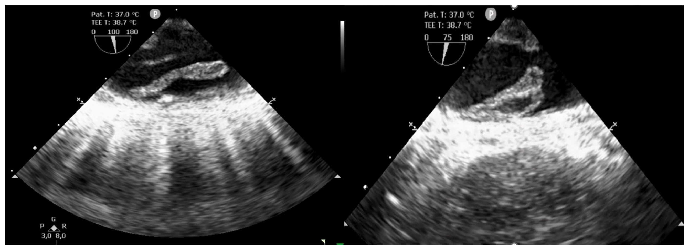
Figure 1. Transesophageal echocardiography showing multiple thrombi of various forms, highly mobile, attached to the wall of the descending thoracic aorta.
Biomedicines 13 02543 g001
Figure 2. Computed tomography of the aorta, oblique sagittal images in arterial time, showing the presence of multiple thrombi (red arrows) attached to the wall of both descending, and abdominal aorta.
Biomedicines 13 02543 g002
Figure 3. Computed tomography showing almost complete thrombosis of the left renal artery; (left): coronal oblique image in arterial time, (right): axial oblique image in arterial time. Red arrows show the left renal artery.
Biomedicines 13 02543 g003
Figure 4. Computed tomography, oblique axial images in portal time, showing a completely obstructive thrombosis of the left portal vein extended to the portal vein trunk (red arrows). PV: portal vein.
Biomedicines 13 02543 g004
Figure 5. The dynamic evolution of the thrombocytes, leucocytes, hemoglobin and reactive C protein during 2 weeks of medical treatment consisting of anticoagulant, antiplatelet (aspirin) and hydroxyurea.
Biomedicines 13 02543 g005
Figure 6. Computed tomography (CT) of the aorta, oblique sagittal images in arterial time, showing the dynamic evolution of the multiple thrombi attached to the wall of both the descending and abdominal aorta. We observe the disappearance of aortic thrombi at 3-month CT follow-up.
Biomedicines 13 02543 g006
Figure 7. The main pathophysiological mechanisms of thrombosis in myeloproliferative neoplasms.
Biomedicines 13 02543 g007
Table 1. The relevant risk scores for thrombotic events in myeloproliferative diseases.
ScoreMPN VariablesCategoriesReference
Conventional approachETage > 60 years
history of thrombosis
Low risk (no risk factors)
High risk
Barbui et al. (2011) [49]
IPSETETage ≥ 60 years (2 points)
history of thrombosis (1 point)
leukocyte count ≥ 11 × 109/L. (1 point)
Low (points 0)
Intermediate (points 1–2)
High (points 3–4)
Passamonti et al. (2012) [96]
IPSET-thrombosisETage > 60 years (1 point)
thrombosis history (2 points)
cardiovascular risk factors (1 point)
JAK2V617F (2 points)
-
Low-risk < 2 points;
Intermediate risk 2 points
High-risk > 2 points
Barbui et al. (2012) [97]
pre-PMFGuglielmelli et al. (2020) [98]
Revised IPSET-thrombosisETage > 60 years
history of thrombosis
JAK2V617F
Very low (age ≤ 60 years, JAK2 wild type, no prior thrombosis)
Low (age ≤ 60 years, JAK2 V617F+, no prior thrombosis)
Intermediate (age > 60 years, JAK2 wild type, no prior thrombosis)
High (age > 60 years and JAK2 V617F+, or prior thrombosis history regardless of other factors)
Barbui et al. (2015) [99]
Machine learningPVage ≥ 60 years—1 point;
prior thrombosis—1 point;
leukocyte count ≥ 12 × 10 9/L—1 point
peri-diagnosis thrombosis (<2 years from diagnosis)—1 point
peri-thrombosis (<2 years from last thrombosis)—1 point
High-risk (score ≥ 2)
Intermediate-risk (score = 1)
Low-risk (score = 0)
Abu-Zeinah et al. (2021) [100]
Machine learningPVintrinsic factors (age, blood type), disease events (time since diagnosis/thrombosis), and short-term changes (body mass index)-Krichevsky et al. (2023) [101]
Machine learning
(PV-AIM)
ETincluded demographic, clinical, and laboratory data Verstovsek et al. (2023) [102]
MFPS-PV PVage ≥ 60 years
cardiovascular risk factors
mutation for thrombosis (DNMT3A, ASXL1, or BCOR/BCORL1) previous thrombosis
-Gu et al. (2023) [61]
Arterial Risk Score (ARTS)ET
PV
MF
MDS
unclassified
cardiovascular risk factors (male sex, tobacco use, hypertension, diabetes, or hypercholesterolemia), 1 point;
TET2 or DNMT3A mutation, 1 point;
age at diagnosis > 60 years, 2 points;
arterial thrombosis prior to or at diagnosis, 2 points.
Low risk (score 0–1)
High risk (score 2–6).
Pasquer et. al. (2024) [91]
Venous Risk ScoreET
PV
MF
MDS
unclassified
venous thrombosis prior to or at diagnosis, 1 point;
JAK2 V617F mutation, 1 point.
Low risk (score 0)
High risk (score 1–2).
Pasquer et al. (2024) [91]
Abbreviations: ET, essential thrombocythemia; IPSET-thrombosis—International Prognostic Score of thrombosis; pre-PMF, pre-fibrotic myelofibrosis; PV, polycythemia vera; MDS, myelodisplazic syndrome; MF, myelofibrosis; MPN, myeloproliferative neoplasms.
Disclaimer/Publisher’s Note: The statements, opinions and data contained in all publications are solely those of the individual author(s) and contributor(s) and not of MDPI and/or the editor(s). MDPI and/or the editor(s) disclaim responsibility for any injury to people or property resulting from any ideas, methods, instructions or products referred to in the content.