
Deep Learning for MRI Segmentation and Molecular Subtyping in Glioblastoma: Critical Aspects from an Emerging Field
 , ,
, ,  , and
, and
Abstract
1. Introduction
2. Technical Challenges
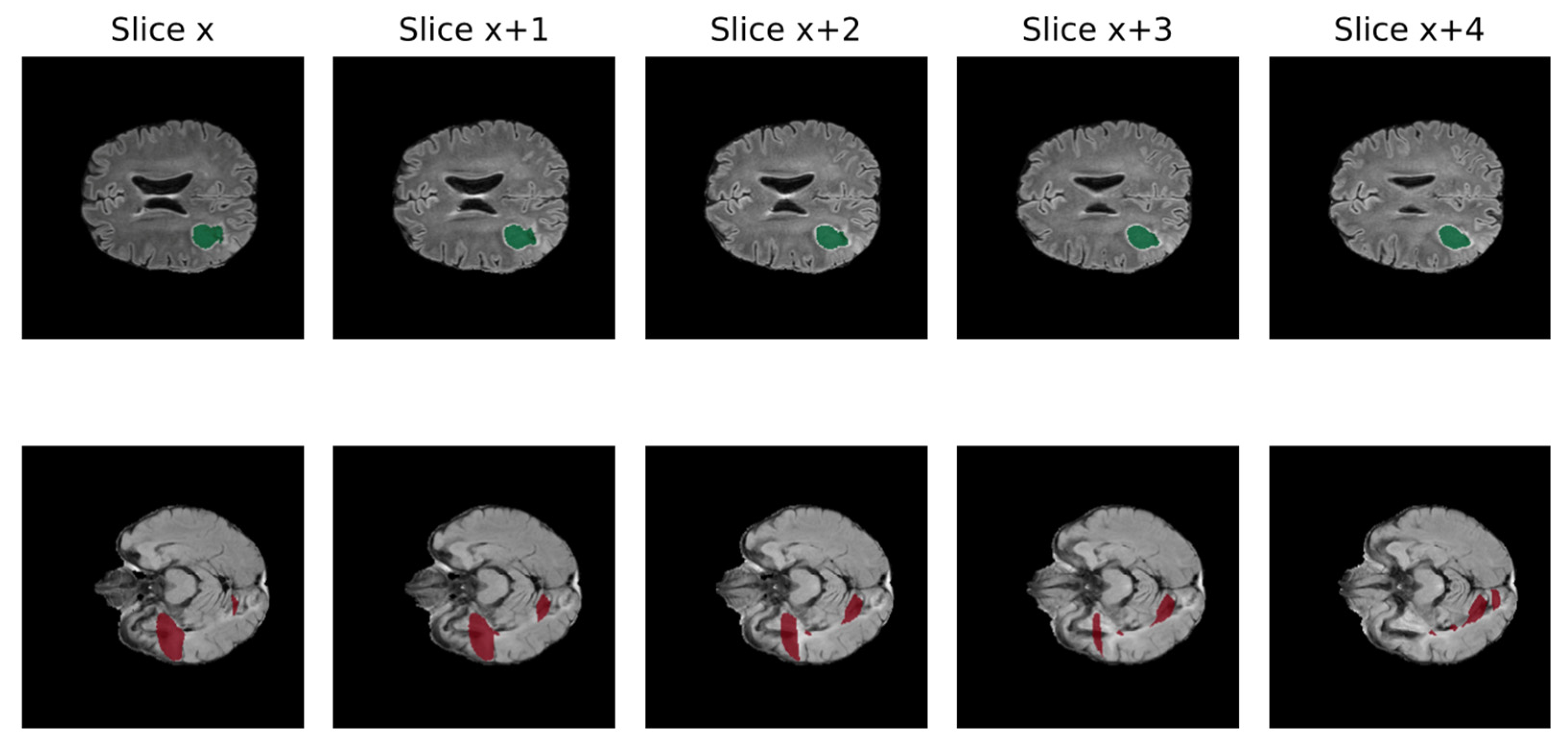
2.1. MRI Imaging Heterogeneity
2.2. Missing MRI Sequences
2.3. Deployment Issues
2.4. Performance Evaluation
3. Application to a Real-Word Scenario
3.1. Limited Number of Patients
3.2. Data Quality
3.3. Data Selection
3.4. Focus on Preoperative Scenario
4. Molecular Subtyping
4.1. IDH Mutation
4.2. P/19q Codeletion
4.3. MGMT Methylation
5. Ethical Concerns
5.1. Lack of Standard Guidelines for Clinical Studies
5.2. Lack of Transparency
5.3. Privacy and Data Protection
6. Conclusions
Author Contributions
Funding
Conflicts of Interest
References
- Ostrom, Q.T.; Gittleman, H.; Liao, P.; Vecchione-Koval, T.; Wolinsky, Y.; Kruchko, C.; Barnholtz-Sloan, J.S. CBTRUS Statistical Report: Primary brain and other central nervous system tumors diagnosed in the United States in 2010–2014. Neuro-Oncology 2017, 19, v1–v88. [Google Scholar] [CrossRef]
- Martucci, M.; Russo, R.; Giordano, C.; Schiarelli, C.; D’apolito, G.; Tuzza, L.; Lisi, F.; Ferrara, G.; Schimperna, F.; Vassalli, S.; et al. Advanced Magnetic Resonance Imaging in the Evaluation of Treated Glioblastoma: A Pictorial Essay. Cancers 2023, 15, 3790. [Google Scholar] [CrossRef]
- Bianconi, A.; Palmieri, G.; Aruta, G.; Monticelli, M.; Zeppa, P.; Tartara, F.; Melcarne, A.; Garbossa, D.; Cofano, F. Updates in Glioblastoma Immunotherapy: An Overview of the Current Clinical and Translational Scenario. Biomedicines 2023, 11, 1520. [Google Scholar] [CrossRef]
- Morello, A.; Bianconi, A.; Rizzo, F.; Bellomo, J.; Meyer, A.C.; Garbossa, D.; Regli, L.; Cofano, F. Laser Interstitial Thermotherapy (LITT) in Recurrent Glioblastoma: What Window of Opportunity for This Treatment? Technol. Cancer Res. Treat. 2024, 23. [Google Scholar] [CrossRef] [PubMed]
- Li, R.; Ye, J.; Huang, Y.; Jin, W.; Xu, P.; Guo, L. A continuous learning approach to brain tumor segmentation: Integrating multi-scale spatial distillation and pseudo-labeling strategies. Front. Oncol. 2024, 13, 1247603. [Google Scholar] [CrossRef] [PubMed]
- Gillies, R.J.; Kinahan, P.E.; Hricak, H. Radiomics: Images Are More than Pictures, They Are Data. Radiology 2016, 278, 563–577. [Google Scholar] [CrossRef]
- Nakamori, S.; Bui, A.H.; Jang, J.; El-Rewaidy, H.A.; Kato, S.; Ngo, L.H.; Josephson, M.E.; Manning, W.J.; Nezafat, R. Increased myocardial native T1 relaxation time in patients with nonischemic dilated cardiomyopathy with complex ventricular arrhythmia. J. Magn. Reson. Imaging 2017, 47, 779–786. [Google Scholar] [CrossRef]
- Pandey, U.; Saini, J.; Kumar, M.; Gupta, R.; Ingalhalikar, M. Normative Baseline for Radiomics in BrainMRI: Evaluating the Robustness, Regional Variations, and Reproducibility onFLAIRImages. J. Magn. Reson. Imaging 2021, 53, 394–407. [Google Scholar] [CrossRef]
- Li, Y.; Ammari, S.; Balleyguier, C.; Lassau, N.; Chouzenoux, E. Impact of Preprocessing and Harmonization Methods on the Removal of Scanner Effects in Brain MRI Radiomic Features. Cancers 2021, 13, 3000. [Google Scholar] [CrossRef]
- Orlhac, F.; Lecler, A.; Savatovski, J.; Goya-Outi, J.; Nioche, C.; Charbonneau, F.; Ayache, N.; Frouin, F.; Duron, L.; Buvat, I. How Can We Combat Multicenter Variability in MR Radiomics? Validation of a Correction Procedure. Eur. Radiol. 2021, 31, 2272–2280. [Google Scholar] [CrossRef] [PubMed]
- Fatania, K.; Mohamud, F.; Clark, A.; Nix, M.; Short, S.C.; O’Connor, J.; Scarsbrook, A.F.; Currie, S. Intensity Standardization of MRI Prior to Radiomic Feature Extraction for Artificial Intelligence Research in Glioma—A Systematic Review. Eur. Radiol. 2022, 32, 7014–7025. [Google Scholar] [CrossRef] [PubMed]
- Da-Ano, R.; Visvikis, D.; Hatt, M. Harmonization strategies for multicenter radiomics investigations. Phys. Med. Biol. 2020, 65, 24TR02. [Google Scholar] [CrossRef]
- Renard, F.; Guedria, S.; De Palma, N.; Vuillerme, N. Variability and reproducibility in deep learning for medical image segmentation. Sci. Rep. 2020, 10, 13724. [Google Scholar] [CrossRef] [PubMed]
- Marzi, C.; Giannelli, M.; Barucci, A.; Tessa, C.; Mascalchi, M.; Diciotti, S. Efficacy of MRI data harmonization in the age of machine learning: A multicenter study across 36 datasets. Sci. Data 2024, 11, 115. [Google Scholar] [CrossRef]
- Carré, A.; Klausner, G.; Edjlali, M.; Lerousseau, M.; Briend-Diop, J.; Sun, R.; Ammari, S.; Reuzé, S.; Alvarez Andres, E.; Estienne, T.; et al. Standardization of Brain MR Images across Machines and Protocols: Bridging the Gap for MRI-Based Radiomics. Sci. Rep. 2020, 10, 12340. [Google Scholar] [CrossRef] [PubMed]
- Wu, J.; Guo, D.; Wang, L.; Yang, S.; Zheng, Y.; Shapey, J.; Vercauteren, T.; Bisdas, S.; Bradford, R.; Saeed, S.; et al. TISS-net: Brain tumor image synthesis and segmentation using cascaded dual-task networks and error-prediction consistency. Neurocomputing 2023, 544, 126295. [Google Scholar] [CrossRef]
- Conte, G.M.; Weston, A.D.; Vogelsang, D.C.; Philbrick, K.A.; Cai, J.C.; Barbera, M.; Sanvito, F.; Lachance, D.H.; Jenkins, R.B.; Tobin, W.O.; et al. Generative Adversarial Networks to Synthesize Missing T1 and FLAIR MRI Sequences for Use in a Multisequence Brain Tumor Segmentation Model. Radiology 2021, 299, 313–323. [Google Scholar] [CrossRef] [PubMed]
- Choi, Y.; Al-Masni, M.A.; Jung, K.-J.; Yoo, R.-E.; Lee, S.-Y.; Kim, D.-H. A single stage knowledge distillation network for brain tumor segmentation on limited MR image modalities. Comput. Methods Programs Biomed. 2023, 240, 107644. [Google Scholar] [CrossRef]
- Cofano, F.; Bianconi, A.; De Marco, R.; Consoli, E.; Zeppa, P.; Bruno, F.; Pellerino, A.; Panico, F.; Salvati, L.F.; Rizzo, F.; et al. The Impact of Lateral Ventricular Opening in the Resection of Newly Diagnosed High-Grade Gliomas: A Single Center Experience. Cancers 2024, 16, 1574. [Google Scholar] [CrossRef]
- Kamnitsas, K.; Ledig, C.; Newcombe, V.F.; Simpson, J.P.; Kane, A.D.; Menon, D.K.; Rueckert, D.; Glocker, B. Efficient multi-scale 3D CNN with fully connected CRF for accurate brain lesion segmentation. Med. Image Anal. 2017, 36, 61–78. [Google Scholar] [CrossRef]
- Zhou, C.; Ding, C.; Lu, Z.; Wang, X.; Tao, D. One-Pass Multi-Task Convolutional Neural Networks for Efficient Brain Tumor Segmentation. In Proceedings of the Medical Image Computing and Computer Assisted Intervention—MICCAI 2018, Granada, Spain, 16–20 September 2018; Lecture Notes in Computer Science (including subseries Lecture Notes in Artificial Intelligence and Lecture Notes in Bioinformatics). Volume 11072, pp. 637–645. [Google Scholar] [CrossRef]
- Zhou, T.; Ruan, S.; Canu, S. A review: Deep learning for medical image segmentation using multi-modality fusion. Array 2019, 3–4, 100004. [Google Scholar] [CrossRef]
- Al-Masni, M.A.; Kim, D.-H. CMM-Net: Contextual multi-scale multi-level network for efficient biomedical image segmentation. Sci. Rep. 2021, 11, 10191. [Google Scholar] [CrossRef] [PubMed]
- Yu, F.; Koltun, V. Multi-Scale Context Aggregation by Dilated Convolutions. In Proceedings of the 4th International Conference on Learning Representations, ICLR 2016-Conference Track Proceedings, San Juan, Puerto Rico, 2–4 May 2016. [Google Scholar] [CrossRef]
- Xu, C.; Xu, L.; Ohorodnyk, P.; Roth, M.; Chen, B.; Li, S. Contrast agent-free synthesis and segmentation of ischemic heart disease images using progressive sequential causal GANs. Med. Image Anal. 2020, 62, 101668. [Google Scholar] [CrossRef] [PubMed]
- Wang, G.; Song, T.; Dong, Q.; Cui, M.; Huang, N.; Zhang, S. Automatic ischemic stroke lesion segmentation from computed tomography perfusion images by image synthesis and attention-based deep neural networks. Med. Image Anal. 2020, 65, 101787. [Google Scholar] [CrossRef] [PubMed]
- Osman, A.F.I.; Tamam, N.M. Deep learning-based convolutional neural network for intramodality brain MRI synthesis. J. Appl. Clin. Med. Phys. 2022, 23, e13530. [Google Scholar] [CrossRef]
- Yang, F.; Dogan, N.; Stoyanova, R.; Ford, J.C. Evaluation of radiomic texture feature error due to MRI acquisition and reconstruction: A simulation study utilizing ground truth. Phys. Medica 2018, 50, 26–36. [Google Scholar] [CrossRef]
- Erickson, B.J.; Cai, J. Magician’s Corner: 5. Generative Adversarial Networks. Radiol. Artif. Intell. 2020, 2, e190215. [Google Scholar] [CrossRef] [PubMed]
- Goodfellow, I.J.; Pouget-Abadie, J.; Mirza, M.; Xu, B.; Warde-Farley, D.; Ozair, S.; Courville, A.; Bengio, Y. Generative adversarial networks. Commun. ACM 2020, 63, 139–144. [Google Scholar] [CrossRef]
- Marcadent, S.; Hofmeister, J.; Preti, M.G.; Martin, S.P.; Van De Ville, D.; Montet, X. Generative Adversarial Networks Improve the Reproducibility and Discriminative Power of Radiomic Features. Radiol. Artif. Intell. 2020, 2, e190035. [Google Scholar] [CrossRef]
- Sharma, A.; Hamarneh, G. Missing MRI Pulse Sequence Synthesis Using Multi-Modal Generative Adversarial Network. IEEE Trans. Med. Imaging 2019, 39, 1170–1183. [Google Scholar] [CrossRef]
- Chen, C.; Dou, Q.; Jin, Y.; Liu, Q.; Heng, P.A. Learning With Privileged Multimodal Knowledge for Unimodal Segmentation. IEEE Trans. Med. Imaging 2021, 41, 621–632. [Google Scholar] [CrossRef] [PubMed]
- Wang, L.; Yoon, K.-J. Knowledge Distillation and Student-Teacher Learning for Visual Intelligence: A Review and New Outlooks. IEEE Trans. Pattern Anal. Mach. Intell. 2021, 44, 3048–3068. [Google Scholar] [CrossRef]
- Bhalerao, M.; Thakur, S. Brain Tumor Segmentation Based on 3D Residual U-Net. In Proceedings of the 5th International Workshop, BrainLes 2019, Held in Conjunction with MICCAI 2019, Shenzhen, China, 17 October 2019; Lecture Notes in Computer Science (including subseries Lecture Notes in Artificial Intelligence and Lecture Notes in Bioinformatics). Volume 11993, pp. 218–225. [Google Scholar] [CrossRef]
- Isensee, F.; Jaeger, P.F.; Kohl, S.A.A.; Petersen, J.; Maier-Hein, K.H. NnU-Net: A Self-Configuring Method for Deep Learning-Based Biomedical Image Segmentation. Nat. Methods 2021, 18, 203–211. [Google Scholar] [CrossRef]
- Thakur, S.P.; Pati, S.; Panchumarthy, R.; Karkada, D.; Wu, J.; Kurtaev, D.; Sako, C.; Shah, P.; Bakas, S. Optimization of Deep Learning Based Brain Extraction in MRI for Low Resource Environments. In Proceedings of the 7th International Workshop, BrainLes 2021, Held in Conjunction with MICCAI 2021, Virtual Event, 27 September 2021; pp. 151–167. [Google Scholar] [CrossRef]
- Ito, Y.; Imai, H.; Le Duc, T.; Negishi, Y.; Kawachiya, K.; Matsumiya, R.; Endo, T. Profiling based out-of-core Hybrid method for large neural networks. In Proceedings of the PPoPP ‘19: 24th ACM SIGPLAN Symposium on Principles and Practice of Parallel Programming, Washington, DC, USA, 16–20 February 2019; pp. 399–400. [Google Scholar] [CrossRef]
- Huang, B.; Reichman, D.; Collins, L.M.; Bradbury, K.; Malof, J.M. Tiling and Stitching Segmentation Output for Remote Sensing: Basic Challenges and Recommendations. arXiv 2018, arXiv:1805.12219. [Google Scholar] [CrossRef]
- Pinckaers, H.; Litjens, G. Training Convolutional Neural Networks with Megapixel Images. arXiv 2018, arXiv:1804.05712. [Google Scholar] [CrossRef]
- Roth, H.R.; Oda, H.; Zhou, X.; Shimizu, N.; Yang, Y.; Hayashi, Y.; Oda, M.; Fujiwara, M.; Misawa, K.; Mori, K. An Application of Cascaded 3D Fully Convolutional Networks for Medical Image Segmentation. Comput. Med. Imaging Graph. 2018, 66, 90–99. [Google Scholar] [CrossRef] [PubMed]
- Reina, G.A.; Panchumarthy, R.; Thakur, S.P.; Bastidas, A.; Bakas, S. Systematic Evaluation of Image Tiling Adverse Effects on Deep Learning Semantic Segmentation. Front. Neurosci. 2020, 14, 65. [Google Scholar] [CrossRef]
- Shazeer, N.; Cheng, Y.; Parmar, N.; Tran, D.; Vaswani, A.; Koanantakool, P.; Hawkins, P.; Lee, H.J.; Hong, M.; Young, C.; et al. Mesh-TensorFlow: Deep Learning for Supercomputers. Adv. Neural Inf. Process. Syst. 2018, 31, 10414–10423. [Google Scholar] [CrossRef]
- Sergeev, A.; Del Balso, M. Horovod: Fast and Easy Distributed Deep Learning in TensorFlow. arXiv 2018, arXiv:1802.05799. [Google Scholar] [CrossRef]
- Revesz, G.; Kundel, H.L.; Bonitatibus, M. The Effect of Verification on the Assessment of Imaging Techniques. Investig. Radiol. 1983, 18, 194–198. [Google Scholar] [CrossRef]
- Bianconi, A.; Rossi, L.F.; Bonada, M.; Zeppa, P.; Nico, E.; De Marco, R.; Lacroce, P.; Cofano, F.; Bruno, F.; Morana, G.; et al. Deep learning-based algorithm for postoperative glioblastoma MRI segmentation: A promising new tool for tumor burden assessment. Brain Inform. 2023, 10, 26. [Google Scholar] [CrossRef] [PubMed]
- Zając, H.D.; Avlona, N.R.; Kensing, F.; Andersen, T.O.; Shklovski, I. Ground Truth or Dare: Factors Affecting the Creation of Medical Datasets for Training AI. In Proceedings of the AIES ‘23: AAAI/ACM Conference on AI, Ethics, and Society, Montreal, QC, Canada, 8–10 August 2023; pp. 351–362. [Google Scholar] [CrossRef]
- Sylolypavan, A.; Sleeman, D.; Wu, H.; Sim, M. The impact of inconsistent human annotations on AI driven clinical decision making. npj Digit. Med. 2023, 6, 26. [Google Scholar] [CrossRef]
- Aganj, I.; Harisinghani, M.G.; Weissleder, R.; Fischl, B. Unsupervised Medical Image Segmentation Based on the Local Center of Mass. Sci. Rep. 2018, 8, 13012. [Google Scholar] [CrossRef]
- Kiyasseh, D.; Cohen, A.; Jiang, C.; Altieri, N. A framework for evaluating clinical artificial intelligence systems without ground-truth annotations. Nat. Commun. 2024, 15, 1808. [Google Scholar] [CrossRef] [PubMed]
- Liu, C.; Amodio, M.; Shen, L.L.; Gao, F.; Avesta, A.; Aneja, S.; Wang, J.C.; Del Priore, L.V.; Krishnaswamy, S. CUTS: A Deep Learning and Topological Framework for Multigranular Unsupervised Medical Image Segmentation. arXiv 2022, arXiv:2209.11359. [Google Scholar] [CrossRef]
- Tillmanns, N.; Lum, A.E.; Cassinelli, G.; Merkaj, S.; Verma, T.; Zeevi, T.; Staib, L.; Subramanian, H.; Bahar, R.C.; Brim, W.; et al. Identifying clinically applicable machine learning algorithms for glioma segmentation: Recent advances and discoveries. Neuro-Oncol. Adv. 2022, 4, vdac093. [Google Scholar] [CrossRef] [PubMed]
- Jekel, L.; Brim, W.R.; von Reppert, M.; Staib, L.; Petersen, G.C.; Merkaj, S.; Subramanian, H.; Zeevi, T.; Payabvash, S.; Bousabarah, K.; et al. Machine Learning Applications for Differentiation of Glioma from Brain Metastasis—A Systematic Review. Cancers 2022, 14, 1369. [Google Scholar] [CrossRef]
- Aboian, M.; Bousabarah, K.; Kazarian, E.; Zeevi, T.; Holler, W.; Merkaj, S.; Cassinelli Petersen, G.; Bahar, R.; Subramanian, H.; Sunku, P.; et al. Clinical implementation of artificial intelligence in neuroradiology with development of a novel workflow-efficient picture archiving and communication system-based automated brain tumor segmentation and radiomic feature extraction. Front. Neurosci. 2022, 16, 860208. [Google Scholar] [CrossRef]
- Dieckhaus, H.B.; Meijboom, R.; Okar, S.; Wu, T.; Parvathaneni, P.; Mina, Y.; Chandran, S.; Waldman, A.D.; Reich, D.S.; Nair, G. Logistic Regression–Based Model Is More Efficient Than U-Net Model for Reliable Whole Brain Magnetic Resonance Imaging Segmentation. Top. Magn. Reson. Imaging 2022, 31, 31–39. [Google Scholar] [CrossRef]
- Sheller, M.J.; Edwards, B.; Reina, G.A.; Martin, J.; Pati, S.; Kotrotsou, A.; Milchenko, M.; Xu, W.; Marcus, D.; Colen, R.R.; et al. Federated learning in medicine: Facilitating multi-institutional collaborations without sharing patient data. Sci. Rep. 2020, 10, 12598. [Google Scholar] [CrossRef]
- Bakas, S.; Akbari, H.; Sotiras, A.; Bilello, M.; Rozycki, M.; Kirby, J.S.; Freymann, J.B.; Farahani, K.; Davatzikos, C. Advancing the Cancer Genome Atlas glioma MRI collections with expert segmentation labels and radiomic features. Sci. Data 2017, 4, 170117. [Google Scholar] [CrossRef] [PubMed]
- Anazodo, U.C.; Ng, J.J.; Ehiogu, B.; Obungoloch, J.; Fatade, A.; Mutsaerts, H.J.M.M.; Secca, M.F.; Diop, M.; Opadele, A.; Alexander, D.C.; et al. A framework for advancing sustainable magnetic resonance imaging access in Africa. NMR Biomed. 2022, 36, e4846. [Google Scholar] [CrossRef]
- Bakas, S.; Reyes, M.; Jakab, A.; Bauer, S.; Rempfler, M.; Crimi, A.; Shinohara, R.T.; Berger, C.; Ha, S.M.; Rozycki, M.; et al. Identifying the Best Machine Learning Algorithms for Brain Tumor Segmentation, Progression Assessment, and Overall Survival Prediction in the BRATS Challenge. arXiv 2018, arXiv:1811.02629. [Google Scholar] [CrossRef]
- Raghu; Sriraam, N.; Temel, Y.; Rao, S.V.; Kubben, P.L. A Convolutional Neural Network Based Framework for Classification of Seizure Types. In Proceedings of the 2019 41st Annual International Conference of the IEEE Engineering in Medicine and Biology Society (EMBC), Berlin, Germany, 23–27 July 2019; pp. 2547–2550. [Google Scholar] [CrossRef]
- Ibrahim, M.; Muhammad, Q.; Zamarud, A.; Eiman, H.; Fazal, F. Navigating Glioblastoma Diagnosis and Care: Transformative Pathway of Artificial Intelligence in Integrative Oncology. Cureus 2023, 15, e44214. [Google Scholar] [CrossRef] [PubMed]
- Ermiş, E.; Jungo, A.; Poel, R.; Blatti-Moreno, M.; Meier, R.; Knecht, U.; Aebersold, D.M.; Fix, M.K.; Manser, P.; Reyes, M.; et al. Fully automated brain resection cavity delineation for radiation target volume definition in glioblastoma patients using deep learning. Radiat. Oncol. 2020, 15, 100. [Google Scholar] [CrossRef]
- Lin, B.; Tan, Z.; Mo, Y.; Yang, X.; Liu, Y.; Xu, B.; Lin, B.; Tan, Z.; Mo, Y.; Yang, X.; et al. Intelligent oncology: The convergence of artificial intelligence and oncology. J. Natl. Cancer Cent. 2023, 3, 83–91. [Google Scholar] [CrossRef] [PubMed]
- Adewole, M.; Rudie, J.D.; Gbadamosi, A.; Toyobo, O.; Raymond, C.; Zhang, D.; Omidiji, O.; Akinola, R.; Suwaid, M.A.; Emegoakor, A.; et al. The Brain Tumor Segmentation (BraTS) Challenge 2023: Glioma Segmentation in Sub-Saharan Africa Patient Population (BraTS-Africa). arXiv 2023, arXiv:2305.19369v1. Available online: https://pubmed-ncbi-nlm-nih-gov.bibliopass.unito.it/37396608/ (accessed on 16 February 2024).
- Lin, H.; Figini, M.; Tanno, R.; Blumberg, S.B.; Kaden, E.; Ogbole, G.; Brown, B.J.; D’Arco, F.; Carmichael, D.W.; Lagunju, I.; et al. Deep Learning for Low-Field to High-Field MR: Image Quality Transfer with Probabilistic Decimation Simulator. In Proceedings of the Machine Learning for Medical Image Reconstruction, Second International Workshop, MLMIR 2019, Held in Conjunction with MICCAI 2019, Shenzhen, China, 17 October 2019; pp. 58–70. [Google Scholar] [CrossRef]
- Chang, K.; Beers, A.L.; Bai, H.X.; Brown, J.M.; Ina Ly, K.; Li, X.; Senders, J.T.; Kavouridis, V.K.; Boaro, A.; Su, C.; et al. Automatic Assessment of Glioma Burden: A Deep Learning Algorithm for Fully Automated Volumetric and Bidimensional Measurement. Neuro-Oncology 2019, 21, 1412–1422. [Google Scholar] [CrossRef] [PubMed]
- Subramanian, H.; Dey, R.; Brim, W.R.; Tillmanns, N.; Cassinelli Petersen, G.; Brackett, A.; Mahajan, A.; Johnson, M.; Malhotra, A.; Aboian, M. Trends in Development of Novel Machine Learning Methods for the Identification of Gliomas in Datasets That Include Non-Glioma Images: A Systematic Review. Front. Oncol. 2021, 11, 788819. [Google Scholar] [CrossRef]
- Zeppa, P.; Neitzert, L.; Mammi, M.; Monticelli, M.; Altieri, R.; Castaldo, M.; Cofano, F.; Borrè, A.; Zenga, F.; Melcarne, A.; et al. How Reliable Are Volumetric Techniques for High-Grade Gliomas? A Comparison Study of Different Available Tools. Neurosurgery 2020, 87, E672–E679. [Google Scholar] [CrossRef]
- Kommers, I.; Bouget, D.; Pedersen, A.; Eijgelaar, R.S.; Ardon, H.; Barkhof, F.; Bello, L.; Berger, M.S.; Conti Nibali, M.; Furtner, J.; et al. Glioblastoma Surgery Imaging-Reporting and Data System: Standardized reporting of tumor volume, location, and resectability based on automated segmentations. Cancers 2021, 13, 2854. [Google Scholar] [CrossRef]
- Visser, M.; Müller, D.; van Duijn, R.; Smits, M.; Verburg, N.; Hendriks, E.; Nabuurs, R.; Bot, J.; Eijgelaar, R.; Witte, M.; et al. Inter-rater agreement in glioma segmentations on longitudinal MRI. NeuroImage Clin. 2019, 22, 101727. [Google Scholar] [CrossRef]
- Cordova, J.S.; Schreibmann, E.; Hadjipanayis, C.G.; Guo, Y.; Shu, H.-K.G.; Shim, H.; Holder, C.A. Quantitative Tumor Segmentation for Evaluation of Extent of Glioblastoma Resection to Facilitate Multisite Clinical Trials. Transl. Oncol. 2014, 7, 40–47, W1–W5. [Google Scholar] [CrossRef] [PubMed]
- Miao, X.; Chen, H.; Tang, M.; Chen, Y. P13.01.A Post-Operative MRI Synthesis from Pre-Operative MRI and Post-Operative CT Using Conditional Gan for the Assessment of Degree of Resection. Neuro Oncol. 2023, 25 (Suppl. 2), ii100. [Google Scholar] [CrossRef]
- Bianconi, A.; Bonada, M.; Zeppa, P.; Colonna, S.; Tartara, F.; Melcarne, A.; Garbossa, D.; Cofano, F. How Reliable Is Fluorescence-Guided Surgery in Low-Grade Gliomas? A Systematic Review Concerning Different Fluorophores. Cancers 2023, 15, 4130. [Google Scholar] [CrossRef]
- Ducray, F.; Idbaih, A.; Wang, X.-W.; Cheneau, C.; Labussiere, M.; Sanson, M. Predictive and prognostic factors for gliomas. Expert Rev. Anticancer. Ther. 2011, 11, 781–789. [Google Scholar] [CrossRef]
- Saaid, A.; Monticelli, M.; Ricci, A.A.; Orlando, G.; Botta, C.; Zeppa, P.; Bianconi, A.; Osella-Abate, S.; Bruno, F.; Pellerino, A.; et al. Prognostic Analysis of the IDH1 G105G (rs11554137) SNP in IDH-Wildtype Glioblastoma. Genes 2022, 13, 1439. [Google Scholar] [CrossRef] [PubMed]
- Bianconi, A.; Koumantakis, E.; Gatto, A.; Zeppa, P.; Saaid, A.; Nico, E.; Bruno, F.; Pellerino, A.; Rizzo, F.; Junemann, C.V.; et al. Effects of Levetiracetam and Lacosamide on survival and seizure control in IDH-wild type glioblastoma during temozolomide plus radiation adjuvant therapy. Brain Spine 2024, 4, 102732. [Google Scholar] [CrossRef]
- Kickingereder, P.; Sahm, F.; Radbruch, A.; Wick, W.; Heiland, S.; Von Deimling, A.; Bendszus, M.; Wiestler, B. IDH Mutation Status Is Associated with a Distinct Hypoxia/Angiogenesis Transcriptome Signature Which Is Non-Invasively Predictable with RCBV Imaging in Human Glioma. Sci. Rep. 2015, 5, 16238. [Google Scholar] [CrossRef]
- Law, M.; Young, R.J.; Babb, J.S.; Peccerelli, N.; Chheang, S.; Gruber, M.L.; Miller, D.C.; Golfinos, J.G.; Zagzag, D.; Johnson, G. Gliomas: Predicting Time to Progression or Survival with Cerebral Blood Volume Measurements at Dynamic Susceptibility-weighted Contrast-enhanced Perfusion MR Imaging. Radiology 2008, 247, 490–498. [Google Scholar] [CrossRef]
- Beiko, J.; Suki, D.; Hess, K.R.; Fox, B.D.; Cheung, V.; Cabral, M.; Shonka, N.; Gilbert, M.R.; Sawaya, R.; Prabhu, S.S.; et al. IDH1 Mutant Malignant Astrocytomas Are More Amenable to Surgical Resection and Have a Survival Benefit Associated with Maximal Surgical Resection. Neuro-Oncology 2013, 16, 81–91. [Google Scholar] [CrossRef] [PubMed]
- Liang, S.; Zhang, R.; Liang, D.; Song, T.; Ai, T.; Xia, C.; Xia, L.; Wang, Y. Multimodal 3D DenseNet for IDH Genotype Prediction in Gliomas. Genes 2018, 9, 382. [Google Scholar] [CrossRef] [PubMed]
- Zlochower, A.; Chow, D.S.; Chang, P.; Khatri, D.; Boockvar, J.A.; Filippi, C.G. Deep Learning AI Applications in the Imaging of Glioma. Top. Magn. Reson. Imaging 2020, 29, 115–121. [Google Scholar] [CrossRef] [PubMed]
- Hegi, M.E.; Diserens, A.-C.; Gorlia, T.; Hamou, M.-F.; de Tribolet, N.; Weller, M.; Kros, J.M.; Hainfellner, J.A.; Mason, W.; Mariani, L.; et al. MGMT Gene Silencing and Benefit from Temozolomide in Glioblastoma. N. Engl. J. Med. 2005, 352, 997–1003. [Google Scholar] [CrossRef] [PubMed]
- Bianconi, A.; Prior, A.; Zona, G.; Fiaschi, P. Anticoagulant therapy in high grade gliomas: A systematic review on state of the art and future perspectives. J. Neurosurg. Sci. 2023, 67, 236–240. [Google Scholar] [CrossRef] [PubMed]
- Han, L.; Kamdar, M.R. MRI to MGMT: Predicting Methylation Status in Glioblastoma Patients Using Convolutional Recurrent Neural Networks. Pac. Symp. Biocomput. 2018, 23, 331–342. [Google Scholar] [CrossRef] [PubMed]
- Korfiatis, P.; Kline, T.L.; Lachance, D.H.; Parney, I.F.; Buckner, J.C.; Erickson, B.J. Residual Deep Convolutional Neural Network Predicts MGMT Methylation Status. J. Digit. Imaging 2017, 30, 622–628. [Google Scholar] [CrossRef] [PubMed]
- Chang, P.; Grinband, J.; Weinberg, B.D.; Bardis, M.; Khy, M.; Cadena, G.; Su, M.-Y.; Cha, S.; Filippi, C.G.; Bota, D.; et al. Deep-Learning Convolutional Neural Networks Accurately Classify Genetic Mutations in Gliomas. Am. J. Neuroradiol. 2018, 39, 1201–1207. [Google Scholar] [CrossRef]
- Li, Z.; Wang, Y.; Yu, J.; Guo, Y.; Cao, W. Deep Learning Based Radiomics (DLR) and Its Usage in Noninvasive IDH1 Prediction for Low Grade Glioma. Sci. Rep. 2017, 7, 5467. [Google Scholar] [CrossRef]
- Kahn, C.E. Artificial Intelligence, Real Radiology. Radiol. Artif. Intell. 2019, 1, e184001. [Google Scholar] [CrossRef]
- Bossuyt, P.M.; Reitsma, J.B.; Bruns, D.E.; Gatsonis, C.A.; Glasziou, P.P.; Irwig, L.; Lijmer, J.G.; Moher, D.; Rennie, D.; de Vet, H.C.; et al. STARD 2015: An Updated List of Essential Items for Reporting Diagnostic Accuracy Studies. Radiology 2015, 277, 826–832. [Google Scholar] [CrossRef]
- Lambin, P.; Leijenaar, R.T.H.; Deist, T.M.; Peerlings, J.; de Jong, E.E.C.; van Timmeren, J.; Sanduleanu, S.; Larue, R.T.H.M.; Even, A.J.G.; Jochems, A.; et al. Radiomics: The Bridge between Medical Imaging and Personalized Medicine. Nat. Rev. Clin. Oncol. 2017, 14, 749–762. [Google Scholar] [CrossRef] [PubMed]
- Cohen, J.F.; Korevaar, D.A.; Altman, D.G.; Bruns, D.E.; Gatsonis, C.A.; Hooft, L.; Irwig, L.; Levine, D.; Reitsma, J.B.; de Vet, H.C.W.; et al. STARD 2015 Guidelines for Reporting Diagnostic Accuracy Studies: Explanation and Elaboration. BMJ Open 2016, 6, e012799. [Google Scholar] [CrossRef] [PubMed]
- Bossuyt, P.M.; Reitsma, J.B. The STARD Initiative. Lancet 2003, 361, 71. [Google Scholar] [CrossRef] [PubMed]
- Vandenbroucke, J.P.; von Elm, E.; Altman, D.G.; Gøtzsche, P.C.; Mulrow, C.D.; Pocock, S.J.; Poole, C.; Schlesselman, J.J.; Egger, M. Strengthening the Reporting of Observational Studies in Epidemiology (STROBE): Explanation and Elaboration. PLoS Med. 2007, 4, e297. [Google Scholar] [CrossRef] [PubMed]
- Schulz, K.F.; Altman, U.G.; Moher, D.; CONSORT Group. CONSORT 2010 Statement: Updated guidelines for reporting parallel group randomised trials. BMJ 2010, 340, c332. [Google Scholar] [CrossRef] [PubMed]
- Begg, C. Improving the quality of reporting of randomized controlled trials. The CONSORT statement. JAMA 1996, 276, 637–639. [Google Scholar] [CrossRef] [PubMed]
- Mongan, J.; Moy, L.; Kahn, C.E. Checklist for Artificial Intelligence in Medical Imaging (CLAIM): A Guide for Authors and Reviewers. Radiol. Artif. Intell. 2020, 2, e200029. [Google Scholar] [CrossRef]
- Reyes, M.; Meier, R.; Pereira, S.; Silva, C.A.; Dahlweid, F.M.; von Tengg-Kobligk, H.; Summers, R.M.; Wiest, R. On the Interpretability of Artificial Intelligence in Radiology: Challenges and Opportunities. Radiol. Artif. Intell. 2020, 2, e190043. [Google Scholar] [CrossRef]
- Selvaraju, R.R.; Cogswell, M.; Das, A.; Vedantam, R.; Parikh, D.; Batra, D. Grad-CAM: Visual Explanations from Deep Networks via Gradient-Based Localization. Int. J. Comput. Vis. 2020, 128, 336–359. [Google Scholar] [CrossRef]
- Kim, B.; Wattenberg, M.; Gilmer, J.; Cai, C.; Wexler, J.; Viegas, F.; Sayres, R. Interpretability beyond Feature Attribution: Quantitative Testing with Concept Activation Vectors (TCAV). In Proceedings of the 35th International Conference on Machine Learning, ICML 2018, Stockholm, Sweden, 10–15 July 2018; Volume 80, pp. 4186–4195. [Google Scholar]
- Chaddad, A.; Peng, J.; Xu, J.; Bouridane, A. Survey of Explainable AI Techniques in Healthcare. Sensors 2023, 23, 634. [Google Scholar] [CrossRef] [PubMed]
- Band, S.S.; Yarahmadi, A.; Hsu, C.-C.; Biyari, M.; Sookhak, M.; Ameri, R.; Dehzangi, I.; Chronopoulos, A.T.; Liang, H.-W. Application of explainable artificial intelligence in medical health: A systematic review of interpretability methods. Inform. Med. Unlocked 2023, 40, 101286. [Google Scholar] [CrossRef]
- Geis, J.R.; Brady, A.P.; Wu, C.C.; Spencer, J.; Ranschaert, E.; Jaremko, J.L.; Langer, S.G.; Kitts, A.B.; Birch, J.; Shields, W.F.; et al. Ethics of Artificial Intelligence in Radiology: Summary of the Joint European and North American Multisociety Statement. J. Am. Coll. Radiol. 2019, 16, 1516–1521. [Google Scholar] [CrossRef] [PubMed]
- Lotan, E.; Tschider, C.; Sodickson, D.K.; Caplan, A.L.; Bruno, M.; Zhang, B.; Lui, Y.W. Medical Imaging and Privacy in the Era of Artificial Intelligence: Myth, Fallacy, and the Future. J. Am. Coll. Radiol. 2020, 17, 1159–1162. [Google Scholar] [CrossRef] [PubMed]


| Section | Limitation | Domain | Definition | Possible Solution(s) |
|---|---|---|---|---|
| 2.1 | imaging heterogeneity | technical | scanner-dependent variation in image signal intensity | intensity standardization |
| rescanning data | ||||
| 2.2 | missing MRI sequences | technical | unavaiable modality/ies (T1, T2, FLAIR, T1CE) | inter-modality translation |
| knowledge distillation | ||||
| 2.3 | deployment issues | technical | limited computational resources and memory constraints | tiling |
| quantization | ||||
| 2.4 | performance evaluation | technical | subjective reference standards | cross-validation |
| unsupervised training | ||||
| 3.1 | limited number of patients | application | low number of data publicly avaiable | transfer learning |
| 3.2 | data quality | application | suboptimal quality of data (non-volumetric scans) | pre-processing |
| inclusion of complex scenarios | ||||
| 3.3 | data selection | application | selection bias and reduced applicability | inclusive database |
| 3.4 | focus on preoperative scenario | application | logistical and technical issues for postop. MRIs | multi-modality and multi-institutional data |
| 4 | exclusion of molecular data | molecular | limited consideration of IDH—1p/19q—MGMT | new coder architecture |
| large-scale data-sharing | ||||
| 5.1 | lack of standard guidelines | ethical | scientific integrity not definable | checklist |
| 5.2 | lack of transparency | ethical | limited understanding of the results | interpretability methods |
| interdisciplinary collaboration | ||||
| 5.3 | privacy and data protection | ethical | difficulty to obtain complete anonimization | skull-stripping |
Disclaimer/Publisher’s Note: The statements, opinions and data contained in all publications are solely those of the individual author(s) and contributor(s) and not of MDPI and/or the editor(s). MDPI and/or the editor(s) disclaim responsibility for any injury to people or property resulting from any ideas, methods, instructions or products referred to in the content. |
© 2024 by the authors. Licensee MDPI, Basel, Switzerland. This article is an open access article distributed under the terms and conditions of the Creative Commons Attribution (CC BY) license (https://creativecommons.org/licenses/by/4.0/).
Share and Cite
Bonada, M.; Rossi, L.F.; Carone, G.; Panico, F.; Cofano, F.; Fiaschi, P.; Garbossa, D.; Di Meco, F.; Bianconi, A. Deep Learning for MRI Segmentation and Molecular Subtyping in Glioblastoma: Critical Aspects from an Emerging Field. Biomedicines 2024, 12, 1878. https://doi.org/10.3390/biomedicines12081878
Bonada M, Rossi LF, Carone G, Panico F, Cofano F, Fiaschi P, Garbossa D, Di Meco F, Bianconi A. Deep Learning for MRI Segmentation and Molecular Subtyping in Glioblastoma: Critical Aspects from an Emerging Field. Biomedicines. 2024; 12(8):1878. https://doi.org/10.3390/biomedicines12081878
Chicago/Turabian StyleBonada, Marta, Luca Francesco Rossi, Giovanni Carone, Flavio Panico, Fabio Cofano, Pietro Fiaschi, Diego Garbossa, Francesco Di Meco, and Andrea Bianconi. 2024. "Deep Learning for MRI Segmentation and Molecular Subtyping in Glioblastoma: Critical Aspects from an Emerging Field" Biomedicines 12, no. 8: 1878. https://doi.org/10.3390/biomedicines12081878
APA StyleBonada, M., Rossi, L. F., Carone, G., Panico, F., Cofano, F., Fiaschi, P., Garbossa, D., Di Meco, F., & Bianconi, A. (2024). Deep Learning for MRI Segmentation and Molecular Subtyping in Glioblastoma: Critical Aspects from an Emerging Field. Biomedicines, 12(8), 1878. https://doi.org/10.3390/biomedicines12081878