Article

Healing of Recurrent Aphthous Stomatitis by Non-Thermal Plasma: Pilot Study

by
Norma Guadalupe Ibáñez-Mancera
1,
Régulo López-Callejas
2,
Víctor Hugo Toral-Rizo
3,
Benjamín Gonzalo Rodríguez-Méndez
2,*,
Edith Lara-Carrillo
3,
Rosendo Peña-Eguiluz
2,
Regiane Cristina do Amaral
4,
Antonio Mercado-Cabrera
2 and
Raúl Valencia-Alvarado
2
1
Interdisciplinary Center for Health Sciences CICS-UST, Instituto Politécnico Nacional, Av. Luis Enrique Erro S/N, Unidad Profesional Adolfo López Mateos, Zacatenco 07738, Mexico
2
Plasma Physics Laboratory, Instituto Nacional de Investigaciones Nucleares, Carretera México Toluca S/N, Ocoyoacac 52750, Mexico
3
Orocenter Clinic, Facultad de Odontología, Universidad Autónoma del Estado de México, Av. Paseo Tollocan esq. Jesús Carranza, Colonia Universidad, Toluca de Lerdo 50130, Mexico
4
Departamento de Odontología, Universidade Federal de Sergipe, Aracajú 49060-108, SE, Brazil
*
Author to whom correspondence should be addressed.
Biomedicines 2023, 11(1), 167; https://doi.org/10.3390/biomedicines11010167
Submission received: 15 November 2022 / Revised: 29 December 2022 / Accepted: 31 December 2022 / Published: 9 January 2023
(This article belongs to the Special Issue Plasma Applications in Biomedicine)

Abstract

:
Recurrent aphthous stomatitis (RAS) is a common disease in the oral cavity characterized by recurrent ulcers (RU). Usually, these cause acute pain without definitive treatment. The present study determines the efficacy of non-thermal plasma (NTP) for treating RU. NTP is applied to the patient’s RU using a radiofrequency generator connected to a point reactor. The power density applied to the ulcer is 0.50 W/cm2, less than 4 W/cm2, which is the maximum value without biological risk. Each patient received two treatments of three minutes each and spaced 60 min apart at a distance of 5 mm from the RU. From a sample of 30 ulcers in patients treated for RU with an average age of 37 years, they stated that the pain decreased considerably and without the need for ingestion of analgesics and antibiotics. Regeneration took place in an average of three days. The NTP proved to be an excellent therapeutic alternative for the treatment of RU since it has a rapid effect of reducing pain and inflammation, as well as adequate tissue regeneration.

1. Introduction

Recurrent aphthous stomatitis (RAS) is a disease that affects approximately 20% of the population, although the impact varies according to ethnic group and socioeconomic status [1,2]. It is characterized by lesions with a loss of continuity of the epithelium that manifest repeatedly, recurrent ulcers (RU), which are representative due to their frequency and symptomatology, constituting 20% of stomatological emergencies [3]. RAS is not a deadly disease but affects the patient’s quality of life. The lesions observed clinically are round or oval ulcers on the buccal mucosa and are covered by a pale or yellowish membrane with an erythematous outline. According to the size of the ulcers, they are classified into three types (Figure 1): minor RAS is the most common type where there are 1 to 5 ulcers less than one centimeter in diameter and they take seven to fourteen days to heal; major RAS, corresponds to 1 to 10 ulcers of more than one centimeter in diameter, which heal in up to thirty days; and herpetiform RAS corresponds to multiple ulcers (from 10 to 100) of 1 to 3 mm in diameter, which take between fifteen and thirty days to heal [1,4,5,6,7]. The RU compromises the mucosa of the lips, cheeks, background of the sack, oropharynx, tongue, the floor of the mouth, palate and gums [4,5].
The etiology of RAS remains unclear, so various factors have been proposed: immunological, hereditary, genetic, allergic, nutritional and microbial, as causative agents and there are no efficient curative treatments. Various treatments for RUs have been reported to heal these types of injuries or reduce pain [8,9,10,11]. Regarding pharmacological therapies prescribed, many medications include antiseptics, anti-inflammatories, antibiotics, corticosteroids, or thalidomide [2,12,13,14,15] and plant-based medicine [16]. In patients with constant and aggressive outbreaks (major canker sores), the pain is severe and typical treatment cannot provide symptom relief; therapy is indicated via corticosteroids (prednisone), among other drugs [15]. There are currently other alternatives, such as laser therapy [17,18,19,20,21], ozone therapy [22,23,24]; biological treatments such as Anti-TNF-alpha [6] and probiotics [25], which provide good results but are still need not an ideal alternative.
From the physical point of view, plasma is defined as the fourth state of matter. Depending on the thermal balance between electrons and atoms or gas molecules, it can be classified as a thermal plasma or non-thermal plasma [26]. Non-thermal plasma (NTP) can be generated by electrical discharges or gas bombardment with a high-energy electron beam at atmospheric pressure and, therefore, at room temperature [27]. In non-thermal plasmas, the temperature of ions, atoms and/or molecules, both excited and neutral, is close to room temperature. In contrast, the temperature of electrons is in the order of thousands of Kelvin. Thus, the electrons strengthen kinetic energy and the existence of the plasma through the formation of different energy states of heavy particles under the direct influence of the ionization process of some atoms and/or molecules present in the gas. In addition, high-temperature electrons can separate molecular gases such as oxygen and nitrogen, generating various chemical reactions. Chemical species in NTP include: the reactive oxygen species (ROS), such as atomic oxygen (O2), superoxide (O2), ozone (O3), hydroxyl radical (OH) and hydrogen peroxide (H2O2); and the reactive nitrogen species (RNS), such as nitric oxide (NO), nitrogen dioxide (NO2), nitrogen trioxide (NO3), nitrous oxide (N2O), nitrogen oxide (N2O4) and positive ions such as N2+, of considerable importance in biomedicine [28,29,30,31,32].
Nowadays, application of NTP is used in biomedical procedures [33], such as disinfection [34], coagulation [35] and wound healing [36], among others. In particular, various research groups have reported the effects of reducing the healing time of wounds and ulcers. The present study applied NTP to RU to identify tissue repair time and symptomatology in patients with RAS.

2. Materials and Methods

Fourteen patients were selected who attended the Orocentro Clinic of the Dentistry Faculty from the Mexico State Autonomous University due to ulcers diagnosed as RU. The patients were over 18 years old, of both genders, without systemic compromise and agreed to participate in the study by signing an informed consent. Patients with oral soft tissue diseases, maxillary orthopedic, orthodontic treatment and those with maladjusted dental prostheses were excluded.
A longitudinal study was carried out to evaluate the tissue repair time of recurrent ulcers and the presence of pain. Once it was completed and the patient medical history had established the diagnosis of RU, data of size and location of ulcer were recorded, as well as the evaluation time and pain intensity. Pain intensity was measured using the 10-point visual analog scale (0 = “no pain at all”, 10 = “worst possible pain”) [37]. Once the patient had agreed to participate in the study and the informed consent was signed, the application of NTP with helium gas continued for three minutes at a distance of 5 mm, the ulcer was measured again and the intensity of the pain was assessed. After the first application, the NTP application was repeated under the same conditions (three minutes at a distance of 5 mm) and the ulcer was subsequently measured. Pain intensity was assessed similarly. The patients were evaluated every 24 h after the NTP application, with ulcer measurement and pain assessment, until tissue regeneration with surface continuity was identified.
The proposed atmospheric pressure NTP system is constituted by a radiofrequency (RF) power generator directly coupled to an LC resonant load, avoiding use of a matching network, where L is a non–linear inductor and C is the resultant capacitance of a coaxial cable connected to an ergonomic plasma reactor. The RF power generator is based on the 13.56 MHz class E RF module, which is supplied by three dc voltage sources, one of them providing an adjustable voltage magnitude represented by VDC. The available electric power at the RF module output terminals PRF is controlled by the magnitude of the applied dc voltage (VDC) [38].
For the NTP application generated with helium gas, the operating parameters reported in previous works were considered [34,36]. These are the radiofrequency (RF) and an electrical power generator of 20 W at a frequency of 13.56 MHz was applied to the plasma reactor, with which a power density applied to the ulcer is established based on the geometry of the reactor. The irradiance applied to the patient was 0.50 W/cm2, less than 4 W/cm2. With this irradiance, it is guaranteed that there is no biological risk, according to the data of the International Commission on Non-Ionizing Radiation Protection [39]. In the applications, 0.75 LPM of helium gas flows through the reactor and the exposure time in each of the two sessions per patient was 180 s. Once the plasma was generated, the tip of the reactor was kept at a distance of 5 mm from the ulcer (see Figure 2).
For the statistical analysis, descriptive and comparative studies were performed on ulcer type (Fisher’s exact test). Comparative analyzes (ANOVA one-way test, followed by Tukey) for pain and lesion size were also achieved.

3. Results

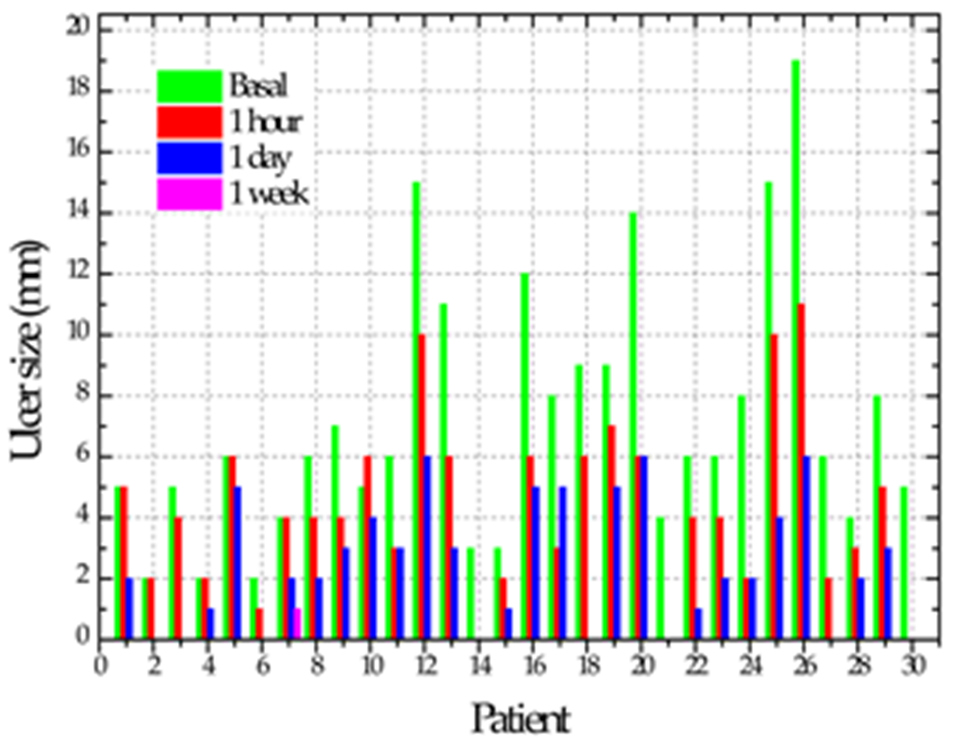
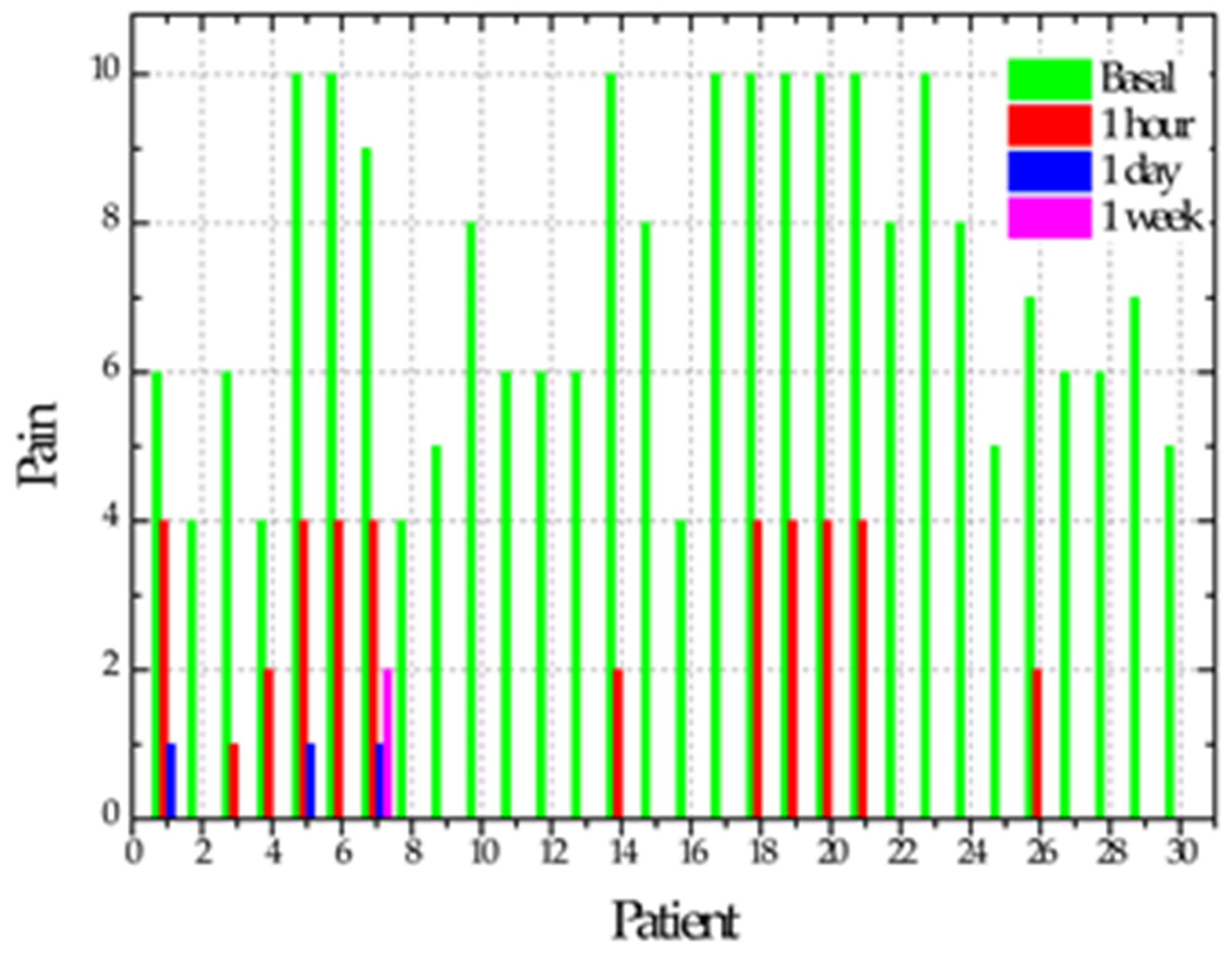
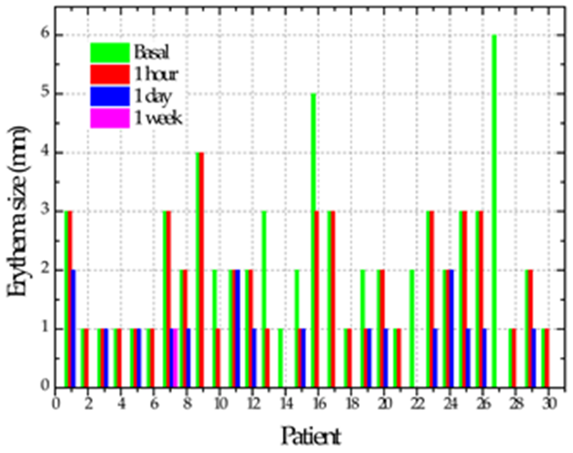
We measured the size of the ulcer and the erythematous contour in each case and took a photographic record. The sample consisted of 14 patients, of whom nine were women (64.3%) and five were men (35.7%). The age range was from 18 to 67 years old, with a mean of 37 years old. The patients presented from one to five ulcers, resulting in 30 ulcers. Of these, 19 (64%) were minor RU, 10 (33%) corresponded to major RU and 1 (3%) was herpetiform RU. The distribution of oral RU cases is shown in Table 1. The size of the ulcers ranged between 2 and 19 mm, with a mean of 7 mm. Before the application of the NTP, all patients reported pain on a numerical verbal scale between 0 and 10 (where 0 is no pain and 10 is the most intense pain); 75% of the patients reported pain, with a mean of 7.
When socioeconomic and risk factors are associated with the type of ulcer, a statistically significant association is verified for recurrence, alcohol use, medication use, occupation, education, marital status, age and gender (p < 0.05—Fisher’s exact) (see Table 1).
After performing the previous review in all cases after the second NTP application, six cases (20%) showed tissue regeneration in the ulcer, considering them repaired at zero days. In this exact measurement, 18 (60%) of the cases reported asymptomatic and the other 12 (40%) cases reported decreased pain with a mean of 4. At 24 h after the two applications with NTP, 11 cases (37%) showed total repair and the remaining 19 cases (63%) showed a reduction in the ulcer size of between 1 and 6 mm with a mean of 3 mm. Regarding the sensation of pain, when assessing the 30 cases, 27 (90%) reported asymptomatic, two cases (7%) reported pain with a value of 1 and the other case (3%) presented pain decreasing from a value from 7 to 4 on the visual analog scale. Ulcer repair time ranged from one hour to 7 days, with a mean of 3 days. In a single case, the ulcer persisted after applying NTP; in addition to the patient reporting pain, the patient reported positive for smoking. The time it took for the ulcers to regenerate showed a correlation only with the ulcer’s initial size of 0.703 (p = 0.000). Table 2 shows the evolution of the cases regarding ulcer size, pain and erythema size.
Figure 3 shows, in the usual manner, the evolution of three cases with RU treated with NTP, which resolved within a maximum of 24 h.
Figure 4 shows three cases of patients with RU who healed on average after three days of NTP application.

4. Discussion

Today, no specific drug or treatment is available for recurrent aphthous stomatitis (RAS). RAS healing is a highly specialized process for repairing damaged/injured tissues through various therapies. Failures in the normal healing process of these ulcers lead to abnormal scar formation and a chronic state that is more susceptible to infection. Wounds caused by RU affect patients’ quality of life, which is why there is an urgent demand for the development of competitive therapies. There are new developments in advanced technologies for the care of RU, for example, laser technology [40,41,42], nano-therapy [43,44], etc. Those studies can improve therapeutic results by focusing on tissue regeneration with minimal side effects. Reports show that evolution time and recurrence periods have been reduced [45,46]. The pain caused due to the ulcers experienced by patients with RAS turns out to be the main problem and usually is treated with antihistaminic agents, analgesics, or even anesthetics to reduce pain and promote patients’ oral function [18]. Recent research suggests that ROS/RNS are central players in the actions of antimicrobial and antiparasitic drugs, cancer therapies, wound healing therapies and treatments involving the cardiovascular system. Understanding how ROS/RNS act in established therapies may help guide future efforts in exploiting novel non-thermal plasma-based medical therapies [47,48].
NTP at atmospheric pressure was applied to the ulcer in the cheek, labial mucosa, tongue, oropharynx, mouth floor, soft palate and gingiva. In, patients with RAS and treated with NTP, its anti-inflammatory and healing effect is evident since ulcer size and pain are significantly reduced. A notable improvement was observed since 86% of the patients did not report pain after one hour of application, being statistically significant (p = 0.0001). Additionally, at 24 h, 90% of the cases reported being asymptomatic, 7% reported pain with a value of 1 and 3% (one case) reported pain with a value of 4. In this regard, it is essential to point out that the two cases that reported pain with a 1 value correspond to a patient with a significant RAS whose ulcers measured 15 and 19 mm. The case that reported pain with a value of 4 is a smoker and the only case that took seven days to heal; therefore, it is crucial to advise smoking patients with RAS to stop smoking during treatment.
With an average time to repair of three days, considering that more than 50% of the cases healed within two days, it was observed that the application of NTP is efficient for RU management. In contrast, studies using laser treatments reported ten days as a time of recovery [20,49] and others 15 days [50]. Some authors report a decrease in pain and erythema after three days of RU laser treatment; in 75% of cases, they have identified total re-epithelialization five days after beginning treatment [51,52]. However, with these results using lasers, there are still three to five days of epithelial exposure, pain and decreased function remaining for RAS patients. Another of the most widely used treatments for RU is topical corticosteroids, which take five to six days to heal a mouth ulcer [45,53].
Regarding the use of these medications, it is essential to consider the adverse effects that can develop as candidiasis. In addition to treatments based on biological products, they have recently been used for wound healing, reducing pain and recurrence frequency in patients with RAS [54,55]. The NTP application highlights that no drug was applied as an adjuvant.
The initial size of the RU showed a correlation with the time elapsed for tissue repair. That is, the larger the initial size of the ulcer, the longer the repair took, which is explained by the amount of epithelium that needs to be formed. Regarding the type of RAS, it is suggested to repeat the treatment with two applications of NTP at 24 h because the significant RU and herpetiform took four to six days to heal completely.
The findings of this study demonstrated the absence of adverse reactions in the tissues. That is, managing recurrent ulcers with NTP was safe for all treated patients since no case showed adverse effects at six months of follow-up and there was significantly reduced pain levels and repair time after the application of NTP. This could be explained due to the mechanism of action of NTP. Most authors agree that treatment with NTP accelerates wound healing by decreasing the level of inflammatory factors and bacterial load and has anti-neoplastic effects [56]. Reactive oxygen and nitrogen species (RONS) generated by NTP can stimulate blood circulation, causing more significant cellular activity. Thus, restoration potentials are improved by accelerating tissue repair and promoting accelerated tissue regeneration. Several studies demonstrate that the role of RONS, particularly H2O2, superoxide (O2) and nitric oxide (NO), is essential in wound healing beyond the simple oxidative elimination of pathogens. The importance of these elements lies in infection prevention, the induction of angiogenesis, the keratinocyte’s increase in differentiation and migration, and the increase in the proliferation of fibroblasts and collagen synthesis. Furthermore, NO has been demonstrated to participate in an intercellular communication network during the healing process, regulating the behavior of macrophages, keratinocytes and fibroblasts [57]. That is, tissue repair and regeneration steps are highly influenced by these intracellular RONS. Therefore, the application of NTP can be considered to cure RU.

5. Conclusions

The study showed that the application of non-thermal plasma in recurrent aphthous stomatitis reduces the levels of pain sensation in the patient, accelerates the healing of the RU and does not produce adverse effects. The technology allowed a quick recovery of the patient and, consequently, a reactivation of daily life. The NTP application in the RUs enables its repair. NTP is an excellent therapeutic alternative for RUs that afflict patients since it provides rapid control of pain and inflammation, accelerating the natural healing process. Due to its application characteristic, it does not bother patients.

Author Contributions

N.G.I.-M., contributed to the conception, experimental design, NTP application, data acquisition, interpretation, writing and critical review of the manuscript; R.L.-C., contributed to the conception, experimental design, NTP application, data acquisition, interpretation, writing and critical review of the manuscript; V.H.T.-R. contributed to the conception, data acquisition and interpretation, performed statistical analyses and drafted and critically reviewed the manuscript; E.L.-C., contributed to the conception, design and data acquisition, performed statistical analysis; B.G.R.-M., experimental design and writing of the manuscript; R.C.d.A. contributed to the statistical analysis, R.P.-E., experimental design and writing of the manuscript; A.M.-C., experimental design and writing of the manuscript; R.V.-A., data acquisition and interpretation and drafted the manuscript; all authors gave final approval and agree to be accountable for all aspects of the work. All authors have read and agreed to the published version of the manuscript.

Funding

This research received no external funding.

Institutional Review Board Statement

The study was conducted in accordance with the Declaration of Helsinki and approved by the Research Ethics Committee of the Center for Research and Advanced Studies in Dentistry of the Faculty of Dentistry of the Autonomous University of the State of Mexico (CEICIEAO-2018-001).

Informed Consent Statement

Informed consent was obtained from all subjects involved in the study.

Data Availability Statement

Not applicable.

Acknowledgments

We appreciate the work of the technicians M.T. Torres-Martínez, P. Angeles-Espinosa, I. Contreras-Villa and M. Lugo-Hernández in the construction of the different equipment systems used in this project. As well as the OROCENTRO Clinic patients who gave their consent for the application of atmospheric pressure non-thermal plasma in the treatment. This study was financially supported by research project CB-001 from National Institute of Nuclear Research and OROCENTRO Clinic of Odontology Faculty from UAEMex. The authors declare no potential conflicts of interest with respect to the authorship and/or publication of this article.

Conflicts of Interest

The authors declare no conflict of interest.

References

  1. Akintoye, S.; Greenberg, M. Recurrent aphthous stomatitis. Dent. Clin. N. Am. 2014, 58, 281–297. [Google Scholar] [CrossRef] [PubMed] [Green Version]
  2. Lau, C.B.; Smith, G.P. Recurrent aphthous stomatitis: A comprehensive review and recommendations on therapeutic options. Dermatol. Ther. 2022, 35, e15500. [Google Scholar] [CrossRef] [PubMed]
  3. Borilova Linhartova, P.; Janos, J.; Slezakova, S.; Bartova, J.; Petanova, J.; Kuklinek, P.; Fassmann, A.; Dusek, L.; Izakovicova Holla, L. Recurrent aphthous stomatitis and gene variability in selected interleukins: A case–control study. Eur. J. Oral Sci. 2018, 126, 485–492. [Google Scholar] [CrossRef] [PubMed]
  4. Han, M.; Fang, H.; Li, Q.L.; Cao, Y.; Xia, R.; Zhang, Z.H. Effectiveness of laser therapy in the management of recurrent aphthous stomatitis: A systematic review. Scientifica 2016, 2016, e9062430. [Google Scholar] [CrossRef] [PubMed] [Green Version]
  5. Monteiro, S.I.; Costa, A.M.; de Vasconcelos, B.C.; Amarante, M.V.; Teixeira, P.T.; de Vasconcelos Gurgel, B.C.; Dantas da Silveira, É.J. Recurrent aphthous ulceration: An epidemiological study of etiological factors, treatment and differential diagnosis. An. Bras. Dermatol. 2018, 93, 341–346. [Google Scholar] [CrossRef] [Green Version]
  6. Sánchez-Bernal, J.; Conejero, C.; Conejero, R. Recurrent aphthous stomatiti. Actas Dermosifiliogr. 2020, 111, 471–480. [Google Scholar] [CrossRef]
  7. Gasmi, B.A.; Noor, S.; Menzel, A.; Gasmi, A. Oral aphthous: Pathophysiology, clinical aspects and medical treatment. Arch. Razi Inst. 2021, 76, 1155–1163. [Google Scholar] [CrossRef]
  8. Bergmeier, L.A.; Fortune, F. Clinical management of oral mucosal disease: A literature review. In Oral Mucosa in Health and Disease; Bergmeier, L., Ed.; Springer International Publishing: Cham, Switzerland, 2018; pp. 161–171. [Google Scholar] [CrossRef]
  9. Saikaly, S.K.; Saikaly, T.S.; Saikaly, L.E. Recurrent aphthous ulceration: A review of potential causes and novel treatments. J. Dermatol. Treat. 2018, 29, 542–552. [Google Scholar] [CrossRef]
  10. Zhang, Y.; Ng, K.; Kuo, C.; Wu, D. Chinese herbal medicine for recurrent aphthous stomatitis: A protocol for systematic review and meta-analysis. Medicine 2018, 97, e13681. [Google Scholar] [CrossRef]
  11. Fitzpatrick, S.G.; Cohen, D.M.; Clark, A.N. Ulcerated Lesions of the Oral Mucosa: Clinical and Histologic Review. Head Neck Pathol. 2019, 13, 91–102. [Google Scholar] [CrossRef]
  12. Szyszkowska, B.; Łepecka-Klusek, C.; Kozłowicz, K.; Jazienicka, I.; Krasowska, D. The influence of selected ingredients of dietary supplements on skin condition. Postepy. Dermatol. Alergol. 2014, 31, 174–181. [Google Scholar] [CrossRef] [Green Version]
  13. Belenguer-Guallar, I.; Jiménez-Soriano, Y.; Claramunt-Lozano, A. Treatment of recurrent aphthous stomatitis. A literature review. J. Clin. Exp. Dent. 2014, 6, e168–e174. [Google Scholar] [CrossRef]
  14. Arafa, M.G.; Ghalwash, D.; El-Kersh, D.M.; Elmazar, M.M. Propolis-based niosomes as oromuco-adhesive films: A randomized clinical trial of a therapeutic drug delivery platform for the treatment of oral recurrent aphthous ulcers. Sci. Rep. 2018, 8, 18056. [Google Scholar] [CrossRef] [Green Version]
  15. Hará, Y.; Shiratuchi, H.; Kaneko, T.; Sakagami, H. Search for drugs used in hospitals to treat stomatitis. Medicines 2019, 6, 19. [Google Scholar] [CrossRef] [Green Version]
  16. Rezvaninejad, R.; Navabi, N.; Khoshroo, M.R.; Torabi, N.; Atai, Z. Herbal medicine in treatment of recurrent aphthous stomatitis: A literature review. J. Iran. Dent. Assoc. 2017, 29, 127–134. [Google Scholar] [CrossRef]
  17. Anand, V.; Gulati, M.; Govila, V.; Anand, B. Low level laser therapy in the treatment of aphthous ulcer. J. Dent. Res. 2013, 24, 267–270. [Google Scholar] [CrossRef]
  18. Nasry, S.A.; El Shenawy, H.M.; Mostafa, D.; Ammar, N.M. Different modalities for treatment of recurrent aphthous stomatitis. A randomized clinical trial. J. Clin. Exp. Dent. 2016, 8, e517–e522. [Google Scholar] [CrossRef]
  19. Suter, V.G.A.; Sjölund, S.; Bornstein, M.M. Effect of laser on pain relief and wound healing of recurrent aphthous stomatitis: A systematic review. Lasers Med. Sci. 2017, 32, 953–963. [Google Scholar] [CrossRef]
  20. Jahromi, N.Z.; Ghapanchi, J.; Pourshahidi, S.; Zahed, M.; Ebrahimi, H. Clinical evaluation of high and low-level laser treatment (CO2 vs InGaAlP diode laser) for Recurrent Aphthous Stomatitis. J. Dent. 2017, 18, 17–23. [Google Scholar]
  21. Melis, M.; Di Giosia, M.; Colloca, L. Ancillary factors in the treatment of orofacial pain: A topical narrative review. J. Oral Rehabil. 2019, 46, 200–207. [Google Scholar] [CrossRef]
  22. Tiwari, S.; Avinash, A.; Katiyar, S.; Iyer, A.A.; Jain, S. Dental applications of ozone therapy: A review of literature. Saudi J. Dent. Res. 2017, 8, 105–111. [Google Scholar] [CrossRef] [Green Version]
  23. Sabbah, F.; Nogales, C.G.; Zaremski, E.; Martinez-Sanchez, G. Ozone therapy in dentistry—Where we are and where we are going to? Rev. Esp. Ozonot. 2018, 8, 37–63. [Google Scholar]
  24. Bader, K.A.Z. Management of denture-related traumatic ulcers using ozone. J. Prosth. Dent. 2019, 121, 76–82. [Google Scholar] [CrossRef]
  25. Nirmala, M.; Smitha, S.G.; Kamath, G. A study to assess the efficacy of local application of oral probiotic in treating recurrent aphthous ulcer and oral Candidiasis. Indian J. Otolaryngol. Head Neck Surg. 2019, 71, S113–S117. [Google Scholar] [CrossRef] [PubMed]
  26. Dubin, D.H.E. Effect of correlations on the thermal equilibrium and normal modes of a non-neutral plasma. Phys. Rev. E 1996, 53, 5268–5290. [Google Scholar] [CrossRef] [Green Version]
  27. Hama-Aziz, K.H.; Miessner, H.; Mueller, S.; Kalass, D.; Moeller, D.; Khorshid, I.; Rashid, M.A.M. Degradation of pharmaceutical diclofenac and ibuprofen in aqueous solution, a direct comparison of ozonation, photocatalysis, and non-thermal plasma. Chem. Engen. J. 2017, 313, 1033–1041. [Google Scholar] [CrossRef]
  28. Kajiyama, H.; Utsumi, F.; Nakamura, K.; Tanaka, H.; Toyokuni, S.; Hori, M.; Kikkawa, F. Future perspective of strategic non-thermal plasma therapy for cancer treatment. J. Clin. Biochem. Nutr. 2017, 60, 33–38. [Google Scholar] [CrossRef] [Green Version]
  29. Laroussi, M. Plasma medicine: A brief introduction. Plasma 2018, 1, 47–60. [Google Scholar] [CrossRef] [Green Version]
  30. Dubuc, A.; Monsarrat, P.; Virard, F.; Merbahi, N.; Sarrette, J.P.; Laurencin-Dalicieux, S.; Cousty, S. Use of cold-atmospheric plasma in oncology: A concise systematic review. Ther. Adv. Med. Oncol. 2018, 10, 1–12. [Google Scholar] [CrossRef] [Green Version]
  31. Jang, J.Y.; Hong, Y.J.; Lim, J.; Choi, J.S.; Choi, E.H.; Kang, S.; Rhim, H. Cold atmospheric plasma (CAP), a novel physicochemical source, induces neural differentiation through cross-talk between the specific RONS cascade and Trk/Ras/ERK signaling pathway. Biomaterials 2018, 156, 258–273. [Google Scholar] [CrossRef]
  32. Park, J.; Lee, H.; Lee, H.J.; Kim, G.C.; Kim, S.S.; Han, S.; Song, K. Non-thermal atmospheric pressure plasma is an excellent tool to activate proliferation in various mesoderm-derived human adult stem cells. Free Radic. Biol. Med. 2019, 134, 374–384. [Google Scholar] [CrossRef]
  33. Wu, S.; Cao, Y.; Lu, X. The state of the art of applications of atmospheric-pressure nonequilibrium plasma jets in dentistry. IEEE Trans. Plasma Sci. 2016, 44, 134–151. [Google Scholar] [CrossRef]
  34. Betancourt-Ángeles, M.; Peña-Eguiluz, R.; López-Callejas, R.; Domínguez-Cadena, N.A.; Mercado-Cabrera, A.; Muñoz-Infante, J.; Rodríguez-Méndez, B.G.; Valencia-Alvarado, R.; Moreno-Tapia, J.A. Treatment in the healing of burns with a cold plasma source. Int. J. Burns Trauma 2017, 7, 142–146. [Google Scholar]
  35. de Masi, G.; Gareri, C.; Cordaro, L.; Fassina, A.; Brun, P.; Zaniol, B.; Cavazzana, R.; Martines, E.; Zuin, M.; Marinaro, G.; et al. Plasma coagulation controller: A low-power atmospheric plasma source for accelerated blood coagulation. Plasma Med. 2018, 8, 245–254. [Google Scholar] [CrossRef] [Green Version]
  36. López-Callejas, R.; Peña-Eguiluz, R.; Valencia-Alvarado, R.; Mercado-Cabrera, A.; Rodríguez-Méndez, B.G.; Serment-Guerrero, J.H.; Cabral-Prieto, A.; González-Garduño, A.C.; Domínguez-Cadena, N.A.; Muñoz-Infante, J.; et al. Alternative method for healing the diabetic foot by means of a plasma needle. Clin. Plasma Med. 2018, 9, 19–23. [Google Scholar] [CrossRef]
  37. Martin, W.J.J.M.; Skorpil, N.E.; Ashton-James, C.E.; Tuinzing, D.B.; Forouzanfar, T. Effect of vasoconstriction on pain after mandibular third molar surgery: A single-blind, randomized controlled trial. Quintessence Int. 2016, 47, 589–596. [Google Scholar] [CrossRef]
  38. Peña-Eguiluz, R.; Serment-Guerrero, J.H.; Azorín-Vega, E.P.; Mercado-Cabrera, A.; Flores-Fuentes, A.A.; Jaramillo-Sierra, B.; Hernández-Arias, A.N.; Girón-Romero, K.; López-Callejas, R.; Rodríguez-Méndez, B.G.; et al. Development and characterization of a non-thermal plasma source for therapeutic treatments. IEEE. Trans. Biomed. Eng. 2021, 68, 1467–1476. [Google Scholar] [CrossRef]
  39. International Commission on Non-Ionizing Radiation Protection. Guidelines on limits of exposure to ultraviolet radiation of wavelengths between 180 nm and 400 nm (incoherent optical radiation). Health Phys. 2004, 87, 171–186. [Google Scholar] [CrossRef]
  40. dos Santos, J.A.; Normando, A.G.C.; de Toledo, I.P.; Melo, G.; de Luca Canto, G.; Santos-Silva, A.R.; Silva-Guerra, E.N. Laser therapy for recurrent aphthous stomatitis: An overview. Clin. Oral Investig. 2020, 24, 37–45. [Google Scholar] [CrossRef]
  41. Rao, Z.; Tomar, D.; Kaushik, S.; Mittal, S.; Kumar, P. Application of soft tissue laser in the management of recurrent apthous stomatitis: A placebo controlled study. Int. J. Health Sci. 2022, 6, 2590–2598. [Google Scholar] [CrossRef]
  42. Nagieb, C.S.; Harhash, T.A.E.; Fayed, H.L.; Ali, S. Evaluation of diode laser versus topical corticosteroid in management of Behcet’s disease-associated oral ulcers: A randomized clinical trial. Clin. Oral Investig. 2022, 26, 697–704. [Google Scholar] [CrossRef] [PubMed]
  43. Makvandi, P.; Josic, U.; Delfi, M.; Pinelli, F.; Jahed, V.; Kaya, E.; Ashrafizadeh, M.; Zarepour, A.; Rossi, F.; Zarrabi, A.; et al. Drug delivery (nano) platforms for oral and dental applications: Tissue regeneration, infection control, and cancer management. Adv. Sci. 2021, 8, e2004014. [Google Scholar] [CrossRef] [PubMed]
  44. Gao, H.; Wu, N.; Nini Wang, N.; Li, J.; Sun, J.; Peng, Q. Chitosan-based therapeutic systems and their potentials in treatment of oral diseases. Int. J. Biol. Macromol. 2022, 222, 3178–3194. [Google Scholar] [CrossRef] [PubMed]
  45. Scully, C.; Porter, S. Oral mucosal disease: Recurrent aphthous stomatitis. Br. J. Oral Maxillofac. Surg. 2008, 46, 198–206. [Google Scholar] [CrossRef]
  46. Yarom, N.; Zelig, K.; Epstein, J.B.; Gorsky, M. The efficacy of minocycline mouth rinses on the symptoms associated with recurrent aphthous stomatitis: A randomized, double-blind, crossover study assessing different doses of oral rinse. Oral Surg. Oral Med. Oral Pathol. Oral Radiol. 2017, 123, 675–679. [Google Scholar] [CrossRef]
  47. Pekbağrıyanık, T.; Dadas, F.K.; Enhoş, Ş. Effects of non-thermal atmospheric pressure plasma on palatal wound healing of free gingival grafts: A randomized controlled clinical trial. Clin. Oral Investig. 2021, 25, 6269–6278. [Google Scholar] [CrossRef]
  48. Suresh, M.; Hemalatha, V.T.; Sundar, N.M.; Nisha, A. Applications of cold atmospheric pressure plasma in dentistry—A review. J. Pharm. Res. Int. 2022, 34, 45–55. [Google Scholar] [CrossRef]
  49. Yilmaz, H.G.; Albaba, M.R.; Caygur, A.; Cengiz, E.; Boke-Karacaoglu, F.; Tumer, H. Treatment of recurrent aphthous stomatitis with Er,Cr:YSGG laser irradiation: A randomized controlled split mouth clinical study. J. Photochem. Photobiol. B Biol. 2017, 170, 1–5. [Google Scholar] [CrossRef]
  50. Aggarwal, H.; Singh, M.P.; Nahar, P.; Mathur, H.; Sowmya, G.V. Efficacy of low-level laser therapy in treatment of recurrent aphthous ulcers—A sham controlled, split mouth follow up study. J. Clin. Diagn. Res. 2014, 8, 218–221. [Google Scholar] [CrossRef]
  51. Lalabonova, H.; Daskalov, H. Clinical assessment of the therapeutic effect of low-level laser therapy on chronic recurrent aphthous stomatitis. Biotechnol. Biotechnol. Equip. 2014, 28, 929–933. [Google Scholar] [CrossRef]
  52. de Sousa, A.C.T.; da Rocha, Í.B.P.; de Carvalho, A.F.M.; de Freitas Coelho, N.P.M.; Feitosa, M.C.P.; Barros, E.M.L.; Arisawa, E.A.L.; de Amorim, M.R.L. Comparative study between low level laser and therapeutic ultrasound in second intertion ulcers repair in mice. J. Laser Med. Sci. 2018, 9, 14–38. [Google Scholar] [CrossRef] [Green Version]
  53. Hamishehkar, H.; Nokhodchi, A.; Ghambarzadeh, S.; Kouhsoltani, M. Triamcinolone acetonide oromucoadhesive paste for treatment of aphthous stomatitis. Adv. Pharm. Bull. 2015, 5, 277–282. [Google Scholar] [CrossRef]
  54. Liu, D.; Zhang, T.; Zhou, H.; Meng, Y.; Wu, C.; Sun, Y.; Xu, Y.; Deng, X.; Wang, H.; Jiang, L. Role of biologics in refractory recurrent aphthous stomatitis. J. Oral Path. Med. 2022, 51, 694–701. [Google Scholar] [CrossRef]
  55. Shavakhi, M.; Sahebkar, A.; Shirban, F.; Bagherniya, M. The efficacy of herbal medicine in the treatment of recurrent aphthous stomatitis: A systematic review of randomized clinical trials. Phytother. Res. 2022, 36, 672–685. [Google Scholar] [CrossRef]
  56. Amini, M.R.; Hosseini, M.S.; Fatollah, S.; Mirpour, S.; Ghoranneviss, M.; Larijani, B.; Mohajeri-Tehrani, M.R.; Khorramizadeh, M.R. Beneficial effects of cold atmospheric plasma on inflammatory phase of diabetic foot ulcers; a randomized clinical trial. J. Diabetes Metab. Disord. 2020, 19, 895–905. [Google Scholar] [CrossRef]
  57. Jha, N.; Ryu, J.J.; Choi, E.H.; Kaushik, N.K. Generation and role of reactive oxygen and nitrogen species induced by plasma, lasers, chemical agents, and other systems in dentistry. Oxid. Med. Cell. Longev. 2017, 2017, 7542540. [Google Scholar] [CrossRef]
Figure 1. Clinical characteristics of: (A) minor RAS, (B) major RAS, (C) herpetiform RAS.
Biomedicines 11 00167 g001
Figure 2. Plasma reactor and non-thermal plasma application in the patient.
Biomedicines 11 00167 g002
Figure 3. RU in the buccal mucosa, horizontally showing the evolution of the ulcers in three patients; (a,d,g) correspond to the initial state of the RU; (b,e,h) show regeneration after the first application of NTP; finally, (c,f,i) show the healthy tissue 24 h after starting the treatment.
Biomedicines 11 00167 g003
Figure 4. RU is located in different parts of the oral cavity (indicated with arrows): (a,d,g) at patient admission. (b,e,h) after the first application of the NTP. Finally, (c,f,i) show follow-up when the wound has healed (indicated with arrows).
Biomedicines 11 00167 g004
Table 1. Sociodemographic and risk factors for injuries according to the type of ulcer in this study.
Recurrent
Herpetiform Ulcers
Major Recurrent UlcersMinor Recurrent UlcersFisher’s Exact
Gender
Male 100.01
Female1109
Age
18–30 290.005
31–501 7
51–67 83
Marital status
Married1860.02
Single 213
Scholarship
High school 230.04
Basic186
Bachelor’s degree 10
Occupation
Unemployed 310.003
Student 23
Homework151
Employee 14
Medication consumption
Not12110.08
Yes 88
Smoking
Cigarette 340.73
No1715
Alcohol consumption
2 to 4 times a month1 0.02
Never184
Once a year 18
Once a month 7
Ulcers evolution time
Two to five days14100.69
Six to ten days 68
One day 1
Ulcers recurrence period
Zero 10.07
One for year152
One for month 55
One for week 3
More than a year 8
Table 2. Size, pain and erythema in the initial period and after plasma application, one hour, one day and one week.
Mean and Standard Deviation of Ulcer Size
Initial6.9 ± 4.51 ABiomedicines 11 00167 i001
One hour3.96 ± 3.1 B
One day2 ± 2 BC
One week0.03 ± 0.18 C
p<0.0001
Mean and Standard Deviation of Pain
Initial7.1 ± 2.5 ABiomedicines 11 00167 i002
One hour1.5 ± 2.3 B
One day0.2 ± 0.7 C
One week0.06 ± 0.3 C
p<0.0001
Mean and Standard Deviation of
Erythema Size
Initial2.1 ± 1.1 ABiomedicines 11 00167 i003
One hour1.4 ± 1 B
One day0.5 ± 0.6 C
One week0.03 ± 0.18 C
p<0.0001
Different letters = statistically significant differences. It was verified that, after one hour of plasma application, there was a substantial reduction in size and pain (ANOVA one-way test followed by Tukey).
Disclaimer/Publisher’s Note: The statements, opinions and data contained in all publications are solely those of the individual author(s) and contributor(s) and not of MDPI and/or the editor(s). MDPI and/or the editor(s) disclaim responsibility for any injury to people or property resulting from any ideas, methods, instructions or products referred to in the content.