Abstract
Telomere maintenance is key during cancer development. Malignant cells can either use telomerase or an alternative lengthening of telomere (ALT) pathway to maintain their telomere length. In Hodgkin’s Lymphoma (HL), the presence of telomerase activation is established. The activation of ALT has been reported recently. Our data confirm this notion describing co-localization of the phosphorylated form of telomeric repeat-binding factor 1 (pT371-TRF1) with ALT-associated promyelocytic leukemia bodies. Surprisingly, to our knowledge, there are no published studies targeting both telomere maintenance pathways in HL. Consequently, we investigated, for the first time, the effects of both telomerase and ALT inhibition on HL cell viability: We inhibited telomerase and/or ALT, given either individually, simultaneously, or consecutively. We report that the inhibition of telomerase using BIBR1532 followed by ALT inhibition, using trabectedin, caused a decrease of greater than 90% in cell viability in three patient-derived HL cell lines. Our results suggest that HL cells are most vulnerable to the consecutive inhibition of telomerase followed by ALT inhibition.
1. Introduction
Hodgkin’s lymphoma (HL) is a B-cell malignancy characterized by the presence of mononucleated Hodgkin (H) and multinucleated Reed–Sternberg (RS) cells [1,2], with the RS cell representing the diagnostic cell of the disease [2]. In 2020, more than 83,000 people globally were diagnosed with classical Hodgkin’s Lymphoma (cHL), and more than 23,000 deaths were registered for the same period [3]. HL can affect all age groups [4,5]. There are high cure rates for cHL when diagnosed at early disease stages or in patients presenting with low-risk disease [4,6]. However, patients diagnosed at advanced disease stage have poor prognosis and unfavorable disease outcome [6]. The 5-year relative survival rate is 89.1% (seer.cancer.gov).
Telomeres are protective heterochromatic structures found at the ends of linear eukaryotic chromosomes. Mammalian telomeric DNA is composed of repetitive TTAGGG tandem repeats. The number of times most normal human somatic cells can divide is linked to the status of their telomeres. Telomeric DNA shortens during each round of DNA replication, in part because DNA polymerase is unable to completely replicate the 3′ overhang of the chromosomal ends (the “end replication problem”) [7,8,9,10]. Once telomeric DNA reaches a critical short length, the cells stop dividing and enter a state called senescence. This state of a limited life span was first defined by Hayflick [11,12] and it has also been coined as the Hayflick limit.
In stark contrast to normal cells, tumor cells escape the Hayflick limit of lineage-dependent limited life span. This step involves mutations inactivating cell cycle checkpoint proteins, increased cell divisions and therefore further telomere shortening. These events trigger a “crisis” event where sister chromatin fusions, end-to-end fusions and breakage-bridge-fusion cycles events occur, leading to chromosomal rearrangements and ongoing genome instability. While most cells die during crisis, some survive and activate telomere maintenance mechanisms. These telomere maintenance pathways permit the cells to continue cellular division despite the presence of critically short telomeres. There are two telomere maintenance pathways, telomerase activation and alternative lengthening of telomeres (ALT) [13,14,15]. Telomerase activation is the most common pathway and is employed by 85–90% of all tumor cells, while ALT is used by the remainder. Some tumors, albeit rare, show the co-activation of both pathways. These include rare cases of renal cell carcinoma [16], breast cancer [17] and classical Hodgkin’s Lymphoma [18]. The consequence of continued cell proliferation of tumor cells despite their critically short telomeres allows for ongoing genomic instability and its dynamic propagation to the next generation of cells.
Genomic instability is an enabling hallmark of cancer [19]. The contribution of telomeres to this dynamic process of genomic instability is genomic remodeling through sister chromatid fusion, end-to-end chromosomal fusion, and breakage-bridge-fusion cycles [20,21]. This dynamic process of telomeric changes is also called telomere dysfunction and is a key factor in creating cell-to-cell genetic heterogeneity in cancer and enables tumor cell and clonal evolution [22]: For example, a single telomeric end-to-end chromosomal fusion will generate, following cell division, due to the formation of an anaphase bridge and chromosomal breakage in telophase, one daughter cell with a terminal deletion and one with an unbalanced translocation. Both daughter cells are then genetically distinct and due to the presence of double-strand breaks at the chromosomal break points, they will engage in further fusion/recombination events with other chromosomes. This process will continue and lead to the genetic divergence of the cells and, upon selective pressure, to the generation and survival of clones [22,23].
Hodgkin’s Lymphoma exhibits telomere dysfunction [24], telomerase activation [24,25] and ALT activation [18]. Since tumor cells evolve to become resistant and overcome telomerase inhibition by activating the ALT pathway [26], it is expected that the dual inhibition of both pathways will be more effective than the single pathway inhibition. While the single inhibition of one of the two telomere maintenance pathways has been reported [27,28,29], there is no published work to date demonstrating the dual inhibition of both pathways. Telomerase inhibition is effective in telomerase-activated cancer cells [30,31]. However, as a result of this treatment, the cells commonly become resistant [26]. Inhibition of ALT is currently offered to patients with soft tissue sarcoma [32] and ovarian cancer [33].
The degree of telomere dysfunction in Hodgkin’s Lymphoma [24] is associated with the aggressiveness of disease and response to treatment [34,35]. As we showed, telomere dysfunction (measured by three-dimensional (3D) telomere imaging and dedicated software) in mononucleated Hodgkin’s cells leads to the formation of the diagnostic Reed–Sternberg cell [2,24,36]. Telomere dysfunction in HL includes critically short telomeres, telomeric aggregates, altered telomere numbers and a different 3D spatial organization [24,34]. In addition, the severity of the 3D telomere dysfunction in HL is associated with response to treatment and recurrent/refractory disease [24]. The higher the level of telomere dysfunction, the greater the chance of poor outcome [34,35,36].
The current study was initiated to explore the addiction of Hodgkin’s Lymphoma cells to telomere maintenance. Using the small molecule inhibitor BIBR1532 to inhibit telomerase [37,38] and the alkylating agent trabectedin to inhibit the ALT pathway [27] alone and in combination, as well as with a consecutive addition of either drug, we report that telomerase inhibition for 72 h, followed by ALT inhibition for 72 h exhibits the strongest negative effects on cell viability of HL cells.
2. Materials and Methods
2.1. Cell Lines and Cell Culture Conditions
Three different human-derived HL cell lines were used for this study: HDLM-2, L-428 and L-1236 (DSMZ, Braunschweig, Germany). The HDLM-2 and L-428 cell lines were grown in RPMI-1640 medium, supplemented with 20% fetal bovine serum (FBS), 1% L-glutamine, 1% sodium pyruvate, and 1% penicillin–streptomycin (reagents from Invitrogen/Gibco, Burlington, ON, Canada). The L-1236 cell line was grown in RPMI-1640 medium, supplemented with 10% FBS, 1% L-glutamine, 1% sodium pyruvate, and 1% penicillin–streptomycin. Cells were incubated at 37 °C in a humidified atmosphere containing 5% CO2. A concentration of 5 × 10⁶ cells/tissue culture well in 6-well plates (NuncTM Cell Culture Treated Multidishes, ThermoFisher Scientific, Waltham, MA, USA) were used for all cell lines during the inhibition of telomere maintenance pathways assay. This cell number was chosen to simulate the overall number of lymphocytes residing in a regular lymph node [39].
2.2. Fluorescent Immunocytochemistry
2.2.1. Antibodies
Antibodies used include: primary anti-TRF2 (rabbit polyclonal, Novus (NB110 57130) 1:500 dilution); secondary goat anti-rabbit Alexa 488 (Molecular Probes, Waltham, MA, USA, 1:1000 dilution); primary anti-pT371-TRF1 [40] (rabbit polyclonal, 1:500 dilution); primary anti-PML (mouse monoclonal, Santa Cruz, sc- 966, 1:100 dilution), and sheep anti-mouse Cy3 (AC111C, Sigma Chemical, St. Louis, MO, USA, 1:500 dilution). The antibodies used for immunocytochemistry analyses were diluted in 4% BSA/4X SSC (blocking solution).
2.2.2. Immunocytochemistry
The cells were fixed in 3.7% formaldehyde/1× PBS for 10 min at room temperature (RT) and immunostained as previously published [24,36]. Cell nuclei were counterstained with DAPI 0.1 µg/mL (D9542, Sigma Chemical, St. Louis, MO, USA) for 3 min. The slides were mounted in Vectashield® (Vector Laboratories, Inc., Burlingame, CA, USA).
2.2.3. Three-Dimensional Image Acquisition
Three-dimensional conventional imaging of 90 cells from each cell line was performed using a ZEISS Axio Imager Z2 (Carl Zeiss, Toronto, ON, Canada) with a cooled AxioCam HR B&W, FITC, Cy3 and DAPI filters in combination with a Planapo 63×/1.4 oil objective lens (Carl Zeiss, Jena, Germany). For every fluorophore, 60 z-stacks were imaged with a 200 nm distance between each stack. Images were obtained using ZEN blue 2.3 edition software (Carl Zeiss, Jena, Germany) in multichannel mode, and deconvolved using the constrained iterative restoration algorithm [41] with theoretical PSF and automatic normalization.
2.3. Inhibition of Telomere Maintenance Pathways
The initial drug concentrations were selected based on prior studies reporting the inhibition of ALT and telomerase pathways in different cancer cell lines [27,42]. To define the drug working concentration, the HL cell line HDLM-2 was subjected to individual treatment with telomerase inhibitor BIBR1532 (EMD MilliporeSigma, San Luis, MO, USA) using the following concentrations: 125, 150, 175, 200 µM from 0–144 h and treatment with ALT-pathway inhibitor trabectedin (Apexbio Technology, Houston, TX, USA) using the following concentrations: 0.25, 0.5, 0.75, 1, 1.25 nM from 0–240 h [27]. The concentrations determined with HDLM-2 were applied to the other HL cell lines L-1236 and L-428.
2.4. Cell Viability
The HL-derived cell lines (HDLM-2, L-428 and L-1236) were seeded in 6-well plates (NuncTM Cell Culture Treated Multidishes, ThermoFisher Scientific, Waltham, MA, USA) at 5 × 10⁶/well. The cells were treated with DMSO to a final concentration of 0.02% (control condition), 200 µM of telomerase-inhibitor BIBR1532 and 4 nM of ALT-pathway inhibitor trabectedin in various combinations and orders as shown in Figure 1. The trabectedin and BIBR 1532 treatment concentrations were determined using the HDLM-2 cell line (Figure A1).
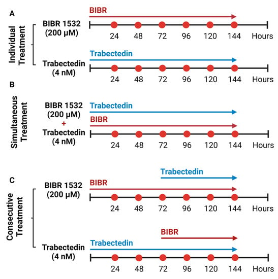
Figure 1.
Inhibition of telomere maintenance pathways in HL cell lines. (A) Individual treatment of HL cell lines with 200 µM of BIBR1532 and 4 nM of trabectedin for 144 h. (B) Simultaneous treatment of HL cell lines with 200 µM of BIBR1532 and 4 nM of trabectedin for 144 h. (C) Consecutive treatment of HL cell lines with BIBR1532 and trabectedin treatment, the latter was added after the first 72 h of BIBR1532. Consecutive treatment of HL cell lines with trabectedin followed by BIBR 1532, which was added at the 72 h time point.
The cell viability before and during treatments was evaluated by trypan-blue exclusion assay every 24 h. Briefly, aliquots containing 15 µL of the cells were taken every 24 h, mixed with 15 µL of trypan blue (1:1 ratio) and submitted (11 µL) to manual counting using a hemocytometer (Hausser Scientific, Horsham, PA, USA).
2.5. Statistical Analysis
Two-way ANOVA followed by Tukey’s test were used to analyze the impact on cellular viability of the HL cell lines (HDLM-2, L-428 and L-1236) treated with telomerase and ALT inhibitors. Significance levels were set as p ≤ 0.05. PRISM Graph Pad v8.0 (San Diego, CA, USA) software was used for illustration.
3. Results
3.1. Hodgkin’s Lymphoma (HL) Shows Hallmarks of an Alternative Telomere Lengthening (ALT) Pathway
As previously reported by us and others, HL cells activate telomerase [24,25]. Recent work described the alternative lengthening of telomeres (ALT) pathway, which involved the presence of promyelocytic leukemia (PML) bodies [18,43]. Since PML bodies can be found in non-malignant cells [44], we examined the presence of both PML and pT371-TRF1 protein in HL cells. The latter does not bind normal telomeres [40] but interacts specifically with dysfunctional telomeres, forming distinct foci in ALT cells [45]. In addition, pT371-TRF1 is a component of APBs, a hallmark of ALT [45].
We performed immunofluorescent analysis of PML and pTRF1 in three HL cell lines (HDLM-2, L-1236 and L-428). First, the presence of PML bodies was confirmed in all HL cell lines, and observed in both mononucleated Hodgkin’s (H) cells and multinucleated Reed–Sternberg (RS) cells as shown in Figure 2A. The presence of the phosphorylated form of the telomeric repeat-binding factor 1 (pT371-TRF1) was also confirmed in all HL cell lines, in both H and RS cells, as shown in Figure 2B. In addition, colocalization of pT371-TRF1 and PML bodies was observed in all HL cell lines (Figure 2C and Appendix A, Figure A2), suggesting the presence of APBs, hallmarks of ALT, in all HL cell lines.
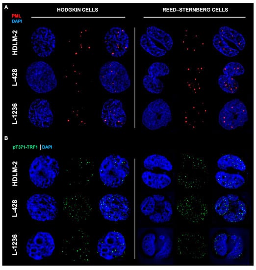
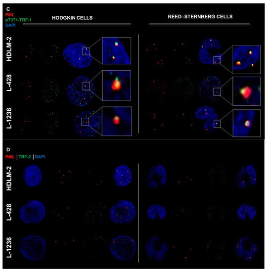
Figure 2.
Hodgkin’s Lymphoma (HL) shows hallmarks of an alternative telomere lengthening (ALT) pathway. (A) HL cell lines show promyelocytic leukemia bodies (PML) in both HL cell phenotypes (Hodgkin cells and Reed–Sternberg cells). (B) HL cell lines show phosphorylated form of telomeric repeat-binding factor 1 (pT371-TRF1) in both HL cell phenotypes. (C) HL cell lines show colocalization (white zoom boxes) of pT371-TRF1 and PML bodies in both HL cell phenotypes. (D) HL cell lines show presence of telomeric repeat-binding factor 2 (TRF2) around PML bodies in both HL cell phenotypes. PML—Red (Cy3), pT371-TRF1—Green (A-488), TRF2—Green (A-488) and DNA—Blue (DAPI). A total of 90 cells were examined in three independent experiments for each of the HL cell lines, and all showed both colocalized (yellow) as well as free signals for pT371-TRF1 and PML in addition to TRF2 and PML in both H and RS cells. For additional information, see Appendix A Figure A2.
The telomeric repeat-binding factor 2 (TRF2) protein is associated with ALT telomere synthesis and works to maintain ALT pathway activity [46,47]. TRF2 protein is seen in all HL cell lines and, in both H and RS cells (Figure 2D). APBs are present in all HL cells.
Analysis of colocalization to pT371-TRF1 and PML as well as of TRF2 and PML in 90 cells and three independent experiments showed for each of the HL cell lines that each cell exhibited both colocalized and free signals of pT371-TFR1/PML and TRF2/PML (Figure 2C,D). The reasons for the heterogeneous signal localization are currently unknown.
3.2. HL Is Sensitive to the Inhibition of Both Telomere Pathways
We hypothesized that the inhibition of both telomere maintenance pathways might be lethal to HL cells. We therefore treated the HL cells with telomerase inhibitor alone, ALT inhibitor alone, both telomerase and ALT inhibitors at the same time or sequentially.
3.3. Independent and Simultaneous Inhibition of Telomere Maintenance Pathways
Exposure of HDLM-2, L-428 and L-1236 HL cell lines to 4 nM of trabectedin [27] (ALT pathway inhibitor) or 200 µM of BIBR1532 (telomerase inhibitor) [48] for 144 h led to a time-dependent decrease in cell viability. As shown in Figure 3, all three cell lines exhibited a reduction in cell viability. At 144 h, trabectedin triggered 60%, 40% and 50% survival of HDLM-2, L-428 and L-1236, respectively. BIBR1532 reduced cell viability by ≥ 90% in all three cell lines during the same period. The simultaneous treatment with both drugs elicited survival similar to the BIBR1532 treatment regimen alone (Figure 3).
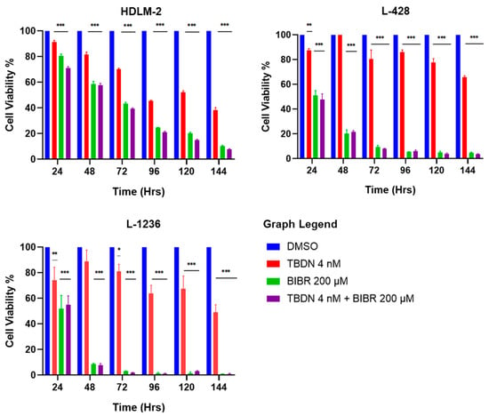
Figure 3.
Combinatory inhibition of telomere maintenance pathways. HL cell lines show decrease in cellular viability upon treatment with trabectedin 4 nM (ALT pathway inhibitor). The effect was time-dependent in all HL cell lines. Incubation of HL cell lines with 200 µM BIBR 1532 (telomerase inhibitor) showed significant time-dependent decrease in cell viability. The combined inhibitory effect on both telomere maintenance pathways resulted in a remarkable time-dependent decrease in cellular viability, in all HL cell lines. Data expressed as mean ± SEM. * p < 0.05, ** p < 0.001 and *** p < 0.0001. TBDN—trabectedin.
3.4. Consecutive Inhibition of Telomere Maintenance Pathways
The consecutive exposure of HDLM-2, L-428 and L-1236 to trabectedin and BIBR1532 for 72 h each (Figure 1) was performed to determine the effects of dual but consecutive telomere maintenance pathway inhibition on HL cells. Trabectedin treatment followed by BIBR1352 treatment induced cell death at 75%, 90% and >95% in HDLM-2, L428 and L-1236, respectively. This effect was stronger than that of trabectedin alone (Figure 4). However, the consecutive treatment of BIBR1352, followed by trabectedin decreased cell survival to 10%, <5% and <5% in HDLM2, L-428 and L-1236, respectively. This consecutive treatment order reduced the viability of HL most dramatically. Collectively, our results indicate that telomerase inhibition followed by ALT inhibition leads to the most potent induction of HL cell death.
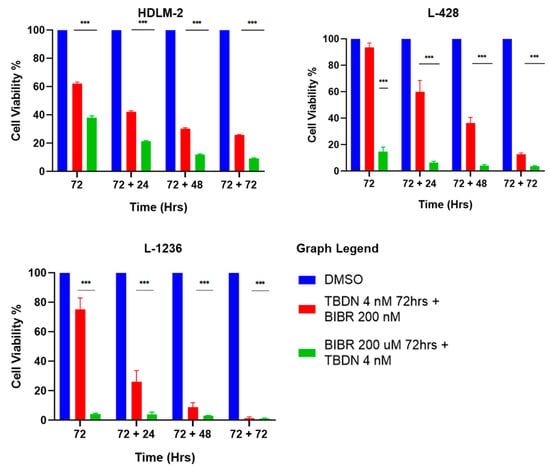
Figure 4.
Consecutive inhibition of telomere maintenance pathways. HL cell lines show pronounced reduction in cellular viability over cell sensitization with trabectedin 4 nM and BIBR 200 µM for 72 h, followed by consecutive treatment with trabectedin 4 nM and BIBR 200 µM for 72 h. Data expressed as mean ± SEM. *** p < 0.0001. TBDN—trabectedin.
4. Discussion
Telomerase activation is key to maintaining an unlimited proliferation potential, which is characteristic of malignant cells [49,50]. However, not all tumor cells express telomerase. Some tumor cells instead activate an alternative pathway for lengthening of telomeres (ALT) and, in some cases, malignant cells activate both pathways [14,49,51,52,53]. In Hodgkin’s Lymphoma, the presence of telomere maintenance pathways through telomerase and ALT was reported for human-derived lymph node samples as well as HL cell lines [18,24,25]. However, only promyelocytic leukemia bodies (PML) were investigated as an ALT marker, and a high frequency of cell-to-cell heterogeneity was observed [18]. Moreover, the presence of PML proteins is also observed in non-malignant cells [44], highlighting the need for further characterization of ALT activation in HL.
Here, we show the presence of ALT-associated promyelocytic leukemia bodies (APBs), TRF2, pT371-TRF1 and for the first time the colocalization of pT371-TRF1 and PML in three patient-derived Hodgkin lymphoma cell lines in both HL and RS cells. Association of TRF2 and APBs are hallmarks of ALT activation in HL [18,53].
The phosphorylation of TRF1 protein on T371 residue promotes its interaction with APBs, therefore causing activation of ALT [45]. Moreover, loss of TRF1 phosphorylation or TRF1 deletion impairs formation of APBs, disrupting ALT pathway activity [45,54]. Here, we show the presence and colocalization of pT371-TRF1 with APBs in HL cells and confirm ALT activation in HL and RS cells of HL.
The activation of telomere maintenance pathways has been correlated with malignant transformation and cancer progression and is also associated with poor prognosis and reduced overall survival [55,56,57,58]. The characterization of telomere maintenance pathways has been used to assess cancer aggressiveness and stratify malignant subgroups [27,56,58]. Although several studies have been targeting telomere maintenance pathways in cancer cells, these inhibitors are not currently used to treat HL in clinical practice [27,32,59,60,61]. Importantly, targeting telomerase alone was shown to induce activation of ALT in cancer cells [26,62], highlighting the need for new therapeutical approaches that target telomerase in addition to ALT.
Here, we show that consecutively targeting telomerase and the ALT pathway induces cell death in HL cell lines synergistically by >90%. Recent studies proposed trabectedin as a co-adjuvant drug to treat classical Hodgkin’s Lymphoma, due to its anti-tumoral activity and tumor microenvironment modulatory ability [28,29]. Additionally, recent clinical trials have explored the potential use of trabectedin, due to its high efficacy and fewer side effects, compared to standard cancer treatments [63,64,65,66,67,68]. Others have reported trabectedin as an effective anti-tumoral drug against advanced soft tissue sarcomas and ovarian cancer in clinical settings [69,70,71,72].
BIBR1532 has been used in several studies as a specific and powerful telomerase inhibitor [48,73,74,75]. It demonstrates anti-migration and anti-proliferation properties in addition to its cytotoxic effect on cancer cells [76,77]. Recent clinical trials using a different telomerase inhibitor (Imetelstat) showed promising results against multiple myeloma, myelodysplasia, acute myeloid leukemia, and myelofibrosis, even in patients presenting resistance to first-line treatment options [78,79,80,81,82,83]. This highlights the potential use of telomerase inhibitors as effective drugs in the treatment of hematological cancers.
Although most cancers activate one of the two telomere maintenance pathways, targeting just one was shown to induce the activation of the other [26]. Here, we highlight that the consecutive use of telomerase inhibitor (BIBR1532) followed by ALT inhibitor (trabectedin) within 72 h is necessary to achieve a high impact on HL cell viability (>90% decrease in cell viability) compared to cell death induced by each inhibitor alone or by combined ALT and telomerase inhibition and consecutive inhibition of first ALT and then telomerase. A limitation of our study is the use of patient-derived cell lines. Future investigations of primary pre-treatment HL lymph node aspirates will be required. The ex vivo treatment of such primary HL cells with the inhibitors of both telomerase maintenance pathways will be absolutely essential prior to any future clinical trials and a potential translation to clinical application. Moreover, future translational studies will determine whether the inhibition of telomere maintenance pathways (alone or in combination with other treatments) could be used as novel therapeutical avenues to treat hematological and solid cancers.
5. Conclusions
Hodgkin’s Lymphoma cells exhibit telomerase and alternative telomere lengthening pathways. The present study investigates whether the inhibition of both telomerase maintenance pathways leads to the death of Hodgkin’s lymphoma cells. Using patient-derived cell lines, we show that the cells are vulnerable to the inhibition of both pathways. The survival of the cells is impaired with either drug alone or in combination; however, the most efficient cell killing was observed during short-term treatment where telomerase inhibition (72 h) was followed by ALT inhibition (72 h). Future work will address ex vivo treatments of primary treatment-naive patient samples and investigate whether this dual treatment will impact the survival of tumor cells in other cancers.
Author Contributions
Writing—original draft preparation, M.F.d.L. and S.M.; M.F.d.L., M.O.F., M.K.H. and A.R.-P. performed laboratory experiments; review and editing, M.F.d.L., A.R.-P., X.-D.Z. and S.M.; visualization, S.M.; supervision, A.R.-P. and S.M.; funding acquisition, S.M. All authors have read and agreed to the published version of the manuscript.
Funding
The authors thank the Canadian Institutes of Health Research (CIHR) for CRC Tier 1 funding (S.M.), the Leukemia and Lymphoma Society of Canada (LLSC) for grant funding (S.M.), and Research Manitoba for funding support (M.F.d.L.).
Institutional Review Board Statement
Not applicable.
Informed Consent Statement
Not applicable.
Data Availability Statement
Not applicable.
Acknowledgments
The authors thank Hans Knecht for critical reading and comments. The authors are grateful to the Leukemia and Lymphoma Society of Canada (LLSC) for grant funding (SM), the Canadian Institutes of Health Research (CIHR) for Canada Research Chair (CRC) Tier 1 support (SM), and Research Manitoba for funding support (MFdL). Imaging was performed at the Genomic Centre for Cancer Research and Diagnosis (GCCRD). The GCCRD is funded by the Canada Foundation for Innovation (CFI) and supported by the CancerCare Manitoba Foundation, the University of Manitoba, and through funds from a Canada Research Chair Tier 1 (SM).
Conflicts of Interest
The authors declare no conflict of interest.
Appendix A
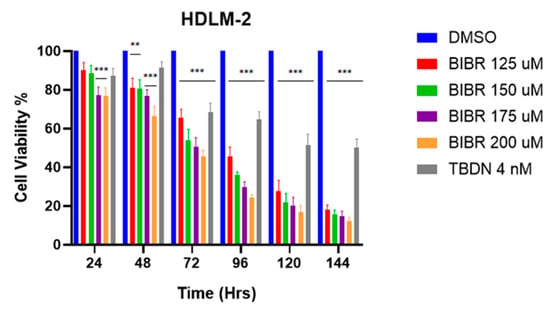
Figure A1.
BIBR1532 concentration titration using HDLM-2 cell line. Decrease in cell viability upon increase in BIBR1532 concentration. The decrease in viability is time-dependent as well as dose-dependent. Data expressed as mean ± SEM. ** p < 0.01 and *** p < 0.001. TBDN—trabectedin.
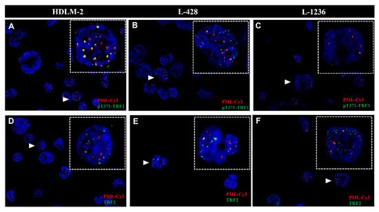
Figure A2.
Hodgkin’s Lymphoma shows ALT activation hallmarks. Analysis of colocalization of PML bodies, pT371-TRF1 and TRF2 in Hodgkin’s Lymphoma cell lines (HDLM-2, L-428 and, L-1236). (A) Analysis of colocalization of PML bodies and pT371-TRF1 in HDLM-2. A white arrow shows the zoomed cell. (B) Analysis of colocalization of PML bodies and pT371-TRF1 in L-248. A white arrow shows the zoomed cell. (C) Analysis of colocalization of PML bodies and pT371-TRF1 in L-1236. A white arrow shows the zoomed cell. (D) Analysis of colocalization of PML bodies and TRF2 in HDLM-2. A white arrow shows the zoomed cell. (E) Analysis of colocalization of PML bodies and TRF2 in L-428. A white arrow shows the zoomed cell. (F) Analysis of colocalization of PML bodies and TRF2 in L-1236. A white arrow shows the zoomed cell. Colocalized signals are shown in yellow. PML (Cy3-Red). pT371-TRF1 (FIT C-Green). TRF2 (FITC-Green). 90 cells were examined in three independent experiments and for each cell line. All cell lines display colocalization of pT371-TRF1 with PML and TRF2 with PML. However not all signals in each cell colocalize.
References
- Mottok, A.; Steidl, C. Biology of Classical Hodgkin Lymphoma: Implications for Prognosis and Novel Therapies. Blood 2018, 131, 1654–1665. [Google Scholar] [CrossRef]
- Bienz, M.; Ramdani, S.; Knecht, H. Molecular Pathogenesis of Hodgkin Lymphoma: Past, Present, Future. Int. J. Mol. Sci. 2020, 21, 6623. [Google Scholar] [CrossRef] [PubMed]
- Sung, H.; Ferlay, J.; Siegel, R.L.; Laversanne, M.; Soerjomataram, I.; Jemal, A.; Bray, F. Global Cancer Statistics 2020: GLOBOCAN Estimates of Incidence and Mortality Worldwide for 36 Cancers in 185 Countries. CA Cancer J. Clin. 2021, 71, 209–249. [Google Scholar] [CrossRef] [PubMed]
- Shamoon, R.P.; Ali, M.D.; Shabila, N.P. Overview and Outcome of Hodgkin’s Lymphoma: Experience of a Single Developing Country’s Oncology Centre. PLoS ONE 2018, 13, e0195629. [Google Scholar] [CrossRef]
- Hodgkin Lymphoma: 2018 Update on Diagnosis, Risk-Stratification, and Management. American Journal of Hematology. Available online: https://onlinelibrary.wiley.com/doi/full/10.1002/ajh.25071 (accessed on 22 June 2022).
- Guru Murthy, G.S.; Szabo, A.; Hamadani, M.; Fenske, T.S.; Shah, N.N. Contemporary Outcomes for Advanced-Stage Classical Hodgkin Lymphoma in the U.S.: Analysis of Surveillance, Epidemiology, and End Results Database. Oncology 2019, 24, 1488–1495. [Google Scholar] [CrossRef] [PubMed]
- Bonnell, E.; Pasquier, E.; Wellinger, R.J. Telomere Replication: Solving Multiple End Replication Problems. Front. Cell Dev. Biol. 2021, 9, 668171. [Google Scholar] [CrossRef] [PubMed]
- Saretzki, G. Telomeres, Telomerase and Ageing. In Biochemistry and Cell Biology of Ageing: Part I Biomedical Science; Harris, J.R., Korolchuk, V.I., Eds.; Subcellular Biochemistry; Springer: Singapore, 2018; pp. 221–308. ISBN 9789811328350. [Google Scholar]
- De Lange, T. Shelterin-Mediated Telomere Protection. Annu. Rev. Genet. 2018, 52, 223–247. [Google Scholar] [CrossRef] [PubMed]
- Lu, W.; Zhang, Y.; Liu, D.; Songyang, Z.; Wan, M. Telomeres—Structure, Function, and Regulation. Exp. Cell Res. 2013, 319, 133–141. [Google Scholar] [CrossRef]
- Hayflick, L. The Limited in Vitro Lifetime of Human Diploid Cell Strains. Exp. Cell Res. 1965, 37, 614–636. [Google Scholar] [CrossRef]
- Hayflick, L.; Moorhead, P.S. The Serial Cultivation of Human Diploid Cell Strains. Exp. Cell Res. 1961, 25, 585–621. [Google Scholar] [CrossRef]
- MacKenzie, D.; Watters, A.K.; To, J.T.; Young, M.W.; Muratori, J.; Wilkoff, M.H.; Abraham, R.G.; Plummer, M.M.; Zhang, D. ALT Positivity in Human Cancers: Prevalence and Clinical Insights. Cancers 2021, 13, 2384. [Google Scholar] [CrossRef]
- Cesare, A.J.; Reddel, R.R. Alternative Lengthening of Telomeres: Models, Mechanisms and Implications. Nat. Rev. Genet. 2010, 11, 319–330. [Google Scholar] [CrossRef] [PubMed]
- Chiodi, I.; Mondello, C. Telomere and Telomerase Stability in Human Diseases and Cancer. Front. Biosci.-Landmark. 2016, 21, 203–224. [Google Scholar] [CrossRef][Green Version]
- Pal, D.; Sharma, U.; Khajuria, R.; Singh, S.K.; Kakkar, N.; Prasad, R. Augmented Telomerase Activity, Reduced Telomere Length and the Presence of Alternative Lengthening of Telomere in Renal Cell Carcinoma: Plausible Predictive and Diagnostic Markers. Gene 2015, 562, 145–151. [Google Scholar] [CrossRef] [PubMed]
- Xu, B.; Peng, M.; Song, Q. The Co-Expression of Telomerase and ALT Pathway in Human Breast Cancer Tissues. Tumor Biol. 2014, 35, 4087–4093. [Google Scholar] [CrossRef] [PubMed]
- M’kacher, R.; Cuceu, C.; Al Jawhari, M.; Morat, L.; Frenzel, M.; Shim, G.; Lenain, A.; Hempel, W.M.; Junker, S.; Girinsky, T.; et al. The Transition between Telomerase and ALT Mechanisms in Hodgkin Lymphoma and Its Predictive Value in Clinical Outcomes. Cancers 2018, 10, 169. [Google Scholar] [CrossRef]
- Hanahan, D.; Weinberg, R.A. Hallmarks of Cancer: The Next Generation. Cell 2011, 144, 646–674. [Google Scholar] [CrossRef] [PubMed]
- McClintock, B. The Stability of Broken Ends of Chromosomes in Zea Mays. Genetics 1941, 26, 234–282. [Google Scholar] [CrossRef]
- Murnane, J.P.; Sabatier, L. Chromosome Rearrangements Resulting from Telomere Dysfunction and Their Role in Cancer. BioEssays 2004, 26, 1164–1174. [Google Scholar] [CrossRef]
- Mai, S. Initiation of Telomere-Mediated Chromosomal Rearrangements in Cancer. J. Cell. Biochem. 2010, 109, 1095–1102. [Google Scholar] [CrossRef]
- Gisselsson, D.; Lichtenzstejn, D.; Kachko, P.; Karlsson, J.; Manor, E.; Mai, S. Clonal Evolution through Genetic Bottlenecks and Telomere Attrition: Potential Threats to in Vitro Data Reproducibility. Genes Chromosomes Cancer 2019, 58, 452–461. [Google Scholar] [CrossRef] [PubMed]
- Knecht, H.; Sawan, B.; Lichtensztejn, D.; Lemieux, B.; Wellinger, R.J.; Mai, S. The 3D Nuclear Organization of Telomeres Marks the Transition from Hodgkin to Reed–Sternberg Cells. Leukemia 2009, 23, 565–573. [Google Scholar] [CrossRef]
- Brousset, P.; Chaouche, N.; Al Saati, T.; Zenou, R.C.; Delsol, G. Telomerase Activity in Hodgkin’s Disease. Leuk. Lymphoma 1998, 30, 189–192. [Google Scholar] [CrossRef]
- De Vitis, M.; Berardinelli, F.; Sgura, A. Telomere Length Maintenance in Cancer: At the Crossroad between Telomerase and Alternative Lengthening of Telomeres (ALT). Int. J. Mol. Sci. 2018, 19, 606. [Google Scholar] [CrossRef]
- Pompili, L.; Leonetti, C.; Biroccio, A.; Salvati, E. Diagnosis and Treatment of ALT Tumors: Is Trabectedin a New Therapeutic Option? J. Exp. Clin. Cancer Res. 2017, 36, 189. [Google Scholar] [CrossRef] [PubMed]
- Casagrande, N.; Borghese, C.; Favero, A.; Vicenzetto, C.; Aldinucci, D. Trabectedin Overcomes Doxorubicin-Resistance, Counteracts Tumor-Immunosuppressive Reprogramming of Monocytes and Decreases Xenograft Growth in Hodgkin Lymphoma. Cancer Lett. 2021, 500, 182–193. [Google Scholar] [CrossRef] [PubMed]
- Casagrande, N.; Borghese, C.; Aldinucci, D. In Classical Hodgkin Lymphoma the Combination of the CCR5 Antagonist Maraviroc with Trabectedin Synergizes, Enhances DNA Damage and Decreases Three-Dimensional Tumor-Stroma Heterospheroid Viability. Haematologica 2021, 107, 287–291. [Google Scholar] [CrossRef]
- Jackson, M.R.; Bavelaar, B.M.; Waghorn, P.A.; Gill, M.R.; El-Sagheer, A.H.; Brown, T.; Tarsounas, M.; Vallis, K.A. Radiolabeled Oligonucleotides Targeting the RNA Subunit of Telomerase Inhibit Telomerase and Induce DNA Damage in Telomerase-Positive Cancer Cells. Cancer Res. 2019, 79, 4627–4637. [Google Scholar] [CrossRef]
- Ellingsen, E.B.; Mangsbo, S.M.; Hovig, E.; Gaudernack, G. Telomerase as a Target for Therapeutic Cancer Vaccines and Considerations for Optimizing Their Clinical Potential. Front. Immunol. 2021, 12, 682492. [Google Scholar] [CrossRef]
- Nakamura, T.; Matsumine, A.; Sudo, A. The Value of Trabectedin in the Treatment of Soft Tissue Sarcoma. Clin. Risk Manag. 2016, 12, 73–79. [Google Scholar] [CrossRef]
- Pignata, S.; Cecere, S.C.; Du Bois, A.; Harter, P.; Heitz, F. Treatment of Recurrent Ovarian Cancer. Ann. Oncol. 2017, 28, viii51–viii56. [Google Scholar] [CrossRef] [PubMed]
- Knecht, H.; Kongruttanachok, N.; Sawan, B.; Brossard, J.; Prévost, S.; Turcotte, E.; Lichtensztejn, Z.; Lichtensztejn, D.; Mai, S. Three-Dimensional Telomere Signatures of Hodgkin- and Reed-Sternberg Cells at Diagnosis Identify Patients with Poor Response to Conventional Chemotherapy. Transl. Oncol. 2012, 5, 269–277. [Google Scholar] [CrossRef] [PubMed]
- Louis, S.; Johnson, N.A.; Brousset, P.; Ludkovski, O.; Shifrin, Y.; Mai, S.; Knecht, H. Three-Dimensional Telomere Analysis Using Teloview® Technology Predicts the Response of Classic Hodgkin’s Lymphoma Patients to First Line Therapy at Point of Diagnosis. Blood 2020, 136, 36–37. [Google Scholar] [CrossRef]
- Knecht, H.; Johnson, N.A.; Haliotis, T.; Lichtensztejn, D.; Mai, S. Disruption of Direct 3D Telomere–TRF2 Interaction through Two Molecularly Disparate Mechanisms Is a Hallmark of Primary Hodgkin and Reed–Sternberg Cells. Lab. Investig. 2017, 97, 772–781. [Google Scholar] [CrossRef] [PubMed]
- Barma, D.K.; Elayadi, A.; Falck, J.R.; Corey, D.R. Inhibition of Telomerase by BIBR 1532 and Related Analogues. Bioorganic Med. Chem. Lett. 2003, 13, 1333–1336. [Google Scholar] [CrossRef]
- Bryan, C.; Rice, C.; Hoffman, H.; Harkisheimer, M.; Sweeney, M.; Skordalakes, E. Structural Basis of Telomerase Inhibition by the Highly Specific BIBR1532. Structure 2015, 23, 1934–1942. [Google Scholar] [CrossRef]
- Wendland, M.; Willenzon, S.; Kocks, J.; Davalos-Misslitz, A.C.; Hammerschmidt, S.I.; Schumann, K.; Kremmer, E.; Sixt, M.; Hoffmeyer, A.; Pabst, O.; et al. Lymph Node T Cell Homeostasis Relies on Steady State Homing of Dendritic Cells. Immunity 2011, 35, 945–957. [Google Scholar] [CrossRef]
- McKerlie, M.; Zhu, X.-D. Cyclin B-Dependent Kinase 1 Regulates Human TRF1 to Modulate the Resolution of Sister Telomeres. Nat. Commun. 2011, 2, 371. [Google Scholar] [CrossRef]
- Schaefer, L.H.; Schuster, D.; Herz, H. Generalized Approach for Accelerated Maximum Likelihood Based Image Restoration Applied to Three-Dimensional Fluorescence Microscopy. J. Microsc. 2001, 204, 99–107. [Google Scholar] [CrossRef] [PubMed]
- Shammas, M.A.; Koley, H.; Bertheau, R.C.; Neri, P.; Fulciniti, M.; Tassone, P.; Blotta, S.; Protopopov, A.; Mitsiades, C.; Batchu, R.B.; et al. Telomerase Inhibitor GRN163L Inhibits Myeloma Cell Growth in Vitro and in Vivo. Leukemia 2008, 22, 1410–1418. [Google Scholar] [CrossRef]
- Gambacorta, M.; Flenghi, L.; Fagioli, M.; Pileri, S.; Leoncini, L.; Bigerna, B.; Pacini, R.; Tanci, L.N.; Pasqualucci, L.; Ascani, S.; et al. Heterogeneous Nuclear Expression of the Promyelocytic Leukemia (PML) Protein in Normal and Neoplastic Human Tissues. Am. J. Pathol. 1996, 149, 2023–2035. [Google Scholar] [PubMed]
- Marchesini, M.; Matocci, R.; Tasselli, L.; Cambiaghi, V.; Orleth, A.; Furia, L.; Marinelli, C.; Lombardi, S.; Sammarelli, G.; Aversa, F.; et al. PML Is Required for Telomere Stability in Non-Neoplastic Human Cells. Oncogene 2016, 35, 1811–1821. [Google Scholar] [CrossRef] [PubMed]
- Wilson, F.R.; Ho, A.; Walker, J.R.; Zhu, X.-D. Cdk-Dependent Phosphorylation Regulates TRF1 Recruitment to PML Bodies and Promotes C-Circle Production in ALT Cells. J. Cell Sci. 2016, 129, 2559–2572. [Google Scholar] [CrossRef]
- Zhang, J.-M.; Genois, M.-M.; Ouyang, J.; Lan, L.; Zou, L. Alternative Lengthening of Telomeres Is a Self-Perpetuating Process in ALT-Associated PML Bodies. Mol. Cell 2021, 81, 1027–1042.e4. [Google Scholar] [CrossRef]
- Lajoie, V.; Lemieux, B.; Sawan, B.; Lichtensztejn, D.; Lichtensztejn, Z.; Wellinger, R.; Mai, S.; Knecht, H. LMP1 Mediates Multinuclearity through Downregulation of Shelterin Proteins and Formation of Telomeric Aggregates. Blood 2015, 125, 2101–2110. [Google Scholar] [CrossRef]
- Tawfik, H.O.; El-Hamaky, A.A.; El-Bastawissy, E.A.; Shcherbakov, K.A.; Veselovsky, A.V.; Gladilina, Y.A.; Zhdanov, D.D.; El-Hamamsy, M.H. New Genetic Bomb Trigger: Design, Synthesis, Molecular Dynamics Simulation, and Biological Evaluation of Novel BIBR1532-Related Analogs Targeting Telomerase against Non-Small Cell Lung Cancer. Pharmaceuticals 2022, 15, 481. [Google Scholar] [CrossRef]
- Dunham, M.A.; Neumann, A.A.; Fasching, C.L.; Reddel, R.R. Telomere Maintenance by Recombination in Human Cells. Nat. Genet. 2000, 26, 447–450. [Google Scholar] [CrossRef]
- Perera, O.N.; Sobinoff, A.P.; Teber, E.T.; Harman, A.; Maritz, M.F.; Yang, S.F.; Pickett, H.A.; Cesare, A.J.; Arthur, J.W.; MacKenzie, K.L.; et al. Telomerase Promotes Formation of a Telomere Protective Complex in Cancer Cells. Sci. Adv. 2019, 5, eaav4409. [Google Scholar] [CrossRef]
- Henson, J.D.; Hannay, J.A.; McCarthy, S.W.; Royds, J.A.; Yeager, T.R.; Robinson, R.A.; Wharton, S.B.; Jellinek, D.A.; Arbuckle, S.M.; Yoo, J.; et al. A Robust Assay for Alternative Lengthening of Telomeres in Tumors Shows the Significance of Alternative Lengthening of Telomeres in Sarcomas and Astrocytomas. Clin. Cancer Res. 2005, 11, 217–225. [Google Scholar] [CrossRef]
- Barthel, F.P.; Wei, W.; Tang, M.; Martinez-Ledesma, E.; Hu, X.; Amin, S.B.; Akdemir, K.C.; Seth, S.; Song, X.; Wang, Q.; et al. Systematic Analysis of Telomere Length and Somatic Alterations in 31 Cancer Types. Nat. Genet. 2017, 49, 349–357. [Google Scholar] [CrossRef]
- Zhang, H.; Zhao, R.; Tones, J.; Liu, M.; Dilley, R.L.; Chenoweth, D.M.; Greenberg, R.A.; Lampson, M.A. Nuclear Body Phase Separation Drives Telomere Clustering in ALT Cancer Cells. MBoC 2020, 31, 2048–2056. [Google Scholar] [CrossRef] [PubMed]
- Jiang, W.-Q.; Zhong, Z.-H.; Henson, J.D.; Reddel, R.R. Identification of Candidate Alternative Lengthening of Telomeres Genes by Methionine Restriction and RNA Interference. Oncogene 2007, 26, 4635–4647. [Google Scholar] [CrossRef] [PubMed]
- Minasi, S.; Baldi, C.; Pietsch, T.; Donofrio, V.; Pollo, B.; Antonelli, M.; Massimino, M.; Giangaspero, F.; Buttarelli, F.R. Telomere Elongation via Alternative Lengthening of Telomeres (ALT) and Telomerase Activation in Primary Metastatic Medulloblastoma of Childhood. J. Neurooncol. 2019, 142, 435–444. [Google Scholar] [CrossRef]
- Roderwieser, A.; Sand, F.; Walter, E.; Fischer, J.; Gecht, J.; Bartenhagen, C.; Ackermann, S.; Otte, F.; Welte, A.; Kahlert, Y.; et al. Telomerase Is a Prognostic Marker of Poor Outcome and a Therapeutic Target in Neuroblastoma. JCO Precis. Oncol. 2019, 3, 1–20. [Google Scholar] [CrossRef]
- Job, S.; Draskovic, I.; Burnichon, N.; Buffet, A.; Cros, J.; Lépine, C.; Venisse, A.; Robidel, E.; Verkarre, V.; Meatchi, T.; et al. Telomerase Activation and ATRX Mutations Are Independent Risk Factors for Metastatic Pheochromocytoma and Paraganglioma. Clin. Cancer Res. 2019, 25, 760–770. [Google Scholar] [CrossRef]
- Kim, J.Y.; Brosnan-Cashman, J.A.; An, S.; Kim, S.J.; Song, K.-B.; Kim, M.-S.; Kim, M.-J.; Hwang, D.W.; Meeker, A.K.; Yu, E.; et al. Alternative Lengthening of Telomeres in Primary Pancreatic Neuroendocrine Tumors Is Associated with Aggressive Clinical Behavior and Poor Survival. Clin. Cancer Res. 2017, 23, 1598–1606. [Google Scholar] [CrossRef]
- Röth, A.; Harley, C.B.; Baerlocher, G.M. Imetelstat (GRN163L)—Telomerase-Based Cancer Therapy. In Small Molecules in Oncology; Martens, U.M., Ed.; Recent Results in Cancer Research; Springer: Berlin/Heidelberg, Germany, 2010; pp. 221–234. ISBN 978-3-642-01222-8. [Google Scholar]
- Kawai, A.; Yonemori, K.; Takahashi, S.; Araki, N.; Ueda, T. Systemic Therapy for Soft Tissue Sarcoma: Proposals for the Optimal Use of Pazopanib, Trabectedin, and Eribulin. Adv. Ther. 2017, 34, 1556–1571. [Google Scholar] [CrossRef]
- Guterres, A.N.; Villanueva, J. Targeting Telomerase for Cancer Therapy. Oncogene 2020, 39, 5811–5824. [Google Scholar] [CrossRef]
- Apte, M.S.; Cooper, J.P. Life and Cancer without Telomerase: ALT and Other Strategies for Making Sure Ends (Don’t) Meet. Crit. Rev. Biochem. Mol. Biol. 2017, 52, 57–73. [Google Scholar] [CrossRef]
- Jones, J.D.; Sinder, B.P.; Paige, D.; Soki, F.N.; Koh, A.J.; Thiele, S.; Shiozawa, Y.; Hofbauer, L.C.; Daignault, S.; Roca, H.; et al. Trabectedin Reduces Skeletal Prostate Cancer Tumor Size in Association with Effects on M2 Macrophages and Efferocytosis. Neoplasia 2019, 21, 172–184. [Google Scholar] [CrossRef]
- Jones, R.L.; Herzog, T.J.; Patel, S.R.; von Mehren, M.; Schuetze, S.M.; Van Tine, B.A.; Coleman, R.L.; Knoblauch, R.; Triantos, S.; Hu, P.; et al. Cardiac Safety of Trabectedin Monotherapy or in Combination with Pegylated Liposomal Doxorubicin in Patients with Sarcomas and Ovarian Cancer. Cancer Med. 2021, 10, 3565–3574. [Google Scholar] [CrossRef] [PubMed]
- Endo, M.; Takahashi, S.; Araki, N.; Sugiura, H.; Ueda, T.; Yonemoto, T.; Takahashi, M.; Morioka, H.; Hiraga, H.; Hiruma, T.; et al. Time Lapse Analysis of Tumor Response in Patients with Soft Tissue Sarcoma Treated with Trabectedin: A Pooled Analysis of Two Phase II Clinical Trials. Cancer Med. 2020, 9, 3656–3667. [Google Scholar] [CrossRef] [PubMed]
- Rubio, M.J.; Lecumberri, M.J.; Varela, S.; Alarcón, J.; Ortega, M.E.; Gaba, L.; Espinós, J.; Calzas, J.; Barretina, P.; Ruiz, I.; et al. Efficacy and Safety of Trabectedin in Metastatic Uterine Leiomyosarcoma: A Retrospective Multicenter Study of the Spanish Ovarian Cancer Research Group (GEICO). Gynecol. Oncol. Rep. 2020, 33, 100594. [Google Scholar] [CrossRef] [PubMed]
- Grosso, F.; D’Ambrosio, L.; Zucchetti, M.; Ibrahim, T.; Tamberi, S.; Matteo, C.; Rulli, E.; Comandini, D.; Palmerini, E.; Baldi, G.G.; et al. Pharmacokinetics, Safety, and Activity of Trabectedin as First-Line Treatment in Elderly Patients Who Are Affected by Advanced Sarcoma and Are Unfit to Receive Standard Chemotherapy: A Phase 2 Study (TR1US Study) from the Italian Sarcoma Group. Cancer 2020, 126, 4726–4734. [Google Scholar] [CrossRef]
- Romero, I.; López-Guerrero, J.A.; Pignata, S. Real-World Experience with Trabectedin for the Treatment of Recurrent Ovarian Cancer. Expert Rev. Anticancer Ther. 2021, 21, 1089–1095. [Google Scholar] [CrossRef]
- Martínez-Trufero, J.; De Sande-González, L.M.; Luna, P.; Martin-Broto, J.; Álvarez, R.; Marquina, G.; Diaz-Beveridge, R.; Poveda, A.; Cano, J.M.; Cruz-Jurado, J.; et al. A Growth Modulation Index-Based GEISTRA Score as a New Prognostic Tool for Trabectedin Efficacy in Patients with Advanced Soft Tissue Sarcomas: A Spanish Group for Sarcoma Research (GEIS) Retrospective Study. Cancers 2021, 13, 792. [Google Scholar] [CrossRef]
- Mannarino, L.; Craparotta, I.; Ballabio, S.; Frapolli, R.; Meroni, M.; Bello, E.; Panini, N.; Callari, M.; Sanfilippo, R.; Casali, P.G.; et al. Mechanisms of Responsiveness to and Resistance against Trabectedin in Murine Models of Human Myxoid Liposarcoma. Genomics 2021, 113, 3439–3448. [Google Scholar] [CrossRef]
- Pignata, S.; Scambia, G.; Villanucci, A.; Naglieri, E.; Ibarbia, M.A.; Brusa, F.; Bourgeois, H.; Sorio, R.; Casado, A.; Reichert, D.; et al. A European, Observational, Prospective Trial of Trabectedin Plus Pegylated Liposomal Doxorubicin in Patients with Platinum-Sensitive Ovarian Cancer. Oncologist 2021, 26, e658–e668. [Google Scholar] [CrossRef]
- Kokkali, S.; Boukovinas, I.; Samantas, E.; Papakotoulas, P.; Athanasiadis, I.; Andreadis, C.; Makrantonakis, P.; Samelis, G.; Timotheadou, E.; Vassilopoulos, G.; et al. A Multicenter, Prospective, Observational Study to Assess the Clinical Activity and Impact on Symptom Burden and Patients’ Quality of Life in Patients with Advanced Soft Tissue Sarcomas Treated with Trabectedin in a Real-World Setting in Greece. Cancers 2022, 14, 1879. [Google Scholar] [CrossRef]
- Lavanya, C.; Venkataswamy, M.M.; Sibin, M.K.; Srinivas Bharath, M.M.; Chetan, G.K. Down Regulation of Human Telomerase Reverse Transcriptase (HTERT) Expression by BIBR1532 in Human Glioblastoma LN18 Cells. Cytotechnology 2018, 70, 1143–1154. [Google Scholar] [CrossRef]
- Ding, X.; Cheng, J.; Pang, Q.; Wei, X.; Zhang, X.; Wang, P.; Yuan, Z.; Qian, D. BIBR1532, a Selective Telomerase Inhibitor, Enhances Radiosensitivity of Non-Small Cell Lung Cancer Through Increasing Telomere Dysfunction and ATM/CHK1 Inhibition. Int. J. Radiat. Oncol. Biol. Phys. 2019, 105, 861–874. [Google Scholar] [CrossRef] [PubMed]
- Pourbagheri-Sigaroodi, A.; Bashash, D.; Safaroghli-Azar, A.; Farshi-Paraasghari, M.; Momeny, M.; Mansoor, F.N.; Ghaffari, S.H. Contributory Role of MicroRNAs in Anti-Cancer Effects of Small Molecule Inhibitor of Telomerase (BIBR1532) on Acute Promyelocytic Leukemia Cell Line. Eur. J. Pharmacol. 2019, 846, 49–62. [Google Scholar] [CrossRef] [PubMed]
- Bu, R.; Siraj, A.K.; Divya, S.P.; Kong, Y.; Parvathareddy, S.K.; Al-Rasheed, M.; Al-Obaisi, K.A.S.; Victoria, I.G.; Al-Sobhi, S.S.; Al-Dawish, M.; et al. Telomerase Reverse Transcriptase Mutations Are Independent Predictor of Disease-Free Survival in Middle Eastern Papillary Thyroid Cancer. Int. J. Cancer 2018, 142, 2028–2039. [Google Scholar] [CrossRef] [PubMed]
- Rafat, A.; Dizaji Asl, K.; Mazloumi, Z.; Movassaghpour, A.A.; Talebi, M.; Shanehbandi, D.; Farahzadi, R.; Nejati, B.; Nozad Charoudeh, H. Telomerase Inhibition on Acute Myeloid Leukemia Stem Cell Induced Apoptosis with Both Intrinsic and Extrinsic Pathways. Life Sci. 2022, 295, 120402. [Google Scholar] [CrossRef]
- Huff, C.A.; Wang, Q.; Badros, A.Z.; Jones, R.J.; Reddy, A.; Nishimoto, K.; Stuart, M.J.; Kelsey, S.M.; Matsui, W. The Telomerase Inhibitor, Imetelstat, Rapidly Reduces Myeloma Cancer Stem Cells (CSCs) in a Phase II Trial. Blood 2012, 120, 4898. [Google Scholar] [CrossRef]
- Barwe, S.P.; Huang, F.; Kolb, E.A.; Gopalakrishnapillai, A. Imetelstat Induces Leukemia Stem Cell Death in Pediatric Acute Myeloid Leukemia Patient-Derived Xenografts. J. Clin. Med. 2022, 11, 1923. [Google Scholar] [CrossRef]
- Mascarenhas, J.; Komrokji, R.S.; Palandri, F.; Martino, B.; Niederwieser, D.; Reiter, A.; Scott, B.L.; Baer, M.R.; Hoffman, R.; Odenike, O.; et al. Randomized, Single-Blind, Multicenter Phase II Study of Two Doses of Imetelstat in Relapsed or Refractory Myelofibrosis. J. Clin. Oncol. 2021, 39, 2881–2892. [Google Scholar] [CrossRef]
- Santini, V.; Fenaux, P.; Van Eygen, K.; Raza, A.; Germing, U.; Font, P.; Diez-Campelo, M.; Thepot, S.; Vellenga, E.; Patnaik, M.M.; et al. On-Target Activity of Imetelstat Correlates with Clinical Benefits, Including Overall Survival (OS), in Heavily Transfused Non-Del(5q) Lower Risk MDS (LR-MDS) Relapsed/Refractory (R/R) to Erythropoiesis Stimulating Agents (ESAs). Blood 2021, 138, 2598. [Google Scholar] [CrossRef]
- Steensma, D.P.; Fenaux, P.; Van Eygen, K.; Raza, A.; Santini, V.; Germing, U.; Font, P.; Diez-Campelo, M.; Thepot, S.; Vellenga, E.; et al. Imetelstat Achieves Meaningful and Durable Transfusion Independence in High Transfusion-Burden Patients with Lower-Risk Myelodysplastic Syndromes in a Phase II Study. J. Clin. Oncol. 2021, 39, 48–59. [Google Scholar] [CrossRef]
- Kuykendall, A.T.; Sun, L.; Mascarenhas, J.; Kiladjian, J.-J.; Vannucchi, A.M.; Wang, J.; Xia, Q.; Zhu, E.; Feller, F.; Rizo, A.; et al. Favorable Overall Survival with Imetelstat in Relapsed/Refractory Myelofibrosis Patients Compared with Real-World Data. Ann. Hematol. 2022, 101, 139–146. [Google Scholar] [CrossRef]
Publisher’s Note: MDPI stays neutral with regard to jurisdictional claims in published maps and institutional affiliations. |
© 2022 by the authors. Licensee MDPI, Basel, Switzerland. This article is an open access article distributed under the terms and conditions of the Creative Commons Attribution (CC BY) license (https://creativecommons.org/licenses/by/4.0/).