Article

The Antibacterial Effect of Platelets on Escherichia coli Strains

by
Amina Ezzeroug Ezzraimi
1,2,
Nadji Hannachi
1,3,
Antoine Mariotti
1,2,4,
Clara Rolland
2,5,
Anthony Levasseur
2,5,
Sophie Alexandra Baron
1,2,
Jean-Marc Rolain
1,2 and
Laurence Camoin-Jau
1,2,4,*
1
Aix Marseille University, IRD, APHM, MEPHI, IHU Méditerranée Infection, 13385 Marseille, France
2
IHU Méditerranée Infection, Boulevard Jean Moulin, 13385 Marseille, France
3
Département de Pharmacie, Faculté de Médecine, Université Ferhat Abbas Sétif I, Sétif 19000, Algeria
4
Laboratoire d’Hématologie, Hôpital de la Timone, APHM, Boulevard Jean-Moulin, 13385 Marseille, France
5
Aix Marseille University, IRD, SSA, APHM, VITROME, IHU Méditerranée Infection, 13385 Marseille, France
*
Author to whom correspondence should be addressed.
Biomedicines 2022, 10(7), 1533; https://doi.org/10.3390/biomedicines10071533
Submission received: 28 April 2022 / Revised: 23 June 2022 / Accepted: 24 June 2022 / Published: 28 June 2022

Abstract

:
Platelets play an important role in defense against pathogens; however, the interaction between Escherichia coli and platelets has not been well described and detailed. Our goal was to study the interaction between platelets and selected strains of E. coli in order to evaluate the antibacterial effect of platelets and to assess bacterial effects on platelet activation. Washed platelets and supernatants of pre-activated platelets were incubated with five clinical colistin-resistant and five laboratory colistin-sensitive strains of E. coli in order to study bacterial growth. Platelet activation was measured with flow cytometry by evaluating CD62P expression. To identify the difference in strain behavior toward platelets, a pangenome analysis using Roary and O-antigen serotyping was carried out. Both whole platelets and the supernatant of activated platelets inhibited growth of three laboratory colistin-sensitive strains. In contrast, platelets promoted growth of the other strains. There was a negative correlation between platelet activation and bacterial growth. The Roary results showed no logical clustering to explain the mechanism of platelet resistance. The diversity of the responses might be due to strains of different types of O-antigen. Our results show a bidirectional interaction between platelets and E. coli whose expression is dependent on the bacterial strain involved.

1. Introduction

Platelets have been widely described as the main actors in hemostasis and thrombosis. More recently, the important role they play in inflammation and defense against pathogens has been highlighted [1,2]. Bacteria interact with platelets through three main mechanisms: (i) by direct binding: bacteria can express surface proteins which allow them to interact directly with surface receptors on platelets and bind to them, as in the case of Streptococcus sanguinis, which showed the ability to bind directly to GPIbα [3]; (ii) by binding through plasma proteins: bacterial proteins are capable of binding to fibrinogen and vWF (von Willebrand Factor), serving as a bridge between two cells, as in the example of Staphylococcus aureus expressing surface protein A (SpA) that binds to vWf and which in turn binds to platelet GPIbα [4]; (iii) and by binding through the secretion of bacterial products, such as toxins, which interact with platelets and activate them. Among these molecules, the Shiga toxin secreted by Escherichia coli induces platelet aggregation by binding to glycosphingolipid receptors on the surface of platelets [5,6,7,8,9,10]. This difference in interaction mechanisms, which is mainly dependent on the bacteria, induces distinct platelet responses. In most cases, this interaction leads to platelet activation, followed by a release of their granular contents composed of, among other things, microbicidal proteins and chemokines that facilitate the destruction of pathogens, signal immune cells and promote inflammation [11,12].
Platelet microbicidal effects have been extensively studied for Gram-positive bacteria. It has been demonstrated that platelets decrease the growth of Staphylococcus aureus [13,14,15]. In contrast, this effect has been less studied for Gram-negative bacteria, and such data are scarce. Platelets have been shown to interact with Escherichia coli through the platelet Toll-Like Receptor 4 (TLR4) and bacterial lipopolysaccharide (LPS) [5,16,17]. The initiation of the pro-inflammatory signal by LPS depends on the interaction between the TLR4 complex and lipid A, a fragment of LPS [18]. However, other research has shown that platelet activation and aggregation occurs through FcγRIIA without the involvement of TLR4 [19,20]. In contrast, Matus et al. demonstrated that platelet activation is dependent on TLR4 but without FcγRIIA engagement [21]. It is important to note that studies have been carried out to investigate the effects of E. coli on the activation and aggregation of platelets, specifically in the case of hemolytic uremic syndrome (HUS), but very few data are available on the effects of platelets on E. coli. These studies have tested distinct strains and serotypes and different operating protocols, such as the platelet-bacteria ratio and platelet form [21,22,23].
The aim of this study was to investigate the interaction between platelets and different human E. coli strains by initially evaluating the antibacterial effect of platelets, and then evaluating the effect of E. coli strains on platelet activation. Moreover, our objective was to compare ten strains of E coli having different characteristics, including their colistin sensitivity profile and their pathogenic capability.

2. Materials and Methods

2.1. Platelet Preparation

Blood was drawn by venepuncture in sodium citrate from healthy subjects who were not receiving antibiotics, anti-inflammatory, or anti-platelet drugs. Platelet rich plasma (PRP) was prepared according to International Society on Thrombosis and Hemostasis (ISTH) recommendations [24]. A platelet count was performed using a hematology analyzer. PRP was again centrifuged at 1100 g for ten minutes to obtain a platelet pellet that was suspended in phosphate buffered saline (PBS) to obtain a solution of 4 × 10⁹/L. Platelets were then kept at 37 °C in order to prevent activation. The protocol was approved by the ethics committee of the IHU Méditérrannée Infection (Reference 2016–002). All of the subjects gave their written informed consent in accordance with the Declaration of Helsinki.

2.2. Bacterial Preparation

In order to test strains of E. coli against platelets and to see if there is a possible cross-resistance between resistance to colistin and resistance to platelet antimicrobial peptides, ten strains of E. coli were selected. Five laboratory colistin-sensitive strains were used (ATCC 25922, ATCC 11303, K12, J53 and BL21DE3). Five colistin-resistant human isolates stored at the IHU were also tested (LH1, LH30, Q1065, Q1066 and Q6269) (Table 1). Identification was confirmed using matrix-assisted laser desorption/ionization time-of-flight (MALDI-TOF) mass spectrometry and the Biotyper database (Bruker, Dresden, Germany). Strains were grown at 37 °C in an overnight culture of Columbia agar +5% sheep blood (bioMérieux, Marcy l’Etoile, France). After 18 h of incubation at 37 °C, the colonies were removed and suspended in 0.9% NaCl medium to obtain the required concentrations: 1 × 10⁸ CFU (Colony Forming Unit)/mL for flow cytometry and 3 × 10⁸ CFU/mL for growth test.
For each strain, the minimum inhibitory concentration (MIC) of colistin (Table 1) was tested by microdilution in accordance with the recommendations of the European Committee on Antimicrobial Susceptibility Testing (EUCAST).

2.3. Analysis of Platelet Activation by Flow Cytometry

The washed platelets were used at a concentration of 2.5 × 10⁹/L, adjusting the concentration with PBS. They were incubated with bacteria in a 1:2.5 bacteria-platelet ratio for one hour at 37 °C and identified by expression of the CD41-FITC antibody (4 μL, IgG, Beckman Coulter, Villepinte, France), as previously described [31]. To determine possible platelet activation following incubation with the bacteria, the expression of the CD62-PC5 antibody (4 μL, IgG, monoclonal, BD sciences, San Jose, CA, USA) on their surface was measured using flow cytometry (Beckman Coulter, FC500, Fullerton, CA, USA). The platelet activator, Thrombin receptor-activating peptide 6 (TRAP) (STAGO®, Asnières, France) (10 μM), and untreated platelets were used as controls. The results were represented by a Mean fluorescence intensity (MFI) percentage of P-Selectin; the expression was calculated using the following equation: (MFI of platelets incubated with bacteria) × 100/(MFI of platelets alone).

2.4. Platelet Supernatant Effect on Bacterial Growth

To obtain the supernatant from the activated platelets, the activated platelets were incubated with the J53 strain for one hour at 37 °C. The mixture underwent three successive centrifugation rounds (1300× g, 5000× g and 5000× g; ten minutes each) and the supernatant was recovered and filtered (0.22 μm), then incubated with bacteria for four hours at 37 °C. Mixtures were serially diluted as described above, then spread on agar and the colonies were counted the following day.

2.5. Pangenome Analysis

The genomes of the seven strains of Escherichia coli (ATCC25922, ATCC11303, K12, J53, BL21 DE3, LH1 and LH30) were retrieved from the National Centre for Biotechnology Information (NCBI) database. For the three other strains, Q1065 (unpublished data), Q1066 (unpublished data) and Q6269 (JAIBLN000000000), the genomes were obtained from the IHU sequencing platform by Illumina MiSeq according to a paired-end strategy.
The first step in performing the pangenome was to predict the ORFs (Open Reading Frame) for each strain with PROKKA software using the default parameters [32]. Then, Roary software was used to build the pangenome with the core genome alignment default parameters [33]. A graphic representation of the pangenome results was prepared using the roary_plots.py script provided on the Roary website.

2.6. O-antigen Strain Serotyping

The serotyping of the E. coli strains (Table 1) used in this study was performed in silico from the genomes obtained by high-throughput sequencing. The FASTA sequences of these genomes were analyzed using ECTyper software [34], which makes it possible to serotype the O and H antigens of E. coli and Shigella spp. The bioinformatic predictions made with this software were then compared with those obtained by the Serotype Finder 2.0 prediction module of the Centre for Genomic Epidemiology developed by the Technical University of Denmark [35]. The results of all of the strains were concordant after analysis by both databases.

2.7. Statistical Analysis

Statistical analyses were performed using GraphPad Prism 9 for Windows (GraphPad Software, La Jolla, CA, USA). Significant differences (for bacterial growth and flow cytometry) between the two groups were determined using the two-tailed, paired Student’s t-test. The statistical significance was set at p < 0.05. A test of normality was applied on the effect of platelets on bacterial growth data, and it turned out that samples (negative control and platelets + bacteria for each strain) show a Gaussian distribution following a verification by the Shapiro-Wilk test (p > 0.05).
While for supernatants effect, they were determined using the Bonferroni’s multiple comparisons test precede by Two-way ANOVA test, considering p < 0.05 as statistically significant. The correlation between bacterial growth and MFI percentage values was determined using the Pearson test.

3. Results

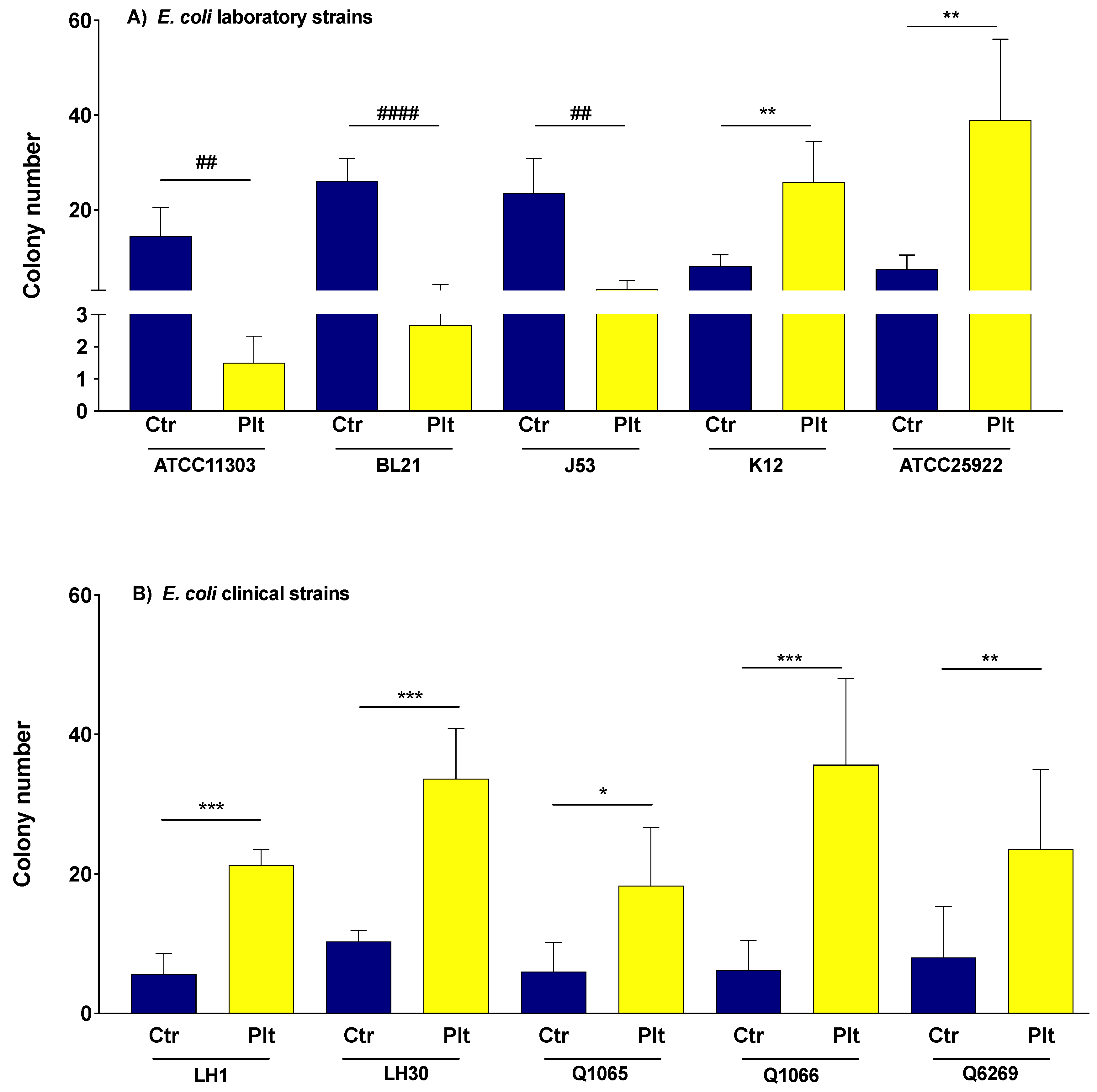
3.1. The Effect of Platelets on the Growth of E. coli Strains

After four hours of incubation, the platelets significantly decreased bacterial growth of three E. coli laboratory strains, as compared to the controls (n = 5 for each strain: p = 0.0036, <0.0001 and 0.0017 for ATCC11303, BL21DE3 and J53, respectively, paired Student’s t-test) (Figure 1). These strains were referred to as “platelet sensitive”.
In contrast, the platelets significantly promoted the growth of E. coli K12 and E. coli ATCC25922 (n = 5 for each strain: p = 0.0013 and 0.0065, respectively). Likewise, the growth of all the clinical strains was enhanced with platelets (n = 5 for each strain: p = 0.0001, 0.0005, 0.0105, 0.0009 and 0.0079, for LH1, LH30, Q1065, Q1066, and Q6269, respectively, paired Student’s t-test) (Figure 1). These seven strains were referred to as “platelet resistant”.

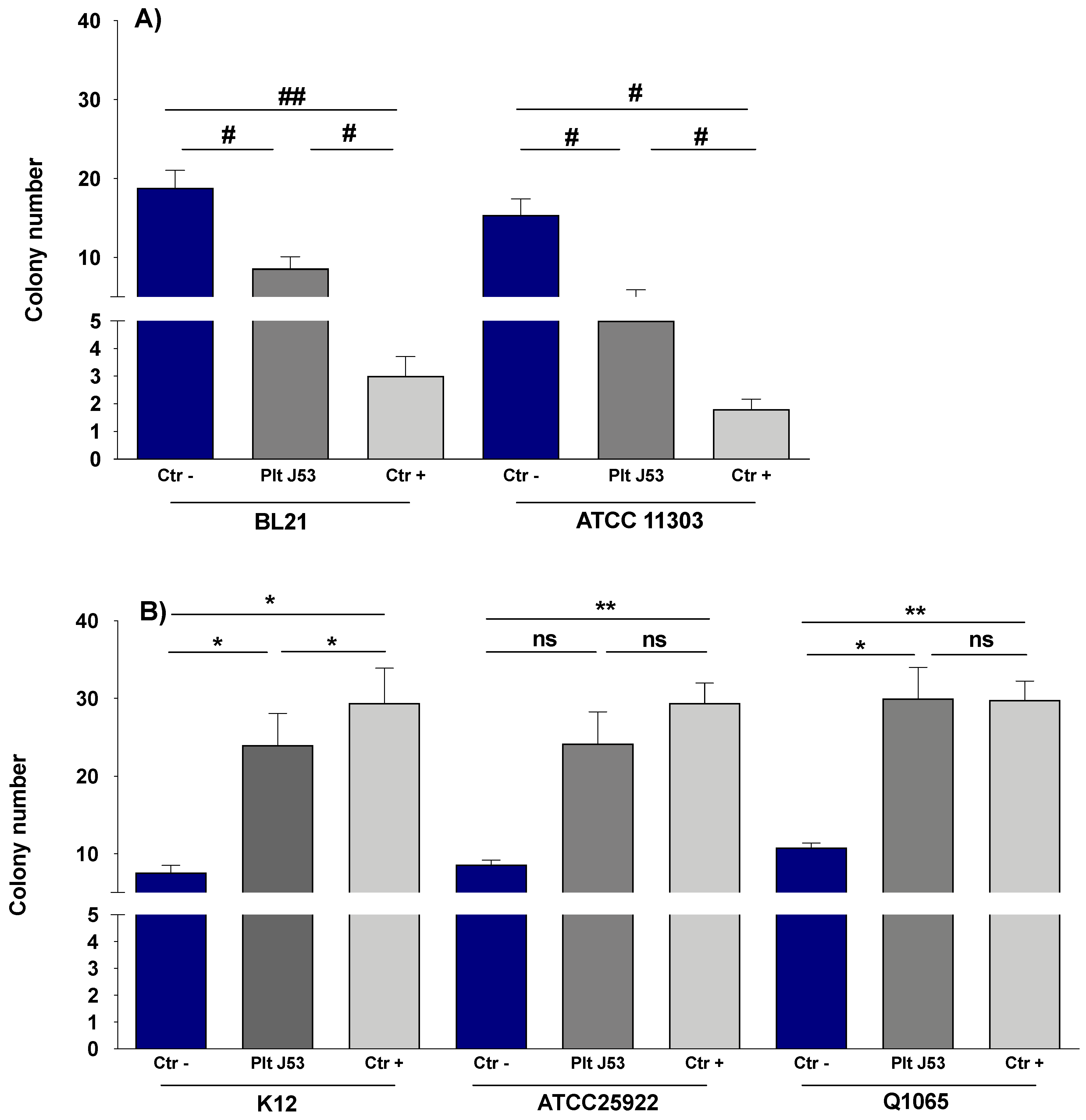
3.2. The Effect of Platelet Supernatant on the Growth of E. coli Strains

Mixes of the supernatants of platelets previously stimulated by the E. coli J53 strain were prepared, then re-incubated again with bacteria, as described in the Section 2. The supernatant of the platelets treated by TRAP was used as a positive control.
The supernatants showed effects similar to whole platelets on the growth of tested strains. Indeed, supernatants of platelets stimulated with E. coli J53 and TRAP significantly inhibited the growth of E. coli BL21DE3 and E. coli ATCC 11303 strains ((n = 5: p= 0.0223, 0.0293 and p = 0.0381, 0.0048 for BL21 and J53, respectively, Figure 2A). In contrast, the supernatants of platelets stimulated with E. coli J53 and TRAP continued to promote the growth of K12 (n = 5: p = 0.0203 and 0.0332, Figure 2B). Q1065 growth was also enhanced by supernatants of platelets stimulated with E. coli J53 (n = 5: p = 0.0233). On the other hand, platelet supernatants had no significant effect on ATCC 25922 growth.

3.3. The Effect of E. coli Strains on Platelet Activation

In order to assess whether E. coli strains induce platelet activation, P-selectin was measured using flow cytometry. After one hour of co-incubation, five strains (ATCC11303, J53, BL21DE3, K12 and ATCC25922) significantly increased P-selectin compared to the controls (platelets alone). All of the other strains tested did not increase the expression of P-selectin. Significant differences between platelets alone and platelets treated by TRAP as well as between platelets alone and platelets infected with bacteria were determined using the two-tailed, paired Student’s t-test. Statistical significance was set at p < 0.05 (Table 2).
The Pearson test was applied to test the correlation between colony count values and the MFI percentage of the different strains in co-incubation with platelets. A negative significant correlation was obtained (Pearson r = −0.6795, p-value = 0.0307).

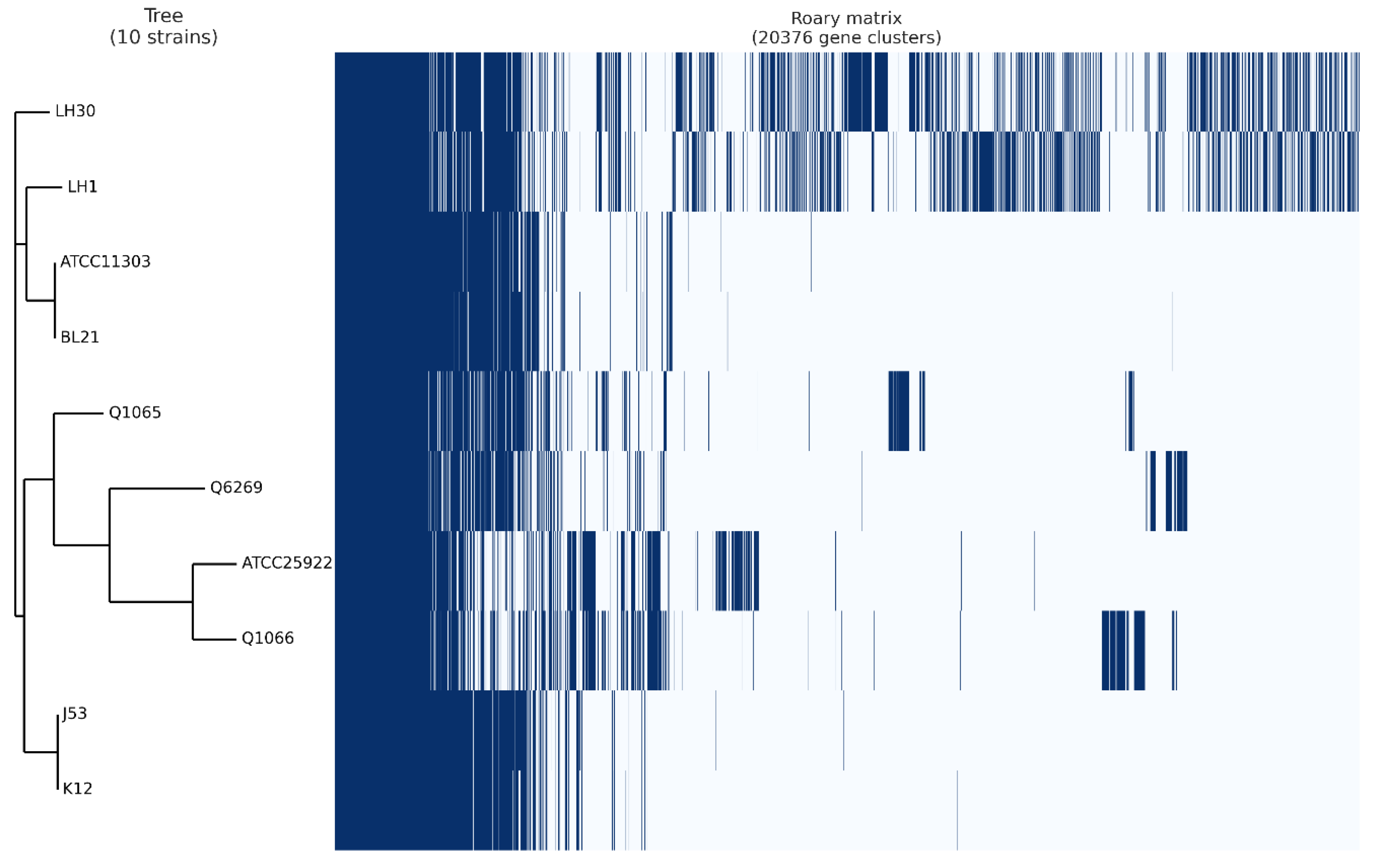
4. Pangenome Analysis

In order to better understand the difference in platelet activation between the E. coli strains as previously shown, a pangenome analysis was performed (Figure 3). This study was designed to identify the difference between platelet-activating and non-platelet-activating strains. We were particularly interested in genes present in only one or the other groups of bacteria. In the non-platelet-activating group that consisted of E. coli strains LH1, LH30, Q1065, Q1066 and Q6269, no genes shared by all strains were found. In contrast, in the platelet-activating group, six common genes were identified. Of them, two were annotated as elongation factor Tu (elongation factor Tu 1 and 2), two others were annotated as transposase (IS3 family transposase IS911, IS4 family transposase IS4), another was annotated as Outer membrane porin protein OmpD, and the last was annotated as a lactose phosphotransferase system repressor. None of these functions showed a direct link to platelet activation. Furthermore, the analysis of the classification of strains following the pangenome study did not indicate a clustering of bacteria according to platelet activation. Indeed, the clustering put E. coli strains BL21 and ATCC11303 in a first group and E. coli strains J53, K12 and ATCC25922 in the second (Figure 3).
Thus, the bioinformatic analyses did not identify a gene or a cluster of genes as being at the origin of the difference in platelet activation.

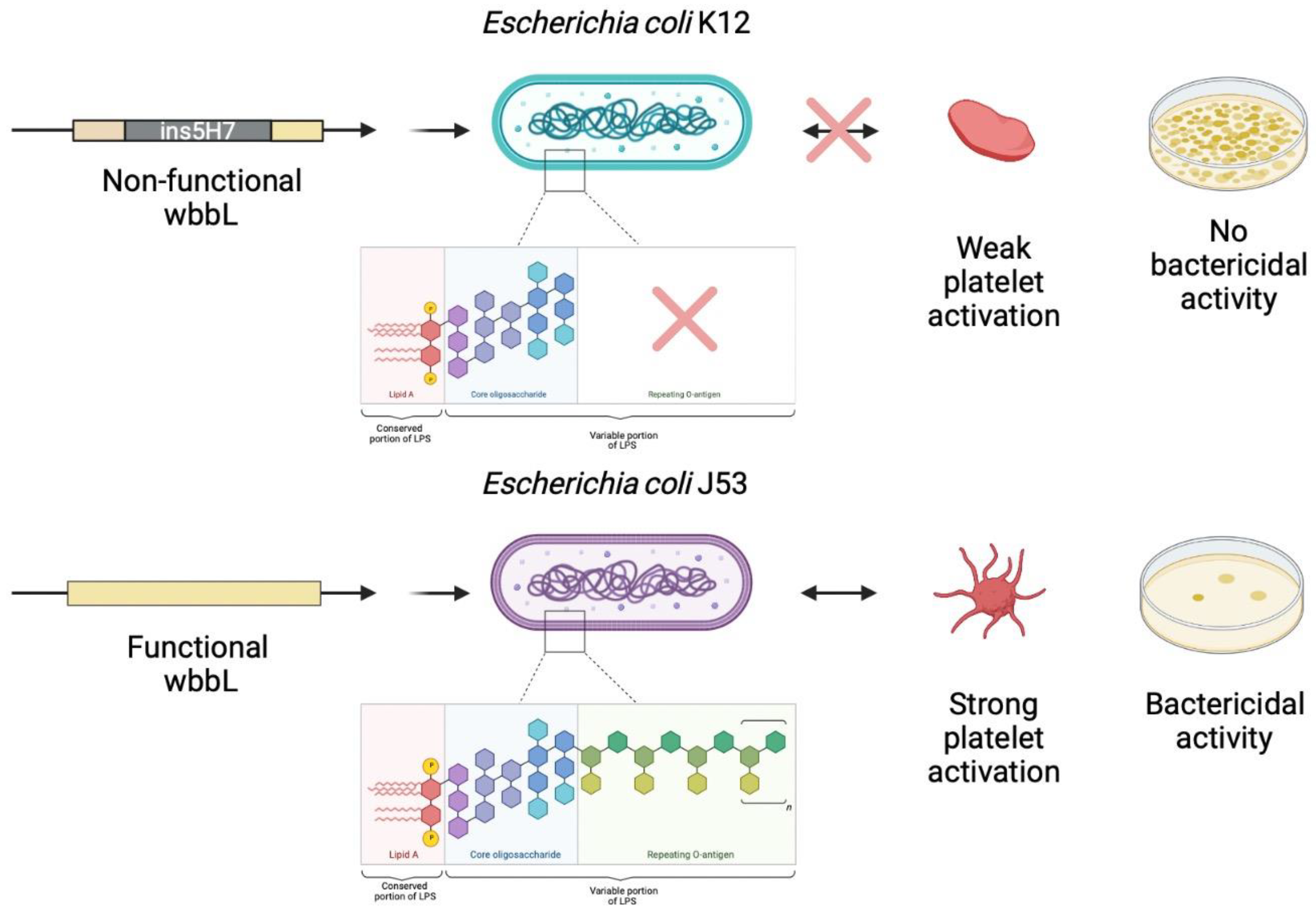
5. O-antigen Strain Serotyping

We investigated the difference in behavior between two genetically related strains, K12 and its mutant strain J53. Using the Genome Mapper of the EcoCyc database (ecocyc.org) and searching for genes coding for proteins involved in the biosynthesis of the O antigen, and thus in the formation of LPS, we found that the wbbL gene had an IS5 insertion in its sequence, making it non-functional, and thus able to modify the structure of LPS (Figure 4).

6. Discussion

In this study, we evaluated the consequences of interactions between platelets and ten strains of Escherichia coli. Our results showed that platelets had an antibacterial activity on three laboratory strains among the ten tested. Genomic comparison of two strains with different susceptibility profiles revealed a difference in the genomic cluster coding for the O-antigen.
To our knowledge, few studies have evaluated the bactericidal effect of platelets against Escherichia coli strains [19,36]. Moreover, the originality of our study resides in the fact that we tested a large panel of strains, including both laboratory and clinical strains, which are further distinguished by their colistin-resistance profile.
The bactericidal effect of platelets has been previously tested in two studies, where two of the five colistin-sensitive laboratory strains selected in our study were used. Our results confirmed that E. coli ATCC1130 growth inhibition was inhibited by platelets, as previously demonstrated by Tohidnezhad et al. [37]. Moreover, as described by Cieślik-Bielecka et al., we confirmed that the growth of the E. coli ATCC25922 strain was not inhibited by platelets [38]. The concordance of these results validated the choice of our experimental model.
Among the ten strains tested, only three were sensitive to platelet bactericidal activity. The interaction of platelets with these strains induced platelet activation responsible for a secretion process of platelet granule content, as evidenced by the increased expression of P-selectin. The bactericidal activity observed is probably due to the action of microbicidal platelet peptides released, since the same effect is observed with the supernatant of activated platelets. We have already described this mechanism for Staphylococcus aureus [13]. These three strains of E. coli, which were sensitive to platelets, are all laboratory strains that do not express resistance mutations and are not responsible for human infectious pathologies. Interestingly, the two other E. coli laboratory strains tested (K12 and ATCC 25922) also induced platelet activation. Regarding this last point, our results are in line with those of Fejes et al., who also demonstrated that the K12 strain induces an increase in P-selectin and CD63 [22]. However, they are insensitive to platelet bactericidal activity. This lack of effect could be the consequence of weak platelet activation induced by these strains, as we have demonstrated a negative correlation between the inhibitory effect of platelets and the activating effect of E. coli strains, which means the more the strains increase platelet granule release, the less bacterial growth decreases in the presence of platelets. However, regarding the E. coli K12 strain, Palankar et al. found that the bactericidal effect was only obtained with the LPS mutant E. coli strains, but not on the wild strain (K12) [36].
Previous studies have suggested that the difference between the profiles of the E. coli strains regarding platelets can be explained by the existence of two types of LPS and their interactions with immune cells, which may be the same mechanism for platelets [22]. Indeed, “rough” LPS could activate a wider range of cells with greater efficiency compared to the “smooth” form [39]. Macrophages have been shown to be able to respond to “rough” LPS and lipid A, but not to “smooth” LPS. Furthermore, the “smooth” form requires CD14 to activate immune cells [40].
When looking at the overall effect of each strain on the different parameters studied (platelet activation and inhibitory effect), different platelet interaction profiles can be determined. It could be hypothesized that these profiles are dependent on the structure of the LPS O-antigen of each E. coli strain. Indeed, it has long been shown that platelets expose TLR-4 on their surface, which is involved in the recognition of LPS [41]. It could be hypothesized that structural abnormalities of LPS might induce an alteration of the phenomenon of recognition of bacterial structural patterns through this TLR-4.
Resistance to colistin can also be implicated in generating a difference in responses to platelets, since LPS more precisely lipid A, represents the target of colistin, which is also the principal element that interacts with the platelet receptor TLR4. A possible modification of LPS can cause a defect in the interaction with platelets, which can lead to platelet non-activation and resistance to platelet peptides, especially cationic ones which share several characteristics with colistin, namely their polarity and their modes of action [42,43]. Moreover, the 5 colistin resistant strains could not activate the platelets and they are all resistant to the platelets, which can constitute a problem during an E. coli infection which should not be neglected.
Conclusions could not be drawn from the pangenome results as to the gene(s) responsible for platelet activation or strain sensitivity toward platelets. We therefore turned to the prediction of the O-antigen type and were mainly interested in the comparison between K12 and its mutant J53, which have distinct profiles based on bacterial growth results. The E. coli K12 strain is known to lack O-antigen, secondary to the presence of mutations, including an IS5 insertion (Figure 3) in the gene cluster involved in O-antigen biosynthesis, as well as core LPS [44]. From this reference K-12 strain, a mutant was developed (K-12 W3110) by transposing the rfb gene cluster from the WG1strain. This K-12 W3110 strain was shown to express an O16-type O-antigen [45]. The J53 strain, which is derived from K12, has a deletion of IS5 in this gene cluster, which may indicate that this J53 strain has a functional O-antigen [46]. Our data, as shown above, demonstrate that the K12 and J53 strains seem to have an opposite profile in terms of platelet bactericidal effect. This could be explained by the changes in the structure of the LPS, which is support by data from the genetic database. This hypothesis, that platelet activation is dependent upon the O-antigen carried by bacteria, can also be supported by the fact that TLR-4-dependent signaling pathways leading to platelet activation and aggregation have been shown to exist [47].
In summary, based on data from the literature and our results, we hypothesize that the platelet activation and the antibacterial effect against E.coli originate from the same mechanism of action, potentially the bacterial LPS, and that a variability or a structural modification of the LPS, leads to both a defect in platelet activation and resistance to platelet peptides [48]. We did not confirm the hypothesis that this difference in behavior could be related to colistin resistance, because 2 colistin sensitive strains have the same profile as colistin resistant strains but we are convinced that this notion of cross-resistance should be more emphasized.
We believe that these new observations are worth sharing. However, we are aware that further studies, involving proteomic and genomic analyses, are needed to better explain the molecular basis of the differential behavior of E. coli strains towards platelets.

7. Conclusions

In conclusion, our work evaluated the bactericidal effect of platelets on ten E. coli strains with different characteristics (clinical strains/laboratory strains, colistin resistance profile). On one hand, we have demonstrated a correlation between platelet activation induced by E coli and bactericidal activity. On the other hand, our preliminary data, obtained by studying the structure of the O antigen of two laboratory strains, suggest that modification of the O antigen would be responsible for this sensitivity to the bactericidal activity of platelets.
Since researchers have described the interaction between platelets and E. coli as a complex interaction, and the fact that all of the clinical strains tested in our study showed resistance to platelet peptides and that their bacterial growth is increased in the presence of platelets, it is important to further investigate the mechanisms of this interaction. Further work should be conducted by testing other clinical strains and by targeting other phenomena such as platelet aggregation to fully understand and identify all the factors involved in this interaction. This will be necessary in order to establish a clinical model of sepsis and HUS for subsequent optimal use of existing drugs and possible development of new drugs.

Author Contributions

Conceptualization, N.H. and L.C.-J.; Formal analysis, A.E.E., C.R. and A.L.; Funding acquisition, A.E.E. and A.M.; Methodology, N.H., A.M. and A.L.; Project administration, L.C.-J.; Resources, S.A.B. and J.-M.R.; Validation, L.C.-J.; Writing—original draft, A.E.E. and N.H.; Writing—review & editing, L.C.-J. All authors have read and agreed to the published version of the manuscript.

Funding

This work was supported by IHU Méditerranée infection.

Institutional Review Board Statement

The study was conducted in accordance with the Declaration of Helsinki, and approved by the Ethics Committee of the IHU Méditérrannée Infection (Reference 2016–002).

Informed Consent Statement

All of the subjects gave their written informed consent in ac-cordance with the Declaration of Helsinki.

Data Availability Statement

The data that support the findings of this study are available from the corresponding author upon reasonable request.

Conflicts of Interest

The authors declare no conflict of interest.

References

  1. Nurden, A. Platelets, Inflammation and Tissue Regeneration. Thromb. Haemost. 2011, 105, S13–S33. [Google Scholar] [CrossRef] [PubMed]
  2. Thomas, M.R.; Storey, R.F. The Role of Platelets in Inflammation. Thromb. Haemost. 2015, 114, 449–458. [Google Scholar] [CrossRef] [PubMed]
  3. Kerrigan, S.W.; Douglas, I.; Wray, A.; Heath, J.; Byrne, M.F.; Fitzgerald, D.; Cox, D. A Role for Glycoprotein Ib in Streptococcus Sanguis-Induced Platelet Aggregation. Blood 2002, 100, 509–516. [Google Scholar] [CrossRef] [PubMed]
  4. O’Seaghdha, M.; van Schooten, C.J.; Kerrigan, S.W.; Emsley, J.; Silverman, G.J.; Cox, D.; Lenting, P.J.; Foster, T.J. Staphylococcus Aureus Protein A Binding to von Willebrand Factor A1 Domain Is Mediated by Conserved IgG Binding Regions. FEBS J. 2006, 273, 4831–4841. [Google Scholar] [CrossRef]
  5. Cox, D.; Kerrigan, S.W.; Watson, S.P. Platelets and the Innate Immune System: Mechanisms of Bacterial-Induced Platelet Activation. J. Thromb. Haemost. JTH 2011, 9, 1097–1107. [Google Scholar] [CrossRef]
  6. Hamzeh-Cognasse, H.; Damien, P.; Chabert, A.; Pozzetto, B.; Cognasse, F.; Garraud, O. Platelets and Infections—Complex Interactions with Bacteria. Front. Immunol. 2015, 6, 82. [Google Scholar] [CrossRef] [Green Version]
  7. Kerrigan, S.W. Platelet Interactions with Bacteria. In The Non-Thrombotic Role of Platelets in Health and Disease; Kerrigan, S.W., Moran, N., Eds.; InTech: Dublin, Ireland, 2015. [Google Scholar]
  8. Rose, P.E.; Armour, J.A.; Williams, C.E.; Hill, F.G. Verotoxin and Neuraminidase Induced Platelet Aggregating Activity in Plasma: Their Possible Role in the Pathogenesis of the Haemolytic Uraemic Syndrome. J. Clin. Pathol. 1985, 38, 438–441. [Google Scholar] [CrossRef] [Green Version]
  9. Ghosh, S.A.; Polanowska-Grabowska, R.K.; Fujii, J.; Obrig, T.; Gear, A.R.L. Shiga Toxin Binds to Activated Platelets. J. Thromb. Haemost. 2004, 2, 499–506. [Google Scholar] [CrossRef]
  10. Proulx, F.; Seidman, E.G.; Karpman, D. Pathogenesis of Shiga Toxin-Associated Hemolytic Uremic Syndrome. Pediatr. Res. 2001, 50, 163–171. [Google Scholar] [CrossRef] [Green Version]
  11. Flaumenhaft, R. Platelet Alpha-Granules: Basic Biology and Clinical Correlates. Blood Rev. 2009, 23, 177–189. [Google Scholar] [CrossRef] [Green Version]
  12. Portier, I.; Campbell, R.A. Role of Platelets in Detection and Regulation of Infection. Arterioscler. Thromb. Vasc. Biol. 2021, 41, 70–78. [Google Scholar] [CrossRef] [PubMed]
  13. Hannachi, N.; Fournier, P.-E.; Martel, H.; Habib, G.; Camoin-Jau, L. Statins Potentiate the Antibacterial Effect of Platelets on Staphylococcus Aureus. Platelets 2021, 32, 671–676. [Google Scholar] [CrossRef] [PubMed]
  14. Kraemer, B.F.; Campbell, R.A.; Schwertz, H.; Cody, M.J.; Franks, Z.; Tolley, N.D.; Kahr, W.H.A.; Lindemann, S.; Seizer, P.; Yost, C.C.; et al. Novel Anti-Bacterial Activities of β-Defensin 1 in Human Platelets: Suppression of Pathogen Growth and Signaling of Neutrophil Extracellular Trap Formation. PLoS Pathog. 2011, 7, e1002355. [Google Scholar] [CrossRef] [PubMed]
  15. Yeaman, M.R. Platelets in Defense against Bacterial Pathogens. Cell. Mol. Life Sci. CMLS 2010, 67, 525–544. [Google Scholar] [CrossRef] [PubMed] [Green Version]
  16. Akashi, S.; Saitoh, S.; Wakabayashi, Y.; Kikuchi, T.; Takamura, N.; Nagai, Y.; Kusumoto, Y.; Fukase, K.; Kusumoto, S.; Adachi, Y.; et al. Lipopolysaccharide Interaction with Cell Surface Toll-like Receptor 4-MD-2: Higher Affinity than That with MD-2 or CD14. J. Exp. Med. 2003, 198, 1035–1042. [Google Scholar] [CrossRef] [PubMed] [Green Version]
  17. Park, B.S.; Lee, J.-O. Recognition of Lipopolysaccharide Pattern by TLR4 Complexes. Exp. Mol. Med. 2013, 45, e66. [Google Scholar] [CrossRef] [Green Version]
  18. Vallance, T.M.; Zeuner, M.-T.; Williams, H.F.; Widera, D.; Vaiyapuri, S. Toll-Like Receptor 4 Signalling and Its Impact on Platelet Function, Thrombosis, and Haemostasis. Mediat. Inflamm. 2017, 2017, 9605894. [Google Scholar] [CrossRef] [Green Version]
  19. Moriarty, R.D.; Cox, A.; McCall, M.; Smith, S.G.J.; Cox, D. Escherichia Coli Induces Platelet Aggregation in an FcγRIIa-Dependent Manner. J. Thromb. Haemost. 2016, 14, 797–806. [Google Scholar] [CrossRef] [Green Version]
  20. Watson, C.N.; Kerrigan, S.W.; Cox, D.; Henderson, I.R.; Watson, S.P.; Arman, M. Human Platelet Activation by Escherichia Coli: Roles for FcγRIIA and Integrin AIIbβ3. Platelets 2016, 27, 535–540. [Google Scholar] [CrossRef] [Green Version]
  21. Matus, V.; Valenzuela, J.G.; Hidalgo, P.; Pozo, L.M.; Panes, O.; Wozniak, A.; Mezzano, D.; Pereira, J.; Sáez, C.G. Human Platelet Interaction with E. Coli O111 Promotes Tissue-Factor-Dependent Procoagulant Activity, Involving Toll like Receptor 4. PLoS ONE 2017, 12, e0185431. [Google Scholar] [CrossRef]
  22. Fejes, A.V.; Best, M.G.; van der Heijden, W.A.; Vancura, A.; Verschueren, H.; de Mast, Q.; Wurdinger, T.; Mannhalter, C. Impact of Escherichia Coli K12 and O18:K1 on Human Platelets: Differential Effects on Platelet Activation, RNAs and Proteins. Sci. Rep. 2018, 8, 16145. [Google Scholar] [CrossRef] [PubMed]
  23. Ståhl, A.; Svensson, M.; Mörgelin, M.; Svanborg, C.; Tarr, P.I.; Mooney, J.C.; Watkins, S.L.; Johnson, R.; Karpman, D. Lipopolysaccharide from Enterohemorrhagic Escherichia Coli Binds to Platelets through TLR4 and CD62 and Is Detected on Circulating Platelets in Patients with Hemolytic Uremic Syndrome. Blood 2006, 108, 167–176. [Google Scholar] [CrossRef] [PubMed] [Green Version]
  24. Cattaneo, M.; Cerletti, C.; Harrison, P.; Hayward, C.P.M.; Kenny, D.; Nugent, D.; Nurden, P.; Rao, A.K.; Schmaier, A.H.; Watson, S.P.; et al. Recommendations for the Standardization of Light Transmission Aggregometry: A Consensus of the Working Party from the Platelet Physiology Subcommittee of SSC/ISTH. J. Thromb. Haemost. JTH 2013, 11, 1183–1189. [Google Scholar] [CrossRef] [PubMed]
  25. Hadjadj, L.; Riziki, T.; Zhu, Y.; Li, J.; Diene, S.M.; Rolain, J.-M. Study of Mcr-1 Gene-Mediated Colistin Resistance in Enterobacteriaceae Isolated from Humans and Animals in Different Countries. Genes 2017, 8, 394. [Google Scholar] [CrossRef] [Green Version]
  26. Venter, P.; Lues, J.F.R. Extraction Methods for Lipopolysaccharides from Escherichia Coli ATCC 25922 for Quantitative Analysis by Capillary Electrophoresis. Int. J. Food Microbiol. 2003, 84, 245–250. [Google Scholar] [CrossRef]
  27. Pacífico, C.; Hilbert, M.; Sofka, D.; Dinhopl, N.; Pap, I.-J.; Aspöck, C.; Carriço, J.A.; Hilbert, F. Natural Occurrence of Escherichia Coli-Infecting Bacteriophages in Clinical Samples. Front. Microbiol. 2019, 10, 2484. [Google Scholar] [CrossRef]
  28. Serres, M.H.; Gopal, S.; Nahum, L.A.; Liang, P.; Gaasterland, T.; Riley, M. A Functional Update of the Escherichia Coli K-12 Genome. Genome Biol. 2001, 2, RESEARCH0035. [Google Scholar] [CrossRef]
  29. Yi, H.; Cho, Y.-J.; Yong, D.; Chun, J. Genome Sequence of Escherichia Coli J53, a Reference Strain for Genetic Studies. J. Bacteriol. 2012, 194, 3742–3743. [Google Scholar] [CrossRef] [Green Version]
  30. Jeong, H.; Kim, H.J.; Lee, S.J. Complete Genome Sequence of Escherichia Coli Strain BL21. Genome Announc. 2015, 3, e00134-15. [Google Scholar] [CrossRef] [Green Version]
  31. Hannachi, N.; Grac, L.; Baudoin, J.-P.; Fournier, P.-E.; Habib, G.; Camoin-Jau, L. Effect of Antiplatelet Agents on Platelet Antistaphylococcal Capacity: An in Vitro Study. Int. J. Antimicrob. Agents 2020, 55, 105890. [Google Scholar] [CrossRef]
  32. Seemann, T. Prokka: Rapid Prokaryotic Genome Annotation. Bioinformatics 2014, 30, 2068–2069. [Google Scholar] [CrossRef] [PubMed]
  33. Page, A.J.; Cummins, C.A.; Hunt, M.; Wong, V.K.; Reuter, S.; Holden, M.T.G.; Fookes, M.; Falush, D.; Keane, J.A.; Parkhill, J. Roary: Rapid Large-Scale Prokaryote Pan Genome Analysis. Bioinformatics 2015, 31, 3691–3693. [Google Scholar] [CrossRef] [PubMed]
  34. Bessonov, K.; Laing, C.; Robertson, J.; Yong, I.; Ziebell, K.; Gannon, V.P.J.; Nichani, A.; Arya, G.; Nash, J.H.E.; Christianson, S. ECTyper: In Silico Escherichia Coli Serotype and Species Prediction from Raw and Assembled Whole-Genome Sequence Data. Microb. Genom. 2021, 7, 12. [Google Scholar] [CrossRef] [PubMed]
  35. Joensen, K.G.; Tetzschner, A.M.M.; Iguchi, A.; Aarestrup, F.M.; Scheutz, F. Rapid and Easy in Silico Serotyping of Escherichia Coli Isolates by Use of Whole-Genome Sequencing Data. J. Clin. Microbiol. 2015, 53, 2410–2426. [Google Scholar] [CrossRef] [Green Version]
  36. Palankar, R.; Kohler, T.P.; Krauel, K.; Wesche, J.; Hammerschmidt, S.; Greinacher, A. Platelets Kill Bacteria by Bridging Innate and Adaptive Immunity via Platelet Factor 4 and FcγRIIA. J. Thromb. Haemost. JTH 2018, 16, 1187–1197. [Google Scholar] [CrossRef] [Green Version]
  37. Tohidnezhad, M.; Varoga, D.; Wruck, C.J.; Podschun, R.; Sachweh, B.H.; Bornemann, J.; Bovi, M.; Sönmez, T.T.; Slowik, A.; Houben, A.; et al. Platelets Display Potent Antimicrobial Activity and Release Human Beta-Defensin 2. Platelets 2012, 23, 217–223. [Google Scholar] [CrossRef]
  38. Cieślik-Bielecka, A.; Bold, T.; Ziółkowski, G.; Pierchała, M.; Królikowska, A.; Reichert, P. Antibacterial Activity of Leukocyte- and Platelet-Rich Plasma: An in Vitro Study. BioMed Res. Int. 2018, 2018, 9471723. [Google Scholar] [CrossRef]
  39. Jiang, Z.; Georgel, P.; Du, X.; Shamel, L.; Sovath, S.; Mudd, S.; Huber, M.; Kalis, C.; Keck, S.; Galanos, C.; et al. CD14 Is Required for MyD88-Independent LPS Signaling. Nat. Immunol. 2005, 6, 565–570. [Google Scholar] [CrossRef]
  40. Huber, M.; Kalis, C.; Keck, S.; Jiang, Z.; Georgel, P.; Du, X.; Shamel, L.; Sovath, S.; Mudd, S.; Beutler, B.; et al. R-Form LPS, the Master Key to the Activation OfTLR4/MD-2-Positive Cells. Eur. J. Immunol. 2006, 36, 701–711. [Google Scholar] [CrossRef]
  41. Andonegui, G.; Kerfoot, S.M.; McNagny, K.; Ebbert, K.V.J.; Patel, K.D.; Kubes, P. Platelets Express Functional Toll-like Receptor-4. Blood 2005, 106, 2417–2423. [Google Scholar] [CrossRef]
  42. Andersson, D.I.; Hughes, D.; Kubicek-Sutherland, J.Z. Mechanisms and Consequences of Bacterial Resistance to Antimicrobial Peptides. Drug Resist. Updates 2016, 26, 43–57. [Google Scholar] [CrossRef] [PubMed]
  43. Hancock, R.E.; Diamond, G. The Role of Cationic Antimicrobial Peptides in Innate Host Defences. Trends Microbiol. 2000, 8, 402–410. [Google Scholar] [CrossRef]
  44. Jeong, H.; Barbe, V.; Lee, C.H.; Vallenet, D.; Yu, D.S.; Choi, S.-H.; Couloux, A.; Lee, S.-W.; Yoon, S.H.; Cattolico, L.; et al. Genome Sequences of Escherichia Coli B Strains REL606 and BL21(DE3). J. Mol. Biol. 2009, 394, 644–652. [Google Scholar] [CrossRef] [PubMed]
  45. Bilge, S.S.; Vary, J.C.; Dowell, S.F.; Tarr, P.I. Role of the Escherichia Coli O157:H7 O Side Chain in Adherence and Analysis of an Rfb Locus. Infect. Immun. 1996, 64, 4795–4801. [Google Scholar] [CrossRef] [PubMed] [Green Version]
  46. Matsumura, Y.; Peirano, G.; Pitout, J.D.D. Complete Genome Sequence of Escherichia Coli J53, an Azide-Resistant Laboratory Strain Used for Conjugation Experiments. Genome Announc. 2018, 6, e00433-18. [Google Scholar] [CrossRef] [PubMed] [Green Version]
  47. Niklaus, M.; Klingler, P.; Weber, K.; Koessler, A.; Boeck, M.; Kobsar, A.; Koessler, J. The Involvement of Toll-like Receptors 2 and 4 in Human Platelet Signalling Pathways. Cell. Signal. 2020, 76, 109817. [Google Scholar] [CrossRef]
  48. Baron, S.; Hadjadj, L.; Rolain, J.-M.; Olaitan, A.O. Molecular Mechanisms of Polymyxin Resistance: Knowns and Unknowns. Int. J. Antimicrob. Agents 2016, 48, 583–591. [Google Scholar] [CrossRef]
Figure 1. The effect of platelets on the growth of E. coli strains. Bacteria (20 μL, 3 × 10⁸ CFU) were added to platelets (180 μL, 4 × 10⁹/L) and incubated together at 37 °C for four hours while being rotated. Bars represent Mean with SD. Ctr = control: bacteria alone (blue column). Plt = bacteria-platelet mixture 1:10 ratio (yellow column). *: significant increase; #: significant decrease. Significant differences between the two groups were determined using the two-tailed, paired Student’s t-test. *: p < 0.05, ** and ##: p < 0.01, ***: p < 0.001, ####: p < 0.0001.
Biomedicines 10 01533 g001
Figure 2. Effect of platelet supernatant on the growth of E. coli strains. Bacteria (20 μL, 3 × 10⁸ CFU) were added to filtered supernatant (180 μL) and incubated together at 37 °C for four hours while being rotated. (A) Supernatant effect of E. coli platelet-sensitive strains BL21DE3 and ATCC11303 (n = 5). (B) Supernatant effect of E. coli platelet-resistant strains K12, ATCC 25922 and Q1065 (n = 5). Ctr−: bacteria alone (blue column); Plt J53: bacteria incubated with supernatant of platelets stimulated by J53 (dark grey column); Ctr+: bacteria incubated with supernatant of platelets treated by TRAP (light grey column). Bars represent Mean with SD. *: significant increase; #: significant decrease, ns: non-significant. Significant differences between the two groups were determined by Bonferroni test preceded by two-way ANOVA. * and #: p < 0.05, ** and ##: p < 0.01, ns: p > 0.05.
Biomedicines 10 01533 g002
Figure 3. Visualization of pangenome analysis by Roary software of 10 E. coli strains. Pangenome analysis of the ten E. coli strains using Roary software. Whole genomes of the strains were clustered according to the presence/absence of core genes. Blue: presence of gene, white: absence of gene.
Biomedicines 10 01533 g003
Figure 4. Comparison between the O-antigen biosynthesis cluster of K12 and J53. Comparison between the O-antigen biosynthesis cluster of K12 and its mutant J53. The E. coli K12 strain lacks O-antigen, secondary to the presence of mutations, including an IS5 insertion in the gene cluster involved in O-antigen biosynthesis (created with BioRender.com, accessed on 27 April 2022).
Biomedicines 10 01533 g004
Table 1. Origins and characteristics of E. coli strains used in this study.
Escherichia coli
Strain
OriginO-Antigen TypeColistin Resistance MechanismMICReferences
IHU clinical isolates
LH 1HumanO174mcr-1 gene7.8 mg/L[25]
LH 30 HumanO8mcr-1 gene3.9 mg/L[25]
Q1066HumanO25Unknow mechanism7.8 mg/LUnpublished
Q1065Human
(Pharyngeal swab)
O9Unknow mechanism3.9 mg/LUnpublished
Q6269Human (urine)O175Unknow mechanism3.9 mg/LUnpublished
Laboratory strains
ATCC 25922Reference strain O6-0.97 mg/L[26]
ATCC 11303Reference strainO7-0.48 mg/L[27]
K12Human--1.95 mg/L[28]
J53Laboratory mutant of K12O16-0.97 mg/L[29]
BL 21 DE3Laboratory mutant of K12O7-0.97 mg/L[30]
Table 2. Mean Fluorescence Intensity (MFI) percentage of P-selectin expression of platelets infected with E. coli strains.
Mean ± SD of MFI % of Platelets Stimulated with E. coli Strains and TRAP
E. coli
Strains
Mean SD
of Plt-E. coli
p-Value
Plt-E. coli Compared to Plt
p-Value Summary of Plt-TRAP Compared to Plt
ATCC11303124.9 ± 14.30.007**
J53190.7 ± 40.50.017**
BL21DE3134.5 ± 22.50.026**
K12106.3 ± 40.024**
ATCC25922109.1 ± 5.40.019**
LH1104.2 ± 17.9-**
LH3096.5 ± 7.4-**
Q106599.4 ± 9.4-*
Q1066102.3 ± 7.8-*
Q6269100.4 ± 9-*
Plt: Platelets alone (100%). Plt-E. coli: platelets stimulated with E. coli strains. Plt-TRAP: platelets treated by TRAP. Percentage of P-Selectin expression in treated and stimulated platelets was calculated by the following equation: (MFI of platelets infected with bacteria) × 100/(MFI of platelets alone).*: p < 0.05, **: p < 0.01.
Publisher’s Note: MDPI stays neutral with regard to jurisdictional claims in published maps and institutional affiliations.

