Review

What We Have Learned from Animal Models to Understand the Etiology and Pathology of Endometrioma-Related Infertility

1
Department of Obstetrics and Gynaecology, The Chinese University of Hong Kong, Hong Kong
2
Reproduction and Development, Li Ka Shing Institute of Health Sciences, The Chinese University of Hong Kong, Hong Kong
3
School of Biomedical Sciences, The Chinese University of Hong Kong, Hong Kong
4
Sichuan University-Chinese University of Hong Kong Joint Laboratory in Reproductive Medicine, The Chinese University of Hong Kong, Hong Kong
*
Author to whom correspondence should be addressed.
These authors contributed equally to this work.
Biomedicines 2022, 10(7), 1483; https://doi.org/10.3390/biomedicines10071483
Submission received: 31 May 2022 / Revised: 16 June 2022 / Accepted: 20 June 2022 / Published: 23 June 2022
(This article belongs to the Special Issue Animal Models of Endometriosis, from the Bench to the Clinic)

Abstract

:
Endometrioma (OMA) is the most common subtype of endometriosis, in which the endometriotic lesions are implanted in the ovary. Women with OMA are usually associated with infertility, presenting with reduced ovarian reserve, low oocyte quantity and quality, and poor fertility outcomes. However, the underlying pathological mechanisms in OMA-related infertility are still unclear. Due to the limitations and ethical issues of human studies in reproduction, animal models that recapitulate OMA characteristics and its related infertility are critical for mechanistic studies and subsequent drug development, preclinical testing, and clinical trials. This review summarized the investigations of OMA-related infertility based on previous and latest endometrioma models, providing the possible pathogenesis and potential therapeutic targets for further studies.

Graphical Abstract

1. Introduction

Endometrioma (OMA) occurs within ovaries and manifests as single or multiple distinct cysts that are also vividly named chocolate cysts because of their brown, tar-like appearance, similar to melting chocolate [1]. It is the most common subtype, affecting up to 40% of patients with endometriosis [2]. There are other two subtypes of endometriosis: (1) superficial peritoneal endometriosis (SUP) and (2) deep infiltrating endometriosis (DIE). The former lies on the lining of the peritoneum and the latter is characterized by lesions infiltrating over 5 mm under the peritoneal surface [3]. On account of heterogeneity in location, these three forms of endometriosis present vast variations in color, size, and invasion depth, which may contribute to the disparity in related symptoms and severity of the diseases [4,5]. OMA always manifests in moderate or severe stages, is associated with infertility, ovarian or pelvic adhesions, and the risk of ovarian cancer [6,7,8,9,10].
A positive correlation between endometriosis and infertility has been shown in several publications [9,11,12,13,14,15]. However, the pathophysiology of impaired fertility in patients with endometriosis remains unclear [11]. From current studies, several possible causes may contribute to the reduced fecundity in women with endometriosis, depending on endometriosis subtypes [16]. When endometriosis extends to the ovaries to form OMA, it is speculated to damage functional ovarian tissue via space-occupying effects and local reactions. This results in reduced ovarian reserve as assessed by ultrasound and follicle-stimulating hormone levels [17]. Although there are various possible mechanisms for reduced fertility in women with OMA, up to now, the primary cause has yet to be identified.
Current therapeutic options for endometriosis usually aim to ameliorate symptoms i.e., pain relief [18]. Hung et al. have well summarized the current medical treatments for endometriosis [19]. Thereinto, the first-line agents including combined oral contraceptives (COC) and progestin are under the mechanism of reducing estrogen effects and suppressing ovulation. These drugs significantly reduced the recurrence of dysmenorrhea [20]. While their application leads to the risk of impaired infertility, symptoms related to hypoestrogenism, and thromboembolism [19]. Gonadotropin-releasing hormone (GnRH) agonist is the second-line therapy of endometriosis to ameliorate the symptoms and prevent recurrence after surgery by suppressing ovarian production of estrogen and creating a hypoestrogenic state. Similarly, it also brings long-term side effects such as osteoporosis because of the increased bone turnover and subsequent reduction in bone mineral density [19,21]. Therefore, the current medicines may not be suitable for patients with OMA-associated infertility who have to conceive desire [22]. Surgical treatments including laparoscopy and laparotomy are widely used for the diagnosis or lesion resection [23]. However, it is still under debate whether surgical treatment of OMA removal will benefit pregnancy outcomes. On one hand, a meta-analysis analyzed 13 studies and reported that ovarian reserve assessed by antral follicle count (AFC) was not reduced with surgical treatment of OMA [24]. On the other hand, a systematic review and prospective cohort studies reported a decreased ovarian reserve as the consequence of surgical excision of OMA [25,26,27]. The discrepancy may come from variations in surgical skills between surgeons and different stages of endometriotic lesions. Instead of the possibility of ovarian injury [28], surgical interventions may lead to other potential side effects, such as postoperative adhesions, surgical complications, and delayed infertility treatment [29], yet, there was also a high possibility of endometriosis recurrence in second-year post-operation [30]. To maximize and restore sub-fertility in OMA treatment, artificial reproduction technology (ART) was proposed to apply solely or combined with surgery [29,30,31,32]. A systematic review reported that during sole application of ART, patients with OMA had a higher cycle cancellation rate and lower oocyte yield [33]. The option of OMA surgery could be considered before ART, while whether the surgical removal of OMA benefits ART result remains risks. It was indicated that women with surgical removed OMA, compared with untreated women with OMA, had comparable oocyte retrieval rate, clinical pregnancy rate, and live birth rate [33]. Moreover, according to the ESHRE guideline, there was no evidence to show that the removal of OMA lesion larger than 3 cm before ART could improve pregnancy rates [34]. In addition, ART is not cost-effective, which restricts its availability and affordability [35].
It has no doubt that both medical treatment and surgical intervention are not satisfactory. Further research on the therapeutic targets and novel treatments for OMA-related infertility is urgently needed. Nevertheless, it raises ethical concerns to study OMA-related infertility in humans due to the limited sample access. As well, the clinical trials in humans have been strictly controlled especially on infertility treatment [36]. Therefore, different animal models have been established to recapitulate the key features of the OMA condition, which aids to understand the impact and etiology of OMA on fecundity parameters. Here our review aims to summarize how different animal models could help to study therapeutic agents for inhibiting OMA development and to improve fertility outcomes. We also proposed the need of further establishment of a more proper OMA-related infertility model and suggest the potential treatment for enhancing the pregnancy outcome.

2. Methodology

A search was performed on Pubmed using the search terms ((“Endometriosis”[Mesh]) AND ((“Fertility”[Mesh]) OR (“Infertility”[Mesh]) OR (“Reproduction”[Mesh]))) AND (“Models, Animal”[Mesh]) on 5 February 2022. All titles and abstracts were screened to verify the mentioned animal model of endometriosis-related infertility and 128 articles were screened from the beginning. Thirty-nine related articles were extracted from other reviews. After screening out reviews, irrelevant and duplicate articles, 82 articles were identified. Thereinto, 3 articles indicated the animal models for OMA in endometriosis-associated infertility.

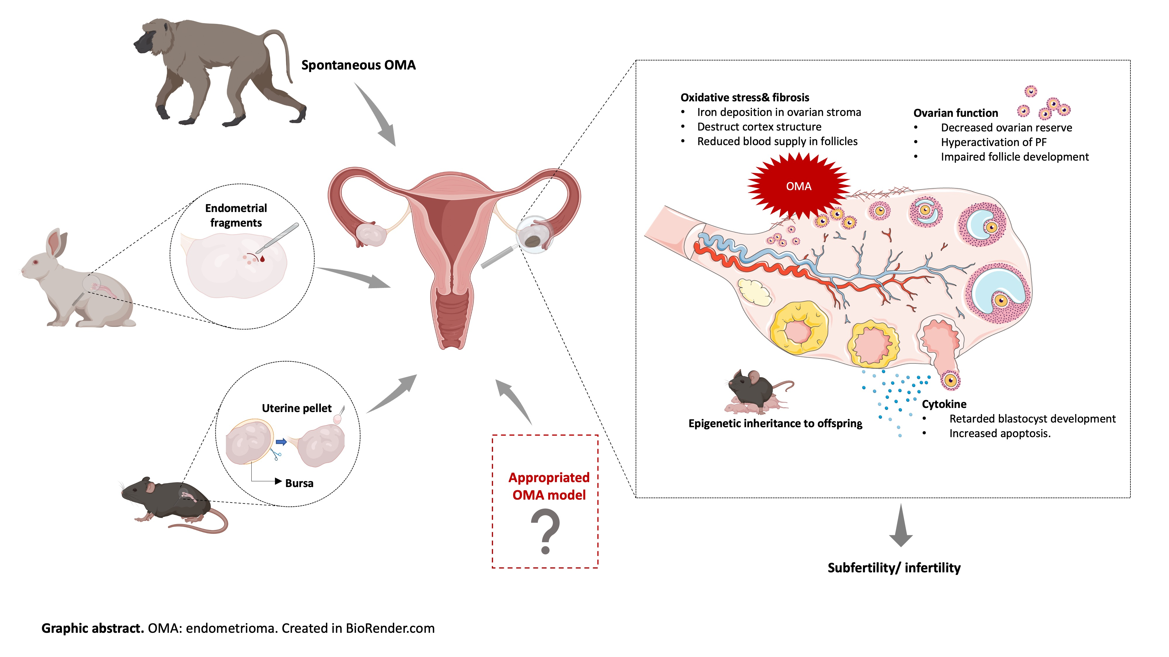
3. Animal Models of Endometriosis-Associated Infertility and OMA-Related Infertility

Currently, the mainstream study of the mechanisms underlying endometriosis-associated infertility is based on limited human samples, such as tissue fragments, primary cells, cell lines, and fluids (i.e., peritoneal fluids, endometriotic fluids, follicle fluids), obtained through surgery or assisted reproductive technology (ART) [37]. Although some mechanisms (i.e., adhesion, hormonal milieu, inflammation, and immunology) are regarded as possible pathophysiology of endometriosis-associated infertility [38], the intertwined relationships between various pathophysiological disturbances are also highlighted [8]. In vitro studies fail to mimic the in vivo microenvironments including cell-cell/cell-extracellular matrix communication, as well as cellular diversity in the natural cell environment. Therefore, in vivo experimental models that simulate the main features of diseases and microenvironments are emergently needed. In recent 40 years, animal models of endometriosis had been widely developed [39]. Nevertheless, most of these models mimic subtypes of SUP and DIE, and few mimic OMA. The lack of specific in vivo models which resemble characteristics of OMA and its related infertility may impede the progress of these studies. This section provides in-depth investigations of the methods of the existed endometriosis and OMA models which were applied to study the associated infertility.

3.1. Endometriosis Models and Infertility

3.1.1. Non-Human Primates

The most widely accepted pathophysiology of endometriosis is retrograde menstruation, proposed by Sampson in 1921, in which endometriosis arises from endometrial cells/debris shedding from the uterus via the fallopian tubes into the ovary or other sites in the pelvic cavity [40,41,42]. Non-human primates (NHPs) like baboons and cynomolgus monkeys have absolute advantages as they are phylogenetically proximate to humans, with similarities in aspects of anatomy, physiology, as well as pathology [43]. NHPs have cyclic menstruation and can develop endometriosis spontaneously. Therefore, they are extensively used as experimental endometriosis animal models to study its associated infertility [44]. Endometriosis symptoms in cynomolgus monkeys are similar to those in women [45,46]. In 1992, Ami et al. demonstrated that the majority of cynomolgus monkeys had lesion cysts and mostly were accompanied by uterine adenomyosis [45]. Implications of sub-infertility in the NHP model of endometriosis are widely investigated to evaluate the disease progression and to monitor drug efficacy [41].
However, the shortcomings of the endometriosis model in NHPs cannot be ignored. Firstly, NHPs develop endometriosis at a lower rate than human beings and cannot be diagnosed until the related signs and symptoms develop severely [47,48]. Many researchers tried other methods to induce endometriotic lesions artificially in NHP. Notably, D’Hopghe et al. and Fazleabas et al. proposed the seeding and inoculation method to induce peritoneal endometriosis autologously in baboons [49,50]. The induced endometriosis model reveals a similar appearance as spontaneous endometriosis, better yet with higher efficiency [49]. The high expense, strict facilities, and ethical issues still need to be taken into account, which have severely hampered its wide application [51].

3.1.2. Rodents

Rodents such as mice, hamsters, rats, and rabbits have numerous advantages such as cost-effectiveness, small size and large litter size, and the short gestation which enables transgenerational study [51,52,53,54,55]. Furthermore, the wide accessibility of genetically modified mice, abundance of antibodies against rat and murine proteins, and the comprehensive genomic profiles of rodents make them extensively applied to explore the fundamental mechanisms of many diseases, including endometriosis [56]. However, rodents do not get menstruation, which hinders them to develop endometriosis spontaneously. They cannot mimic human conditions as accurately as NHPs. Consequently, the existed rodent models of endometriosis are artificially induced. There are two approaches mostly used to induce endometriosis in rodents: homologous and heterologous. In the homologous model, uteri or the two uterine horns from a donor are implanted in the same or syngeneic recipients. In heterologous models, human endometrium tissues are transplanted into rodents [57,58]. Transplantation by intraperitoneal injection [59] or suturing around the mesentery artery [60] manifests the subtypes of SPE or DIE [61,62,63,64,65]. In rabbits with induced SUP, a decrease in pregnancy rate was reported [61]. A significant reduction in litter size and embryo weights had been observed in the rats model of SUP [62]. It was also indicated that surgically induced SUP in rats resulted in impaired folliculogenesis and oocyte quality [63,64,65]. These models were proved to mimic some characteristics of endometriosis in human cases and provided the insights that endometriotic lesions can negatively affect fecundity.

3.1.3. Organoids

Recent years, organoid, as an in vitro model derived from stem cells, displays the 3D structure and recapitulates biological and pathological features of the original organs [66,67]. They are genetically stable and highly committed to the original tissue during long-term expansion in culture, which enables them to substitute in vitro models, and on the interface of in vitro and in vivo models [67]. Endometrial organoids were firstly cultured by Turco et al. and Boretto et al. in 2017 [68,69], they developed similar characteristics to original tissue, such as hormonal responses during early pregnancy and menstruation [68,69]. For infertility studies, a 3-D organoid model of human endometrium could be developed to study the endometrial interface during embryo implantation [70]. When it was exposed to trophoblast in the implantation window, it showed the process and underlying mechanism of normal or pathological endometrium-embryo crosstalk [70]. For endometriosis research, organoids derived from ectopic endometrium could provide a tool to understand the aetiopathogenesis of endometriosis by comparing the one from normal endometrium [68]. Organoids of endometriosis show the characteristics of endometriosis such as luminal invasion, proliferation, and increased CA-125 level. Combing with genome engineering, it allows genetic intervention and modification for identifying therapeutic targets for further investigation. Besides, the lineage tracing in organoids provides information on cell dynamics, which may be related to the onset, progression, and pathogenesis of endometriosis [71]. Despite the recent development of this technology, there is a lack of studies investigating endometriosis-associated infertility based on organoids.

3.2. OMA Models for Infertility

3.2.1. The Current OMA Animal Models

The first description of OMA was in 1899 by Russel. He described the formation of an endometriotic cyst containing glands and uterine epithelium on an ovary [72]. Nearly 100 years later, OMA was first artificially induced in rabbits to explore the effect of OMA on postovulatory events [73]. In this study, the endometrium was separated from the uterus and relocated to one ovary with a fresh incision. Under the same conditions, adipose tissues were transplanted to the contralateral ovary, acting as control. Seven weeks later, nearly 88% of rabbits formed viable implants according to histological analysis [73]. To evaluate the ovulatory function, they counted ovulation points before and after the placement of endometrial or adipose tissues. Results showed a significantly reduced number of ovulation points after endometrial tissue implantation, which indicated that the experimental OMA implied a detrimental effect on the ovulation process [73]. In addition, they graded the periovarian adhesions according to the adhesion density and the proportion of affected ovarian surface. It indicated a higher distribution of extensive adhesions for ovaries with endometrial tissue compared to those with adipose tissue. The ovulation points were significantly decreased in both groups with adhesions, while in the adipose group without adhesions, the number of ovulation points was not affected by the implantation process [73]. This study evaluated the role of periovarian adhesions in OMA and suggested that there might be other factors leading to ovulatory dysfunction in OMA. However, this model and study design remained under some restrictions. Firstly, laparostomy was performed to inspect the ovulation points and periovarian adhesion before implantation. Although the second laparoscopy of tissue placement was arranged three weeks later, the repetitive surgeries in the short term may also be the cause of adhesions. Secondly, the ovulation point was an indirect index for assessing ovulatory function, which was unstable and could impose heterogeneity and subjectivity. With the advancement in modern research, there are lots of tests for ovulation in the clinic, i.e., ovulation hormone test, which can also be applied in animal models for a more accurate result [74]. Besides, there is no further study to explore the subsequent effect on fertility based on this model.
Before 2003, spontaneous OMA was presented as rare in baboons, which was not corresponding to the high frequency in human manifestation, accounting for half of the patients with endometriosis [75,76]. However, a significant prevalence of spontaneous OMA in baboons was reported by Dick et al. in 2003, accounting for 37% of all subjects with endometriosis [77]. The differences in genetics and husbandry environment might contribute to the differences between the two colonies [75,76,78,79]. In Dick’s study, these baboons with OMA had a longer interval between pregnancies, which was considered a potential index of reduced fecundity [77]. This paper demonstrates the feasibility of baboons as a spontaneous model for OMA-associated infertility. Compared to artificially induced animal models, spontaneous models follow a natural routine, which closely resembles human disease. They have the benefits to discover novel molecules associated with traits or pathologies. However, in addition to late manifestation, these models may have uncertain etiology and multifarious causes [80,81].
Most recently, Hayashi et al. established a novel OMA model in mice by attaching uterine tissue around the ovary after the removal of the ovarian bursa which was a membranous structure surrounding the ovaries to protect it from infiltration of ectopic implants [82,83]. Four weeks after implantation, the success rate of the OMA establishment reached 85.7%. Based on this model, the suppressed expression of follicle-stimulating hormone receptor (FSHR) and reduced litter size were found in OMA mice compared with control, indicating that infertility was impaired in OMA mice [82]. However, there were some limitations to this model. Instead of ovaries, implants were also found in nearby tissues and organs, such as the pancreas, muscle, and intestine, manifesting the subtype of SUP and DIE [82]. The involvement of multiple subtypes would cause confounding effects and interact with the specific pathogenesis of OMA-related infertility. Moreover, although the success rate was high, it could not reach 100%, which increased the workload of researchers as histological confirmation of endometriotic lesions was required with this model. Moreover, this model failed to provide the availability of genetic modifications, which could be a strong tool to identify the genetic regulators and therapeutic targets of OMA-related infertility.
Table 1 summaries the current animal models of OMA for studying the related infertility.

3.2.2. The Urge to Establish a Proper OMA Animal Models

Up to now, the current animal models for OMA are quite rare and with slow development. From the above information, it takes more than 10 years to propose a new animal model of OMA. OMA, which act as the most common subtype of endometriosis, may lead to severe dysmenorrhea, infertility, and increases the risk of ovarian cancer [16,84]. While unlike other subtypes of endometriosis, studies on specific OMA models are scarce, which hinders the development of OMA-related studies. It is urgent to develop an appropriate model of OMA for studying its pathology and underlying mechanisms. There is no doubt that these current models of OMA provided us with new insights into the OMA-related infertility, whereas their applications are restrained due to various flaws.
A proper animal model of OMA should be with a promising successful rate, with no ectopic lesion found in surrounding organs except ovaries, and available for genetic modification. In addition, the standard for a proper OMA model should be issued emergently. It is undecided for the type of implants (whole uterus or sole endometrium), transplantation methods, and duration after implantation. According to the ‘3Rs’ principle in animal experiments, it is important to define the suitable implant types, transplantation methods, and duration which might maximize the success rate and minimize the number of animals needed and their suffering time.

4. Pathology and Underlying Mechanisms of OMA-Related Infertility Based on OMA Models

4.1. Periovarian Adhesions

According to the American Society for Reproductive Medicine (ASRM) scoring, patients with OMA are always staged as moderate or severe as they are usually accompanied by adhesions of surrounding tissues and organs [7,8]. In rabbits with induced OMA, the reduced ovulatory function was significantly correlated with pelvic adhesions at the ovary. Its severity was positively related to decreased ovulation points [73], suggesting that the peri-ovarian adhesions of OMA might impair fecundity. In addition, the surgical treatment for ectopic lesions in the ovary could further facilitate pelvic adhesion and cul-de-sac obliteration, therefore resulting in reduced fertility due to impeded sperm passaging, compromised egg release, and blocked oocyte pickup through fallopian tubes when ovaries embedded within adhesions [38,85].

4.2. Ovarian Function

Ovarian reserve reflects ovarian functions from the production of qualified eggs for fertilization, and secretion of ovarian hormones for homeostasis [86]. The reproductive potential comes from dormant primordial follicles activity that is repressed until folliculogenesis takes place in the ovarian cortex [87]. Folliculogenesis is a process that involves follicle activation, development, maturation, and either ovulation of qualified oocyte or follicle atresia. Disturbance of it may lead to oocytes with a reduced number, and poor quality, and subsequently impairs pregnancy outcome [88,89]. Prior clinical studies indicated that the ovarian cortex derived from ovaries with OMA showed a decrease in follicular density compared to that from healthy ovaries [90]. Ovarian response to ovulation stimulation is an indirect indicator of ovarian reserve [91]. Women with unilateral OMA had significantly lower ovarian responsiveness compared to women with contralateral healthy ovaries, demonstrating the presence of OMA lesions may lead to an impaired ovarian reserve [92].
AMH is expressed by GC of developing follicles during folliculogenesis [93]. It prevented primordial follicles from activation and assisted follicle selection for ovulation to maintain ovarian reserve [94]. Serum AMH level is a commonly used marker of ovarian reserve [95]. Its downregulated expression implicated the decline of ovarian reserve, which was found associated with the severity of endometriosis [96]. A study indicated a significantly reduced AMH level in women with previous endometrioma resection, regardless of the current endometrioma lesion [34]. As mentioned before, surgery may reduce ovarian reserve by resecting healthy ovarian tissue [26,28]. Hence, whether it is OMA per se or its resection, or both that damage ovarian reserve is still unclear. As AMH is produced by the preantral and early antral follicles through a dynamic process, its serum level reflects ovarian reserve indirectly [97]. In vivo OMA models provide an opportunity to detect its expression level in ovarian follicles directly. A study on this is still lacking. Follicle-stimulating hormone (FSH) is critical to female fertility via folliculogenesis. The development and maturation of pre-ovulatory follicles are dependent on FSH secretion. FSH is also important for oocyte developmental competence and regulates its subsequent ovulation. It was reported that large follicle survival rate and oocyte quality were significantly decreased in the absence of FSH in vitro [98]. It was also proved that FSH could increase primordial follicle dormancy and ovarian reserve [1]. FSH acts on gonadal target cells by specifically binding to its G protein-coupled receptor, FSHR. The downstream transduction of FSH signaling relies on the receptors [99]. Decreased FSHR distribution leads to a lower response of endogenous FSH, therefore resulting in incomplete reproductive functions [99]. In OMA murine model, FSHR was found to have a dramatically lower expression level in pre-antral, antral, and pre-ovulatory follicles, which might decrease the transduction of FSH signaling reached follicles during folliculogenesis [82]. Less FSH binding to FSHR leads to its accumulation in serum. Thus, a higher serum concentration of FSH was observed to be associated with larger endometriotic lesion size, correlated with lower antral follicle count and oocyte retrieval rate in women with OMA [100].
Ovarian tissues of OMA women were found with a low distribution of primordial follicles, high distribution of growing follicles. It was suggested that premature activation of the ovarian cortex could be the underlying mechanism of declined ovarian reserve caused by OMA [101]. Extensive activation of primordial follicles in a mouse model of peritoneal endometriosis was confirmed to be regulated by the PI3K-PTEN-Akt-Foxo3 signaling pathway [102]. However, the PI3K-PTEN-Akt-Foxo3 pathway was not found significantly different between women with and without OMA [102], yet it was limited by the small sample size of clinical data and the lack of an OMA animal model.

4.3. Oxidative Stress

Oxidative stress as a result of excess reactive oxygen species (ROS) and the aberrant antioxidants in the follicular microenvironment, was regarded as essential to induce follicle senescence. This led to disturbed reproductive endocrinology, reduced follicle quality, and density, and ended in subfertility and infertility [103]. More recent studies speculated that excessive oxidative stress and reduced ability of GC might act as causative factors of endometriosis-related infertility via regulating folliculogenesis, oocyte development, ovulation, and embryogenesis [104]. In one study, GCs from infertile patients with or without OMA were collected. Intracellular ROS levels were measured and the result demonstrated increased ROS and excessive oxidative stress in GCs from women with OMA. Their activities impaired ovarian function and were negatively related to oocyte retrieval rate and proportion of mature oocytes [105,106]. The role of oxidative stress in OMA-related infertility had been proved in the murine model of OMA [82]. With endometriotic lesions introduced to the mouse ovaries, fibrosis and iron deposition were found in the endometriotic stroma, representing extensive oxidative stress in the pathogenesis [82]. Though the underlying mechanism of how ROS leads to OMA-related infertility is yet to be understood, it was hypothesized that the toxic contents inside ovarian cyst might induce excessive ROS to increase fibrosis and destruct cortex structure. The blood supply to follicles was reduced, eventually leading to impaired follicle development and decreased follicle count [107]. To prove the hypothesis and explore the potential therapeutic agents that might improve fertility outcomes caused by oxidative stress in OMA, more research should be launched based on OMA models.

5. Potential Therapeutics

Therapeutic targets aim at restoring fertility, especially in the aspect of ovarian function are pivotal for the development of novel drugs. Based on the pathogenesis of OMA-related infertility described in part 4, 10 drugs targeting the potential signaling pathways below were screened from the Therapeutic Target Database and listed in Table 2 [108]. These drugs are already available in the market for other diseases, while they could also provide a possibility for their application in the area of OMA-related infertility.

5.1. Management of Periovarian Adhesions

Up to now, surgery is the most common management of periovarian adhesions which lead to infertility [109]. Laparoscopy enables aspiration of fresh adhesions, which improves infertility caused by tubal-ovarian adhesion [110]. While there are limited therapeutic targets to remove periovarian adhesions. Some adhesion-reducing substances, i.e., ferric hyaluronic acid, dextran sulfate, and fibrin, decrease post-surgical adhesions only but do not prevent their formation [109]. Furthermore, both surgery and adhesion-reducing substances treatments cannot avoid the recurrence of adhesions [109,110].

5.2. Antioxidants

Antioxidants could inactivate, immobilize, or paralyze the ROS before they damage the cells [111]. They can be synthesized endogenously for normal cellular function or derived exogenously to deal with the overabundance of ROS [112]. Quercetin and baicalin, which belong to the flavonoid class, are common-used antioxidants during oocyte development and embryo in vitro maturation [113,114]. As maturation-promoting factor (MPF) and Sirtuins can protect oocytes from aging by decreasing ROS generation, Quercetin delayed oocyte aging after ovulation by attenuating the reduction of MPF activity and regulating the expression of Sirtuins [115]. Mitochondria produce ATP and some ROS, and ROS adversely affects mitochondria function [116]. The abundance of ROS can destroy mitochondria in embryos and thereby decline ATP production, which impairs embryo quality [117]. The Baicalin supplement significantly decreased the ROS level and apoptosis during in vitro embryonic development by improving the mitochondrial membrane potential and ATP level [118]. N-acetyl-cysteine (NAC), an amino thiol with antioxidant properties, prevented damage to bovine embryos when exposed to FF from endometriosis patients with infertility [119]. Melatonin, another antioxidant, is synthesized in the human body naturally. Melatonin regulated against mitochondrial oxidative damage and therefore ameliorated oocyte aging and ovarian reserve exhaustion in mice [108,120,121,122,123,124]. Tafenoquine (TFQ), an FDA-approved antimalarial drug, is applied to treat Trypanosoma brucei infection by targeting ROS and electron transport complex III [125,126]. TFQ showed effectiveness in killing Trypanosoma brucei by increasing ROS production in parasites, thereby leading to programmed cell death by necrosis [127,128]. While TFQ induces cell death by increasing ROS in cells, it should be with limitation in OMA-related infertility as extensive ROS is the possible underlying mechanism to inducing infertility [82].

5.3. Restoration of Ovarian Function

MTORC1 and PI3K-PTEN-Akt-Foxo3 pathways were involved in OMA-related infertility [102]. FOXO3, located in the oocyte nucleus of dormant primordial follicles, inhibited activation of primordial follicles and whereby maintained ovarian reserve in the ovarian cortex. FOXO3 phosphorylated through the PI3K-Akt pathway, was transited from nucleus to cytoplasm to initiate primordial follicles activation. The expression level of FOXO3 decreased in ovaries of OMA, leading to premature activation of primordial follicles and subsequent follicle loss. There is no drug reported to increase the FOXO3 level. While a recent study mentioned that melatonin administration inhibited FOXO3 from nuclear transitions [124]. Moreover, FOXO3 is the major target of Akt and can be inactivated by Akt. Melatonin intake was reported to reduce the phosphorylation of Akt, potentially could inhibit Akt activation, and enhanced FOXO3 expression [124]. Akt inhibitor, such as ARQ-751, was also reported to upregulate the expression of FOXO3 [129,130]. Administration of ammonium trichloro (dioxoethylene-o,o’) tellurate (AS101), a modulator of the PI3K-PTEN-Akt pathway, was also observed to prevent hyperactivation of primordial follicles and preserve ovarian reserve in ovarian endometriosis of mice model [102]. There are some drugs (i.e., Sirolimus, Everolimus, Novolimus) commercially available for treating organ transplant rejection, carcinoma, artery stenosis, leukemia, and solid tumor/cancer through regulating mTOR and PI3K-PTEN-Akt pathways [131,132,133,134,135,136,137,138]. Their application and effect on OMA-related infertility could be further investigated.
On the other hand, AMH, which inhibits primordial activation and assists in selective ovulation, was found to lower serum levels in patients with OMA [34,94]. No drug has been reported to improve AMH levels. Vitamin D could significantly increase the serum level of AMH in ovulatory women [139]. Although the correlation between vitamin D and AMH is sophisticated and not fully known, it could be considered a potential nutrient to improve ovarian reserve in infertile women with OMA [139]. GATA binding protein 4 (GATA-4) was identified as the regulatory element located in the AMH promoter sequence [139]. It activates AMH transcriptional activity and regulates folliculogenesis of goose [139]. Its overexpression increases AMH expression levels, which may benefit fertility in OMA patients. FSH also plays a central role in regulating the development and maturation of pre-ovulatory follicles, thus helping the folliculogenesis and maintaining mammalian reproduction [140]. Its transduction relies on its receptor, FSHR. There is no drug approved for increasing the FSHR level. Three drugs, Follitropin beta, Menotropins, and Urofollitropin, were recombinant FSH approved for treating female infertility targeting FSHR. While a decreased expression level of FSHR has been reported in mouse models of OMA [82]. The exogenous FSH might have a subtle effect as the signal cannot be transferred through receptors. More investigations on novel drugs are needed for increasing the expression level of FSHR thus improving fecundity in infertile women with OMA.

6. Summary and Future Perspective

OMA-related infertility imposes a huge burden on not only individuals and families but also the health care system and society. In order to improve the understanding of fecundity affected by OMA, the establishment of suitable animal models is of great importance to the related studies. Up to now, three animal models of OMA were induced to unmask the key factors responsible for OMA-related infertility, including periovarian adhesion, ovarian dysfunction, and oxidative stress. They constructed a contour about how OMA lesions lead to infertility. In addition, the related molecules and pathways (i.e., PI3K-PTEN-Akt-Foxo3 pathway, excess ROS) provide potential therapeutic targets for OMA-related infertility. It is undisputed that none of these factors can cause OMA-related infertility alonely, and there might be other factors still under investigation.
However, there are limited studies of OMA models available and there is a long-time interval between each study. Except for the one published in 2021, the other two studies were published nearly 20 years ago, which failed to provide trendy perspectives in this area. As well, the lack of further studies based on these two models decreased their conviction.
Moreover, the limitations of current OMA models include the unavailability of genetic modifications or the difficulty to mimic the subtype of OMA hinders the research progress in the pathogenesis of OMA-related infertility. NHP is most proximate to human beings, while its high cost, long period of disease manifestation, difficult access, and maintenance issues prevent it to be the most ideal model of OMA [46,77]. Though the rabbit model of OMA presented a high success rate (88%), it is with a long generation time, long estrus cycle, and is not available for gene modification compared to the mouse model, which impeded its wide application in reproductive diseases [61,73]. An OMA model with a promising success rate, exclusive location of ectopic lesions, and vast availability of genetic modification are desirable. Based on this model, future research can be applied to study fertility indexes, including follicle development, oocyte selection, ovulation, zygote quality, embryo implantation, pregnancy outcomes, etc. Furthermore, a number of on-market drugs which regulate the associated pathways can be repurposed as potential new treatments for OMA-related infertility, followed by adequate preclinical studies and clinical trials that need to be conducted to assess their effectiveness and safety. A proper model can act as an ideal platform for preclinical testing of potential therapeutics, which can accelerate the development of scientific research and novel therapies for OMA-associated infertility.

Author Contributions

Z.T., S.-W.H., C.-C.W. and T.Z. participated in the research design. Z.T. participated in data evaluation, extraction, and interpretation. Z.T., S.-W.H. and X.Z. participated in data validation and in drafting the manuscript. Z.T., S.-W.H., T.Z., J.P.-W.C. and C.-C.W. critically revised the manuscript. All authors have read and agreed to the published version of the manuscript.

Funding

This research received no external funding.

Institutional Review Board Statement

Not applicable.

Informed Consent Statement

Not applicable.

Data Availability Statement

Not applicable.

Conflicts of Interest

The authors declare no conflict of interest.

References

  1. Exacoustos, C.; De Felice, G.; Pizzo, A.; Morosetti, G.; Lazzeri, L.; Centini, G.; Piccione, E.; Zupi, E. Isolated Ovarian Endometrioma: A History Between Myth and Reality. J. Minim. Invasive Gynecol. 2018, 25, 884–891. [Google Scholar] [CrossRef] [PubMed]
  2. Fuldeore, M.; Yang, H.; Du, E.X.; Soliman, A.M.; Wu, E.Q.; Winkel, C. Healthcare utilization and costs in women diagnosed with endometriosis before and after diagnosis: A longitudinal analysis of claims databases. Fertil. Steril. 2015, 103, 163–171. [Google Scholar] [CrossRef] [PubMed]
  3. Fonseca, M.A.S.; Wright, K.N.; Lin, X.; Abbasi, F.; Haro, M.; Sun, J.; Hernandez, L.; Orr, N.L.; Hong, J.; Choi-Kuaea, Y.; et al. A cellular and molecular portrait of endometriosis subtypes. bioRxiv 2021, arXiv:2021.05.20.445037. [Google Scholar]
  4. Redwine, D.B. Age-related evolution in color appearance of endometriosis. Fertil. Steril. 1987, 48, 1062–1063. [Google Scholar] [CrossRef]
  5. Harirchian, P.; Gashaw, I.; Lipskind, S.T.; Braundmeier, A.G.; Hastings, J.M.; Olson, M.R.; Fazleabas, A.T. Lesion kinetics in a non-human primate model of endometriosis. Hum. Reprod. 2012, 27, 2341–2351. [Google Scholar] [CrossRef] [PubMed] [Green Version]
  6. Holt, V.L.; Weiss, N.S. Recommendations for the Design of Epidemiologic Studies of Endometriosis. Epidemiology 2000, 11, 654–659. [Google Scholar] [CrossRef] [Green Version]
  7. Adamson, G.D.; Pasta, D.J. Endometriosis fertility index: The new, validated endometriosis staging system. Fertil. Steril. 2010, 94, 1609–1615. [Google Scholar] [CrossRef]
  8. Dai, Y.; Li, X.; Shi, J.; Leng, J. A review of the risk factors, genetics and treatment of endometriosis in Chinese women: A comparative update. Reprod. Health 2018, 15, 82. [Google Scholar] [CrossRef] [PubMed]
  9. Bulletti, C.; Coccia, M.E.; Battistoni, S.; Borini, A. Endometriosis and infertility. Fertil. Steril. 2006, 86 (Suppl. S5), S156–S160. [Google Scholar] [CrossRef]
  10. Kajiyama, H.; Suzuki, S.; Yoshihara, M.; Tamauchi, S.; Yoshikawa, N.; Niimi, K.; Shibata, K.; Kikkawa, F. Endometriosis and cancer. Free Radic. Biol. Med. 2019, 133, 186–192. [Google Scholar] [CrossRef]
  11. Filip, L.; Duică, F.; Prădatu, A.; Crețoiu, D.; Suciu, N.; Crețoiu, S.M.; Predescu, D.V.; Varlas, V.N.; Voinea, S.C. Endometriosis Associated Infertility: A Critical Review and Analysis on Etiopathogenesis and Therapeutic Approaches. Medicina 2020, 56, 460. [Google Scholar] [CrossRef] [PubMed]
  12. Senapati, S.; Barnhart, K. Managing endometriosis-associated infertility. Clin. Obstet. Gynecol. 2011, 54, 720–726. [Google Scholar] [CrossRef] [PubMed] [Green Version]
  13. Hughes, E.G.; Fedorkow, D.M.; Collins, J.A. A quantitative overview of controlled trials in endometriosis-associated infertility. Fertil. Steril. 1993, 59, 963–970. [Google Scholar] [CrossRef]
  14. Naples, J.D.; Batt, R.E.; Sadigh, H. Spontaneous abortion rate in patients with endometriosis. Obstet. Gynecol. 1981, 57, 509–512. [Google Scholar]
  15. Groll, M. Endometriosis and spontaneous abortion. Fertil. Steril. 1984, 41, 933–935. [Google Scholar] [CrossRef]
  16. De Ziegler, D.; Borghese, B.; Chapron, C. Endometriosis and infertility: Pathophysiology and management. Lancet 2010, 376, 730–738. [Google Scholar] [CrossRef]
  17. Broekmans, F.J.; Soules, M.R.; Fauser, B.C. Ovarian aging: Mechanisms and clinical consequences. Endocr. Rev. 2009, 30, 465–493. [Google Scholar] [CrossRef] [Green Version]
  18. Hickey, M.; Ballard, K.; Farquhar, C. Endometriosis. BMJ 2014, 348, g1752. [Google Scholar] [CrossRef]
  19. Hung, S.W.; Zhang, R.; Tan, Z.; Chung, J.P.W.; Zhang, T.; Wang, C.C. Pharmaceuticals targeting signaling pathways of endometriosis as potential new medical treatment: A review. Med. Res. Rev. 2021, 41, 2489–2564. [Google Scholar] [CrossRef]
  20. Muzii, L.; Di Tucci, C.; Achilli, C.; Di Donato, V.; Musella, A.; Palaia, I.; Panici, P.B. Continuous versus cyclic oral contraceptives after laparoscopic excision of ovarian endometriomas: A systematic review and metaanalysis. Am. J. Obstet. Gynecol. 2016, 214, 203–211. [Google Scholar] [CrossRef]
  21. Smith, M.R.; Lee, W.C.; Brandman, J.; Wang, Q.; Botteman, M.; Pashos, C.L. Gonadotropin-releasing hormone agonists and fracture risk: A claims-based cohort study of men with nonmetastatic prostate cancer. J. Clin. Oncol. 2005, 23, 7897–7903. [Google Scholar] [CrossRef] [PubMed]
  22. Hughes, E.; Brown, J.; Collins, J.J.; Farquhar, C.; Fedorkow, D.M.; Vandekerckhove, P. Ovulation suppression for endometriosis. Cochrane Database Syst. Rev. 2007, 2007, Cd000155. [Google Scholar]
  23. Ahmad, G.; Gent, D.; Henderson, D.; O’Flynn, H.; Phillips, K.; Watson, A. Laparoscopic entry techniques. Cochrane Database Syst. Rev. 2015, 8, Cd006583. [Google Scholar] [CrossRef] [PubMed]
  24. Muzii, L.; Di Tucci, C.; Di Feliciantonio, M.; Marchetti, C.; Perniola, G.; Panici, P.B. The effect of surgery for endometrioma on ovarian reserve evaluated by antral follicle count: A systematic review and meta-analysis. Hum. Reprod. 2014, 29, 2190–2198. [Google Scholar] [CrossRef] [Green Version]
  25. Seyhan, A.; Ata, B.; Uncu, G. The Impact of Endometriosis and Its Treatment on Ovarian Reserve. Semin. Reprod. Med. 2015, 33, 422–428. [Google Scholar] [CrossRef] [Green Version]
  26. Goodman, L.R.; Goldberg, J.M.; Flyckt, R.L.; Gupta, M.; Harwalker, J.; Falcone, T. Effect of surgery on ovarian reserve in women with endometriomas, endometriosis and controls. Am. J. Obstet. Gynecol. 2016, 215, 589.e1–589.e6. [Google Scholar] [CrossRef]
  27. Mehdizadeh Kashi, A.; Chaichian, S.; Ariana, S.; Fazaeli, M.; Moradi, Y.; Rashidi, M.; Najmi, Z. The impact of laparoscopic cystectomy on ovarian reserve in patients with unilateral and bilateral endometrioma. Int. J. Gynaecol. Obstet. 2017, 136, 200–204. [Google Scholar] [CrossRef]
  28. Donnez, J.; Nisolle, M.; Gillet, N.; Smets, M.; Bassil, S.; Casanas-Roux, F. Large ovarian endometriomas. Hum. Reprod. 1996, 11, 641–646. [Google Scholar] [CrossRef]
  29. Rajab, T.K.; Ahmad, U.N.; Kelly, E. Implications of late complications from adhesions for preoperative informed consent. J. R. Soc. Med. 2010, 103, 317–321. [Google Scholar] [CrossRef] [Green Version]
  30. Hui, Y.; Zhao, S.; Gu, J.; Hang, C. Analysis of factors related to fertility after endometriosis combined with infertility laparoscopic surgery. Medicine 2020, 99, e20132. [Google Scholar] [CrossRef]
  31. Bourdon, M.; Raad, J.; Dahan, Y.; Marcellin, L.; Maignien, C.; Even, M.; Pocate-Cheriet, K.; Lamau, M.C.; Santulli, P.; Chapron, C. Endometriosis and ART: A prior history of surgery for OMA is associated with a poor ovarian response to hyperstimulation. PLoS ONE 2018, 13, e0202399. [Google Scholar]
  32. Coccia, M.E.; Rizzello, F.; Cammilli, F.; Bracco, G.L.; Scarselli, G. Endometriosis and infertility Surgery and ART: An integrated approach for successful management. Eur. J. Obstet. Gynecol. Reprod. Biol. 2008, 138, 54–59. [Google Scholar] [CrossRef] [PubMed]
  33. Hamdan, M.; Dunselman, G.; Li, T.C.; Cheong, Y. The impact of endometrioma on IVF/ICSI outcomes: A systematic review and meta-analysis. Hum. Reprod. Update 2015, 21, 809–825. [Google Scholar] [CrossRef] [PubMed] [Green Version]
  34. Dunselman, G.A.; Vermeulen, N.; Becker, C.; Calhaz-Jorge, C.; D’Hooghe, T.; De Bie, B.; Heikinheimo, O.; Horne, A.W.; Kiesel, L.; Nap, A.; et al. ESHRE guideline: Management of women with endometriosis. Hum. Reprod. 2014, 29, 400–412. [Google Scholar] [PubMed]
  35. Zaliska, O.; Stasiv, K.; Maksymovych, N.; Hrynkiv, Y. The trends of assisted reproductive technologies and cost for ovarian stimulation protocols in Ukraine. Pharmacia 2020, 67, 269–277. [Google Scholar] [CrossRef]
  36. Shreffler, K.M.; Johnson, D.R.; Scheuble, L.K. Ethical Problems with Infertility Treatments: Attitudes and Explanations. Soc. Sci. J. 2010, 47, 731–746. [Google Scholar] [CrossRef] [Green Version]
  37. Malvezzi, H.; Marengo, E.B.; Podgaec, S.; Piccinato, C.A. Endometriosis: Current challenges in modeling a multifactorial disease of unknown etiology. J. Transl. Med. 2020, 18, 311. [Google Scholar]
  38. Tanbo, T.; Fedorcsak, P. Endometriosis-associated infertility: Aspects of pathophysiological mechanisms and treatment options. Acta Obstet. Gynecol. Scand. 2017, 96, 659–667. [Google Scholar]
  39. Burns, K.A.; Pearson, A.M.; Slack, J.L.; Por, E.D.; Scribner, A.N.; Eti, N.A.; Burney, R.O. Endometriosis in the Mouse: Challenges and Progress Toward a ’Best Fit’ Murine Model. Front. Physiol. 2021, 12, 806574. [Google Scholar]
  40. Sampson, J.A. Heterotopic or misplaced endometrial tissue. Am. J. Obstet. Gynecol. 1925, 10, 649–664. [Google Scholar] [CrossRef]
  41. Braundmeier, A.G.; Fazleabas, A.T. The non-human primate model of endometriosis: Research and implications for fecundity. Mol. Hum. Reprod. 2009, 15, 577–586. [Google Scholar] [CrossRef] [PubMed] [Green Version]
  42. Vercellini, P.; Viganò, P.; Somigliana, E.; Fedele, L. Endometriosis: Pathogenesis and treatment. Nat. Rev. Endocrinol. 2014, 10, 261–275. [Google Scholar] [PubMed]
  43. Zhang, X.-L.; Pang, W.; Hu, X.-T.; Li, J.-L.; Yao, Y.-G.; Zheng, Y.-T. Experimental primates and non-human primate (NHP) models of human diseases in China: Current status and progress. Dongwuxue Yanjiu 2014, 35, 447–464. [Google Scholar] [PubMed]
  44. D’Hooghe, T.M.; Kyama, C.M.; Chai, D.; Fassbender, A.; Vodolazkaia, A.; Bokor, A.; Mwenda, J.M. Nonhuman primate models for translational research in endometriosis. Reprod. Sci. 2009, 16, 152–161. [Google Scholar] [CrossRef]
  45. Ami, Y.; Suzaki, Y.; Goto, N. Endometriosis in cynomolgus monkeys retired from breeding. J. Vet. Med. Sci. 1993, 55, 7–11. [Google Scholar] [CrossRef] [Green Version]
  46. Nishimoto-Kakiuchi, A.; Netsu, S.; Okabayashi, S.; Taniguchi, K.; Tanimura, H.; Kato, A.; Suzuki, M.; Sankai, T.; Konno, R. Spontaneous endometriosis in cynomolgus monkeys as a clinically relevant experimental model. Hum. Reprod. 2018, 33, 1228–1236. [Google Scholar] [CrossRef]
  47. Simitsidellis, I.; Gibson, D.A.; Saunders, P.T.K. Animal models of endometriosis: Replicating the aetiology and symptoms of the human disorder. Best Pract. Res. Clin. Endocrinol. Metab. 2018, 32, 257–269. [Google Scholar] [CrossRef] [Green Version]
  48. Yamanaka, A.; Kimura, F.; Takebayashi, A.; Kita, N.; Takahashi, K.; Murakami, T. Primate model research for endometriosis. Tohoku J. Exp. Med. 2012, 226, 95–99. [Google Scholar] [CrossRef] [Green Version]
  49. D’Hooghe, T.M.; Bambra, C.S.; Raeymaekers, B.M.; De Jonge, I.; Lauweryns, J.M.; Koninckx, P.R. Intrapelvic injection of menstrual endometrium causes endometriosis in baboons (Papio cynocephalus and Papio anubis). Am. J. Obstet. Gynecol. 1995, 173, 125–134. [Google Scholar] [CrossRef]
  50. Fazleabas, A.T.; Brudney, A.; Gurates, B.; Chai, D.; Bulun, S. A modified baboon model for endometriosis. Ann. N. Y. Acad. Sci. 2002, 955, 308–317. [Google Scholar]
  51. Grümmer, R. Animal models in endometriosis research. Hum. Reprod. Update 2006, 12, 641–649. [Google Scholar] [CrossRef] [PubMed] [Green Version]
  52. Story, L.; Kennedy, S. Animal studies in endometriosis: A review. ILAR J. 2004, 45, 132–138. [Google Scholar] [CrossRef] [PubMed] [Green Version]
  53. Sharpe-Timms, K.L. Using rats as a research model for the study of endometriosis. Ann. N. Y. Acad. Sci. 2002, 955, 318–327. [Google Scholar] [CrossRef] [PubMed]
  54. Tirado-González, I.; Barrientos, G.; Tariverdian, N.; Arck, P.C.; García, M.G.; Klapp, B.F.; Blois, S.M. Endometriosis research: Animal models for the study of a complex disease. J. Reprod. Immunol. 2010, 86, 141–147. [Google Scholar] [CrossRef]
  55. Dinulescu, D.M.; Ince, T.A.; Quade, B.J.; Shafer, S.A.; Crowley, D.; Jacks, T. Role of K-ras and Pten in the development of mouse models of endometriosis and endometrioid ovarian cancer. Nat. Med. 2005, 11, 63–70. [Google Scholar] [CrossRef]
  56. Bruner-Tran, K.L.; Mokshagundam, S.; Herington, J.L.; Ding, T.; Osteen, K.G. Rodent Models of Experimental Endometriosis: Identifying Mechanisms of Disease and Therapeutic Targets. Curr. Womens Health Rev. 2018, 14, 173–188. [Google Scholar] [CrossRef]
  57. Martinez, J.; Bisbal, V.; Marin, N.; Cano, A.; Gómez, R. Noninvasive Monitoring of Lesion Size in a Heterologous Mouse Model of Endometriosis. J. Vis. Exp. 2019, 144, e58358. [Google Scholar] [CrossRef] [Green Version]
  58. Tejada, M.A.; Santos-Llamas, A.I.; Escriva, L.; Tarin, J.J.; Cano, A.; Fernández-Ramírez, M.J.; Nunez-Badinez, P.; De Leo, B.; Saunders, P.T.K.; Vidal, V.; et al. Identification of Altered Evoked and Non-Evoked Responses in a Heterologous Mouse Model of Endometriosis-Associated Pain. Biomedicines 2022, 10, 501. [Google Scholar] [CrossRef]
  59. Nowak, N.M.; Fischer, O.M.; Gust, T.C.; Fuhrmann, U.; Habenicht, U.F.; Schmidt, A. Intraperitoneal inflammation decreases endometriosis in a mouse model. Hum. Reprod. 2008, 23, 2466–2474. [Google Scholar] [CrossRef] [Green Version]
  60. Berkley, K.J.; Dmitrieva, N.; Curtis, K.S.; Papka, R.E. Innervation of ectopic endometrium in a rat model of endometriosis. Proc. Natl. Acad. Sci. USA 2004, 101, 11094–11098. [Google Scholar] [CrossRef] [Green Version]
  61. Schenken, R.S.; Asch, R.H. Surgical induction of endometriosis in the rabbit: Effects on fertility and concentrations of peritoneal fluid prostaglandins. Fertil. Steril. 1980, 34, 581–587. [Google Scholar] [CrossRef]
  62. Vernon, M.W.; Wilson, E.A. Studies on the surgical induction of endometriosis in the rat. Fertil. Steril. 1985, 44, 684–694. [Google Scholar] [CrossRef]
  63. Moon, C.E.; Bertero, M.C.; Curry, T.E.; London, S.N.; Muse, K.N.; Sharpe, K.L.; Vernon, M.W. The presence of luteinized unruptured follicle syndrome and altered folliculogenesis in rats with surgically induced endometriosis. Am. J. Obstet. Gynecol. 1993, 169, 676–682. [Google Scholar] [CrossRef]
  64. Pal, A.K.; Biswas, S.; Goswami, S.K.; Kabir, S.N. Effect of pelvic endometrial implants on overall reproductive functions of female rats. Biol. Reprod. 1999, 60, 954–958. [Google Scholar] [CrossRef] [PubMed] [Green Version]
  65. Stilley, J.A.; Woods-Marshall, R.; Sutovsky, M.; Sutovsky, P.; Sharpe-Timms, K.L. Reduced fecundity in female rats with surgically induced endometriosis and in their daughters: A potential role for tissue inhibitors of metalloproteinase 1. Biol. Reprod. 2009, 80, 649–656. [Google Scholar] [PubMed]
  66. Huch, M.; Gehart, H.; van Boxtel, R.; Hamer, K.; Blokzijl, F.; Verstegen, M.M.; Ellis, E.; van Wenum, M.; Fuchs, S.A.; de Ligt, J.; et al. Long-term culture of genome-stable bipotent stem cells from adult human liver. Cell 2015, 160, 299–312. [Google Scholar] [CrossRef] [PubMed] [Green Version]
  67. Heidari-Khoei, H.; Esfandiari, F.; Hajari, M.A.; Ghorbaninejad, Z.; Piryaei, A.; Baharvand, H. Organoid technology in female reproductive biomedicine. Reprod. Biol. Endocrinol. 2020, 18, 64. [Google Scholar]
  68. Turco, M.Y.; Gardner, L.; Hughes, J.; Cindrova-Davies, T.; Gomez, M.J.; Farrell, L.; Hollinshead, M.; Marsh, S.G.E.; Brosens, J.J.; Critchley, H.O.; et al. Long-term, hormone-responsive organoid cultures of human endometrium in a chemically defined medium. Nat. Cell Biol. 2017, 19, 568–577. [Google Scholar]
  69. Boretto, M.; Cox, B.; Noben, M.; Hendriks, N.; Fassbender, A.; Roose, H.; Amant, F.; Timmerman, D.; Tomassetti, C.; Vanhie, A.; et al. Development of organoids from mouse and human endometrium showing endometrial epithelium physiology and long-term expandability. Development 2017, 144, 1775–1786. [Google Scholar]
  70. Luddi, A.; Pavone, V.; Semplici, B.; Governini, L.; Criscuoli, M.; Paccagnini, E.; Gentile, M.; Morgante, G.; Leo, V.; Belmonte, G.; et al. Organoids of Human Endometrium: A Powerful In Vitro Model for the Endometrium-Embryo Cross-Talk at the Implantation Site. Cells 2020, 9, 1121. [Google Scholar] [CrossRef]
  71. Betjes, M.A.; Zheng, X.; Kok, R.N.U.; van Zon, J.S.; Tans, S.J. Cell Tracking for Organoids: Lessons From Developmental Biology. Front. Cell Dev. Biol. 2021, 9, 675013. [Google Scholar] [CrossRef] [PubMed]
  72. Longo, L.D. Classic pages in obstetrics and gynecology. Aberrant portions of the müllerian duct found in an ovary: William Wood Russell Johns Hopkins Hospital Bulletin, vol. 10, pp. 8–10, 1899. Am. J. Obstet. Gynecol. 1979, 134, 225–226. [Google Scholar] [PubMed]
  73. Kaplan, C.R.; Eddy, C.A.; Olive, D.L.; Schenken, R.S. Effect of ovarian endometriosis on ovulation in rabbits. Am. J. Obstet. Gynecol. 1989, 160, 40–44. [Google Scholar] [CrossRef]
  74. Su, H.-W.; Yi, Y.-C.; Wei, T.-Y.; Chang, T.-C.; Cheng, C.-M. Detection of ovulation, a review of currently available methods. Bioeng. Transl. Med. 2017, 2, 238–246. [Google Scholar] [CrossRef] [PubMed]
  75. D’Hooghe, T.M. Clinical relevance of the baboon as a model for the study of endometriosis. Fertil. Steril. 1997, 68, 613–625. [Google Scholar] [CrossRef]
  76. D’Hooghe, T.M.; Bambra, C.S.; Cornillie, F.J.; Isahakia, M.; Koninckx, P.R. Prevalence and laparoscopic appearance of spontaneous endometriosis in the baboon (Papio anubis, Papio cynocephalus). Biol. Reprod. 1991, 45, 411–416. [Google Scholar] [CrossRef] [PubMed]
  77. Dick, E.J., Jr.; Hubbard, G.B.; Martin, L.J.; Leland, M.M. Record review of baboons with histologically confirmed endometriosis in a large established colony. J. Med. Primatol. 2003, 32, 39–47. [Google Scholar] [CrossRef]
  78. D’Hooghe, T.M.; Bambra, C.S.; Isahakia, M.; Koninckx, P.R. Evolution of spontaneous endometriosis in the baboon (Papio anubis, Papio cynocephalus) over a 12-month period. Fertil. Steril. 1992, 58, 409–412. [Google Scholar]
  79. D’Hooghe, T.M.; Bambra, C.S.; Raeymaekers, B.M.; Riday, A.M.; Suleman, M.A.; Koninckx, P.R. The cycle pregnancy rate is normal in baboons with stage I endometriosis but decreased in primates with stage II and stage III-IV disease. Fertil. Steril. 1996, 66, 809–813. [Google Scholar] [CrossRef]
  80. Onaciu, A.; Munteanu, R.; Munteanu, V.C.; Gulei, D.; Raduly, L.; Feder, R.-I.; Pirlog, R.; Atanasov, A.G.; Korban, S.S.; Irimie, A.; et al. Spontaneous and Induced Animal Models for Cancer Research. Diagnostics 2020, 10, 660. [Google Scholar]
  81. Francia, G.; Cruz-Munoz, W.; Man, S.; Xu, P.; Kerbel, R.S. Mouse models of advanced spontaneous metastasis for experimental therapeutics. Nat. Rev. Cancer 2011, 11, 135–141. [Google Scholar] [CrossRef] [PubMed] [Green Version]
  82. Hayashi, S.; Nakamura, T.; Motooka, Y.; Ito, F.; Jiang, L.; Akatsuka, S.; Iwase, A.; Kajiyama, H.; Kikkawa, F.; Toyokuni, S. Novel ovarian endometriosis model causes infertility via iron-mediated oxidative stress in mice. Redox. Biol. 2020, 37, 101726. [Google Scholar] [CrossRef] [PubMed]
  83. Motohara, T.; Masuko, S.; Ishimoto, T.; Yae, T.; Onishi, N.; Muraguchi, T.; Hirao, A.; Matsuzaki, Y.; Tashiro, H.; Katabuchi, H.; et al. Transient depletion of p53 followed by transduction of c-Myc and K-Ras converts ovarian stem-like cells into tumor-initiating cells. Carcinogenesis 2011, 32, 1597–1606. [Google Scholar] [CrossRef] [PubMed] [Green Version]
  84. Somigliana, E.; Vigano, P.; Parazzini, F.; Stoppelli, S.; Giambattista, E.; Vercellini, P. Association between endometriosis and cancer: A comprehensive review and a critical analysis of clinical and epidemiological evidence. Gynecol. Oncol. 2006, 101, 331–341. [Google Scholar] [CrossRef] [PubMed]
  85. Grümmer, R. Translational animal models to study endometriosis-associated infertility. Semin. Reprod. Med. 2013, 31, 125–132. [Google Scholar] [CrossRef]
  86. Fauser, B.C.; Van Heusden, A.M. Manipulation of human ovarian function: Physiological concepts and clinical consequences. Endocr. Rev. 1997, 18, 71–106. [Google Scholar]
  87. Kawamara, K.; Kelsey, T.; Hiraike, O. Editorial: Ovarian Ageing: Pathophysiology and Recent Development of Maintaining Ovarian Reserve. Front. Endocrinol. 2020, 11, 591764. [Google Scholar] [CrossRef]
  88. Hsueh, A.J.; Kawamura, K.; Cheng, Y.; Fauser, B.C. Intraovarian control of early folliculogenesis. Endocr. Rev. 2015, 36, 1–24. [Google Scholar]
  89. Titus, S.; Li, F.; Stobezki, R.; Akula, K.; Unsal, E.; Jeong, K.; Dickler, M.; Robson, M.; Moy, F.; Goswami, S.; et al. Impairment of BRCA1-related DNA double-strand break repair leads to ovarian aging in mice and humans. Sci. Transl. Med. 2013, 5, 172ra21. [Google Scholar] [CrossRef] [Green Version]
  90. Kitajima, M.; Defrère, S.; Dolmans, M.M.; Colette, S.; Squifflet, J.; Van Langendonckt, A.; Donnez, J. Endometriomas as a possible cause of reduced ovarian reserve in women with endometriosis. Fertil. Steril. 2011, 96, 685–691. [Google Scholar] [CrossRef]
  91. Mutlu, M.F.; Erdem, A. Evaluation of ovarian reserve in infertile patients. J. Turk. Ger. Gynecol. Assoc. 2012, 13, 196–203. [Google Scholar] [CrossRef] [PubMed]
  92. Somigliana, E.; Infantino, M.; Benedetti, F.; Arnoldi, M.; Calanna, G.; Ragni, G. The presence of ovarian endometriomas is associated with a reduced responsiveness to gonadotropins. Fertil. Steril. 2006, 86, 192–196. [Google Scholar] [CrossRef] [PubMed]
  93. Moolhuijsen, L.M.E.; Visser, J.A. Anti-Müllerian Hormone and Ovarian Reserve: Update on Assessing Ovarian Function. J. Clin. Endocrinol. Metab. 2020, 105, 3361–3373. [Google Scholar] [CrossRef] [PubMed]
  94. Visser, J.A.; de Jong, F.H.; Laven, J.S.; Themmen, A.P. Anti-Müllerian hormone: A new marker for ovarian function. Reproduction 2006, 131, 1–9. [Google Scholar] [CrossRef] [PubMed] [Green Version]
  95. Weenen, C.; Laven, J.S.; Von Bergh, A.R.; Cranfield, M.; Groome, N.P.; Visser, J.A.; Kramer, P.; Fauser, B.C.; Themmen, A.P. Anti-Müllerian hormone expression pattern in the human ovary: Potential implications for initial and cyclic follicle recruitment. Mol. Hum. Reprod. 2004, 10, 77–83. [Google Scholar] [CrossRef]
  96. Pacchiarotti, A.; Frati, P.; Milazzo, G.N.; Catalano, A.; Gentile, V.; Moscarini, M. Evaluation of serum anti-Mullerian hormone levels to assess the ovarian reserve in women with severe endometriosis. Eur. J. Obstet. Gynecol. Reprod. Biol. 2014, 172, 62–64. [Google Scholar] [CrossRef]
  97. Visser, J.A.; Themmen, A.P. Anti-Müllerian hormone and folliculogenesis. Mol. Cell. Endocrinol. 2005, 234, 81–86. [Google Scholar] [CrossRef]
  98. Adriaens, I.; Cortvrindt, R.; Smitz, J. Differential FSH exposure in preantral follicle culture has marked effects on folliculogenesis and oocyte developmental competence. Hum. Reprod. 2004, 19, 398–408. [Google Scholar] [CrossRef] [Green Version]
  99. Casarini, L.; Crépieux, P. Molecular Mechanisms of Action of FSH. Front. Endocrinol. 2019, 10, 305. [Google Scholar] [CrossRef]
  100. Tian, Z.; Zhang, Y.; Zhang, C.; Wang, Y.; Zhu, H.-L. Antral follicle count is reduced in the presence of endometriosis: A systematic review and meta-analysis. Reprod. BioMed. Online 2021, 42, 237–247. [Google Scholar] [CrossRef]
  101. Kitajima, M.; Dolmans, M.M.; Donnez, O.; Masuzaki, H.; Soares, M.; Donnez, J. Enhanced follicular recruitment and atresia in cortex derived from ovaries with endometriomas. Fertil. Steril. 2014, 101, 1031–1037. [Google Scholar] [CrossRef] [PubMed]
  102. Takeuchi, A.; Koga, K.; Satake, E.; Makabe, T.; Taguchi, A.; Miyashita, M.; Takamura, M.; Harada, M.; Hirata, T.; Hirota, Y.; et al. Endometriosis Triggers Excessive Activation of Primordial Follicles via PI3K-PTEN-Akt-Foxo3 Pathway. J. Clin. Endocrinol. Metab. 2019, 104, 5547–5554. [Google Scholar] [CrossRef] [PubMed]
  103. Qiao, J.; Wang, Z.B.; Feng, H.L.; Miao, Y.L.; Wang, Q.; Yu, Y.; Wei, Y.C.; Yan, J.; Wang, W.H.; Shen, W.; et al. The root of reduced fertility in aged women and possible therapentic options: Current status and future perspects. Mol. Asp. Med. 2014, 38, 54–85. [Google Scholar] [CrossRef] [PubMed]
  104. Da Broi, M.G.; Jordão, A.A., Jr.; Ferriani, R.A.; Navarro, P.A. Oocyte oxidative DNA damage may be involved in minimal/mild endometriosis-related infertility. Mol. Reprod. Dev. 2018, 85, 128–136. [Google Scholar] [CrossRef]
  105. Lin, X.; Dai, Y.; Tong, X.; Xu, W.; Huang, Q.; Jin, X.; Li, C.; Zhou, F.; Zhou, H.; Lin, X.; et al. Excessive oxidative stress in cumulus granulosa cells induced cell senescence contributes to endometriosis-associated infertility. Redox. Biol. 2020, 30, 101431. [Google Scholar] [CrossRef]
  106. Notarstefano, V.; Gioacchini, G.; Byrne, H.; Zacà, C.; Sereni, E.; Vaccari, L.; Borini, A.; Carnevali, O.; Giorgini, E. Vibrational characterization of granulosa cells from patients affected by unilateral ovarian endometriosis: New insights from infrared and Raman microspectroscopy. Spectrochim. Acta Part A Mol. Biomol. Spectrosc. 2019, 212, 206–214. [Google Scholar] [CrossRef]
  107. Jiang, D.; Nie, X. Effect of endometrioma and its surgical excision on fertility (Review). Exp. Ther. Med. 2020, 20, 114. [Google Scholar] [CrossRef]
  108. Zhou, Y.; Zhang, Y.; Lian, X.; Li, F.; Wang, C.; Zhu, F.; Qiu, Y.; Chen, Y. Therapeutic target database update 2022: Facilitating drug discovery with enriched comparative data of targeted agents. Nucleic Acids Res. 2022, 50, D1398–D1407. [Google Scholar] [CrossRef]
  109. Al-Jabri, S.; Tulandi, T. Management and prevention of pelvic adhesions. Semin. Reprod. Med. 2011, 29, 130–137. [Google Scholar] [CrossRef]
  110. Henry-Suchet, J.; Tesquier, L. Role of laparoscopy in the management of pelvic adhesions and pelvic sepsis. Baillieres Clin. Obstet. Gynaecol. 1994, 8, 759–772. [Google Scholar] [CrossRef]
  111. Matés, J.M.; Pérez-Gómez, C.; Núñez de Castro, I. Antioxidant enzymes and human diseases. Clin. Biochem. 1999, 32, 595–603. [Google Scholar] [CrossRef]
  112. Alkadi, H. A Review on Free Radicals and Antioxidants. Infect. Disord. Drug Targets 2020, 20, 16–26. [Google Scholar] [CrossRef] [PubMed]
  113. Cao, Y.; Zhao, H.; Wang, Z.; Zhang, C.; Bian, Y.; Liu, X.; Zhang, C.; Zhang, X.; Zhao, Y. Quercetin promotes in vitro maturation of oocytes from humans and aged mice. Cell Death Dis. 2020, 11, 965. [Google Scholar] [CrossRef] [PubMed]
  114. Guo, Q.; Xuan, M.F.; Luo, Z.B.; Wang, J.X.; Jin, S.S.; Yin, X.J.; Kang, J.D. Baicalin improves IVM of pig oocytes and subsequent preimplantation embryo development by inhibiting apoptosis. Reprod. Fertil. Dev. 2019, 31, 983–992. [Google Scholar] [CrossRef]
  115. Wang, H.; Jo, Y.J.; Oh, J.S.; Kim, N.H. Quercetin delays postovulatory aging of mouse oocytes by regulating SIRT expression and MPF activity. Oncotarget 2017, 8, 38631–38641. [Google Scholar] [CrossRef]
  116. Wallace, D.C.; Fan, W.; Procaccio, V. Mitochondrial energetics and therapeutics. Annu. Rev. Pathol. 2010, 5, 297–348. [Google Scholar] [CrossRef] [Green Version]
  117. Fissore, R.A.; Kurokawa, M.; Knott, J.; Zhang, M.; Smyth, J. Mechanisms underlying oocyte activation and postovulatory ageing. Reproduction 2002, 124, 745–754. [Google Scholar] [CrossRef]
  118. Guo, Q.; Xuan, M.F.; Luo, Z.B.; Wang, J.X.; Han, S.Z.; Ri, M.H.; Choe, Y.G.; Hwang, K.M.; Yin, X.J.; Kang, J.D. Baicalin improves the in vitro developmental capacity of pig embryos by inhibiting apoptosis, regulating mitochondrial activity and activating sonic hedgehog signaling. Mol. Hum. Reprod. 2019, 25, 538–549. [Google Scholar] [CrossRef]
  119. Giorgi, V.S.I.; Ferriani, R.A.; Navarro, P.A. Follicular Fluid from Infertile Women with Mild Endometriosis Impairs In Vitro Bovine Embryo Development: Potential Role of Oxidative Stress. Rev. Bras. Ginecol. Obstet. 2021, 43, 119–125. [Google Scholar] [CrossRef]
  120. Song, C.; Peng, W.; Yin, S.; Zhao, J.; Fu, B.; Zhang, J.; Mao, T.; Wu, H.; Zhang, Y. Melatonin improves age-induced fertility decline and attenuates ovarian mitochondrial oxidative stress in mice. Sci. Rep. 2016, 6, 35165. [Google Scholar] [CrossRef]
  121. Cayci, T.; Akgul, E.O.; Kurt, Y.G.; Ceyhan, T.S.; Aydin, I.; Onguru, O.; Yaman, H.; Cakir, E.; Yasar, M.; Bilgi, C.; et al. The levels of nitric oxide and asymmetric dimethylarginine in the rat endometriosis model. J. Obstet. Gynaecol. Res. 2011, 37, 1041–1047. [Google Scholar] [CrossRef] [PubMed]
  122. Lv, D.; Song, H.; Shi, G. Anti-TNF-alpha treatment for pelvic pain associated with endometriosis. Cochrane Database Syst. Rev. 2010, 3, Cd008088. [Google Scholar]
  123. Kwak-Kim, J.Y.; Chung-Bang, H.S.; Ng, S.C.; Ntrivalas, E.I.; Mangubat, C.P.; Beaman, K.D.; Beer, A.E.; Gilman-Sachs, A. Increased T helper 1 cytokine responses by circulating T cells are present in women with recurrent pregnancy losses and in infertile women with multiple implantation failures after IVF. Hum. Reprod. 2003, 18, 767–773. [Google Scholar] [CrossRef] [PubMed] [Green Version]
  124. Yang, C.; Liu, Q.; Chen, Y.; Wang, X.; Ran, Z.; Fang, F.; Xiong, J.; Liu, G.; Li, X.; Yang, L.; et al. Melatonin delays ovarian aging in mice by slowing down the exhaustion of ovarian reserve. Commun. Biol. 2021, 4, 534. [Google Scholar] [CrossRef] [PubMed]
  125. Lu, K.Y.; Derbyshire, E.R. Tafenoquine: A Step toward Malaria Elimination. Biochemistry 2020, 59, 911–920. [Google Scholar] [CrossRef]
  126. Frampton, J.E. Tafenoquine: First Global Approval. Drugs 2018, 78, 1517–1523. [Google Scholar] [CrossRef]
  127. Golstein, P.; Kroemer, G. Cell death by necrosis: Towards a molecular definition. Trends Biochem. Sci. 2007, 32, 37–43. [Google Scholar] [CrossRef]
  128. Smirlis, D.; Duszenko, M.; Ruiz, A.J.; Scoulica, E.; Bastien, P.; Fasel, N.; Soteriadou, K. Targeting essential pathways in trypanosomatids gives insights into protozoan mechanisms of cell death. Parasit. Vectors 2010, 3, 107. [Google Scholar] [CrossRef] [Green Version]
  129. Santo, E.E.; Stroeken, P.; Sluis, P.V.; Koster, J.; Versteeg, R.; Westerhout, E.M. FOXO3a is a major target of inactivation by PI3K/AKT signaling in aggressive neuroblastoma. Cancer Res. 2013, 73, 2189–2198. [Google Scholar] [CrossRef] [Green Version]
  130. ArQule Inc. Vevorisertib (ARQ 751) (4440-001) as a Single Agent or in Combination with Other Anti-Cancer Agents, in Solid Tumors With PIK3CA/AKT/PTEN Mutations; ArQule Inc.: Burlington, MA, USA, 2022. [Google Scholar]
  131. Newman, D.J.; Cragg, G.M. Natural products as sources of new drugs over the last 25 years. J. Nat. Prod. 2007, 70, 461–477. [Google Scholar] [CrossRef] [Green Version]
  132. Hughes, B. 2009 FDA drug approvals. Nat. Rev. Drug Discov. 2010, 9, 89–92. [Google Scholar] [CrossRef] [PubMed]
  133. Elixir Medical Corporation. Post Marketing Study of the Elixir DeSyne® Novolimus Eluting Coronary Stent System and the Desyne® BD Novolimus Eluting Coronary Stent System in the Treatment of Patients with de Novo Native Coronary Artery Lesions; Elixir Medical Corporation: Milpitas, CA, USA, 2021. [Google Scholar]
  134. Mullard, A. 2018 FDA Drug Approvals. Nat. Rev. Drug Discov. 2019, 18, 85–89. [Google Scholar] [CrossRef] [PubMed]
  135. Rask-Andersen, M.; Zhang, J.; Fabbro, D.; Schiöth, H.B. Advances in kinase targeting: Current clinical use and clinical trials. Trends Pharmacol. Sci. 2014, 35, 604–620. [Google Scholar] [CrossRef] [PubMed]
  136. Southan, C.; Sharman, J.L.; Benson, H.E.; Faccenda, E.; Pawson, A.J.; Alexander, S.P.; Buneman, O.P.; Davenport, A.P.; McGrath, J.C.; Peters, J.A.; et al. The IUPHAR/BPS Guide to PHARMACOLOGY in 2016: Towards curated quantitative interactions between 1300 protein targets and 6000 ligands. Nucleic Acids Res. 2016, 44, D1054–D1068. [Google Scholar] [CrossRef] [Green Version]
  137. US Food and Drug Administration. Drugs@FDA: FDA-Approved Drugs. Available online: https://www.accessdata.fda.gov/scripts/cder/daf (accessed on 2 May 2022).
  138. CardioVascular Research Foundation. Triple Versus Dual Antiplatelet Therapy after ABT578-Eluting Stent (DECLARELONG). Available online: https://www.clinicaltrials.gov/ct2/show/NCT00589927 (accessed on 31 May 2022).
  139. Moridi, I.; Chen, A.; Tal, O.; Tal, R. The Association between Vitamin D and Anti-Müllerian Hormone: A Systematic Review and Meta-Analysis. Nutrients 2020, 12, 1567. [Google Scholar] [CrossRef]
  140. Messinis, I.E.; Messini, C.I.; Dafopoulos, K. Novel aspects of the endocrinology of the menstrual cycle. Reprod. Biomed. Online 2014, 28, 714–722. [Google Scholar] [CrossRef] [Green Version]
Table 1. OMA-associated infertility models summary.
TypeSpecies/SourcesYear MethodFertility ParametersLimitationsRef
In vivoNHP2003BaboonSpontaneouslyTotal pregnancies1. Low successful rate of spontaneous model;
2. Long period for disease manifestation;
3. Maintenance issues;
4. Ethical concerns;
5. High cost
[46,77]
Rodent1989RabbitPlace endometrial tissue in ovaries after incisionOvulation points1. Do not develop endometriosis spontaneously
2. Autologous transplantation limited
3. Long generation time;
4. Long estrus cycle
5. Not available for gene modification
[61,73]
2020MouseUterine tissue pellet was placed to ovaries after removing ovarian bursaFSHR, pup numbers1. Species differences between murine and human;
2. Successful rate of OMA model cannot reach 100%.
3. Lesions was not exclusively located in ovaries.
[82]
NHP: Non-human primates; OMA: endometrioma; FSHR: Follicle-stimulating hormone receptor.
Table 2. Signaling and potential therapeutic targets explored on OMA models.
TargetsDrug Name 1ApprovedDisease
FSHRFollitropin betaYFemale infertility
FSHRMenotropinsYFemale infertility
FSHRUrofollitropinYFemale infertility
ROSTafenoquineYPlasmodium vivax malaria
Electron transport complex III (Complex III)TafenoquineYPlasmodium vivax malaria
PI3K/AKT/mTOR pathwaySirolimusYOrgan transplant rejection
Serine/threonine-protein kinase mTOR (mTOR)EverolimusYRenal cell carcinoma
Serine/threonine-protein kinase mTOR (mTOR)NovolimusYArtery stenosis
Serine/threonine-protein kinase mTOR (mTOR)PF-04449913YChronic myelomonocytic leukaemia
Serine/threonine-protein kinase mTOR (mTOR)SirolimusYOrgan transplant rejection
Serine/threonine-protein kinase mTOR (mTOR)TemsirolimusYRenal cell carcinoma
Serine/threonine-protein kinase mTOR (mTOR)ZotarolimusYSolid tumour/cancer
1 Data were extracted from clinicaltrials.gov.
Publisher’s Note: MDPI stays neutral with regard to jurisdictional claims in published maps and institutional affiliations.

