Article

Vascular Endothelial Growth Factor as a Potential Biomarker of Neuroinflammation and Frontal Cognitive Impairment in Patients with Alcohol Use Disorder

by
Nerea Requena-Ocaña
1,2,
María Flores-Lopez
1,
Esther Papaseit
3,4,
Nuria García-Marchena
1,5,
Juan Jesús Ruiz
6,
Jesús Ortega-Pinazo
1,
Antonia Serrano
1,
Francisco Javier Pavón-Morón
1,7,8,
Magí Farré
3,4,
Juan Suarez
1,9,*,
Fernando Rodríguez de Fonseca
1,* and
Pedro Araos
1,10,*
1
Laboratorio de Medicina Regenerativa (LMR), Unidad de Gestión Clínica de Salud Mental, Instituto de Investigación Biomédica de Málaga (IBIMA), Hospital Regional Universitario de Málaga, Avda. Carlos Haya 82, Sótano, 29010 Malaga, Spain
2
Departamento de Psicobiología, Facultad de Psicología, Universidad Complutense de Madrid, Campus de Somosaguas, 28040 Madrid, Spain
3
Department of Clinical Pharmacology, Hospital Universitari Trias I Pujol and Institut de Recerca Germans Trias I Pujol (HUGTiP-IGTP), 08916 Badalona, Spain
4
Department of Pharmacology, Therapeutics and Toxicology, Universitat Autònoma de Barcelona, 08913 Cerdanyola del Vallès, Spain
5
Institut D, Investigació en Ciències de la Salut Germans Trias i Pujol (IGTP), Unidad de Adicciones-Servicio de Medicina Interna, Campus Can Ruti, Carrer del Canyet s/n, 08916 Badalona, Spain
6
Centro Provincial de Drogodependencias (CPD) de Málaga, Diputación de Málaga, C/Ana Solo de Zaldívar, n° 3, 29010 Malaga, Spain
7
Instituto de Investigación Biomédica de Málaga (IBIMA), Unidad de Gestión Clínica del Corazón, Hospital Universitario Virgen de la Victoria de Málaga, Planta 5a-Sección Central, 29010 Malaga, Spain
8
Centro de Investigación Biomédica en Red Enfermedades Cardiovasculares (CIBERCV), Instituto de Salud Carlos III, Calle de Melchor Fernández Almagro, 3, 28029 Madrid, Spain
9
Department of Anatomy, Legal Medicine and History of Science, School of Medicine, University of Malaga, Boulevard Louis Pasteur 32, 29071 Malaga, Spain
10
Departamento de Psicobiología y Metodología de las CC del Comportamiento, Facultad de Psicología, Universidad de Málaga, 29016 Malaga, Spain
*
Authors to whom correspondence should be addressed.
Biomedicines 2022, 10(5), 947; https://doi.org/10.3390/biomedicines10050947
Submission received: 24 February 2022 / Revised: 14 April 2022 / Accepted: 16 April 2022 / Published: 20 April 2022
(This article belongs to the Special Issue Biological Aspects of Drug Addiction)

Abstract

:
(1) Background: Alcohol Use Disorder (AUD) is associated with functional disruption of several brain structures that may trigger cognitive dysfunction. One of the mechanisms of alcohol-associated cognitive impairment has been proposed to arise from its direct impact on the immune system, which culminates in the release of cytokines and chemokines which can eventually reach the brain. Alcohol can also disrupt the blood–brain barrier, facilitating the penetration of pro-inflammatory molecules throughout vascular endothelial growth factor A (VEGFA). Thus, alcohol-induced alterations in chemokines and VEGFA might contribute to the neuroinflammation and cognitive impairment associated with AUD. (2) Methods: The present cross-sectional study investigates whether patients with AUD (n = 86) present cognitive disability associated to alterations in plasma concentration of SDF-1, fractalkine, eotaxin, MCP-1, MIP-1α and VEGFA when compared to control subjects (n = 51). (3) Results: The analysis indicated that SDF-1 and MCP-1 concentrations were higher in AUD patients than in controls. Concentrations of VEGFA were higher in AUD patients with severe frontal deficits, and the score of frontal lobe functions was negatively correlated with VEGFA and fractalkine. Acute alcohol effects on VEGFA plasma levels in healthy volunteers demonstrated the induction of VEGFA release by heavy alcohol drinking. VEGFA was positively correlated with pro-inflammatory chemokines in AUD patients with frontal cognitive impairment. (4) Conclusions: we propose VEGFA/chemokine monitoring as biomarkers of potential cognitive impairment in AUD patients.

1. Introduction

Alcohol use disorder (AUD) is one of the main global health problems, carrying a significant social and economic burden. Alcohol abuse is responsible for more than 3 million deaths annually in the world, with the highest rates of alcohol consumption being in the European Union [1]. In Spain, alcohol is the most consumed drug among the general population (15–64 years): 91.2% at some time in their life, 75.2% in the last year, 62.7% in the last 30 days and 7.4% daily during the last month [2].
Among medical consequences related to AUD, we can highlight the induction of liver and pancreatic disease [3,4], psychiatric comorbidities, and other substance use disorders throughout life [5]. Major depressive disorders and anxiety disorders are the most prevalent comorbid psychiatric disorders, whereas cocaine and cannabis misuse are the most frequent comorbid substance use disorders associated with AUD [6,7]. Furthermore, a growing number of studies indicate that alcohol abuse is a major contributor to the development of any type of dementia, especially when there is an early onset of the cognitive impairment [8,9]. In addition, lifetime presence of chronic alcohol dependence has been suggested as an independent risk factor for the development of dementia. Thus, alcohol-related dementia (ARD) has been reported to be one the most prevalent, especially in young men (from 8.27 per 100,000 to 25.6% in several study populations) [10]. Moreover, heavy alcohol consumption has been related with a rapid progression of cognitive decline in aging [11]. According to these data, it is widely known that chronic alcohol consumption has a profound impact on brain structures that support higher cognitive functions [12,13,14].
Regarding molecular mechanisms mediating alcohol abuse-associated cognitive impairment, it is thought that it is derived from alcohol-induced neuroinflammation mediated by oxidative stress and by the release of proinflammatory signaling molecules (i.e., chemokines/cytokines), ultimately leading to neuronal apoptosis and even necrosis. This process may eventually lead to a permanent derangement of cognition, resulting in dementia. Increasing evidence supports an essential role for Toll-like receptors (TLRs) in alcohol-induced neurodegenerative disease [15]. Recent studies have observed that alcohol stimulates brain immune cells (microglia and astrocytes) by activating TLR (mainly TLR4) and NOD-like receptors. This activation culminates in the production of proinflammatory cytokines and chemokines, leading to neuroinflammation and neuronal damage in the cortex and the hippocampus. Hence, activation of TLR4 by ethanol triggers fast downstream signaling pathways such as mitogen-activated protein kinases (MAPKs) and nuclear factor-kappa B (NF-kB), eventually promoting the expression and release of cytokines, chemokines, and inflammatory mediators [16,17]. These events can be potentiated by alcohol-induced neuroinflammation inflammatory signals at peripheral tissues (i.e., gut, liver or pancreas) reaching the brain through ethanol’s disruption of the blood–brain barrier [18]. Thus, substantial evidence suggests that alcohol impacts the immune system and induces an up-regulation of cytokines and chemokines which are associated with behavioral changes and cognitive impairment [19,20].
Chemokines (chemotactic cytokines) are immune signals involved in cellular migration and intercellular communication. These proteins also act as modulators in neuronal transmission and contribute to communication between glia and neuronal cells [21,22]. In addition, cytokines are involved in the regulation of cell development, survival, and regeneration of the central nervous system [23,24]. These signals are important components of the neuroimmune system that contribute to neuronal activity, neuroendocrine function, brain development, synaptic plasticity, and circuity of mood in drug addiction [25,26]. Furthermore, chemokine decompensation described in plasma, serum and cerebrospinal fluid has been associated with several psychiatric and neurodegenerative diseases such as mild cognitive impairment, Alzheimer’s disease, Parkinson’s disease, schizophrenia, bipolar disorder and major depression [27,28,29,30]. However, despite the interaction between alcohol and immunological mediators having been well investigated [31,32], little is known about whether these immunoinflammatory signals impact the development of AUD-associated cognitive impairment. Long-lasting brain induction of the proinflammatory cytokines tumor necrosis factor alpha (TNFα), interleukin (IL)-1β and monocyte chemoattractant protein-1 (MCP-1) and the anti-inflammatory cytokine IL-10 have been related to microglial activation and reduced neurogenesis in mice exposed to LPS endotoxin after ethanol treatment [33]. However, plasma chemokines have been evaluated almost exclusively in the context of liver disease in alcohol-dependent patients [34,35].
On the other hand, Vascular Endothelial Growth Factor A (VEGFA) could be a potential candidate for explaining how alcohol facilitates both infiltration and inflammation in the brain. VEGFA is a protein that belongs to the family of growth factors and is commonly known for its role in angiogenesis and vascular permeability. In addition, VEGFA plays a fundamental role in the development and adult nervous system since it is involved in the extension and complexity of the microvasculature that supplies the necessary nutrients and oxygen in the brain. In this way, the effects of VEGFA on the nervous system have been related to neuroprotection, neurogenesis, and synaptic plasticity through the stimulation of neural stem cells and safeguarding the integrity of the blood–brain barrier [36]. Therefore, changes in VEGFA concentrations could affect the function and survival of neurons by not providing enough nutrients or producing hypoxia, which has been related to the deterioration of cognitive function [37]. Furthermore, VEGFA is a potent vasodilator and angiogenic factor released under hypoxic and stressful conditions via endothelial nitric oxide synthase [38,39]. Altered levels of VEGFA have been related to several neurodegenerative and neurological disorders, such as Alzheimer’s disease, vascular dementia and stroke [36]. Nevertheless, VEGFA is still poorly understood in the field of substance use disorders. Heberlein (2010) observed that VEGFA serum levels increase during alcohol withdrawal, and it might be intimately associated with alcohol intoxication and the severity of the addiction reflected by recurrent episodes of alcohol intoxication [40]. In addition, augmented VEGFA levels have been found in alcoholic liver disease patients compared to controls, showing a positive association with cholestatic enzymes [41].
Considering the previous antecedents, in the present study, we investigated the potential association of plasma concentrations of the chemokines stromal cell-derived factor 1 (SDF-1), fractalkine, eotaxin, MCP-1, macrophage inflammatory protein 1 alpha (MIP-1α) and the trophic factor VEGFA to frontal cognitive impairment in AUD patients. In addition, to fully understand the effects of alcohol on VEGFA, we studied the plasma concentration of this trophic factor after acute alcohol intake. The ultimate goal was to identify a potential link between AUD-associated cognitive impairment and plasma levels of VEGFA and chemokines that might be eventually useful for clinical purposes.

2. Materials and Methods

2.1. Recruitment and Screening of Participants

The cross-sectional study included 137 Caucasian volunteers divided into two groups: 86 abstinent AUD patients (alcohol group) in outpatient treatment and 51 control subjects (control group) matched by age, body mass index (BMI) and proportion of sex. Patients were recruited at the Psychiatry Service of the Hospital Universitario 12 de Octubre (Madrid, Spain) and Centro Provincial de Drogodependencias (Málaga, Spain). Control participants were included from databases of healthy subjects (without presence of cognitive impairment, medical diseases and substance use disorders) of the Biobanco Nacional de DNA. In addition, we performed a brief frontal neuropsychological evaluation in 59% of AUD patients (Figure 1).

AUD Patients and Control Volunteers

To be included in the present study, participants had to meet the following inclusion criteria: people aged 18 to 65 years in the abstinence phase, being in outpatient treatment and willingness to participate by signing the informed consent. As we wanted to control for potential interferences in plasma concentrations of chemokines and VEGFA, the exclusion criteria included: use of anti-inflammatory drugs or MAOI’s, personal history of long-term inflammatory disease or cancer, pregnant or breast-feeding women and infectious diseases such as Hepatitis C, Hepatitis B and HIV.
An additional group of healthy subjects was recruited at Hospital German Trias I Pujol from Badalona, Spain, to investigate the acute actions of alcohol on VEGFA plasma concentrations. The study design was simple blind, non-randomized, non-controlled study of the experimental administration of alcohol simulating a “binge drinking” episode. Ten healthy male subjects were recruited and administrated an alcoholic beverage containing 100 g of alcohol (312 mL vodka Absolut®, Ahus, Sweden) were mixed with 588 mL of orange soda [Trina® Orange No gas. Suntury Limited, Dōjimahama, Japan], total volume 900 mL. The alcoholic beverage was distributed in 6 identical glasses (volume 150 mL) and consumed continuously over a 2 h period (15 min per glass). Participants were selected after a general medical examination to exclude any psychopathological condition. Subjects signed an informed consent prior to participation and were economically compensated for any inconvenience caused during the trial. The participants had a mean age of 22 ± 2 years, mean weight 73.0 ± 9.2 kg, mean height 180 ± 6.5 cm and index body mass (IBM) 22.5 ± 1.9 kg/m2. They drank an average of 13.7 ± 8.3 g of alcohol per day and reported a mean 1.3 ± 1.7 alcohol binge episodes per month.

2.2. Ethical Statement

Written informed consent was obtained from each participant after a complete description of the study. All participants had the opportunity to discuss any questions or problems. For the cross-sectional study, the design and the recruitment protocols were approved by the Ethics Committee of the Hospital Regional Universitario de Málaga (PND 2019/040). The acute alcohol administration experiment protocol was approved by the local Human Research Ethics Committee (CEI Hospital Universitari Germans Trias i Pujol, Badalona, Spain) and registered at ClinicalTrials.gov (NCT02232789). All procedures were in strict accordance with the Ethical Principles for Medical Research with Human Subjects adopted in the Declaration of Helsinki by the World Medical Association (64th General Assembly of the WMA, Fortaleza, Brazil, October 2013) and Recommendation No. R (97) 5 of the Committee of Ministers to the Member States on the protection of medical data (1997), and the Spanish law on data protection [Regulation (EU) 2016/679 of the European Parliament and of the Council of 27 April 2016 on the protection of natural persons with regard to the processing of personal data and the free circulation of such data, and repealing Directive 95/46/EC (General Data Protection Regulation)]. All collected data received code numbers to maintain privacy and confidentiality.

2.3. Psychiatric and Neuropsychological Evaluation

The Spanish version of the PRISM (Psychiatric Research Interview for Substance and Mental Diseases) diagnostic interview was used for the evaluation of substance use disorders and other psychiatric disorders according to the criteria of the DSM-IV-TR (Diagnostic and Statistical Manual of Mental Disorders, 4th edition). The PRISM is a semi-structured interview with good psychometric properties in the evaluation of substance use disorders and in the main comorbid psychiatric disorders related to the substance use population [42,43].
The neuropsychological evaluation was performed using the Spanish version of the Frontal Assessment Battery (FAB) for the diagnosis related to frontal lobe dysfunctions [44] that have demonstrated reliability and good psychometric properties. The total FAB score was obtained from 0 to 18 evaluating the subdomains respectively: grasp, go-no-go, conflictive, lexical fluency, and motor skills. A cut-off point lower than 16 separates normal frontal deficits from mild ones, and a cut-off point lower than 13 separates mild and severe frontal syndrome.

2.4. Obtaining Plasma Samples

Blood samples were obtained in the morning after fasting for 8–12 h (before psychiatric interviews). Venous blood was extracted into 10 mL K2 EDTA tubes (BD, Franklin Lakes, NJ, USA) and immediately processed to obtain plasma. Blood samples are centrifuged at 2200 g for 15 min (4 °C) and individually tested for infectious diseases using 3 commercial rapid tests for HIV, Hepatitis B and Hepatitis C (Strasbourg, Cedex, France). Finally, the plasma samples were individually aliquoted, recorded and stored at −80 °C until further analysis.

2.5. Multiplexed Bead Immunoassay

Plasma concentrations of SDF-1, eotaxin, MIP-1, MCP-1, fractalkine and VEGFA were measured by using a human custom 7-ProcartaPlex bead immunoassay kit (Invitrogen, cat. no. PPX-07-MXH6ANW, Waltham, MA, USA) in a Luminex xMAP®® technology—MAGPIG system (ThermoFisher, Waltham, MA, USA). Sensitivity was approximately 13, 33, 12, 51, 39 and 78 pg/mL for SDF-1, eotaxin, MIP-1α, MCP-1, fractalkine and VEGFA, respectively. Mean intra-assay variation (%CV replicates) was 5.3, 9.3, 10.3, 7.1, 11.1 and 12.1%, respectively, and mean inter-assay variation (%CV) was 29.7, 30.1, 44.6, 48.5, 36.7 and 19.9%, respectively, for all analyses. The minimum detectable concentration values were attributed to missing values that were under the standard curve.

2.6. Statistical Analysis

All data in tables are expressed as numbers and percentage of subjects [n (%)] or means and standard deviations (SD). The significance of the differences in the qualitative variables was determined through Fisher’s exact test (Chi-square). The normal distribution of the variables was assessed using Lilliefors corrected Kolmogorov-Smirnov test. For continuous variables that did not meet the assumption of normality, statistical analyses were performed using non-parametric Mann–Whitney U-test for comparisons between two groups and Spearman for correlations. For continuous variables that met the assumption of normality, we used an ANOVA with repeated measures. Lastly, a principal components analysis with varimax rotation and bivariate relationships (correlation) was performed to determine the different profiles of alcohol-abstinent patients with cognitive decline. Only variables with a factor load of at least 0.3 (i.e., those that share at least 10% of the variance with a factor) were used for the interpretation. A p-value less than 0.05 was considered statistically significant. Statistical analyses were carried out using GraphPad Prism version 5.04 and IBM SPSS Statistical version 22 (IBM, Armonk, NY, USA). For the time-course analysis of the acute effects of alcohol on VEGFA, ANOVA with repeated measures design was selected. In the case of plasma concentrations of VEGFA a non-parametric Friedman test for repeated measures was selected. A value of p < 0.05 was considered statistically significant.

3. Results

3.1. Sociodemographic Characteristics

Table 1 shows a socio-demographic description of the total sample. We selected 86 abstinent patients with AUD diagnosis and 51 healthy control subjects matched for sex, age and BMI. The mean age of the AUD group was 44 years and the 81% were men with a BMI index of 26. A significant difference was observed between the two sample groups when educational level and occupation was analyzed (p < 0.022, p < 0.001).

3.2. Alcohol-Related Variables in AUD Group

The variables related to the AUD group were evaluated and are described in Table 2. The mean age at first drink of alcohol was 15 years, while the average age of the AUD onset was 26 years with 15 years of problematic alcohol use. The mean of severity criteria of addiction was 8 (based on DSM-5) and they had a length of 322 days of abstinence at the moment of the evaluation.
Regarding other substance use disorders, tobacco (77%) and cocaine (48%) were the most prevalent drugs among AUD patients. In addition, an elevated prevalence of other comorbid psychiatric disorders was observed, with lifetime mood and anxiety disorders being the most frequently diagnosed, in 49% and 27%, respectively. Furthermore, 87% of the abstinent alcohol patients received psychiatric medication during the last year: anxiolytics (63%), antidepressants (52%), antipsychotics (11%) and anticraving (10%). Finally, 39% of the AUD group was treated with disulfiram.
The neuropsychological evaluation revealed that 55% of the AUD group showed some deficits related to frontal cognition (assessed by FAB): 45% did not have frontal deficits, 31% had mild cognitive deficits, and 23% showed severe cognitive impairment. We observed a high prevalence of sedative use disorders in patients without frontal cognitive impaired compared to patients with cognitive impairment (U = 5.180, p = 0.023). However, we did not find significant differences in other alcohol-related variables between patients with and without frontal cognitive impairment.

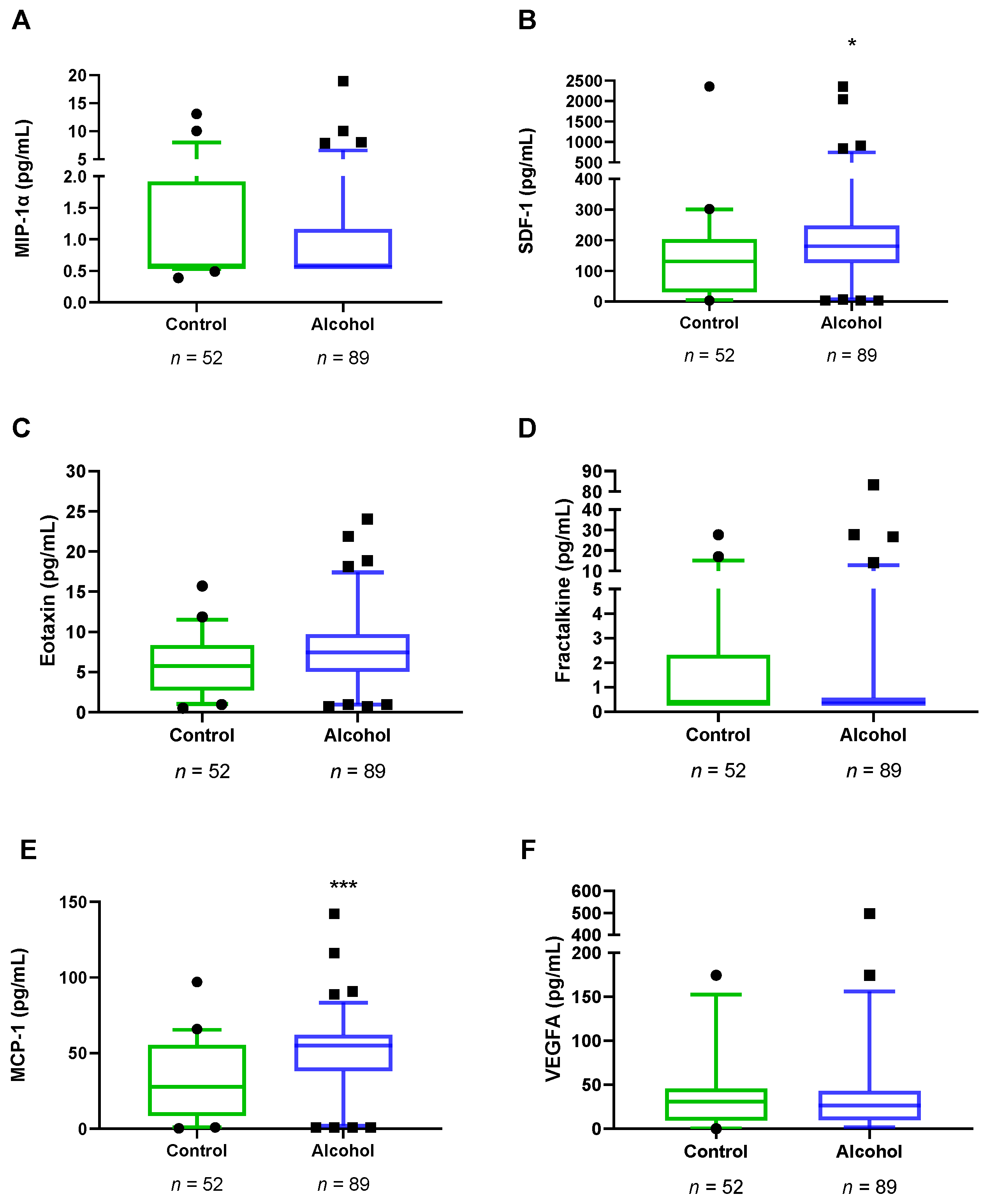
3.3. Plasma Concentrations of VEGFA and Chemokines in Abstinent AUD Patients

The impact of alcohol dependence on plasma concentrations of VEGFA and chemokines was studied in the total sample using Mann–Whitney U-test. Plasma concentrations of SDF-1 (U = 1615, p = 0.010) and MCP-1 (U = 1354, p < 0.001) were significantly higher in the alcohol group compared to the control group (Figure 2). However, we did not observe major differences in MIP-1α, eotaxine, fractalkine and VEGFA between the alcohol group and the control group (Table S1).
Moreover, correlation analysis between plasma concentrations of VEGFA and chemokines and age at first alcohol use, age at onset of AUD, length of AUD diagnosis, severity criteria, and length of abstinence were conducted in these AUD patients. Plasma levels of SDF-1, eotaxin and VEGFA were found to be significantly correlated with alcohol addiction severity based on alcohol criteria (rho = 0.211, p < 0.048; rho = 0.250, p < 0.018; rho = 0.234, p < 0.027, respectively) (Table S2).

3.4. Plasma Concentrations of VEGFA and Chemokines in Abstinent AUD Patients with Frontal Cognitive Impairment

To explore the influence of frontal cognition integrity on plasma concentrations of VEGFA and chemokines, we performed a Kruskal Wallis test with “frontal cognitive impairment” (no, mild, and severe) as a factor. We did not find a significant effect of “frontal cognitive impairment” on plasma concentrations of VEGFA and chemokines (Table S3). However, as shown in Figure 3, circulant levels of VEGFA almost reached the significance (K = 5.404, p = 0.067), having higher plasma concentrations of VEGFA the AUD patients with severe frontal cognitive impairment than those without frontal deficits (U = 72.50, p = 0.021).

3.5. Correlation Analyses between Frontal Cognition and Plasma Concentrations of VEGFA and Chemokines in AUD Patients

For deeper analysis, we explored the relationship between frontal lobe functions (evaluated by FAB) and plasma concentrations of VEGFA and chemokines using Spearman correlations (rho). As shown in Table 3, there was a significant and negative correlation between FAB score and plasma concentrations of VEGFA (rho = −0.290, p = 0.039). We also observed a significant and negative correlation between FAB score and plasma concentrations of fractalkine (rho= −0.336, p = 0.016). However, we did not find significant correlations between FAB scores and the plasma concentrations of SDF-1, eotaxin, MIP-1α and MCP-1.

3.6. Correlation Analyses between Plasma Concentrations of VEGFA and Chemokines in AUD Patients with and without Mild Cognitive Impairment

Moreover, we wanted to explore the relationship between plasma concentrations of chemokines and VEGFA depending on the integrity of frontal lobe function (evaluated by FAB) using Spearman correlations (rho). As shown in Figure 4, AUD patients with cognitive impairment displayed strong positive and significant correlations between VEGFA with all chemokines [SDF-1 (rho = 0.787, p < 0.001), eotaxin (rho = 0.678, p < 0.001), MIP-1α (rho = 0.592, p = 0.001), MCP-1 (rho = 0.601, p = 0.001), fractalkine (rho = 0.706, p < 0.001)], while we only observed a positive and significant correlation between VEGFA and SFD-1 (rho = 0.532, p = 0.009) for AUD patients without cognitive impairment (Table S4).

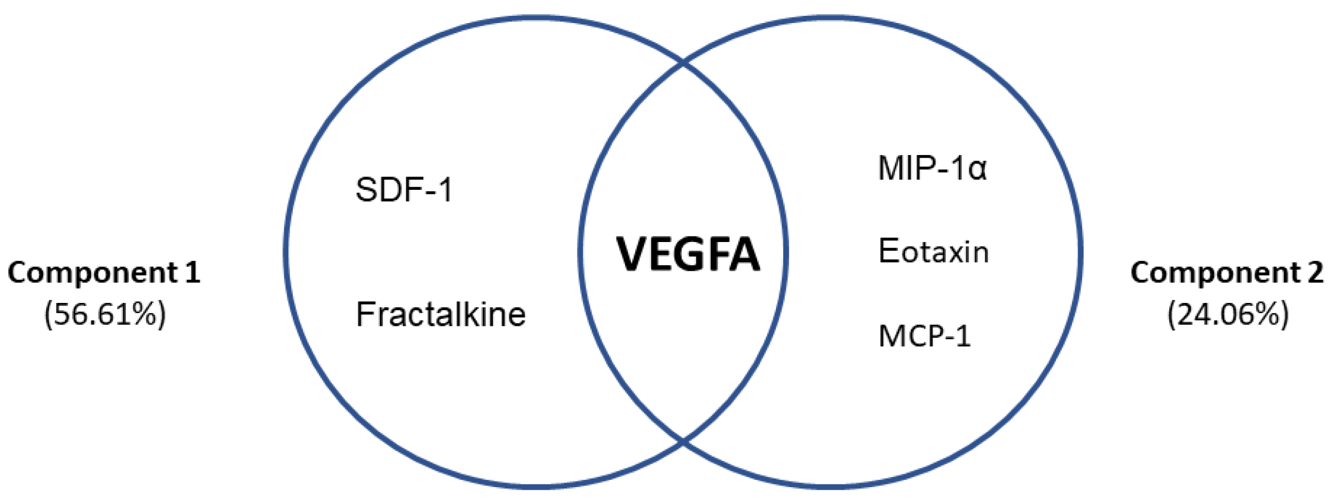
3.7. Differential Profiles Associated with VEGFA and Chemokines in AUD Patients with Frontal Cognitive Impairment

To understand the contribution of VEGFA and chemokines in the frontal cognitive decline of AUD patients, a principal component analysis was performed. Two components together explained 80.67% of the variance associated with cognitive impairment in AUD patients (Figure 5). Component 1 explained 56.61% of the total variance and was composed of SDF-1, fractalkine and VEGFA, which had high factor loads (0.984, 0.983, 0.757, respectively). Component 2 explained 24.06% of the total variance and was composed of MIP-1α, eotaxin, MCP-1 and VEGFA, which had high factor loads (0.669, 0.904, 0.807, 0.582, respectively).

3.8. Plasma Concentrations of VEGFA and Chemokines in Comorbid Medical, Psychiatric Medication and Substance Use Problems in AUD Patients

We wanted to investigate whether medical and psychiatric comorbidity, other concomitant substance use disorder or using psychotropic medication could affect plasma concentrations of VEGFA and chemokines in the alcohol group. Plasma concentrations of MIP-1α (U = 567, p = 0.002) and VEGFA (U = 552, p = 0.005) were significantly higher in AUD patients with comorbid psychiatric disorder compared to those without psychiatric comorbidity (Table 4). Moreover, plasma concentrations of MIP-1α (U = 633, p = 0.005) were significantly higher in AUD patients with comorbid substance use disorder compared to those without comorbid substance use disorder (Table S5). Regarding comorbid medical problems and the use of psychotropic medication, we did not observe major differences in plasma levels of chemokines and VEGFA (Tables S6 and S7).

3.9. Time Course of Plasma Concentrations of VEGFA after an Acute Administration of Alcohol (100 g) in Healthy Male Volunteers

To clarify how an acute intoxicating dose of alcohol affects circulating levels of VEFGA, we administered 100 g of alcohol to male healthy volunteers whose daily average alcohol intake was of 13.7 ± 8.3. Plasma samples were taken previous to (time 0) and 2, 8, and 24 h after oral ingestion of alcohol. As expected, plasma ethanol level peaked at 2 h after ingestion and decreased to a third of the 2 h concentration 8 h after the ingestion, being undetectable 24 h after oral intake (Figure 6A). Levels of VEGFA were very variable in between these subjects so a non-parametric statistics with repeated measures approach was taken. Data indicated that alcohol modified plasma VEGFA, being elevated 8 h (Figure 6B) after the ingestion of alcohol (Friedman’s statistic for n = 9, four groups, was 9.26, p < 0.03). Analysis of the percentage of change from basal values indicated the alcohol induced a 2-fold change on VEGFA circulating concentrations with respect to the basal values (Figure 6C, ANOVA repeated measures F (8,24) = 5.14, p < 0.001). Twenty-four hours after the intake of alcohol, the % of change had a non-significative average fold change of 1.7, suggesting that alcohol induced sustained increases of VEGFA. However, this assumption needs to be conclusively determined.

4. Discussion

While the association between immune signaling and neuropsychiatric disorders has been widely investigated, its link with AUD-related cognitive impairment remains poorly investigated. We lack relevant information concerning how immune mediator-induced proinflammatory states in the brain influence the neuroadaptations derived from chronic alcohol consumption, and how they impact executive functions. In the present study, we unveil a link between plasma concentrations of VEGFA and chemokines with frontal lobe dysfunction in abstinent AUD patients treated in an outpatient setting. The relevance of the present study rests on the confirmation of the link between alcohol-induced dysfunctions in modulators of the blood–brain barrier (i.e., VEGFA) and neuroinflammation (i.e., chemokines) with the presence of cognitive impairment. The main findings are as follows: (i) AUD patients had increased plasma concentrations of SDF-1 and MCP-1 compared to control subjects; (ii) there were higher circulant levels of VEGFA in AUD patients with severe frontal deficits than in those without cognitive impairment; (iii) acute administration of a heavy dose of VEGFA resulted in a delayed increase (8 h after alcohol ingestion) of plasma VEGFA concentration; (iv) the integrity of frontal lobe functions was negatively correlated with VEGFA and fractalkine; (v) plasma concentrations of VEGFA were strongly and positively correlated with all chemokines in AUD patients with frontal deficits but not in those without frontal impairment; (vi) two components together explained 80.67% of the variance associated with frontal deficits in AUD patients, with VEGFA acting as a link between all chemokines; and (vii) plasma concentrations of some chemokines changed in AUD patients with comorbid psychiatric (MIP-1α, VEGFA) and substance use (MIP-1 α) disorders.
A growing literature indicates that the pharmacodynamic action of several drugs involves changes in the neuroimmune signal [45]. Some studies have reported that alcohol-related behaviors are interrupted when the innate immune network is disturbed [46,47]. Thus, Blednov et al. (2005) showed that gene deletion of CCR2, MCP-1 or MIP-1α reduced motivational effects of alcohol consumption in mice [48]. Moreover, Steinar et al. (2019) found an increase in the plasma concentrations of IL-6, IFNγ and MCP-1 in patients with a history of chronic alcohol overconsumption [49]. In agreement with these reports, the AUD patients of the present study displayed higher plasma concentrations of SDF-1 and MCP-1 than control subjects. SDF-1 was also associated (along with eotaxin) with worse severity of alcohol addiction. Even post mortem analysis reported increased expression of MCP-1 in multiple limbic brain regions in alcoholic subjects [50]. Taking into account the high prevalence of cocaine use disorder in AUD patients, it is important to note that extensive preclinical research has reported how the chemokines SDF-1 and MCP-1 promote cocaine-related behaviors in a brain region specific manner [51,52,53]. Our group has described that severity of cocaine consumption is related to IL-1β, SDF-1 and fractalkine in cocaine use disorder patients [54]. Furthermore, we found higher plasma concentrations of fractalkine in abstinent cocaine patients with comorbid major depressive disorder than in those without this psychiatric condition [55]. In addition, we demonstrated the induction of a potent fractalkine signaling associated with cocaine-induced sensitization and extinction in mice [56].
Regarding cognitive function, 55% of the AUD patients recruited in the present study displayed some kind of frontal cognitive impairment. Accordingly, executive functions are particularly affected in AUD patients who show deficits in domains related to cognitive control, flexibility, inhibition, planning and working memory [57,58]. However, there are other neuropsychological processes that could be disrupted, including memory, emotion, and social cognition [59]. It is important to note that despite there being evidence of partial recovery of certain cognitive functions after cessation of alcohol intake [13,60], deficits in other domains may remain stable during sobriety [57,61]. Additionally, cognitive impairment can compromise efforts to initiate and maintain abstinence by impacting treatment effectiveness [62]. It is thought that increased vulnerability to alcohol-induced working memory impairment may impact in the ability to moderate alcohol consumption [63] and cognitive training could reduce the number of beverages in these patients [64]. Furthermore, our results indicate that AUD patients have low educational and occupational attainment, which has been related to worse neuropsychological performance, more indicators of neurocognitive disorders, early drug use onset and development of addiction, high-severity substance-related problems, and worse treatment outcomes [65,66,67]. Thus, excessive alcohol consumption has been linked to worse cognitive functioning in patients with low socioeconomic status operated by educational and occupational achievements [68]. Moreover, a longitudinal study has suggested that an educational level lower than high school and a low job occupation is associated with an increased risk of dementia in alcohol patients [69]. Similarly, our group has recently found that a high educational level could play a protective role in the onset, development, and progression of cocaine use disorders and could also protect against cognitive impairment caused by alcohol consumption throughout life [70,71]. Interestingly, in this study we found a negative association between the state of frontal lobe functions with two signaling molecules, VEGFA and fractalkine, involved in vascular function and neural plasticity.
VEGFA has been related to neuroprotection, neurogenesis, and synaptic plasticity mechanisms in the central nervous system through stimulation of neuronal stem cells and safeguarding the integrity of the blood–brain barrier [36]. Despite VEGFA levels having been linked to several neurodegenerative and neurological disorders [36], their effects could be time dependent. Augmented VEGFA levels in cerebrospinal fluid and plasma have been reported in Alzheimer’s disease and vascular dementia patients [72,73] probably as a consequence of hypoperfusion and hypoxia. Nevertheless, improvement in learning and memory after a bilateral carotid artery occlusion has been associated with an increase in VEGFA expression in the hippocampus, which suggests that VEGFA signaling could compensate for cognitive impairment [74,75]. Similarly, partial increases in VEGFA stimulate vasodilation, angiogenesis and neuroprotection mechanisms, which are beneficial for the brain in later stages after cerebral ischemia [76]. However, early VEGFA increases may lead to undesirable effects in cerebral ischemia, such as an increase in blood–brain barrier permeability and infiltration of immune cells inducing neuroinflammation and edema [77,78,79].
With reference to the latter, in the present study we observed that increases in VEGFA were associated with worse severity of alcohol addiction, severe frontal deficits and the elevation of all chemokines in frontal cognitive impaired AUD patients. Interestingly, in a pilot study with healthy male volunteers with a history of moderate alcohol consumption, we found that acute alcohol administration resulted in a delayed increase in plasma VEGFA, observed 8 h after alcohol intake. Because of the increased vascular permeability induced by VEGFA, it is reasonable to think that this action of alcohol might facilitate neuroinflammation by opening the blood–brain barrier to pro-inflammatory signals originating in peripheral tissues, especially in the intestine. Supporting this hypothesis, in our principal component analysis, we found that the interaction of chemokines and VEGFA explained the 80.67% of the variance associated with frontal deficits in AUD patients, observing that VEGFA has an essential role as a factor interacting with the pro-inflammatory immune response associated with alcohol consumption. Consistent with our results, using cellular and animal models, Muneer et al. (2012) found that chronic alcohol exposure disrupts the blood–brain barrier across degradation of endothelial VEGF receptor 2. This also increases circulating levels of VEGFA leading to neuronal death and inflammation in the brain [80]. Moreover, Louboutin et al. (2012) reported increases in VEGFA levels and blood vessel density in cerebral tissue within two weeks of the onset of ethanol consumption in rats [81]. Moreover, despite VEGFA not being an inflammatory cytokine, it can activate nuclear factor-enhancer of activated B-cell kappa light chains (NF-KB) and nuclear factor of activated T-cells (NFAT) signaling cascades that could promote a chemotaxis response involved in the angiogenic process (although this role remains unknown) [82]. Thus, our results in AUD patients with frontal deficits might suggest two things: (1) chronic alcohol abuse might lead to alterations in the concentrations of VEGFA that increase the permeability of the blood–brain barrier, leading to infiltration of immune cells and inflammation in the brain [80,81], and/or (2) under the presence of hypoperfusion and hypoxia as a result of alcohol-derived brain damage, concentrations of VEGFA might ultimately increase as a compensatory signal in order to form new blood vessels and recruit chemokines to the affected brain area. Lastly, it has been found that higher circulant levels of VEGFA in major depression and its alterations are related to impaired cognitive function in schizophrenia [83,84]. This might explain why psychiatric comorbidity affected plasma concentrations of VEGFA in our study. It is important to note that cognitive deficits found in patients with addictions can often be exacerbated by comorbid psychiatric disorders [85].
Lastly, previous studies have reported that fractalkine (CX3CL1) develops an essential role in the neuronal–microglial intercommunication [86]. This chemokine is expressed from brain neurons that control activation of microglia through its binding receptor CX3CR1. In harmful conditions, neurons release fractalkine in order to stimulate proliferation, activation and migration at the site of the brain injury [87,88]. CX3CL1-KO mice showed altered microglial function and neurotoxicity following LPS injection as well as more neuronal damage in Parkinson’s disease and amyotrophic lateral sclerosis [88]. In accordance with this, Sokolowki et al. (2014) found that CX3CL1-KO mice revealed signals of neuronal apoptosis after ethanol treatment, suggesting a role in the clearance of those apoptotic cells [86]. Moreover, additional studies have reported that mild–moderate Alzheimer’s disease patients had higher plasma levels of CX3CL1 than those with severe Alzheimer’s disease [89,90]. These results suggest that fractalkine and CX3CR1 signaling might act as a neuroprotective mechanism through the microglial activity modulation in the early stage of brain injury while this signal seems to disappear when neuronal damage is established [91]. This may indicate that the AUD patients in this study are actively fighting against cognitive impairment.

5. Conclusions, Limitations, and Future Perspectives

In conclusion, a lifetime of chronic alcohol consumption leads to a proinflammatory systemic condition revealed by enhanced circulating chemokines, and to frontal cognitive impairment. The trophic factor VEGFA appears to be a relevant contributor to alcohol-associated neuroinflammation, probably through its role on controlling blood–brain barrier permeability, ultimately leading to impaired cognition. In addition, fractalkine could act as a signal of brain damage in early stages of cognitive impairment. Potential biomarkers could be useful and reliable tools in patients with AUD for confirming the diagnosis, defining the current stage of the AUD, and diagnosing these patients early.
Nevertheless, this study has several limitations that should be taken into account in future research. First, we do not know the time course of the effects of alcohol on these chemokines, either after acute or chronic alcohol consumption, nor its alterations in early or extended abstinence. We must also investigate whether sociodemographic variables, especially educational level, time of alcohol consumption, age of alcohol drinking initiation, etc., might contribute to the cognitive performance in both control and AUD patients. Finally, we lack significant representation of the female population, which precludes investigation of sex differences in chemokines and VEGFA. However, the data obtained clearly point to the need of considering these immunoinflammatory signals and trophic factors as relevant biomarkers of AUD-associated complications. Moreover, this concept should be extended to the analysis of immunomodulators capable of activating chronic inflammation. There are several biochemical pathways affected by alcohol consumption that need to be considered under the light of the present discoveries. For example, the tryptophan/kynurenine pathway is a potent immunomodulatory system that can modify inflammation, learning and memory [92,93]. The intestinal microbiota (which is determinant for AUD) [94] participates in these pathways, modifying the presence of pro- and anti-inflammatory mediators and eventually growth factors.
As a future perspective, we need to integrate all the information related to this multiplicity of inflammatory signals in a single model of alcohol addiction. Although certain factors such as VEGFA or fractalkine may contribute to important aspects of alcohol addiction, the complexity of the interactions of these inflammatory signaling proteins goes beyond our current technique and knowledge. Further clinical and technological research is necessary to elucidate the role of these factors in the etiology of AUD and associated comorbidities.

Supplementary Materials

The following supporting information can be downloaded at: https://www.mdpi.com/article/10.3390/biomedicines10050947/s1, Table S1: Plasma concentrations of chemokines and VEGFA in AUD group versus control subjects; Table S2: Correlation analysis between FAB score and chemokines in AUD patients; Table S3: Correlation analysis between plasma concentrations of chemokines and VEGFA in AUD patients with and without cognitive impairment; Table S4: Plasma concentrations of chemokines and VEGFA grouped according to comorbid psychiatric disorder; Table S5: Plasma concentrations of chemokines and VEGFA grouped according to comorbid substance use disorder; Table S6: Plasma concentrations of chemokines and VEGFA grouped according to comorbid medical problem; Table S7: Plasma concentrations of chemokines and VEGFA grouped according to the use of psychotropic medication last year.

Author Contributions

Conception and design: J.S., F.R.d.F. and P.A. Data acquisition: N.R.-O., P.A., M.F.-L., N.G.-M., E.P. and J.J.R. Data analysis and interpretation: N.R.-O., P.A., N.G.-M., F.J.P.-M., E.P. and M.F. Draft writing: N.R.-O., J.S., F.R.d.F. and A.S. Review and editing: P.A., J.J.R., J.O.-P., A.S., F.J.P.-M. and M.F. Final approval: All authors. All authors have read and agreed to the published version of the manuscript.

Funding

RETICS Red de Trastornos Adictivos, Instituto de Salud Carlos III (ISCIII), Ministerio de Ciencia e Innovación and European Regional Development Funds—European Union (ERDF-EU) (RD16/0017/0001 and RD16/0017/003); ISCIII, ERDF-EU (PI20/01399, PI19/01577, PI19/00886); Ministerio de Sanidad, Delegación de Gobierno para el Plan Nacional sobre Drogas (PND 2020I048, PND 2019I040, PND 2018I044, PND 2018I033, PND 2016I024 and PND 2018I037) and Consejería de Salud y Familia, Junta de Andalucía (Neuro-RECA, RIC-0111-2019). F.J.P.-M. (CPII19/00022) and A.S. (CPII19/00031) hold “Miguel Servet II” research contracts from the National System of Health, ISCIII, ERDF-EU. F.J.P.-M. also holds a “Nicolas Monardes” contract from Servicio Andaluz de Salud, Consejería de Salud y Familia, Junta de Andalucía (C1-0049-2019). P.A. has a research contract (UMA-FEDERJA-076) funded by the Ministry of Economy and Knowledge—Regional Government of Andalucía and ERDF-EU. “PFIS” Predoctoral research contract (FI18/00249) financed by ISCIII ERDF/ESF and NGM has a ‘Sara Borrell’ Research Contract (CD19/00019) financed by ISCIII and ERDF-EU. The funding sources had no further role in study design; in the collection, analysis, and interpretation of data; in writing of the report; and in the decision to submit the paper for publication.

Institutional Review Board Statement

The study was conducted in accordance with the Declaration of Helsinki and approved by the Ethics Committee of the Portal de Ética de la Investigación Biomédica de Andalucía-PEIBA (Consejería de Salud y Familias, Junta de Andalucía) and Hospital Regional Universitario de Málaga (PND2018I033). The acute alcohol administration experiment protocol was approved by the local Human Research Ethics Committee (CEI Hospital Universitari Germans Trias i Pujol, Badalona, Spain) and registered at ClinicalTrials.gov (NCT02232789). The clinical trial was conducted in accordance with the Declaration of Helsinki and Spanish laws concerning clinical research.

Informed Consent Statement

Informed consent was obtained from all subjects involved in the study.

Data Availability Statement

Not applicable.

Conflicts of Interest

The authors declare no conflict of interest. The funders had no role in the design of the study; in the collection, analyses, or interpretation of data; in the writing of the manu-script, or in the decision to publish the results.

References

  1. World Health Organization. Global Status Report on Alcohol and Health 2018; World Health Organization: Geneva, Switzerland, 2019. [Google Scholar]
  2. OEDA. Observatorio Español de las Drogas y las Adicciones (OEDA) [2017]. Informe Anual 2017. Informe EDADES 2017. Plan Nacional Sobre Drogas. 2019. Available online: https://pnsd.sanidad.gob.es/profesionales/sistemasInformacion/home.htm (accessed on 23 February 2022).
  3. Louvet, A.; Mathurin, P. Alcoholic liver disease: Mechanisms of injury and targeted treatment. Nat. Rev. Gastroenterol. Hepatol. 2015, 12, 231–242. [Google Scholar] [CrossRef] [PubMed]
  4. Setiawan, V.W.; Monroe, K.; Lugea, A.; Yadav, D.; Pandol, S. Uniting Epidemiology and Experimental Disease Models for Alcohol-Related Pancreatic Disease. Alcohol Res. Curr. Rev. 2017, 38, 173–182. [Google Scholar]
  5. Marchena, N.G.; Araos, P.; Pavón, F.J.; Ponce, G.; Pedraz, M.; Serrano, A.; Arias, F.; Romero-Sanchiz, P.; Suárez, J.; Pastor, A.; et al. Psychiatric comorbidity and plasma levels of 2-acyl-glycerols in outpatient treatment alcohol users. Analysis of gender differences. Adicciones 2016, 29, 83. [Google Scholar] [CrossRef] [Green Version]
  6. Boden, J.; Fergusson, D.M. Alcohol and depression. Addiction 2011, 106, 906–914. [Google Scholar] [CrossRef]
  7. Samet, S.; Fenton, M.C.; Nunes, E.; Greenstein, E.; Aharonovich, E.; Hasin, D. Effects of independent and substance-induced major depressive disorder on remission and relapse of alcohol, cocaine and heroin dependence. Addiction 2013, 108, 115–123. [Google Scholar] [CrossRef]
  8. Schwarzinger, M.; Pollock, B.G.; Hasan, O.S.M.; Dufouil, C.; Rehm, J.; Baillot, S.; Guibert, Q.; Planchet, F.; Luchini, S. Contribution of alcohol use disorders to the burden of dementia in France 2008-13: A nationwide retrospective cohort study. Lancet Public Health 2018, 3, e124–e132. [Google Scholar] [CrossRef]
  9. Withall, A.; Draper, B.; Seeher, K.; Brodaty, H. The prevalence and causes of younger onset dementia in Eastern Sydney, Australia. Int. Psychogeriatr. 2014, 26, 1955–1965. [Google Scholar] [CrossRef]
  10. Cheng, C.; Huang, C.-L.; Tsai, C.-J.; Chou, P.-H.; Lin, C.-C.; Chang, C.-K. Alcohol-Related Dementia: A Systemic Review of Epidemiological Studies. J. Psychosom. Res. 2017, 58, 331–342. [Google Scholar] [CrossRef]
  11. Woods, A.J.; Porges, E.C.; Bryant, V.E.; Seider, T.; Gongvatana, A.; Kahler, C.W.; De La Monte, S.; Monti, P.M.; Cohen, R.A. Current Heavy Alcohol Consumption is Associated with Greater Cognitive Impairment in Older Adults. Alcohol. Clin. Exp. Res. 2016, 40, 2435–2444. [Google Scholar] [CrossRef] [Green Version]
  12. Hayes, V.; Demirkol, A.; Ridley, N.; Withall, A.; Draper, B. Alcohol-related cognitive impairment: Current trends and future perspectives. Neurodegener. Dis. Manag. 2016, 6, 509–523. [Google Scholar] [CrossRef]
  13. Ros-Cucurull, E.; Palma-Álvarez, R.F.; Cardona-Rubira, C.; García-Raboso, E.; Jacas, C.; Grau-López, L.; Abad, A.C.; Rodríguez-Cintas, L.; Ros-Montalbán, S.; Casas, M.; et al. Alcohol use disorder and cognitive impairment in old age patients: A 6 months follow-up study in an outpatient unit in Barcelona. Psychiatry Res. 2018, 261, 361–366. [Google Scholar] [CrossRef]
  14. Horton, L.; Duffy, T.; Martin, C. Neurocognitive, psychosocial and functional status of individuals with alco-hol-related brain damage (ARBD) on admission to specialist residential care. Drugs Educ. Prev. Policy 2015, 22, 416–427. [Google Scholar] [CrossRef] [Green Version]
  15. Montesinos, J.; Pascual, M.; Pla, A.; Maldonado, C.; Rodríguez-Arias, M.; Miñarro, J.; Guerri, C. TLR4 elimination prevents synaptic and myelin alterations and long-term cognitive dysfunctions in adolescent mice with intermittent ethanol treatment. Brain Behav. Immun. 2015, 45, 233–244. [Google Scholar] [CrossRef]
  16. Dwivedi, D.K.; Kumar, D.; Kwatra, M.; Pandey, S.N.; Choubey, P.; Lahkar, M.; Jangra, A. Voluntary alcohol consumption exacerbated high fat diet-induced cognitive deficits by NF-κB-calpain dependent apoptotic cell death in rat hippocampus: Ameliorative effect of melatonin. Biomed. Pharmacother. 2018, 108, 1393–1403. [Google Scholar] [CrossRef] [PubMed]
  17. Yang, J.-Y.; Xue, X.; Tian, H.; Wang, X.-X.; Dong, Y.-X.; Wang, F.; Zhao, Y.-N.; Yao, X.-C.; Cui, W.; Wu, C.-F. Role of microglia in ethanol-induced neurodegenerative disease: Pathological and behavioral dysfunction at different developmental stages. Pharmacol. Ther. 2014, 144, 321–337. [Google Scholar] [CrossRef] [PubMed]
  18. Banks, W.A. Peptides and the blood-brain barrier. Peptides 2015, 72, 16–19. [Google Scholar] [CrossRef] [Green Version]
  19. Pascual, M.; Baliño, P.; Aragón, C.M.; Guerri, C. Cytokines and chemokines as biomarkers of ethanol-induced neuroinflammation and anxiety-related behavior: Role of TLR4 and TLR2. Neuropharmacology 2015, 89, 352–359. [Google Scholar] [CrossRef] [PubMed]
  20. Yen, C.-H.; Ho, P.-S.; Yeh, Y.-W.; Liang, C.-S.; Kuo, S.-C.; Huang, C.-C.; Chen, C.-Y.; Shih, M.-C.; Ma, K.-H.; Sung, Y.-F.; et al. Differential cytokine levels between early withdrawal and remission states in patients with alcohol dependence. Psychoneuroendocrinology 2017, 76, 183–191. [Google Scholar] [CrossRef] [PubMed]
  21. Rostène, W.; Kitabgi, P.; Parsadaniantz, S.M. Chemokines: A new class of neuromodulator? Nat. Rev. Neurosci. 2007, 8, 895–903. [Google Scholar] [CrossRef]
  22. Watson, A.E.S.; Goodkey, K.; Footz, T.; Voronova, A. Regulation of CNS precursor function by neuronal chemokines. Neurosci. Lett. 2020, 715, 134533. [Google Scholar] [CrossRef]
  23. Comerford, I.; McColl, S.R. Mini-review series: Focus on chemokines. Immunol. Cell Biol. 2011, 89, 183–184. [Google Scholar] [CrossRef] [PubMed]
  24. Palomino, D.C.T.; Marti, L.C. Chemokines and immunity. Einstein (São Paulo) 2015, 13, 469–473. [Google Scholar] [CrossRef] [PubMed] [Green Version]
  25. Cui, C.; Shurtleff, D.; Harris, R.A. Neuroimmune Mechanisms of Alcohol and Drug Addiction. Int. Rev. Neurobiol. 2014, 118, 1–12. [Google Scholar] [CrossRef] [PubMed] [Green Version]
  26. Lacagnina, M.; Rivera, P.D.; Bilbo, S.D. Glial and Neuroimmune Mechanisms as Critical Modulators of Drug Use and Abuse. Neuropsychopharmacology 2017, 42, 156–177. [Google Scholar] [CrossRef] [PubMed] [Green Version]
  27. Ivanovska, M.; Abdi, Z.; Murdjeva, M.; Macedo, D.; Maes, A.; Maes, M. CCL-11 or Eotaxin-1: An Immune Marker for Ageing and Accelerated Ageing in Neuro-Psychiatric Disorders. Pharmaceuticals 2020, 13, 230. [Google Scholar] [CrossRef]
  28. Wang, R.; Li, H. A focus on CXCR4 in Alzheimer’s disease. Brain Circ. 2017, 3, 199–203. [Google Scholar] [CrossRef]
  29. Stuart, M.; Baune, B. Chemokines and chemokine receptors in mood disorders, schizophrenia, and cognitive impairment: A systematic review of biomarker studies. Neurosci. Biobehav. Rev. 2014, 42, 93–115. [Google Scholar] [CrossRef]
  30. Teixeira, A.L.; Gama, C.S.; Rocha, N.P.; Teixeira, M.M. Revisiting the role of eotaxin-1/CCL11 in psychiatric disorders. Front. Psychiatry 2018, 9, 1–6. [Google Scholar] [CrossRef]
  31. Wei, Z.; Chen, L.; Zhang, J.; Cheng, Y. Aberrations in peripheral inflammatory cytokine levels in substance use disorders: A meta-analysis of 74 studies. Addiction 2020, 115, 2257–2267. [Google Scholar] [CrossRef]
  32. Achur, R.N.; Freeman, W.M.; Vrana, K.E. Circulating Cytokines as Biomarkers of Alcohol Abuse and Alcoholism. J. Neuroimmune Pharmacol. 2010, 5, 83–91. [Google Scholar] [CrossRef] [Green Version]
  33. Qin, L.; He, J.; Hanes, R.N.; Pluzarev, O.; Hong, J.-S.; Crews, F.T. Increased systemic and brain cytokine production and neuroinflammation by endotoxin following ethanol treatment. J. Neuroinflammation 2008, 5, 10. [Google Scholar] [CrossRef] [PubMed] [Green Version]
  34. Gao, B.; Xu, M. Chemokines and alcoholic hepatitis: Are chemokines good therapeutic targets? Gut 2014, 63, 1683–1684. [Google Scholar] [CrossRef] [PubMed] [Green Version]
  35. McClain, C.J.; Shedlofsky, S.; Barve, S.; Hill, D.B. Cytokines and alcoholic liver disease. Alcohol Health Res. World 1997, 21, 317–320. [Google Scholar] [PubMed]
  36. Lange, C.; Storkebaum, E.; de Almodovar, C.R.; Dewerchin, M.; Carmeliet, P. Vascular endothelial growth factor: A neurovascular target in neurological diseases. Nat. Rev. Neurol. 2016, 12, 439–454. [Google Scholar] [CrossRef] [PubMed]
  37. Klintsova, A.Y.; Hamilton, G.F.; Boschen, K.E. Long-Term Consequences of Developmental Alcohol Exposure on Brain Structure and Function: Therapeutic Benefits of Physical Activity. Brain Sci. 2012, 3, 1–38. [Google Scholar] [CrossRef] [Green Version]
  38. Bates, D.O. Vascular endothelial growth factors and vascular permeability. Cardiovasc. Res. 2010, 87, 262–271. [Google Scholar] [CrossRef] [Green Version]
  39. Tammela, T.; Enholm, B.; Alitalo, K.; Paavonen, K. The biology of vascular endothelial growth factors. Cardiovasc. Res. 2005, 65, 550–563. [Google Scholar] [CrossRef]
  40. Heberlein, A.; Muschler, M.; Lenz, B.; Frieling, H.; Büchl, C.; Gröschl, M.; Riera, R.; Kornhuber, J.; Bleich, S.; Hillemacher, T. Serum levels of vascular endothelial growth factor a increase during alcohol withdrawal. Addict. Biol. 2010, 15, 362–364. [Google Scholar] [CrossRef]
  41. Kasztelan-Szczerbinska, B.; Surdacka, A.; Slomka, M.; Rolinski, J.M.; Celinski, K.; Cichoz-Lach, H.; Madro, A.; Szczerbinski, M. Angiogenesis-Related Biomarkers in Patients with Alcoholic Liver Disease: Their Association with Liver Disease Complications and Outcome. Mediat. Inflamm. 2014, 2014, 673032. [Google Scholar] [CrossRef]
  42. Torrens, M.; Serrano, D.; Astals, M.; Pérez-Domínguez, G.; Martín-Santos, R. Diagnosing Comorbid Psychiatric Disorders in Substance Abusers: Validity of the Spanish Versions of the Psychiatric Research Interview for Substance and Mental Disorders and the Structured Clinical Interview for DSM-IV. Am. J. Psychiatry 2004, 161, 1231–1237. [Google Scholar] [CrossRef] [Green Version]
  43. Hasin, D.S.; O’Brien, C.P.; Auriacombe, M.; Borges, G.; Bucholz, K.; Budney, A.; Compton, W.M.; Crowley, T.; Ling, W.; Petry, N.M.; et al. DSM-5 Criteria for Substance Use Disorders: Recommendations and Rationale. Am. J. Psychiatry 2013, 170, 834–851. [Google Scholar] [CrossRef] [PubMed] [Green Version]
  44. Del Alamo, A.R.; Alonso, M.J.C.; Marín, L.C. FAB: A preliminar Spanish application of the frontal assessment battery to 11 groups of patients. Revista de Neurología 2003, 36, 605–608. [Google Scholar]
  45. Ahearn, O.C.; Watson, M.N.; Rawls, S.M. Chemokines, cytokines and substance use disorders. Drug Alcohol Depend. 2021, 220, 108511. [Google Scholar] [CrossRef] [PubMed]
  46. Blednov, Y.; Benavidez, J.; Geil, C.; Perra, S.; Morikawa, H.; Harris, R. Activation of inflammatory signaling by lipopolysaccharide produces a prolonged increase of voluntary alcohol intake in mice. Brain Behav. Immun. 2011, 25, S92–S105. [Google Scholar] [CrossRef] [PubMed] [Green Version]
  47. Blednov, Y.A.; Ponomarev, I.; Geil, C.; Bergeson, S.; Koob, G.F.; Harris, R.A. Neuroimmune regulation of alcohol consumption: Behavioral validation of genes obtained from genomic studies. Addict. Biol. 2012, 17, 108–120. [Google Scholar] [CrossRef] [Green Version]
  48. Blednov, Y.A.; Bergeson, S.E.; Walker, D.; Ferreira, V.M.; Kuziel, W.A.; Harris, R.A. Perturbation of chemokine networks by gene deletion alters the reinforcing actions of ethanol. Behav. Brain Res. 2005, 165, 110–125. [Google Scholar] [CrossRef] [Green Version]
  49. Bjørkhaug, S.T.; Neupane, S.P.; Bramness, J.G.; Aanes, H.; Skar, V.; Medhus, A.W.; Valeur, J. Plasma cytokine levels in patients with chronic alcohol overconsumption: Relations to gut microbiota markers and clinical correlates. Alcohol 2020, 85, 35–40. [Google Scholar] [CrossRef]
  50. He, J.; Crews, F.T. Increased MCP-1 and microglia in various regions of the human alcoholic brain. Exp. Neurol. 2008, 210, 349–358. [Google Scholar] [CrossRef] [Green Version]
  51. Kim, J.; Connelly, K.L.; Unterwald, E.M.; Rawls, S.M. Chemokines and cocaine: CXCR4 receptor antagonist AMD3100 attenuates cocaine place preference and locomotor stimulation in rats. Brain, Behav. Immun. 2017, 62, 30–34. [Google Scholar] [CrossRef] [Green Version]
  52. Trecki, J.; Unterwald, E. Modulation of cocaine-induced activity by intracerebral administration of CXCL12. Neuroscience 2009, 161, 13–22. [Google Scholar] [CrossRef] [Green Version]
  53. Trocello, J.M.; Rostène, W.; Melik-Parsadaniantz, S.; Godefroy, D.; Roze, E.; Kitabgi, P.; Kuziel, W.A.; Chalon, S.; Caboche, J.; Apartis, E. Implication of CCR2 Chemokine Receptor in Cocaine-Induced Sensitization. J. Mol. Neurosci. 2011, 44, 147–151. [Google Scholar] [CrossRef] [PubMed]
  54. Araos, P.; Pedraz, M.; Serrano, A.; Lucena, M.; Barrios, V.; García-Marchena, N.; Campos-Cloute, R.; Ruiz, J.J.; Romero, P.; Suárez, J.; et al. Plasma profile of pro-inflammatory cytokines and chemokines in cocaine users under outpatient treatment: Influence of cocaine symptom severity and psychiatric co-morbidity. Addict. Biol. 2014, 20, 756–772. [Google Scholar] [CrossRef] [PubMed]
  55. García-Marchena, N.; Barrera, M.; Mestre-Pintó, J.I.; Araos, P.; Serrano, A.; Pérez-Mañá, C.; Papaseit, E.; Fonseca, F.; Ruiz, J.J.; De Fonseca, F.R.; et al. Inflammatory mediators and dual depression: Potential biomarkers in plasma of primary and substance-induced major depression in cocaine and alcohol use disorders. PLoS ONE 2019, 14, e0213791. [Google Scholar] [CrossRef] [PubMed] [Green Version]
  56. Montesinos, J.; Castilla-Ortega, E.; Sánchez-Marín, L.; Montagud-Romero, S.; Araos, P.; Pedraz, M.; Porras-Perales, Ó.; García-Marchena, N.; Serrano, A.; Suárez, J.; et al. Cocaine-induced changes in CX3CL1 and inflammatory signaling pathways in the hippocampus: Association with IL1β. Neuropharmacology 2020, 162, 107840. [Google Scholar] [CrossRef]
  57. Romero-Martínez, Á.; Vitoria-Estruch, S.; Moya-Albiol, L. Perfil cognitivo de los alcohólicos abstinentes durante un periodo de tiempo prolongado en comparación con un grupo de hombres que no consumen alcohol. Adicciones 2018, 32, 19–31. [Google Scholar] [CrossRef]
  58. Joos, L.; Schmaal, L.; Goudriaan, A.E.; Fransen, E.; Brink, W.V.D.; Sabbe, B.G.C.; Dom, G. Age of Onset and Neuropsychological Functioning in Alcohol Dependent Inpatients. Alcohol. Clin. Exp. Res. 2013, 37, 407–416. [Google Scholar] [CrossRef]
  59. Freeman, C.R.; Wiers, C.E.; Sloan, M.E.; Zehra, A.; Ramirez, V.; Wang, G.-J.; Volkow, N.D. Emotion Recognition Biases in Alcohol Use Disorder. Alcohol. Clin. Exp. Res. 2018, 42, 1541–1547. [Google Scholar] [CrossRef]
  60. Kopera, M.; Wojnar, M.; Brower, K.; Glass, J.; Nowosad, I.; Gmaj, B.; Szelenberger, W. Cognitive functions in abstinent alcohol-dependent patients. Alcohol 2012, 46, 665–671. [Google Scholar] [CrossRef]
  61. Sachdeva, A.; Chandra, M.; Choudhary, M.; Dayal, P.; Anand, K.S. Alcohol-Related Dementia and Neurocognitive Impairment: A Review Study. Int. J. High Risk Behav. Addict. 2016, 5, e27976. [Google Scholar] [CrossRef] [Green Version]
  62. Le Berre, A.-P.; Fama, R.; Sullivan, E.V. Executive Functions, Memory, and Social Cognitive Deficits and Recovery in Chronic Alcoholism: A Critical Review to Inform Future Research. Alcohol. Clin. Exp. Res. 2017, 41, 1432–1443. [Google Scholar] [CrossRef]
  63. Lechner, W.V.; Day, A.M.; Metrik, J.; Leventhal, A.M.; Kahler, C.W. Effects of alcohol-induced working memory decline on alcohol consumption and adverse consequences of use. Psychopharmacology 2015, 233, 83–88. [Google Scholar] [CrossRef] [PubMed] [Green Version]
  64. Khemiri, L.; Brynte, C.; Stunkel, A.; Klingberg, T.; Jayaram-Lindstrom, N. Working Memory Training in Alcohol Use Disorder: A Randomized Controlled Trial. Alcohol. Clin. Exp. Res. 2019, 43, 135–146. [Google Scholar] [CrossRef] [PubMed]
  65. Toledo-Fernández, A.; Marín-Navarrete, R.; Villalobos-Gallegos, L.; Salvador-Cruz, J.; Benjet, C.; Roncero, C. Testing whether cognitive reserve as measured by self-rating of stimulating activities moderates the association of polysubstance use and neurocognitive disorder. Cogn. Neuropsychiatry 2019, 24, 421–433. [Google Scholar] [CrossRef] [PubMed]
  66. Toledo-Fernández, A.; Villalobos-Gallegos, L.; Salvador-Cruz, J.; Benjet, C.; Roncero, C.; Marín-Navarrete, R. Differential Effects of Cognitive Reserve on the Neurocognitive Functioning of Polysubstance Users: An Exploratory Analysis Using Mixture Regression. Int. J. Ment. Health Addict. 2019, 18, 500–514. [Google Scholar] [CrossRef]
  67. Cutuli, D.; De Guevara-Miranda, D.L.; Castilla-Ortega, E.; Santín, L.; Sampedro-Piquero, P. Highlighting the Role of Cognitive and Brain Reserve in the Substance use Disorder Field. Curr. Neuropharmacol. 2019, 17, 1056–1070. [Google Scholar] [CrossRef]
  68. Sabia, S.; Guéguen, A.; Berr, C.; Berkman, L.; Ankri, J.; Goldberg, M.; Zins, M.; Singh-Manoux, A. High alcohol consumption in middle-aged adults is associated with poorer cognitive performance only in the low socio-economic group. Results from the GAZEL cohort study. Addiction 2011, 106, 93–101. [Google Scholar] [CrossRef] [Green Version]
  69. Sabia, S.; Fayosse, A.; Dumurgier, J.; Dugravot, A.; Akbaraly, T.; Britton, A.; Kivimaki, M.; Singh-Manoux, A. Alcohol consumption and risk of dementia: 23 year follow-up of Whitehall II cohort study. BMJ 2018, 362, k2927. [Google Scholar] [CrossRef] [Green Version]
  70. Requena-Ocaña, N.; Flores-Lopez, M.; Martín, A.S.; García-Marchena, N.; Pedraz, M.; Ruiz, J.J.; Serrano, A.; Suarez, J.; Pavón, F.J.; de Fonseca, F.R.; et al. Influence of gender and education on cocaine users in an outpatient cohort in Spain. Sci. Rep. 2021, 11, 20928. [Google Scholar] [CrossRef]
  71. Requena-Ocaña, N.; Araos, P.; Flores, M.; García-Marchena, N.; Silva-Peña, D.; Aranda, J.; Rivera, P.; Ruiz, J.J.; Serrano, A.; Pavón, F.J.; et al. Evaluation of neurotrophic factors and education level as predictors of cognitive decline in alcohol use disorder. Sci. Rep. 2021, 11, 15583. [Google Scholar] [CrossRef]
  72. Tarkowski, E.; Issa, R.; Sjögren, M.; Wallin, A.; Blennow, K.; Tarkowski, A.; Kumar, P. Increased intrathecal levels of the angiogenic factors VEGF and TGF-β in Alzheimer’s disease and vascular dementia. Neurobiol. Aging 2002, 23, 237–243. [Google Scholar] [CrossRef]
  73. Chiappelli, M.; Borroni, B.; Archetti, S.; Calabrese, E.; Corsi, M.M.; Franceschi, M.; Padovani, A.; Licastro, F. VEGF Gene and Phenotype Relation with Alzheimer’s Disease and Mild Cognitive Impairment. Rejuvenation Res. 2006, 9, 485–493. [Google Scholar] [CrossRef]
  74. Zhang, N.; Xing, M.; Wang, Y.; Liang, H.; Yang, Z.; Shi, F.; Cheng, Y. Hydroxysafflor yellow A improves learning and memory in a rat model of vascular dementia by increasing VEGF and NR1 in the hippocampus. Neurosci. Bull. 2013, 30, 417–424. [Google Scholar] [CrossRef] [PubMed] [Green Version]
  75. Zhang, N.; Xing, M.; Wang, Y.; Tao, H.; Cheng, Y. Repetitive transcranial magnetic stimulation enhances spatial learning and synaptic plasticity via the VEGF and BDNF-NMDAR pathways in a rat model of vascular dementia. Neuroscience 2015, 311, 284–291. [Google Scholar] [CrossRef] [PubMed]
  76. de Almodovar, C.R.; Lambrechts, D.; Mazzone, M.; Carmeliet, P. Role and Therapeutic Potential of VEGF in the Nervous System. Physiol. Rev. 2009, 89, 607–648. [Google Scholar] [CrossRef] [PubMed]
  77. Argaw, A.T.; Gurfein, B.T.; Zhang, Y.; Zameer, A.; John, G.R. VEGF-mediated disruption of endothelial CLN-5 promotes blood-brain barrier breakdown. Proc. Natl. Acad. Sci. USA 2009, 106, 1977–1982. [Google Scholar] [CrossRef] [PubMed] [Green Version]
  78. Chi, O.Z.; Hunter, C.; Liu, X.; Tan, T.; Weiss, H.R. Effects of VEGF on the Blood-Brain Barrier Disruption Caused by Hyperosmolarity. Pharmacology 2008, 82, 187–192. [Google Scholar] [CrossRef]
  79. Argaw, A.T.; Asp, L.; Zhang, J.; Navrazhina, K.; Pham, T.; Mariani, J.N.; Mahase, S.; Dutta, D.J.; Seto, J.; Kramer, E.G.; et al. Astrocyte-derived VEGF-A drives blood-brain barrier disruption in CNS inflammatory disease. J. Clin. Investig. 2012, 122, 2454–2468. [Google Scholar] [CrossRef] [Green Version]
  80. Muneer, P.M.A.; Alikunju, S.; Szlachetka, A.M.; Haorah, J. The Mechanisms of Cerebral Vascular Dysfunction and Neuroinflammation by MMP-Mediated Degradation of VEGFR-2 in Alcohol Ingestion. Arter. Thromb. Vasc. Biol. 2012, 32, 1167–1177. [Google Scholar] [CrossRef] [Green Version]
  81. Louboutin, J.-P.; Marusich, E.; Gao, E.; Agrawal, L.; Koch, W.J.; Strayer, D.S. Ethanol protects from injury due to ischemia and reperfusion by increasing vascularity via vascular endothelial growth factor. Alcohol 2012, 46, 441–454. [Google Scholar] [CrossRef]
  82. Claesson-Welsh, L.; Welsh, M. VEGFA and tumour angiogenesis. J. Intern. Med. 2013, 273, 114–127. [Google Scholar] [CrossRef]
  83. Rampino, A.; Annese, T.; Torretta, S.; Tamma, R.; Falcone, R.M.; Ribatti, D. Involvement of Vascular Endothelial Growth Factor in schizophrenia. Neurosci. Lett. 2021, 760, 136093. [Google Scholar] [CrossRef] [PubMed]
  84. Shi, Y.; Luan, D.; Song, R.; Zhang, Z. Value of peripheral neurotrophin levels for the diagnosis of depression and response to treatment: A systematic review and meta-analysis. Eur. Neuropsychopharmacol. 2020, 41, 40–51. [Google Scholar] [CrossRef] [PubMed]
  85. Yang, B.; Ren, Q.; Zhang, J.-C.; Chen, Q.-X.; Hashimoto, K. Altered expression of BDNF, BDNF pro-peptide and their precursor proBDNF in brain and liver tissues from psychiatric disorders: Rethinking the brain-liver axis. Transl. Psychiatry 2017, 7, e1128. [Google Scholar] [CrossRef] [PubMed]
  86. Sokolowski, J.D.; Chabanon-Hicks, C.N.; Han, C.Z.; Heffron, D.S.; Mandell, J.W. Fractalkine is a “find-me” signal released by neurons undergoing ethanol-induced apoptosis. Front. Cell. Neurosci. 2014, 8, 360. [Google Scholar] [CrossRef]
  87. Hatori, K.; Nagai, A.; Heisel, R.; Ryu, J.K.; Kim, S.U. Fractalkine and fractalkine receptors in human neurons and glial cells. J. Neurosci. Res. 2002, 69, 418–426. [Google Scholar] [CrossRef]
  88. Cardona, A.E.; Pioro, E.P.; Sasse, M.E.; Kostenko, V.; Cardona, S.M.; Dijkstra, I.M.; Huang, D.; Kidd, G.; Dombrowski, S.; Dutta, R.; et al. Control of microglial neurotoxicity by the fractalkine receptor. Nat. Neurosci. 2006, 9, 917–924. [Google Scholar] [CrossRef]
  89. Perea, J.R.; Lleó, A.; Alcolea, D.; Fortea, J.; Avila, J.; Bolós, M. Decreased CX3CL1 Levels in the Cerebrospinal Fluid of Patients with Alzheimer’s Disease. Front. Neurosci. 2018, 12, 609. [Google Scholar] [CrossRef]
  90. Strobel, S.; Grünblatt, E.; Riederer, P.; Heinsen, H.; Arzberger, T.; Al-Sarraj, S.; Troakes, C.; Ferrer, I.; Monoranu, C.M. Changes in the expression of genes related to neuroinflammation over the course of sporadic Alzheimer’s disease progression: CX3CL1, TREM2, and PPARγ. J. Neural Transm. 2015, 122, 1069–1076. [Google Scholar] [CrossRef]
  91. Suresh, P.; Phasuk, S.; Liu, I. Modulation of microglia activation and Alzheimer’s disease: CX3 chemokine ligand 1/CX3CR and P2X7R signaling. Tzu-Chi Med. J. 2021, 33, 1–6. [Google Scholar]
  92. Tanaka, M.; Tóth, F.; Polyák, H.; Szabó, Á.; Mándi, Y.; Vécsei, L. Immune Influencers in Action: Metabolites and Enzymes of the Tryptophan-Kynurenine Metabolic Pathway. Biomedicines 2021, 9, 734. [Google Scholar] [CrossRef]
  93. Battaglia, S. Neurobiological advances of learned fear in humans. Adv. Clin. Exp. Med. 2022, 31, 217–221. [Google Scholar] [CrossRef] [PubMed]
  94. Segovia-Rodríguez, L.; Echeverry-Alzate, V.; Rincón-Pérez, I.; Calleja-Conde, J.; Bühler, K.M.; Giné, E.; Albert, J.; Hinojosa, J.A.; Huertas, E.; Gómez-Gallego, F.; et al. Gut microbiota and voluntary alcohol consumption. Transl. Psychiatry 2022, 12, 1–10. [Google Scholar] [CrossRef] [PubMed]
Figure 1. Diagram showing the design of the cross-sectional study.
Biomedicines 10 00947 g001
Figure 2. Plasma concentrations of VEGFA and chemokines in the alcohol group vs the control group (n = 141). (A) MIP-1α (pg/mL), (B) SDF-1 (pg/mL), (C) Eotaxin (pg/mL), (D) Fractalkine (pg/mL), (E) MCP-1 (pg/mL), and (F) VEGFA (pg/mL). Box and whiskers plotted at the 5–95 percentile. Dots are individual values. Data were analyzed by Mann–Whitney U-test. (*) p < 0.05 and (***) p < 0.001 denote significant differences compared with the control group.
Biomedicines 10 00947 g002
Figure 3. Plasma concentrations of VEGFA and chemokines in AUD patients with frontal cognitive impairment (n = 28). (A) MIP-1α (pg/mL), (B) SDF-1 (pg/mL), (C) Eo-taxin (pg/mL), (D) Fractalkine (pg/mL), (E) MCP-1 (pg/mL), and (F) VEGFA (pg/mL). Box and whiskers plotted at the 5–95 percentile. Dots are individual values. Data were analyzed by Kruskal Wallis test. (*) p < 0.05 denote a significant difference compared with AUD patients without frontal deficits. Abbreviations: FCI = Frontal Cognitive Impairment.
Biomedicines 10 00947 g003
Figure 4. Correlations analysis between VEGFA and chemokines in AUD patients with and without frontal cognitive impairment (n = 51). Colors show Spearman’s rho correlation coefficient.
Biomedicines 10 00947 g004
Figure 5. Exploratory principal component analysis in AUD patients with frontal deficits (n = 51). Two components together explained 80.67% of the variance associated with frontal cognitive impairment in AUD patients.
Biomedicines 10 00947 g005
Figure 6. Plasma concentrations of alcohol (A) and VEGFA (B) at 2, 8 and 24 h after alcohol (100 g) ingestion in male (n = 9–10) healthy volunteers. 0 represents the time of ingestion. Alcohol resulted in increase in plasma concentrations of VEGF 8 h after its ingestion (Friedman’s non-parametric test for repeated measures, * p < 0.05 versus 0 time). (C) Percentage of change of VGEFA calculated over basal concentrations (ANOVA with repeated measures, *** p < 0.001 versus 0 time). ns = non-significant.
Biomedicines 10 00947 g006
Table 1. Socio-demographic characteristics of the total sample.
Total Sample (n = 141)
VariablesControl Group
(n = 52)
Alcohol Group
(n = 89)
Statisticp-Value
Age (Mean ± SD)Years47.14 ± 5.2944.16 ± 11.881867.50 10.056
Body Mass Index
(Mean ± SD)
Kg/m227.15 ± 3.5926.36 ± 4.841907.50 10.117
Sex [n (%)]Women
Men
17 (32.70)
35 (67.30)
17 (19.10)
72 (80.90)
3.313 20.069
Education Degree
[n (%)]
Elementary
Secondary
University
13 (25)
20 (38.50)
19 (36.50)
36 (40.40)
38 (42.70)
15 (16.90)
7.672 20.022
Occupation
[n (%)]
Employed
Unemployed
Retired
Other
45 (86.50)
0
3 (5.80)
4 (7.70)
19 (21.30)
39 (43.80)
13 (14.60)
18 (20.20)
59.081 2<0.001
1 Value was calculated with Mann–Whitney U-test. 2 Value was calculated with Fischer’s exact test. Bold values are statistically significant for p < 0.05.
Table 2. Clinical characteristics of AUD patients with and without frontal cognitive impairment.
VariablesAUD Group
Total AUD
(n = 89)
AUD with FCI
(n = 28)
AUD without FCI
(n = 23)
Statisticp-Value
Age at first alcohol use
(Mean ± SD)
Years14.69 ± 4.02714.42 ± 3.6215.62 ± 3.71271 10.330
Age at onset of AUD
(Mean ± SD)
Years25.99 ± 9.59128.38 ± 12.3126.20 ± 9.58266.50 10.417
Length of AUD diagnosis
(Mean ± SD)
Years15.06 ± 11.31411.46 ± 8.9615.19 ± 11.40228 10.334
Severity criteria
(Mean ± SD)
Criteria [1,2,3,4,5,6,7,8,9,10,11]8.09 ± 2.1147.96 ± 2.208.52 ± 2.32299.50 10.666
Length of abstinence
(Mean ± SD)
Days322.12 ± 908.54563.46 ± 60.69432.95 ± 1069.93305.50 10.961
Comorbid substance use disorders
[n (%)]
Tobacco
Cocaine
Cannabis
Sedatives
69 (77.50)
43 (48.30)
19 (21.30)
7 (7.90)
21 (75)
12 (42.90)
4 (14.30)
-
21 (91.30)
11 (47.80)
4 (17.40)
4 (17.40)
2.264 2
0.126
0.090
5.180
0.132
0.723
0.764
0.023
Comorbid psychiatric disorders
[n (%)]
Mood
Anxiety
ADHD
Personality
Psychotic
44 (49.4)
24 (27)
19 (21.30)
14 (15.70)
8 (9)
14 (50)
6 (21.40)
3 (10.70)
5 (17.90)
3 (10.70)
8 (34.80)
5 (21.70)
2 (8.70)
4 (17.40)
1 (4.30)
1.192 2
0.001
0.057
0.002
0.694
0.275
0.979
0.811
0.966
0.405
Psychiatric medication
[n (%)]
Antidepressants
Anxiolytics
Antipsychotics
Disulfiram
Anticraving
46 (51.70)
56 (62.90)
10 (11.20)
35 (39.30)
9 (10.10)
17 (60.70)
15 (53.60)
2 (7.10)
14 (50)
5 (17.90)
12 (52.20)
18 (78.30)
1 (4.30)
10 (43.50)
1 (4.30)
0.375 2
3.370
0.175
0.216
2.117
0.540
0.066
0.676
0.642
0.204
Abbreviations: FCI = Frontal Cognitive Impairment, ADHD = attention deficit hyperactivity disorder (childhood). 1 Value was calculated with Mann–Whitney U-test. 2 Value was calculated with Fischer’s exact test. Bold values are statistically significant for p < 0.05.
Table 3. Correlation analysis between FAB scores and plasma concentrations of chemokines and VEGFA in AUD patients (n = 51). (rho) Spearman’s correlation coefficient. Bold values are statistically significant for p < 0.05.
VariablesFAB (Score)
Rhop-Value
SDF-1 (pg/mL)−0.2280.111
Eotaxin (pg/mL)−0.1470.303
MIP-1α (pg/mL)−0.1540.280
MCP-1 (pg/mL)−0.2030.152
Fractalkine (pg/mL)−0.3360.016
VEGFA (pg/mL)−0.2900.039
Table 4. Plasma concentrations of chemokines and VEGFA grouped according to comorbid psychiatric disorder. Bold values are statistically significant for p < 0.05.
AUD Group (n = 89)
VariablesComorbid Psychiatric Disorder
(n = 60)
No Comorbid Psychiatric Disorder
(n = 29)
Statistics
U-Valuep-Value
Mean [95% CI]Mean [95% CI]
SDF-1 (pg/mL)271.9046
[165.9141–377.8952]
189.3975
[143.9530–234.8421]
8310.828
Eotaxin (pg/mL)7.77278
[6.57250–8.97306]
7.47531
[5.57394–9.37668]
8060.575
MIP-1α (pg/mL)1.8189
[1.0330–2.6048]
0.6189
[0.5586–0.6793]
5670.002
MCP-1 (pg/mL)48.9765
[42.4613–55.4917]
48.4279
[36.5812–60.2747]
8130.618
Fractalkine (pg/mL)2.0546
[0.6521–3.4570]
0.7399
[0.1778–1.3020]
7410.160
VEGFA (pg/mL)36.4530
[28.3181–44.5879]
20.1710
[14.3319–26.0100]
5520.005
Publisher’s Note: MDPI stays neutral with regard to jurisdictional claims in published maps and institutional affiliations.

