


Toxins, Volume 8, Issue 6 (June 2016) – 33 articles
Cover Story (view full-size image):
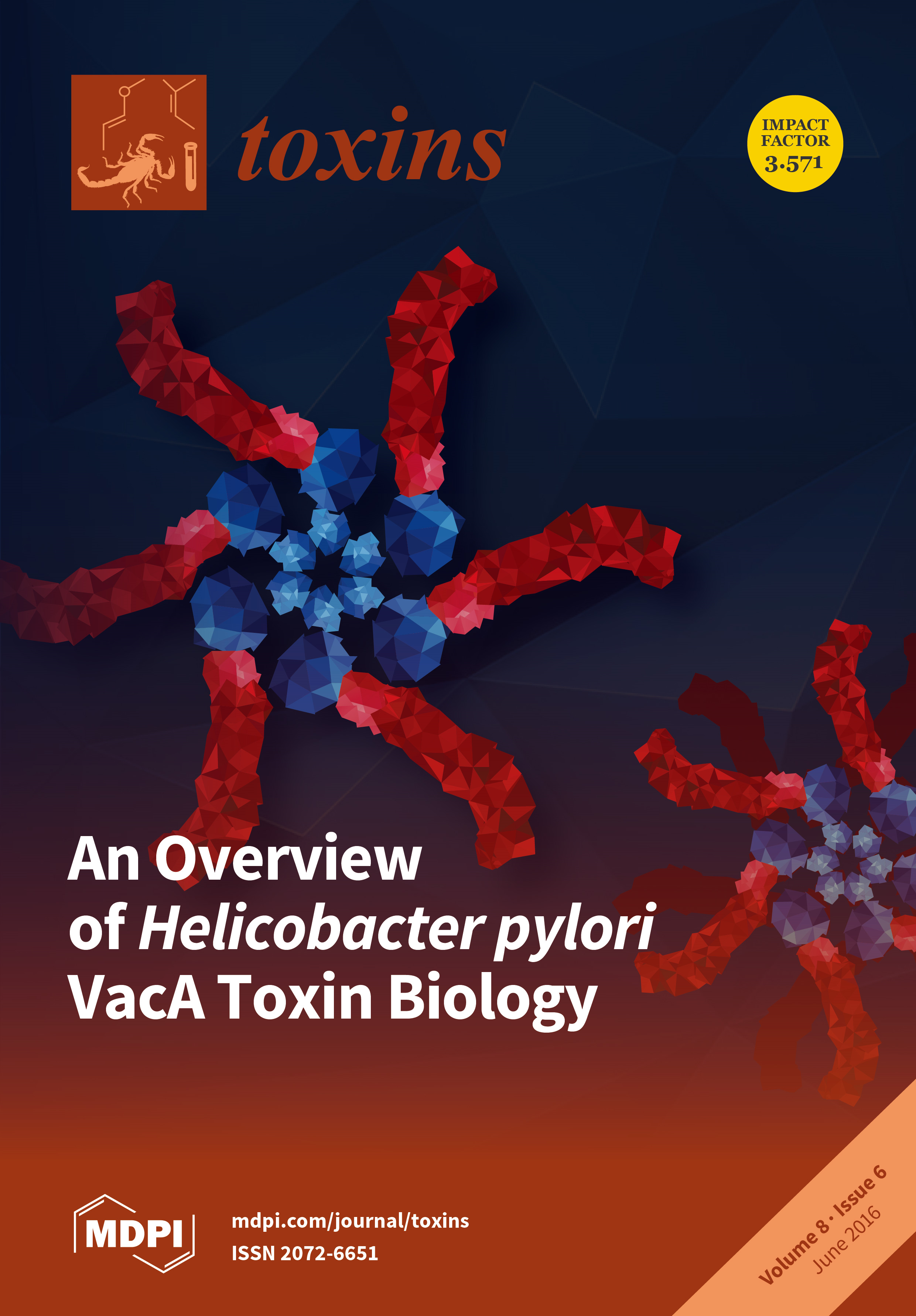
Structural organization of water-soluble oligomers formed by Helicobacter pylori VacA toxin. A hexamer (left) and dodecamer (right) are shown. Within each component p88 monomer, p33 and p55 domains are shown in blue and red, respectively. Water-soluble hexamers are predicted to be structurally similar to membrane channels formed by VacA.
- Issues are regarded as officially published after their release is announced to the table of contents alert mailing list.
- You may sign up for e-mail alerts to receive table of contents of newly released issues.
- PDF is the official format for papers published in both, html and pdf forms. To view the papers in pdf format, click on the "PDF Full-text" link, and use the free Adobe Reader to open them.
Previous Issue
Next Issue