Article

Venomic Analysis of the Poorly Studied Desert Coral Snake, Micrurus tschudii tschudii, Supports the 3FTx/PLA2 Dichotomy across Micrurus Venoms

1
Laboratorio de Venómica Estructural y Funcional, Instituto de Biomedicina de Valencia, CSIC, Jaime Roig 11, Valencia 46010, Spain
2
Departamento Academico de Ciencias Celulares y Moleculares, Facultad de Ciencias y Filosofía, Universidad Peruana Cayetano Heredia, Lima 31, Peru
3
Instituto Nacional de Salud, Ministerio de Salud, Lima 11, Peru
4
Instituto Clodomiro Picado, Facultad de Microbiología, Universidad de Costa Rica, San Jose 11501, Costa Rica
*
Author to whom correspondence should be addressed.
Toxins 2016, 8(6), 178; https://doi.org/10.3390/toxins8060178
Submission received: 23 March 2016 / Revised: 17 May 2016 / Accepted: 1 June 2016 / Published: 7 June 2016
(This article belongs to the Special Issue Venomics, Venom Proteomics and Venom Transcriptomics)

Abstract

:
The venom proteome of the poorly studied desert coral snake Micrurus tschudii tschudii was unveiled using a venomic approach, which identified ≥38 proteins belonging to only four snake venom protein families. The three-finger toxins (3FTxs) constitute, both in number of isoforms (~30) and total abundance (93.6% of the venom proteome), the major protein family of the desert coral snake venom. Phospholipases A2 (PLA2s; seven isoforms, 4.1% of the venom proteome), 1–3 Kunitz-type proteins (1.6%), and 1–2 l-amino acid oxidases (LAO, 0.7%) complete the toxin arsenal of M. t. tschudii. Our results add to the growing evidence that the occurrence of two divergent venom phenotypes, i.e., 3FTx- and PLA2-predominant venom proteomes, may constitute a general trend across the cladogenesis of Micrurus. The occurrence of a similar pattern of venom phenotypic variability among true sea snake (Hydrophiinae) venoms suggests that the 3FTx/PLA2 dichotomy may be widely distributed among Elapidae venoms.

Graphical Abstract

1. Introduction

New World genus Micrurus (Elapidae) (Wagler, 1824) [1] represents a monophyletic clade of some 80 currently recognized species of venomous coral snakes [2,3,4,5,6,7,8], although the topology of the tree is still unresolved. Coral snakes are widely distributed in tropical and subtropical regions from the southern United States to northeastern Argentina, including several continental islands inhabited by endemic forms [2,3]. Coral snakes are considered by herpetologists to be among the most beautiful snakes of the planet, as they are adorned with unique combinations of red-, black-, and yellow-colored banding. Based on their body ring color pattern characteristics, tail proportion with respect to body length, and hemipenial morphology, coral snakes are traditionally arranged in four species groups: a tricolored monadal group, a bicolored group, a Central American tricolored triadal group, and a South American triadal group [3,9,10,11,12]. The monadal group comprises long-tailed species with a single black ring separating the white and red rings; the bicolored group contains short-tailed species with white or red rings separated by black rings; the Central American triadal group taxa are long-tailed species with three black rings separated by white rings between red rings; the South American triadal group is represented by short-tailed species with the same color pattern as the Central American triadal group species but is restricted geographically to South America, extending from Panama to southern Argentina.
In the last decade, the increasing application of omics techniques to the study of snake venoms has greatly enhanced our knowledge on their composition, evolution, biological activities, and clinical effects [13,14,15,16,17]. However, the venoms of only a handful of the vast number (~130) of species and subspecies that constitute the genus Micrurus have been the subject of proteomic studies (consult Table 2 of [18]). To understand this fact it should be taken into consideration that, despite producing among the most potent neurotoxic venoms of any New World snake, bites and fatalities by coral snakes are very rare. This is facilitated by the fact that coral snakes are not aggressive; when confronted by humans, coral snakes will almost always attempt to flee, and bite only as a last resort. On the other hand, coral snakes generally inhabit sparsely populated areas and are thus infrequently encountered, and they have short fangs that cannot penetrate thick leather clothing. Consequently, envenomings by coral snakes are less frequent than those produced by sympatric pitvipers, by far the most dangerous snakes of South America, accounting for less than 3% of the snakebites recorded in the Americas [19,20,21].
The desert coral snake M. tschudii tschudii (January 1858) [22], one of the lesser-studied species of the genus Micrurus, belongs to the South American triad coral snakes [11,12,23]. Named after the Swiss naturalist, explorer and diplomat Johann Jakob von Tschudi (1818–1889), who traveled extensively in South America and published important works in herpetology, M. t. tschudii is a small (adults average 45 to 55 cm in length), tri-colored (Figure 1), mainly diurnal and terrestrial coral snake, found in tropical deciduous forest, dry tropical forest, and thorn scrub, mainly along watercourses, from near sea level to 1450 m elevation, in the western slopes of the Andes on the semi-arid Pacific coast of South America, from southern Ecuador to southwestern Peru [2,3,24]. The desert coral snake feeds on geckkonid lizards (Phyllodactilus spp.), Amphisbaenids (A. occidentalis), and colubrids of the species Mastigodryas haathi [3]. However, available literature about its reproduction and activity is scarce, and in particular virtually nothing (not even a single entry in PubMed) is known about its venom. Here, we have applied a venomics approach [13,14] to gain an insight into the spectrum of toxins that make up the venom proteome of M. t. tschudii.

2. Results and Discussion

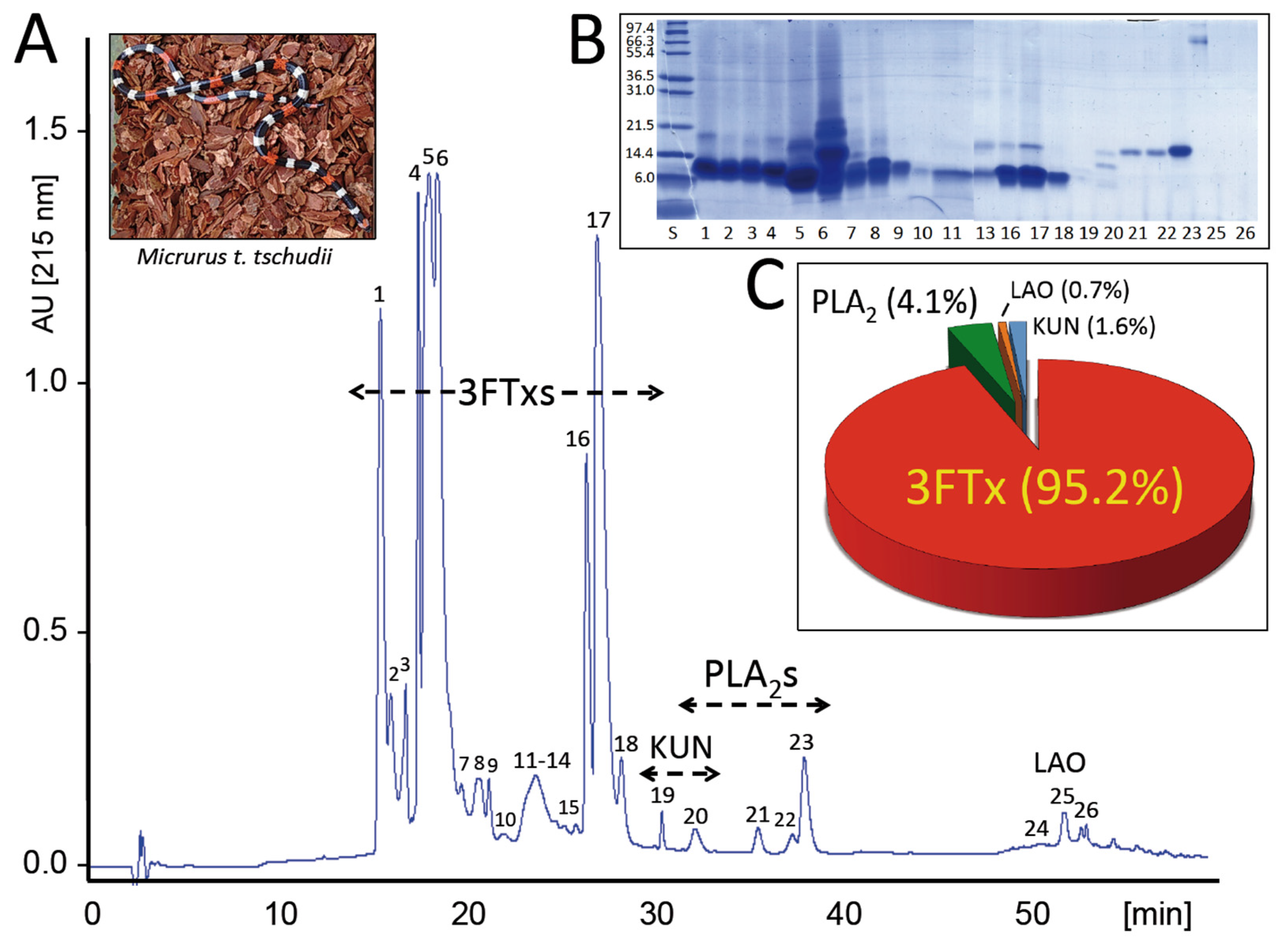
2.1. The Venom Proteome of the Desert Coral Snake

The venom of M. t. tschudii was fractionated into 26 RP-HPLC fractions (Figure 1A). Each chromatographic fraction was analyzed by SDS-PAGE (Figure 1B), and the protein bands were excised and submitted to mass spectrometric analysis. The MS/MS data, listed in Supplementary Table S1, resulted in the identification of ≥38 proteins belonging to only four snake venom protein families: three-finger toxin (3FTx), phospholipase A2 (PLA2), Kunitz-type, and l-amino acid oxidase (LAO) (Table 1). The relative abundances of M. t. tschudii venom proteins and toxin families are displayed, respectively, in Table S1 and Figure 1C. The 3FTxs constitute, by far, both in number of isoforms (~30) and total abundance (93.6% of the total venom proteins), the major protein family of the desert coral snake venom. Seven PLA2 isoforms, 1–3 Kunitz-type proteins, and 1–2 LAO molecules (accounting, respectively, for 4.1%, 1.6%, and 0.7% of the venom proteome) complete the toxin arsenal of the desert coral snake.
The 3FTx and PLA2 molecules are hallmark components of the venoms of Elapidae. Catalytically active PLA2 molecules typically exhibit presynaptic neurotoxic activity, myotoxic activity, or both, although some forms exhibit antiplatelet activity [25,26]. Despite their pronounced structural similarity, members of the 3FTx family exhibit a wide variety of pharmacological effects including postsynaptic neurotoxicity, cytotoxicity, cardiotoxicity, and anticoagulant, and antiplatelet activities [27,28]. In addition, l-type Ca2+ channel antagonists of the 3FTx family may act synergistically with muscarinic three-finger toxins to promote hypotension [29]. Kunitz-type serine protease inhibitor isolated from elapid venoms blocked the activity of a range of serine proteases, producing an antihemorrhagic effect [30]. Non-covalent PLA2-KUN complexes have been characterized from venom of some Micrurus species [18 and references cited]. These complexes, originally discovered in the venom of the Texas coral snake M. tener, are target acid-sensing receptors ASIC1a/2 evoking pain [31,32]. There are many examples of venom-derived toxins that elicit notoriously intense pain [33]. Pain response may cause paralysis and serve as a warning signal for discouraging potentially threatening predators by triggering lasting acute physiological distress. The l-amino acid oxidases are flavoenzymes that catalyze oxidative deamination of l-amino acids to form corresponding α-keto acids, hydrogen peroxide and ammonia. LAOs are thought to contribute to the toxicity of the venom due to the production of hydrogen peroxide during the oxidation reaction [34,35], although their contribution to the envenoming process remains elusive.
The low yield of venom and the challenge of long-term maintenance of the desert coral snake in captivity precluded a detailed toxicovenomics analysis of the venom components of this poorly studied Micrurus species.

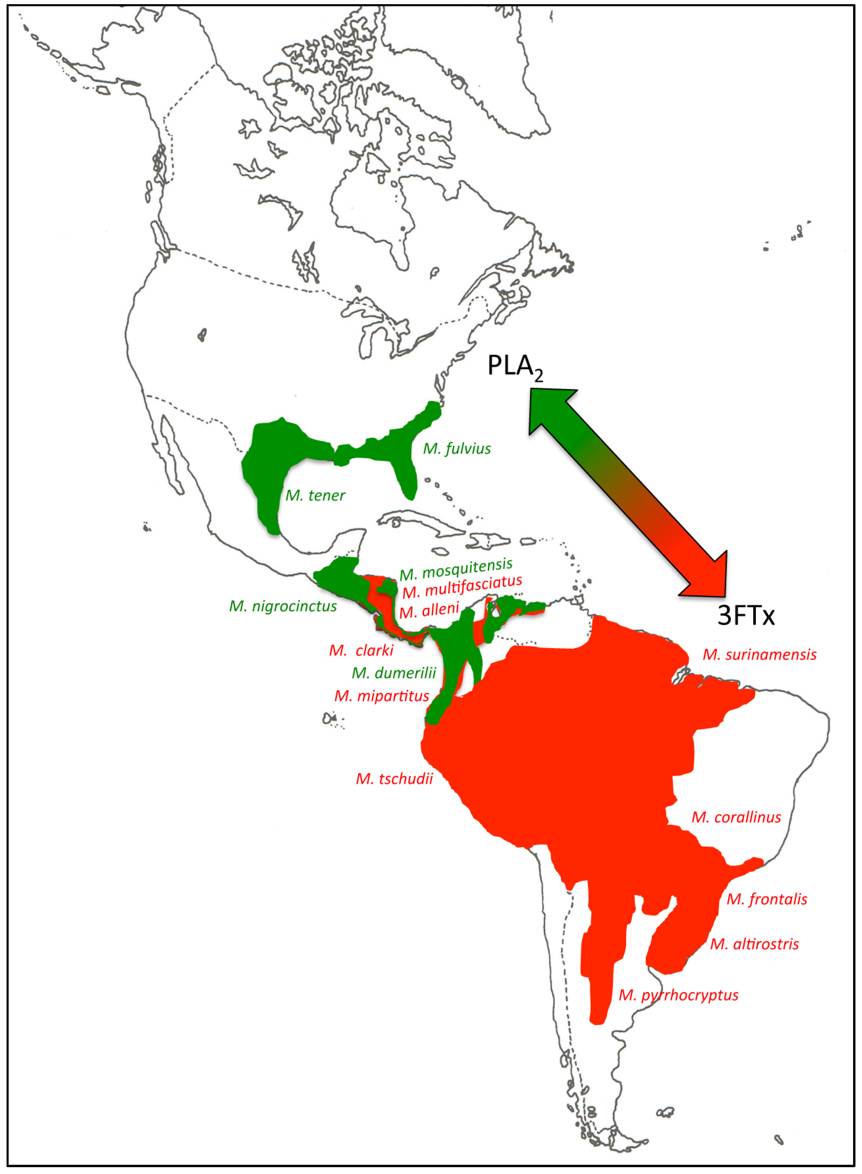
2.2. 3FTx- and PLA2-Predominant Venom Proteomes among Coral Snakes

Despite their extremely adaptive ecological radiation, coral and sea snakes show virtually identical rates of body size evolution to other elapids, suggesting that their diversification may be correlated with phenotypic traits other than body size [36]. Given the central role that diet has played in the adaptive radiation of snakes [37], venom represents a key adaptation that has played an important role in the diversification of these animals [38,39]. Growing evidence, including the venomic analysis of the desert coral snake here reported, supports the view that the occurrence of two divergent venom phenotypes, i.e., 3FTx- and PLA2-predominant venom proteomes, may constitute a general trend across Micrurus cladogenesis [18,40]. Thus, M. alleni (77% 3FTx vs. 11% PLA2) and M. mosquitensis (56% PLA2, 22% 3FTx) [18]; M. corallinus (82% 3FTx, 12% PLA2) [41]; M. nigrocinctus (38% 3FTx, 48% PLA2) [42]; M. fulvius (21% 3FTx, 65% PLA2) [43,44]; M. tener (38% 3FTx, 46% PLA2) [45]; M. dumerilii (28% 3FTx, 52% PLA2) [40]; and M. clarki (48.2% 3FTx, 36.5% PLA2) [46] all are species included in the well-supported monophyletic monadal clade [3,10,47,48]. The 22 valid species of coral snakes classified in the South American triadal group, to which M. t. tschudii belongs, also form a strongly supported clade [3,11,12,23]. Biochemical and/or proteomic analyses have been reported for the venoms of M. altirostris (80% 3FTx, 14% PLA2) [44], M. surinamensis (3FTx-predominant venom) [49], M. frontalis (3FTx-predominant venom) [50], M. pyrrhocryptus (3FTx-rich venom proteome) [51], and M. clarki (moderately 3FTx-predominant venom) [46]. On the other hand, the venom proteomes of M. multifasciatus and M. mipartitus from the bicolor species group are both dominated by 3FTxs [52].
Experiments in mice determined the Median Lethal Dose (LD50) for M. t. tschudii venom at 10.5 µg/mouse (7.4–13.8 µg/mouse, 95% confidence limits) by the i.p. route. This figure corresponds to 0.62 µg venom/g of mouse body weight (0.44–0.81 µg/g, 95% confidence limits) (Table 1). Comparison of reported median lethal doses (LD50) clearly shows that 3FTx-rich and PLA2-rich Micrurus venoms are not statistically different in terms of lethality to mice (Table 1). Although a more meaningful correlation would require comparing LD50 values for their natural prey, current data suggest that Micrurus venoms may have evolved under the action of balancing selection [56].
Margres and coworkers [44] have detected strong evidence of positive selection for the 3FTx and PLA2 toxin families of M. fulvius. This accelerated evolution is most likely due to their direct involvement in fitness. Figure 2 shows a clear geographical distribution of PLA2- and 3FTx-predominant Micrurus venoms along their north-south dispersal axis, further supporting the adaptive nature of this phenotypic dichotomy.

2.3. Tracing the Evolutionary Origin of the 3FTx/PLA2 Dichotomy across Elapidae

Elapid snakes are a relatively young group. The crown elapid radiation is approximately 38 million years (My) old, yet elapid snakes exhibit some of the highest diversification rates in reptiles [57]. In particular, two clades, Hydrophis and Micrurus, show anomalously high rates of diversification within Elapidae [36]. The Asian coral snake genus Calliophis emerges as monophyletic and the sister group to all other American and Asian coral snakes [47], suggesting an Asiatic origin for the common ancestor of these elapids [58]. Coral snakes diversified around 30–25 My ago, and sea snakes (Hydrophiini) are approximately 16 My old [36]. Estimated divergence times suggest that Hydrophiini is a young and rapidly speciating clade that had a common ancestor ~8 My ago, although the majority of extant lineages diversified more recently, over the last ~1.5–3.5 My [36,59].
A similar pattern of venom phenotypic variability has been documented by the proteomic analysis of true sea snake (Hydrophiinae) venoms. Hence, the venom of A. laevis comprises mainly PLA2 (71%) and 3FTxs (25%) [60], whereas the venoms of H. schistosus [61], H. cyanocinctus [62], and P. platura [63] all share 3FTx-rich phenotypes (i.e., 70.5% 3FTx/26.5% PLA2; 81% 3FTx/19% PLA2; and 50% 3FTx/33% PLA2, respectively). Transcripts for 3FTx and PLA2 molecules also represent the two main constituents of the venom gland toxin-encoding transcriptomes of Acalyptophis peronii (77.5% 3FTx/17.5 PLA2) and Lapemis curtus (92.4% 3FTx; 7.5% PLA2) [64].
The evolutionary origin and adaptive relevance of the puzzling 3FTx/PLA2 dichotomy remains elusive since 3FTx- and PLA2-predominant venoms are scattered through the phylogenetic tree of Micrurus. The dominant protein families in the venom proteome of C. bivirgata flaviceps are PLA2 (41%), 3FTx (22.6%) and SVMP (18.7%) [65], suggesting that the ancestor venom phenotype of coral snakes might have been of the PLA2-predominant type. Alternatively, 3FTx- and PLA2-rich venom proteomes may represent pedomorphic and ontogenetic traits, as has been documented in Crotalus. Rattlesnake venoms belong to one of two distinct phenotypes, which broadly correspond to type I (high levels of SVMPs and low toxicity, LD50 1 mg/g mouse body weight) and type II (low metalloproteinase activity and high toxicity, LD50 1 mg/g mouse body weight) [66]. In Neotropical rattlesnakes, the adaptive pressure for type I to type II transition was the gain of neurotoxicity and lethality to rodents, and this transition represents a miRNA-modulated pedomorphic trait that correlates with the increased concentration of crotoxin along the axis of Crotalus radiation in South America [67,68]. In Nearctic species, such as C. s. scutulatus (Css), the venom phenotype changes geographically from SVMP-rich to Mojave toxin-rich (type-II) as one moves from south central to southeastern Arizona, with a transitional zone between the SVMP and Mojave toxin phenotypes [69]. Understanding the phylogenetic origin of the 3FTx-rich and PLA2-predominant venom phenotypes across Micrurus, and whether there is parallelism between the 3FTx/PLA2 (Micrurus) and the type I/type II (Crotalus) venom dichotomies, requires genus-wide profiling of the venom proteomes and the venom gland transcriptomes of adult and juvenile Crotalus and Micrurus congeneric specimens.

3. Concluding Remarks and Perspectives

While genomic studies on model organisms have been widely applied to identify traits involved in maintaining functional genetic variation, few studies have drawn the links between genotype, phenotype, and fitness, and the environmental pressures that act to maintain variation that affects organismal phenotypes [70,71]. Venom represents a key adaptive trophic trait that has played an important role in the radiation of advanced snakes, and thus could be a particularly powerful model system to investigate players and mechanisms of adaptive variation at the phenotype level [17,70,72]. In this regard, structural and functional studies of Micrurus venoms point to balancing selection as the mechanism acting to maintain a 3FTx/PLA2 venom dichotomy. The realization that this 3FTx/PLA2 dichotomy may have deep roots in the evolution of coral snakes may also have important translational implications. Thus, both types of venoms should be part of immunization mixtures aimed at generating broad-spectrum antivenoms.

4. Materials and Methods

4.1. Venom

Adult M. t. tschudii specimens were caught in the Peruvian Pacific coast regions of La Libertad, Ancash, and Lima, and kept in captivity at the serpentarium of the Instituto Nacional de Salud, Lima, Perú. Venom from nine adult M. t. tschudii specimens was collected and pooled during the first three months of captivity. Venom was lyophilized and stored at −20 °C until used.

4.2. Isolation and Characterization of Venom Proteins

First 0.5 milligrams of crude, lyophilized venom were dissolved in 200 μL of 5% acetonitrile in water containing 0.1% trifluoroacetic acid (TFA), centrifuged to remove debris, and separated by reverse-phase HPLC using a Teknokroma Europa Protein 300 C18 (0.4 cm × 25 cm, 5 μm particle size, 300 Å pore size) column and an LC 1100 High Pressure Gradient System (Agilent Technologies, Santa Clara, CA, USA) equipped with DAD detector and micro-Auto-sampler [73]. The flow rate was set to 1 mL/min and the column was developed with a linear gradient of 0.1% TFA in water (solution A) and acetonitrile (solution B) using the following column elution conditions: isocratically (5% B) for 5 min, followed by 5%–25% B for 10 min, 25%–45% B for 60 min, and 45%–70% for 10 min. Protein detection was carried out at 215 nm with a reference wavelength of 400 nm. Fractions were collected manually, dried in a vacuum centrifuge (Savant), and redissolved in water, and submitted to molecular mass determination using a SYNAPT® G2 High Definition Mass Spectrometry System (Waters Corp., Milford, MA, USA), and SDS-PAGE analysis in 15% polyacrylamide gels, under reducing and non-reducing conditions. Gels were stained with Coomassie Brilliant Blue R-250 (Sigma-Aldrich, St. Louis, MO, USA).

4.3. Characterization of the Venom Peptidome and Proteome

Electrophoretic protein bands were excised from a Coomassie Brilliant Blue-stained SDS-PAGE gel and subjected to in-gel reduction (10 mM dithiothreitol) and alkylation (50 mM iodoacetamide), followed by overnight sequencing-grade trypsin digestion (66 ng/μL in 25 mM ammonium bicarbonate, 10% acetonitrile; 0.25 μg/sample) in an automated processor (ProGest Protein Digestion Workstation, Genomic Solution Ltd., Cambridgeshire, UK) following the manufacturer’s instructions. Tryptic digests were dried in a SpeedVac (Savant™, Thermo Scientific Inc., West Palm Beach, FL, USA), redissolved in 15 μL of 0.1% formic acid in water, and submitted to LC-MS/MS. To this end, tryptic peptides were separated by nano-Acquity UltraPerformance LC® (UPLC®, Waters Corporation, Milford, MA, USA) using BEH130 C18 (100 μm × 100 mm, 1.7 μm particle size) column in-line with a SYNAPT® G2 High Definition Mass Spectrometry System (Waters Corp., Milford, MA, USA). The flow rate was set to 0.6 μL/min and the column was developed with a linear gradient of 0.1% formic acid in water (solution A) and 0.1% formic acid in acetonitrile (solution B), isocratically 1% B for 1 min, followed by 1%–12% B for 1 min, 12%–40% B for 15 min, 40%–85% B for 2 min. Doubly and triply charged ions were selected for collision-induced dissociation (CID) MS/MS. Fragmentation spectra were interpreted (a) manually (de novo sequencing); (b) using the on-line form of the MASCOT program at http://www.matrixscience.com against NCBInr database, a comprehensive, non-identical protein database compiled from GenBank CDS translations, PIR, SwissProt, PRF, and PDB; and (c) processed in Waters Corporation’s (Milford, MA, USA) ProteinLynx Global SERVER 2013 version 2.5.2. (with Expression version 2.0) and the generated .pkl peak list files were exported to MASCOT for protein identification against the NCBInr database. MS/MS mass tolerance was set to ±0.6 Da. Carbamidomethyl cysteine and oxidation of methionine were selected as fixed and variable modifications, respectively. Cut-off for MASCOT reporting was set to top 10 hits All MASCOT identifications were manually verified. Amino acid sequence similarity searches were performed against the NCBInr and UniProtKB databases using the BLASTP program implemented in the WU-BLAST2 search engine at http://www.bork.embl-heidelberg.de.
The relative abundances (expressed as percentage of the total venom proteins) of the different protein families were calculated as the ratio of the sum of the areas of the reverse-phase chromatographic peaks containing proteins from the same family to the total area of venom protein peaks in the reverse-phase chromatogram [14,74]. When more than one protein band was present in a reverse-phase fraction, their proportions were estimated by densitometry of Coomassie-stained SDS-polyacrylamide gels using ImageJ version 1.47 (Free Software Foundation, Boston, MA, USA) (http://rsbweb.nih.gov/ij). Conversely, the relative abundances of different proteins contained in the same SDS-PAGE band were estimated based on the relative ion intensities of the three more abundant peptide ions associated with each protein by MS/MS analysis. Finally, protein family abundances were estimated as the percentages of the total venom proteome.

4.4. Determination of LD50 for Mice

Animal experiments were performed in accordance with protocols approved by the Institutional Committee for the Care and Use of Laboratory Animals of the University of Costa Rica (CICUA 041-15, 21 October 2015), using CD-1 mice of either sex, provided by Instituto Clodomiro Picado. To evaluate the lethal activity, various doses of M. t. tschudii venom, dissolved in 200 μL of PBS, were injected into groups of five CD-1 mice (16–18 g body weight), by the intraperitoneal (i.p.) route. Deaths were scored over a 48 h period and the median lethal dose (LD50) was calculated by probits [75].

Supplementary Materials

The following are available online at www.mdpi.com/2072-6651/8/6/178/s1. Table S1: Summary of the MS/MS identification of the SDS-PAGE protein bands of M. t. tschudii venom.

Acknowledgments

This study was conducted as part of an agreement signed between the Instituto Nacional de Salud (Perú) and the Universidad Peruana Cayetano Heredia. Venomic analyses were supported by Grant BFU2013-42833-P from the Ministerio de Economía y Competitividad, Madrid (Spain). Authors gratefully acknowledge Dr. Med. Vet. Gualberto Marcas Cáceres, Centro Nacional de Productos Biológicos, Instituto Nacional de Salud, Ministerio de Salud, Perú, for kindly providing pictures of M. t. tschudii.

Author Contributions

L.S., D.P., B.L., and J.J.C. conceived and designed the experiments; L.S., D.P., and B.L. performed the experiments; A.Z., B.L., and J.J.C. contributed reagents/materials/analysis tools; L.S., D.P., B.L. and J.J.C. analyzed the data. J.J.C. wrote the paper. All authors revised the manuscript.

Conflicts of Interest

The authors declare no conflict of interest.

References

  1. Wagler, J. Serpentum Brasiliensium species novae, ou histoire naturelle des espèces nouvelles de serpens. In Animalia Nova Sive Species Novae; de Spix, J., Ed.; Franc. Seraph. Hübschmanni: Munich, Germany, 1824. [Google Scholar]
  2. Roze, J.A. Coral Snakes of the Americas. Biology, Identification and Venoms; Krieger Publishing Company: Malabar, FL, USA, 1996; p. 328. [Google Scholar]
  3. Campbell, J.A.; Lamar, W.W. The Venomous Reptiles of the Western Hemisphere; Cornell University Press: Ithaca, NY, USA, 2004; p. 476. [Google Scholar]
  4. The Reptile Database. Available online: http://www.reptile-database.org (accessed on 12 February2016).
  5. Pires, M.G.; da Silva, N.J., Jr.; Feitosa, D.T.; Prudente, A.L.; Filho, G.A.; Zaher, H. A new species of triadal coral snake of the genus Micrurus Wagler, 1824 (Serpentes: Elapidae) from northeastern Brazil. Zootaxa 2014, 3811, 569–584. [Google Scholar] [CrossRef] [PubMed]
  6. Feitosa, D.T.; da Silva, N.J.; Pires, M.G.; Zaher, H.; Prudente, A.L. A new species of monadal coral snake of the genus Micrurus (Serpentes, Elapidae) from western Amazon. Zootaxa 2015, 3974, 538–554. [Google Scholar] [CrossRef] [PubMed]
  7. Slowinski, J.B.; Keogh, J.S. Phylogenetic relationship of coral snakes based on cytochrome b mtDNA sequences. Mol. Phylogenet. Evol. 2005, 15, 157–164. [Google Scholar] [CrossRef] [PubMed]
  8. Harvey, M.B.; Gutberlet, R.L. The evolution of New World venomous snakes. In The Venomous Reptiles of the Western Hemisphere; Campbell, J.A., Lamar, W.W., Eds.; Cornell University Press: Ithaca, NY, USA, 2004; pp. 634–682. [Google Scholar]
  9. Campbell, J.A.; Lamar, W.W. The Venomous Reptiles of Latin America; Cornell University Press: Ithaca, NY, USA; London, UK, 1989; p. 425. [Google Scholar]
  10. Savage, J.M.; Slowinski, J.B. The coloration of the venomous coral snakes (family Elapidae) and their mimics (families Aniliidae and Colubridae). Biol. J. Linn. Soc. 1992, 45, 235–254. [Google Scholar] [CrossRef]
  11. Slowinski, J.B. A phylogenetic analysis of the New World coral snakes (Elapidae: Leptomicrurus, Micruroides and Micrurus) based on allozymic and morphological characters. J. Herpetol. 1995, 29, 325–338. [Google Scholar] [CrossRef]
  12. Silva, N.J., Jr.; Sites, J.W., Jr. Phylogeny of South American triad coral snakes (Elapidae: Micrurus) based on molecular characters. Herpetologica 2001, 57, 1–22. [Google Scholar]
  13. Calvete, J.J. Snake venomics: From the inventory of toxins to biology. Toxicon 2013, 75, 44–62. [Google Scholar] [CrossRef] [PubMed]
  14. Calvete, J.J. Next-generation snake venomics: Protein-locus resolution through venom proteome decomplexation. Expert Rev. Proteom. 2014, 11, 315–329. [Google Scholar] [CrossRef] [PubMed]
  15. Brahma, R.K.; McCleary, R.J.; Kini, R.M.; Doley, R. Venom gland transcriptomics for identifying, cataloging, and characterizing venom proteins in snakes. Toxicon 2015, 93, 1–10. [Google Scholar] [CrossRef] [PubMed]
  16. Calvete, J.J.; Lomonte, B. A bright future for integrative venomics. Toxicon 2015, 107, 159–162. [Google Scholar] [CrossRef] [PubMed]
  17. Sunagar, K.; Morgenstern, D.; Reitzel, A.M.; Moran, Y. Ecological venomics: How genomics, transcriptomics and proteomics can shed new light on the ecology and evolution of venom. J. Proteom. 2016, 135, 62–72. [Google Scholar] [CrossRef] [PubMed]
  18. Fernández, J.; Vargas-Vargas, N.; Pla, D.; Sasa, M.; Rey-Suárez, P.; Sanz, L.; Gutiérrez, J.M.; Calvete, J.J.; Lomonte, B. Snake venomics of Micrurus alleni and Micrurus mosquitensis from the Caribbean region of Costa Rica reveals two divergent compositional patterns in New World elapids. Toxicon 2015, 107, 217–233. [Google Scholar] [CrossRef] [PubMed]
  19. Warrell, D.A. Snakebites in central and South America: Epidemiology, clinical features, clinical management. In The Venomous Reptiles of the Western Hemisphere; Campbell, J.A., Lamar, W.W., Eds.; Cornell University Press: Ithaca, NY, USA, 2004; pp. 709–761. [Google Scholar]
  20. Bucaretchi, F.; de Capitani, E.M.; Vieira, R.J.; Rodrigues, C.K.; Zannin, M.; da Silva, N.J., Jr.; Casais-E-Silva, L.L.; Hyslop, S. Coral snake bites (Micrurus spp.) in Brazil: A review of literature reports. Clin. Toxicol. 2016, 25, 1–13. [Google Scholar]
  21. Norris, R.L.; Pfalzgraf, R.R.; Laing, G. Death following coral snake bite in the United States—First documented case (with ELISA confirmation of envenomation) in over 40 years. Toxicon 2009, 53, 693–697. [Google Scholar] [CrossRef] [PubMed]
  22. Jan, G. Plan d’une iconographie descriptive des ophidiens et description sommaire de nouvelles espèces deserpents. Rev. Mag. Zool. Pure Apliquée (Paris) 1858, 2, 438–439 and 514–527. [Google Scholar]
  23. Silva, N.J., Jr.; Sites, J.W., Jr. Revision of the Micrurus frontalis complex (Serpentes: Elapidae). Herpetol. Monogr. 1999, 13, 142–194. [Google Scholar] [CrossRef]
  24. Peters, J.A.; Orejas-Miranda, B. Catalogue of the neotropical Squamata: Part I. Snakes. US Natl. Mus. Bull. 1970, 297, 1–347. [Google Scholar] [CrossRef]
  25. Mackessy, S.P.; Tu, A.T. Biology of the sea snakes and biochemistry of their venoms. In Toxin-Related Diseases: Poisons Originating from Plants, Animals and Spoilage; Tu, A.T., Ed.; Oxford IBH Publishing Co.: New Delhi, India, 1993; pp. 305–351. [Google Scholar]
  26. Jackson, T.N.; Sunagar, K.; Undheim, E.A.; Koludarov, I.; Chan, A.H.; Sanders, K.; Ali, S.A.; Hendrikx, I.; Dunstan, N.; Fry, B.G. Venom down under: Dynamic evolution of Australian elapid snake toxins. Toxins 2013, 5, 2621–2655. [Google Scholar] [CrossRef] [PubMed]
  27. Kini, R.M.; Doley, R. Structure, function and evolution of three-finger toxins: Mini proteins with multiple targets. Toxicon 2010, 56, 855–867. [Google Scholar] [CrossRef] [PubMed]
  28. Utkin, Y.N. Three-finger toxins, a deadly weapon of elapid venom-milestones of discovery. Toxicon 2013, 62, 50–55. [Google Scholar] [CrossRef] [PubMed]
  29. Du, X.-Y.; Clemetson, K.J. Snake venom l-amino acid oxidases. Toxicon 2002, 40, 659–665. [Google Scholar] [CrossRef]
  30. Filippovich, I.; Sorokina, N.; Masci, P.P.; de Jersey, J.; Whitaker, A.N.; Winzor, D.J.; Gaffney, P.J.; Lavin, M.F. A family of textilinin genes, two of which encode proteins with antihaemorrhagic properties. Br. J. Haematol. 2002, 119, 376–384. [Google Scholar] [CrossRef] [PubMed]
  31. Bohlen, C.J.; Chesler, A.T.; Sharif-Naeini, R.; Medzihradszky, K.F.; Zhou, S.; King, D.; Sánchez, E.E.; Burlingame, A.L.; Basbaum, A.I.; Julius, D. A heteromeric Texas coral snake toxin targets acid-sensing ion channels to produce pain. Nature 2011, 479, 410–414. [Google Scholar] [CrossRef] [PubMed]
  32. Baconguis, I.; Bohlen, C.J.; Goehring, A.; Julius, D.; Gouaux, E. X-ray structure of acid-sensing ion channel 1-snake toxin complex reveals open state of a Na(+)-selective channel. Cell 2014, 156, 717–729. [Google Scholar] [CrossRef] [PubMed]
  33. Bohlen, C.J.; Julius, D. Receptor-targeting mechanisms of pain-causing toxins: How ow? Toxicon 2012, 60, 254–264. [Google Scholar] [CrossRef] [PubMed]
  34. Aird, S.D. Ophidian envenomation strategies and the role of purines. Toxicon 2002, 40, 335–393. [Google Scholar] [CrossRef]
  35. Guo, C.; Liu, S.; Yao, Y.; Zhang, Q.; Sun, M.-Z. Past decade study of snake vemom l-amino acid oxidase. Toxicon 2012, 60, 302–311. [Google Scholar] [CrossRef] [PubMed]
  36. Lee, M.S.Y.; Sanders, K.L.; King, B.; Palci, A. Diversification rates and phenotypic evolution in venomous snakes (Elapidae). R. Soc. Open Sci. 2016, 3, 150277. [Google Scholar] [CrossRef] [PubMed]
  37. Greene, H.W. Dietary correlates of the origin and radiation of snakes. Am. Zool. 1983, 23, 431–441. [Google Scholar] [CrossRef]
  38. Daltry, J.C.; Wüster, W.; Thorpe, R.S. Diet and snake venom evolution. Nature 1996, 379, 537–540. [Google Scholar] [CrossRef] [PubMed]
  39. Barlow, A.; Pook, C.E.; Harrison, R.A.; Wüster, W. Coevolution of diet and prey-specific venom activity supports the role of selection in snake venom evolution. Proc. Biol. Sci. 2009, 276, 2443–2449. [Google Scholar] [CrossRef] [PubMed]
  40. Rey-Suárez, P.; Núñez, V.; Fernández, J.; Lomonte, B. Integrative characterization of the venom of the coral snake Micrurus dumerilii (Elapidae) from Colombia: Proteome, toxicity, and cross-neutralization by antivenom. J. Proteom. 2016, 136, 262–273. [Google Scholar] [CrossRef] [PubMed]
  41. Corrêa-Netto, C.; Junqueira-de-Azevedo, I.L.; Silva, D.A.; Ho, P.L.; Leitão-de-Araújo, M.; Alves, M.L.; Sanz, L.; Foguel, D.; Zingali, R.B.; Calvete, J.J. Snake venomics and venom gland transcriptomic analysis of Brazilian coral snakes, Micrurus altirostris and M. corallinus. J. Proteom. 2011, 74, 1795–1809. [Google Scholar] [CrossRef] [PubMed]
  42. Fernández, J.; Alape-Girón, A.; Angulo, Y.; Sanz, L.; Gutiérrez, J.M.; Calvete, J.J.; Lomonte, B. Venomic and antivenomic analyses of the Central American coral snake, Micrurus nigrocinctus (Elapidae). J. Proteome Res. 2011, 10, 1816–1827. [Google Scholar] [CrossRef] [PubMed]
  43. Vergara, I.; Pedraza-Escalona, M.; Paniagua, D.; Restano-Cassulini, R.; Zamudio, F.; Batista, C.V.; Possani, L.D.; Alagón, A. Eastern coral snake Micrurus fulvius venom toxicity in mice is mainly determined by neurotoxic phospholipases A2. J. Proteom. 2014, 105, 295–306. [Google Scholar] [CrossRef] [PubMed]
  44. Margres, M.J.; Aronow, K.; Loyacano, J.; Rokyta, D.R. The venom-gland transcriptome of the eastern coral snake (Micrurus fulvius) reveals high venom complexity in the intragenomic evolution of venoms. BMC Genom. 2013, 14, 531. [Google Scholar] [CrossRef] [PubMed]
  45. Bénard-Valle, M.; Carbajal-Saucedo, A.; de Roodt, A.; López-Vera, E.; Alagón, A. Biochemical characterization of the venom of the coral snake Micrurus tener and comparative biological activities in the mouse and a reptile model. Toxicon 2014, 77, 6–15. [Google Scholar] [CrossRef] [PubMed]
  46. Lomonte, B.; Sasa, M.; Rey-Suárez, P.; Bryan, W.; Gutiérrez, J.M. Venom of the coral snake Micrurus clarki: Proteomic profile, toxicity, immunological cross-neutralization, and characterization of a three-finger toxin. Toxins 2016, 8, 138. [Google Scholar] [CrossRef] [PubMed]
  47. Castoe, T.A.; Smith, E.N.; Brown, R.M.; Parkinson, K.L. Higher-level phylogeny of Asian and American coralsnakes, their placement within the Elapidae (Squamata), and the systematic affinities at the enigmatic Asian coralsnake Hemibungarus calligaster (Weigmann, 1834). Zool. J. Linn. Soc. 2007, 151, 809–831. [Google Scholar] [CrossRef]
  48. Renjifo, C.; Smith, E.N.; Hodgson, W.C.; Renjifo, J.M.; Sanchez, A.; Acosta, R.; Maldonado, J.H.; Rivero, A. Neuromuscular activity of the venoms of the Colombian coral snakes Micrurus dissoleucus and Micrurus mipartitus: An evolutionary perspective. Toxicon 2012, 59, 132–142. [Google Scholar] [CrossRef] [PubMed]
  49. Olamendi-Portugal, T.; Batista, C.; Restano-Cassulini, R.; Pando, V.; Villa-Hernandez, O.; Zavaleta-Martínez-Vargas, A.; Salas-Arruz, M.C.; Rodríguez de la Vega, R.C.; Becerril, B.; Possani, L. Proteomic analysis of the venom from the fish eating coral snake Micrurus surinamensis: Novel toxins, their function and phylogeny. Proteomics 2008, 8, 1919–1932. [Google Scholar] [CrossRef] [PubMed]
  50. Ciscotto, P.H.; Rates, B.; Silva, D.A.; Richardson, M.; Silva, L.P.; Andrade, H.; Donato, M.F.; Cotta, G.A.; Maria, W.S.; Rodrigues, R.J.; et al. Venomic analysis and evaluation of antivenom cross-reactivity of South American Micrurus species. J. Proteom. 2011, 74, 1810–1825. [Google Scholar] [CrossRef] [PubMed]
  51. Dokmetjian, J.C.; Del Canto, S.; Vinzón, S.; de Jiménez Bonino, M.B. Biochemical characterization of the Micrurus pyrrhocryptus venom. Toxicon 2009, 53, 375–382. [Google Scholar] [CrossRef] [PubMed]
  52. Rey-Suárez, P.; Núñez, V.; Gutiérrez, J.M.; Lomonte, B. Proteomic and biological characterization of the venom of the redtail coral snake, Micrurus mipartitus (Elapidae), from Colombia and Costa Rica. J. Proteom. 2011, 75, 655–667. [Google Scholar] [CrossRef] [PubMed]
  53. Tanaka, G.D.; Furtado, M.F.; Portaro, F.C.; Sant’Anna, O.A.; Tambourgi, D.V. Diversity of Micrurus snake species related to their venom toxic effects and the prospective of antivenom neutralization. PLoS Negl. Trop. Dis. 2010, 4, e622. [Google Scholar] [CrossRef] [PubMed]
  54. Salazar, A.M.; Vivas, J.; Sánchez, E.E.; Rodríguez-Acosta, A.; Ibarra, C.; Gil, A.; Carvajal, Z.; Girón, M.E.; Estrella, A.; Navarrete, L.F.; et al. Hemostatic and toxinological diversities in venom of Micrurus tener tener, Micrurus fulvius fulvius and Micrurus isozonus coral snakes. Toxicon 2011, 58, 35–45. [Google Scholar] [CrossRef] [PubMed]
  55. Aguilar, I.; Sánchez, E.E.; Girón, M.E.; Estrella, A.; Guerrero, B.; Rodriguez-Acosta, F.A. Coral snake antivenom produced in chickens (Gallus domesticus). Rev. Inst. Med. Trop. São Paulo 2014, 56, 61–66. [Google Scholar] [CrossRef] [PubMed]
  56. Gloss, A.D.; Whiteman, N.K. Balancing Selection: Walking a Tightrope. Curr. Biol. 2016, 26, R73–R76. [Google Scholar] [CrossRef] [PubMed]
  57. Sanders, K.L.; Lee, M.S.Y. Molecular evidence for a rapid late-Miocene radiation of Australasian venomous snakes. Mol. Phylogenet. Evol. 2008, 46, 1165–1173. [Google Scholar] [CrossRef] [PubMed]
  58. Pyron, R.A.; Burbrink, F.T.; Wiens, J.J. A phylogeny and revised classification of Squamata, including 4161 species of lizards and snakes. BMC Evol. Biol. 2013, 13, 93. [Google Scholar] [CrossRef] [PubMed]
  59. Sanders, K.L.; Lee, M.S.Y.; Mumpuni, B.T.; Rasmussen, A.R. Multilocus phylogeny and recent rapid radiation of the viviparous sea snakes (Elapidae: Hydrophiinae). Mol. Phylogenet. Evol. 2013, 66, 575–591. [Google Scholar] [CrossRef] [PubMed]
  60. Laustsen, A.H.; Gutiérrez, J.M.; Rasmussen, A.R.; Engmark, M.; Gravlund, P.; Sanders, K.L.; Lohse, B.; Lomonte, B. Danger in the reef: Proteome, toxicity, and neutralization of the venom of the olive sea snake, Aipysurus laevis. Toxicon 2015, 107, 187–196. [Google Scholar] [CrossRef] [PubMed] [Green Version]
  61. Tan, C.H.; Tan, K.Y.; Lim, S.E.; Tan, N.H. Venomics of the beaked sea snake, Hydrophis schistosus: A minimalist toxin arsenal and its cross-neutralization by heterologous antivenoms. J. Proteom. 2015, 126, 121–130. [Google Scholar] [CrossRef] [PubMed]
  62. Calvete, J.J.; Ghezellou, P.; Paiva, O.; Matainaho, T.; Ghassempour, A.; Goudarzi, H.; Kraus, F.; Sanz, L.; Williams, D.J. Snake venomics of two poorly known Hydrophiinae: Comparative proteomics of the venoms of terrestrial Toxicocalamus longissimus and marine Hydrophis cyanocinctus. J. Proteom. 2012, 75, 4091–4101. [Google Scholar] [CrossRef] [PubMed]
  63. Lomonte, B.; Pla, D.; Sasa, M.; Tsai, W.C.; Solórzano, A.; Ureña-Díaz, J.M.; Fernández-Montes, M.L.; Mora-Obando, D.; Sanz, L.; Gutiérrez, J.M.; et al. Two color morphs of the pelagic yellow-bellied sea snake, Pelamis platura, from different locations of Costa Rica: Snake venomics, toxicity, and neutralization by antivenom. J. Proteom. 2014, 103, 137–152. [Google Scholar] [CrossRef] [PubMed]
  64. Pahari, S.; Bickford, D.; Fry, B.G.; Kini, R.M. Expression pattern of three-finger toxin and phospholipase A2 genes in the venom glands of two sea snakes, Lapemis curtus and Acalyptophis peronii: Comparison of evolution of these toxins in land snakes, sea kraits and sea snakes. BMC Evol. Biol. 2007, 7, 175. [Google Scholar] [CrossRef] [PubMed]
  65. Tan, C.H.; Fung, S.Y.; Yap, M.K.; Leong, P.K.; Liew, J.L.; Tan, N.H. Unveiling the elusive and exotic: Venomics of the Malayan blue coral snake (Calliophis bivirgata flaviceps). J. Proteom. 2016, 132, 1–12. [Google Scholar] [CrossRef] [PubMed]
  66. Mackessy, S.P. Venom composition in rattlesnakes: trends and biological significance. In The Biology of Rattlesnakes; Hayes, W.K., Beaman, K.R., Cardwell, M.D., Bush, S.P., Eds.; Loma Linda University Press: Loma Linda, CA, USA, 2008; pp. 495–510. [Google Scholar]
  67. Calvete, J.J.; Sanz, L.; Cid, P.; de la Torre, P.; Flores-Díaz, M.; Dos Santos, M.C.; Borges, A.; Bremo, A.; Angulo, Y.; Lomonte, B.; et al. Snake venomics of the Central American rattlesnake Crotalus simus and the South American Crotalus durissus complex points to neurotoxicity as an adaptive paedomorphic trend along Crotalus dispersal in South America. J. Proteome Res. 2010, 9, 528–544. [Google Scholar] [CrossRef] [PubMed]
  68. Durban, J.; Pérez, A.; Sanz, L.; Gómez, A.; Bonilla, F.; Rodríguez, S.; Chacón, D.; Sasa, M.; Angulo, Y.; Gutiérrez, J.M.; et al. Integrated “omics” profiling indicates that miRNAs are modulators of the ontogenetic venom composition shift in the Central American rattlesnake, Crotalus simus simus. BMC Genom. 2013, 14, 234. [Google Scholar] [CrossRef] [PubMed] [Green Version]
  69. Massey, D.J.; Calvete, J.J.; Sánchez, E.E.; Sanz, L.; Richards, K.; Curtis, R.; Boesen, K. Venom variability and envenoming severity outcomes of the Crotalus scutulatus scutulatus (Mojave rattlesnake) from Southern Arizona. J. Proteom. 2012, 75, 2576–2587. [Google Scholar] [CrossRef] [PubMed]
  70. Diz, A.P.; Martínez-Fernández, M.; Rolán-Alvarez, E. Proteomics in evolutionary ecology: Linking the genotype with the phenotype. Mol. Ecol. 2012, 21, 1060–1080. [Google Scholar] [CrossRef] [PubMed]
  71. Baer, B.; Millar, A.H. Proteomics in evolutionary ecology. J. Proteom. 2016, 135, 4–11. [Google Scholar] [CrossRef] [PubMed]
  72. Diz, A.P.; Calvete, J.J. Ecological proteomics: Is the field ripe for integrating proteomics into evolutionary ecology research? J. Proteom. 2016, 135, 1–3. [Google Scholar] [CrossRef] [PubMed]
  73. Eichberg, S.; Sanz, L.; Calvete, J.J.; Pla, D. Constructing comprehensive venom proteome reference maps for integrative venomics. Expert Rev. Proteom. 2015, 12, 557–573. [Google Scholar] [CrossRef] [PubMed]
  74. Calvete, J.J.; Juárez, P.; Sanz, L. Snake venomics, strategy and aplications. J. Mass Spectrom. 2007, 42, 1405–1414. [Google Scholar] [CrossRef] [PubMed]
  75. Finney, D.J. Statistical Methods in Biological Assay; Charles Griffin and Company Ltd.: London, UK, 1971. [Google Scholar]
Figure 1. Panel (A) reverse-phase HPLC separation of the venom proteins from M. t. tschudii. Photo credit: Dr. Med. Vet. Gualberto Marcas Cáceres, Centro Nacional de Productos Biológicos, Instituto Nacional de Salud, Ministerio de Salud, Perú; Panel (B) SDS-PAGE of the isolated chromatographic fractions run under reduced conditions; Panel (C) displays the relative abundance (in % of the total venom proteins) of the toxin families found in M. t. tschudii venom.
Toxins 08 00178 g001
Figure 2. Geographic distribution of Micrurus species for which quantitative estimation of proteome compositions have been reported in the literature (Table 1). Distribution ranges were adapted from [3] and The Reptile Database (http://www.reptile-database.org), and are color-coded: green, PLA2-rich venom phenotype; red, 3FTx-predominant venom composition. The arrow highlights the trend towards diverging venom phenotypes along the Micrurus north-south dispersal, suggesting the epicenter of the divergence in Mesoamerica.
Toxins 08 00178 g002
Table 1. Comparison of reported median lethal doses (LD50, μg venom/g mouse) for mice of 3FTx- and PLA2-predominant Micrurus venoms. Route of venom administration: i.v., intravenous; i.p., intraperitoneal; s.c., sub-cutaneous; tw, this work.
3FTx-Rich VenomLD50ReferencePLA2-Rich VenomLD50References
M. altirostrisi.p. 0.26–0.65[41]M. fulviusi.v. 0.32 ± 0.12[43]
i.p. 2.60–4.40[53]
M. corallinusi.p. 0.25–1.35[41]M. teners.c. 4.4[45]
i.v. 0.78 ± 0.14[54,55]
M. mipartitusi.p. 0.47[52]M. nigrocinctusi.v. 0.3–0.5[45]
i.p. 0.4–1.2
s.c. 1.7–2.5
M. multifasciatusi.p. 1.35[52]M. mosquitensisi.v. 0.20–0.61[18]
M. allenii.v. 0.23–0.55[18]M. dumeriliii.p. 0.8–1.9[40]
i.v. 0.74 ± 0.16
M. tschudiii.p. 0.44–0.81[tw]
M. frontalisi.p. 0.20–1.45[53]
M. pyrrhocryptusi.p. 1.10 ± 0.10[51]
M. surinamensisi.p. 2.15–4.35[53]
M. clarkii.v. 0.42–1.38[46]

