Next Issue
Volume 16, August
Previous Issue
Volume 16, June
 
 
ijms-logo

Journal Browser

Journal Browser

Int. J. Mol. Sci., Volume 16, Issue 7 (July 2015) – 133 articles , Pages 14291-16709

Cover Story: 
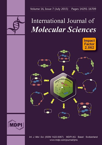
The cover picture shows a schematic representation of the formation of a disulfide combinatorial library. Simple, basic building blocks, from which are formed more complex compounds, are situated in the middle of the picture. Aromatic units of these blocks are represented by hexagons and thiol moieties are represented by yellow dots. Due to the dynamism of the disulfide bond, this bond is able to be broken up and formed again. Consequently, under certain specifically chosen conditions, the mixing of compounds with such bonds contributes to the formation of a dynamic library of disulfide compounds, to which we can include 2-, 3-, and even 4-membered macrocycle rings and catenanes, as presented in the picture. The composition of the library is changeable and can be controlled using both chemical and physical stimuli. View the article.
  • Issues are regarded as officially published after their release is announced to the table of contents alert mailing list.
  • You may sign up for e-mail alerts to receive table of contents of newly released issues.
  • PDF is the official format for papers published in both, html and pdf forms. To view the papers in pdf format, click on the "PDF Full-text" link, and use the free Adobe Reader to open them.
Order results
Result details
Section
Select all
Export citation of selected articles as:
949 KiB  
Article
Characterization of a Functional Role of the Bradyrhizobium japonicum Isocitrate Lyase in Desiccation Tolerance
by Jeong-Min Jeon, Hae-In Lee, Michael J. Sadowsky, Masayuki Sugawara and Woo-Suk Chang
Int. J. Mol. Sci. 2015, 16(7), 16695-16709; https://doi.org/10.3390/ijms160716695 - 22 Jul 2015
Cited by 12 | Viewed by 5014
Abstract
Bradyrhizobium japonicum is a nitrogen-fixing symbiont of soybean. In previous studies, transcriptomic profiling of B. japonicum USDA110, grown under various environmental conditions, revealed the highly induced gene aceA, encoding isocitrate lyase (ICL). The ICL catalyzes the conversion of isocitrate to succinate and [...] Read more.
Bradyrhizobium japonicum is a nitrogen-fixing symbiont of soybean. In previous studies, transcriptomic profiling of B. japonicum USDA110, grown under various environmental conditions, revealed the highly induced gene aceA, encoding isocitrate lyase (ICL). The ICL catalyzes the conversion of isocitrate to succinate and glyoxylate in the glyoxylate bypass of the TCA cycle. Here, we evaluated the functional role of B. japonicum ICL under desiccation-induced stress conditions. We purified AceA (molecular mass = 65 kDa) from B. japonicum USDA110, using a His-tag and Ni-NTA column approach, and confirmed its ICL enzyme activity. The aceA mutant showed higher sensitivity to desiccation stress (27% relative humidity (RH)), compared to the wild type. ICL activity of the wild type strain increased approximately 2.5-fold upon exposure to 27% RH for 24 h. The aceA mutant also showed an increased susceptibility to salt stress. Gene expression analysis of aceA using qRT-PCR revealed a 148-fold induction by desiccation, while other genes involved in the glyoxylate pathway were not differentially expressed in this condition. Transcriptome analyses revealed that stress-related genes, such as chaperones, were upregulated in the wild-type under desiccating conditions, even though fold induction was not dramatic (ca. 1.5–2.5-fold). Full article
(This article belongs to the Section Biochemistry)
Show Figures

Figure 1

698 KiB  
Article
Effects of Plant Growth Hormones on Mucor indicus Growth and Chitosan and Ethanol Production
by Zahra Safaei, Keikhosro Karimi, Poorandokht Golkar and Akram Zamani
Int. J. Mol. Sci. 2015, 16(7), 16683-16694; https://doi.org/10.3390/ijms160716683 - 22 Jul 2015
Cited by 12 | Viewed by 6175
Abstract
The objective of this study was to investigate the effects of indole-3-acetic acid (IAA) and kinetin (KIN) on Mucor indicus growth, cell wall composition, and ethanol production. A semi-synthetic medium, supplemented with 0–5 mg/L hormones, was used for the cultivations (at 32 °C [...] Read more.
The objective of this study was to investigate the effects of indole-3-acetic acid (IAA) and kinetin (KIN) on Mucor indicus growth, cell wall composition, and ethanol production. A semi-synthetic medium, supplemented with 0–5 mg/L hormones, was used for the cultivations (at 32 °C for 48 h). By addition of 1 mg/L of each hormone, the biomass and ethanol yields were increased and decreased, respectively. At higher levels, however, an inverse trend was observed. The glucosamine fraction of the cell wall, as a representative for chitosan, followed similar but sharper changes, compared to the biomass. The highest level was 221% higher than that obtained without hormones. The sum of glucosamine and N-acetyl glucosamine (chitin and chitosan) was noticeably enhanced in the presence of the hormones. Increase of chitosan was accompanied by a decrease in the phosphate content, with the lowest phosphate (0.01 g/g cell wall) being obtained when the chitosan was at the maximum (0.45 g/g cell wall). In conclusion, IAA and KIN significantly enhanced the M. indicus growth and chitosan production, while at the same time decreasing the ethanol yield to some extent. This study shows that plant growth hormones have a high potential for the improvement of fungal chitosan production by M. indicus. Full article
(This article belongs to the Special Issue Chitins 2015)
Show Figures

Graphical abstract

1380 KiB  
Communication
Structural Variation (SV) Markers in the Basidiomycete Volvariella volvacea and Their Application in the Construction of a Genetic Map
by Wei Wang, Bingzhi Chen, Lei Zhang, Junjie Yan, Yuanping Lu, Xiaoyin Zhang, Yuji Jiang, Taju Wu, Arend Frans Van Peer, Shaojie Li and Baogui Xie
Int. J. Mol. Sci. 2015, 16(7), 16669-16682; https://doi.org/10.3390/ijms160716669 - 22 Jul 2015
Cited by 4 | Viewed by 6307
Abstract
Molecular markers and genetic maps are useful tools in genetic studies. Novel molecular markers and their applications have been developed in recent years. With the recent advancements in sequencing technology, the genomic sequences of an increasingly great number of fungi have become available. [...] Read more.
Molecular markers and genetic maps are useful tools in genetic studies. Novel molecular markers and their applications have been developed in recent years. With the recent advancements in sequencing technology, the genomic sequences of an increasingly great number of fungi have become available. A novel type of molecular marker was developed to construct the first reported linkage map of the edible and economically important basidiomycete Volvariella volvacea by using 104 structural variation (SV) markers that are based on the genomic sequences. Because of the special and simple life cycle in basidiomycete, SV markers can be effectively developed by genomic comparison and tested in single spore isolates (SSIs). This stable, convenient and rapidly developed marker may assist in the construction of genetic maps and facilitate genomic research for other species of fungi. Full article
(This article belongs to the Special Issue Microbial Genomics and Metabolomics)
Show Figures

Graphical abstract

1914 KiB  
Article
Fluoxetine Decreases the Proliferation and Adipogenic Differentiation of Human Adipose-Derived Stem Cells
by Bo Kyung Sun, Ji Hye Kim, Joon-Seok Choi, Sung-Joo Hwang and Jong-Hyuk Sung
Int. J. Mol. Sci. 2015, 16(7), 16655-16668; https://doi.org/10.3390/ijms160716655 - 22 Jul 2015
Cited by 19 | Viewed by 5998
Abstract
Fluoxetine was originally developed as an antidepressant, but it has also been used to treat obesity. Although the anti-appetite effect of fluoxetine is well-documented, its potential effects on human adipose-derived stem cells (ASCs) or mature adipocytes have not been investigated. Therefore, we investigated [...] Read more.
Fluoxetine was originally developed as an antidepressant, but it has also been used to treat obesity. Although the anti-appetite effect of fluoxetine is well-documented, its potential effects on human adipose-derived stem cells (ASCs) or mature adipocytes have not been investigated. Therefore, we investigated the mechanisms underlying the inhibitory effects of fluoxetine on the proliferation of ASCs. We also investigated its inhibitory effect on adipogenic differentiation. Fluoxetine significantly decreased ASC proliferation, and signal transduction PCR array analysis showed that it increased expression of autophagy-related genes. In addition, fluoxetine up-regulated SQSTM1 and LC3B protein expression as detected by western blotting and immunofluorescence. The autophagy inhibitor, 3-methyladenine (3-MA), significantly attenuated fluoxetine-mediated effects on ASC proliferation and SQSTM1/LC3B expression. In addition, 3-MA decreased the mRNA expression of two autophagy-related genes, beclin-1 and Atg7, in ASCs. Fluoxetine also significantly inhibited lipid accumulation and down-regulated the levels of PPAR-γ and C/EBP-α in ASCs. Collectively, these results indicate that fluoxetine decreases ASC proliferation and adipogenic differentiation. This is the first in vitro evidence that fluoxetine can reduce fat accumulation by inhibiting ASC proliferation and differentiation. Full article
(This article belongs to the Section Biochemistry)
Show Figures

Graphical abstract

1518 KiB  
Article
Genetically Encoded FRET-Sensor Based on Terbium Chelate and Red Fluorescent Protein for Detection of Caspase-3 Activity
by Alexander S. Goryashchenko, Maria G. Khrenova, Anna A. Bochkova, Tatiana V. Ivashina, Leonid M. Vinokurov and Alexander P. Savitsky
Int. J. Mol. Sci. 2015, 16(7), 16642-16654; https://doi.org/10.3390/ijms160716642 - 22 Jul 2015
Cited by 4 | Viewed by 6180
Abstract
This article describes the genetically encoded caspase-3 FRET-sensor based on the terbium-binding peptide, cleavable linker with caspase-3 recognition site, and red fluorescent protein TagRFP. The engineered construction performs two induction-resonance energy transfer processes: from tryptophan of the terbium-binding peptide to Tb3+ and [...] Read more.
This article describes the genetically encoded caspase-3 FRET-sensor based on the terbium-binding peptide, cleavable linker with caspase-3 recognition site, and red fluorescent protein TagRFP. The engineered construction performs two induction-resonance energy transfer processes: from tryptophan of the terbium-binding peptide to Tb3+ and from sensitized Tb3+ to acceptor—the chromophore of TagRFP. Long-lived terbium-sensitized emission (microseconds), pulse excitation source, and time-resolved detection were utilized to eliminate directly excited TagRFP fluorescence and background cellular autofluorescence, which lasts a fraction of nanosecond, and thus to improve sensitivity of analyses. Furthermore the technique facilitates selective detection of fluorescence, induced by uncleaved acceptor emission. For the first time it was shown that fluorescence resonance energy transfer between sensitized terbium and TagRFP in the engineered construction can be studied via detection of microsecond TagRFP fluorescence intensities. The lifetime and distance distribution between donor and acceptor were calculated using molecular dynamics simulation. Using this data, quantum yield of terbium ions with binding peptide was estimated. Full article
(This article belongs to the Special Issue Förster Resonance Energy Transfer (FRET) 2015)
Show Figures

Figure 1

6689 KiB  
Article
14-3-3γ Regulates Lipopolysaccharide-Induced Inflammatory Responses and Lactation in Dairy Cow Mammary Epithelial Cells by Inhibiting NF-κB and MAPKs and Up-Regulating mTOR Signaling
by Lixin Liu, Ye Lin, Lili Liu, Yanjie Bian, Li Zhang, Xuejun Gao and Qingzhang Li
Int. J. Mol. Sci. 2015, 16(7), 16622-16641; https://doi.org/10.3390/ijms160716622 - 22 Jul 2015
Cited by 28 | Viewed by 7739
Abstract
As a protective factor for lipopolysaccharide (LPS)-induced injury, 14-3-3γ has been the subject of recent research. Nevertheless, whether 14-3-3γ can regulate lactation in dairy cow mammary epithelial cells (DCMECs) induced by LPS remains unknown. Here, the anti-inflammatory effect and lactation regulating ability of [...] Read more.
As a protective factor for lipopolysaccharide (LPS)-induced injury, 14-3-3γ has been the subject of recent research. Nevertheless, whether 14-3-3γ can regulate lactation in dairy cow mammary epithelial cells (DCMECs) induced by LPS remains unknown. Here, the anti-inflammatory effect and lactation regulating ability of 14-3-3γ in LPS-induced DCMECs are investigated for the first time, and the molecular mechanisms responsible for their effects are explored. The results of qRT-PCR showed that 14-3-3γ overexpression significantly inhibited the mRNA expression of tumor necrosis factor-α (TNF-α), interleukin-6 (IL-6), interleukin-1β (IL-1β) and inducible nitric oxide synthase (iNOS). Enzyme-linked immunosorbent assay (ELISA) analysis revealed that 14-3-3γ overexpression also suppressed the production of TNF-α and IL-6 in cell culture supernatants. Meanwhile, CASY-TT Analyser System showed that 14-3-3γ overexpression clearly increased the viability and proliferation of cells. The results of kit methods and western blot analysis showed that 14-3-3γ overexpression promoted the secretion of triglycerides and lactose and the synthesis of β-casein. Furthermore, the expression of genes relevant to nuclear factor-κB (NF-κB) and mitogen-activated protein kinase (MAPKs) and lactation-associated proteins were assessed by western blot, and the results suggested that 14-3-3γ overexpression inactivated the NF-κB and MAPK signaling pathways by down-regulating extracellular signal regulated protein kinase (ERK), p38 mitogen-activated protein kinase (p38MAPK) and inhibitor of NF-κB (IκB) phosphorylation levels, as well as by inhibiting NF-κB translocation. Meanwhile, 14-3-3γ overexpression enhanced the expression levels of β-casein, mammalian target of rapamycin (mTOR), ribosomal protein S6 kinase 1 (S6K1), serine/threonine protein kinase Akt 1 (AKT1), sterol regulatory element binding protein 1 (SREBP1) and peroxisome proliferator-activated receptor gamma (PPARγ). These results suggest that 14-3-3γ was able to attenuate the LPS-induced inflammatory responses and promote proliferation and lactation in LPS-induced DCMECs by inhibiting the activation of the NF-κB and MAPK signaling pathways and up-regulating mTOR signaling pathways to protect against LPS-induced injury. Full article
(This article belongs to the Special Issue Mechanism of Action and Applications of Cytokines in Immunotherapy)
Show Figures

Graphical abstract

870 KiB  
Review
What Do We Know about the Role of miRNAs in Pediatric Sarcoma?
by Lorna C. Kelly, Antonio Lázaro and Maureen J. O'Sullivan
Int. J. Mol. Sci. 2015, 16(7), 16593-16621; https://doi.org/10.3390/ijms160716593 - 22 Jul 2015
Cited by 3 | Viewed by 5598
Abstract
Non-coding RNAs have received a lot of attention in recent years, with especial focus on microRNAs (miRNAs), so much so that in the just over two decades since the first miRNA, Lin4, was described, almost 40,000 publications about miRNAs have been generated. [...] Read more.
Non-coding RNAs have received a lot of attention in recent years, with especial focus on microRNAs (miRNAs), so much so that in the just over two decades since the first miRNA, Lin4, was described, almost 40,000 publications about miRNAs have been generated. Less than 500 of these focus on sarcoma, and only a fraction of those on sarcomas of childhood specifically, with some of these representing observational studies and others containing functionally validated data. This is a group of cancers for which prognosis is often poor and therapeutic options limited, and it is especially in these areas that strides in understanding the role of non-coding RNAs and miRNAs in particular are to be welcomed. This review deals with the main forms of pediatric sarcoma, exploring what is known about the diagnostic and prognostic profiles of miRNAs in these tumours and where novel therapeutic options might present themselves for further exploration. Full article
(This article belongs to the Collection Regulation by Non-coding RNAs)
Show Figures

Graphical abstract

681 KiB  
Review
B Cells and Autoantibodies in Multiple Sclerosis
by Anne-Katrin Pröbstel, Nicholas S. R. Sanderson and Tobias Derfuss
Int. J. Mol. Sci. 2015, 16(7), 16576-16592; https://doi.org/10.3390/ijms160716576 - 21 Jul 2015
Cited by 42 | Viewed by 8172
Abstract
While over the past decades T cells have been considered key players in the pathogenesis of multiple sclerosis (MS), it has only recently become evident that B cells have a major contributing role. Our understanding of the role of B cells has evolved [...] Read more.
While over the past decades T cells have been considered key players in the pathogenesis of multiple sclerosis (MS), it has only recently become evident that B cells have a major contributing role. Our understanding of the role of B cells has evolved substantially following the clinical success of B cell-targeting therapies and increasing experimental evidence for significant B cell involvement. Rather than mere antibody-producing cells, it is becoming clear that they are team players with the capacity to prime and regulate T cells, and function both as pro- and anti-inflammatory mediators. However, despite tremendous efforts, the target antigen(s) of B cells in MS have yet to be identified. The first part of this review summarizes the clinical evidence and results from animal studies pointing to the relevance of B cells in the pathogenesis of MS. The second part gives an overview of the currently known potential autoantigen targets. The third part recapitulates and critically appraises the currently available B cell-directed therapies. Full article
(This article belongs to the Special Issue Advances in Multiple Sclerosis)
2034 KiB  
Article
NOS1AP O-GlcNAc Modification Involved in Neuron Apoptosis Induced by Excitotoxicity
by Liang Zhu, Tao Tao, Dongmei Zhang, Xiaojuan Liu, Kaifu Ke and Aiguo Shen
Int. J. Mol. Sci. 2015, 16(7), 16560-16575; https://doi.org/10.3390/ijms160716560 - 21 Jul 2015
Cited by 11 | Viewed by 6276
Abstract
O-Linked N-acetylglucosamine, or O-GlcNAc, is a dynamic post-translational modification that cycles on and off serine and threonine residues of nucleocytoplasmic and mitochondrial proteins. In addition to cancer and inflammation diseases, O-GlcNAc modification appears to play a critical role during [...] Read more.
O-Linked N-acetylglucosamine, or O-GlcNAc, is a dynamic post-translational modification that cycles on and off serine and threonine residues of nucleocytoplasmic and mitochondrial proteins. In addition to cancer and inflammation diseases, O-GlcNAc modification appears to play a critical role during cell apoptosis and stress response, although the precise mechanisms are still not very clear. Here we found that nitric oxide synthase adaptor (NOS1AP), which plays an important part in glutamate-induced neuronal apoptosis, carries the modification of O-GlcNAc. Mass spectrometry analysis identified Ser47, Ser183, Ser204, Ser269, Ser271 as O-GlcNAc sites. Higher O-GlcNAc of NOS1AP was detected during glutamate-induced neuronal apoptosis. Furthermore, with O-GlcNAc sites of NOS1AP mutated, the interaction of NOS1AP and neuronal nitric oxide syntheses (nNOS) decreases. Finally, during glutamate-induced neuronal apoptosis, decreasing the O-GlcNAc modification of NOS1AP results in more severe neuronal apoptosis. All these results suggest that O-GlcNAc modification of NOS1AP exerts protective effects during glutamate-induced neuronal apoptosis. Full article
(This article belongs to the Special Issue Glycosylation and Glycoproteins)
Show Figures

Graphical abstract

1639 KiB  
Article
High Genetic Diversity of Microbial Cellulase and Hemicellulase Genes in the Hindgut of Holotrichia parallela Larvae
by Ping Sheng, Yushan Li, Sean D. G. Marshall and Hongyu Zhang
Int. J. Mol. Sci. 2015, 16(7), 16545-16559; https://doi.org/10.3390/ijms160716545 - 21 Jul 2015
Cited by 14 | Viewed by 5966
Abstract
In this study, we used a culture-independent method based on library construction and sequencing to analyze the genetic diversity of the cellulase and hemicellulase genes of the bacterial community resident in the hindgut of Holotrichia parallela larvae. The results indicate that there is [...] Read more.
In this study, we used a culture-independent method based on library construction and sequencing to analyze the genetic diversity of the cellulase and hemicellulase genes of the bacterial community resident in the hindgut of Holotrichia parallela larvae. The results indicate that there is a large, diverse set of bacterial genes encoding lignocellulose hydrolysis enzymes in the hindgut of H. parallela. The total of 101 distinct gene fragments (similarity <95%) of glycosyl hydrolase families including GH2 (24 genes), GH8 (27 genes), GH10 (19 genes), GH11 (14 genes) and GH36 (17 genes) families was retrieved, and certain sequences of GH2 (10.61%), GH8 (3.33%), and GH11 (18.42%) families had <60% identities with known sequences in GenBank, indicating their novelty. Based on phylogenetic analysis, sequences from hemicellulase families were related to enzymes from Bacteroidetes and Firmicutes. Fragments from cellulase family were most associated with the phylum of Proteobacteria. Furthermore, a full-length endo-xylanase gene was obtained, and the enzyme exhibited activity over a broad range of pH levels. Our results indicate that there are large number of cellulolytic and xylanolytic bacteria in the hindgut of H. parallela larvae, and these symbiotic bacteria play an important role in the degradation of roots and other organic matter for the host insect. Full article
(This article belongs to the Section Biochemistry)
Show Figures

Graphical abstract

2851 KiB  
Article
Differential Antioxidant Responses and Perturbed Porphyrin Biosynthesis after Exposure to Oxyfluorfen and Methyl Viologen in Oryza sativa
by Nhi-Thi Pham, Jin-Gil Kim and Sunyo Jung
Int. J. Mol. Sci. 2015, 16(7), 16529-16544; https://doi.org/10.3390/ijms160716529 - 21 Jul 2015
Cited by 13 | Viewed by 6104
Abstract
We compared antioxidant responses and regulation of porphyrin metabolism in rice plants treated with oxyfluorfen (OF) or methyl viologen (MV). Plants treated with MV exhibited not only greater increases in conductivity and malondialdehyde but also a greater decline in Fv/Fm [...] Read more.
We compared antioxidant responses and regulation of porphyrin metabolism in rice plants treated with oxyfluorfen (OF) or methyl viologen (MV). Plants treated with MV exhibited not only greater increases in conductivity and malondialdehyde but also a greater decline in Fv/Fm, compared to plants treated with OF. MV-treated plants had greater increases in activities of superoxide dismutase (SOD) and catalase (CAT) as well as transcript levels of SODA and CATA than OF-treated plants after 28 h of the treatments, whereas increases in ascorbate peroxidase (APX) activity and transcript levels of APXA and APXB were greater in OF-treated plants. Both OF- and MV-treated plants resulted in not only down-regulation of most genes involved in porphyrin biosynthesis but also disappearance of Mg-porphyrins during the late stage of photooxidative stress. By contrast, up-regulation of heme oxygenase 2 (HO2) is possibly part of an efficient antioxidant response to compensate photooxidative damage in both treatments. Our data show that down-regulated biosynthesis and degradation dynamics of porphyrin intermediates have important roles in photoprotection of plants from perturbed porphyrin biosynthesis and photosynthetic electron transport. This study suggests that porphyrin scavenging as well as strong antioxidative activities are required for mitigating reactive oxygen species (ROS) production under photooxidative stress caused by OF and MV. Full article
(This article belongs to the Special Issue Plant Molecular Biology)
Show Figures

Figure 1

715 KiB  
Article
Sleep Disorders Reduce Health-Related Quality of Life in Multiple Sclerosis (Nottingham Health Profile Data in Patients with Multiple Sclerosis)
by Christian Veauthier, Gunnar Gaede, Helena Radbruch, Klaus-Dieter Wernecke and Friedemann Paul
Int. J. Mol. Sci. 2015, 16(7), 16514-16528; https://doi.org/10.3390/ijms160716514 - 21 Jul 2015
Cited by 38 | Viewed by 7403
Abstract
Quality of Life (QoL) is decreased in multiple sclerosis (MS), but studies about the impact of sleep disorders (SD) on health-related quality of Life (HRQoL) are lacking. From our original cohort, a cross-sectional polysomnographic (PSG) study in consecutive MS patients, we retrospectively analysed [...] Read more.
Quality of Life (QoL) is decreased in multiple sclerosis (MS), but studies about the impact of sleep disorders (SD) on health-related quality of Life (HRQoL) are lacking. From our original cohort, a cross-sectional polysomnographic (PSG) study in consecutive MS patients, we retrospectively analysed the previously unpublished data of the Nottingham Health Profile (NHP). Those MS patients suffering from sleep disorders (n = 49) showed significantly lower HRQoL compared to MS patients without sleep disorders (n = 17). Subsequently, we classified the patients into four subgroups: insomnia (n = 17), restless-legs syndrome, periodic limb movement disorder and SD due to leg pain (n = 24), obstructive sleep apnea (n = 8) and patients without sleep disorder (n = 17). OSA and insomnia patients showed significantly higher NHP values and decreased HRQoL not only for the sleep subscale but also for the “energy” and “emotional” area of the NHP. In addition, OSA patients also showed increased NHP values in the “physical abilities” area. Interestingly, we did not find a correlation between the objective PSG parameters and the subjective sleep items of the NHP. However, this study demonstrates that sleep disorders can reduce HRQoL in MS patients and should be considered as an important confounder in all studies investigating HRQoL in MS. Full article
(This article belongs to the Special Issue Advances in Multiple Sclerosis)
Show Figures

Graphical abstract

2707 KiB  
Article
Morphogenesis, Flowering, and Gene Expression of Dendranthema grandiflorum in Response to Shift in Light Quality of Night Interruption
by Yoo Gyeong Park, Sowbiya Muneer and Byoung Ryong Jeong
Int. J. Mol. Sci. 2015, 16(7), 16497-16513; https://doi.org/10.3390/ijms160716497 - 21 Jul 2015
Cited by 35 | Viewed by 6973
Abstract
The impact of shifts in the spectral quality of light on morphogenesis, flowering, and photoperiodic gene expression during exposure to light quality of night interruption (NI) was investigated in Dendranthema grandiflorum. The circadian rhythms of plants grown in a closed walk-in growth [...] Read more.
The impact of shifts in the spectral quality of light on morphogenesis, flowering, and photoperiodic gene expression during exposure to light quality of night interruption (NI) was investigated in Dendranthema grandiflorum. The circadian rhythms of plants grown in a closed walk-in growth chamber were interrupted at night for a total of 4 h, using light-emitting diodes with an intensity of 10 μmol·m2·s1 PPF. The light quality of the NI was shifted from one wavelength to another after the first 2 h. Light treatments consisting of all possible pairings of blue (B), red (R), far-red (Fr), and white (W) light were tested. Plants in the NI treatment groups exposed to Fr light grew larger than plants in other treatment groups. Of plants in NI treatment groups, those in the NI-WB treatment grew the least. In addition, the impact of shifts in the light quality of NI on leaf expansion was greater in treatment groups exposed to a combination of either B and R or R and W light, regardless of their order of supply. Flowering was observed in the NI-RB, NI-FrR, NI-BFr, NI-FrB, NI-WB, NI-FrW, NI-WFr, NI-WR, and SD (short-day) treatments, and was especially promoted in the NI-BFr and NI-FrB treatments. In a combined shift treatment of B and R or B and W light, the NI concluded with B light (NI-RB and NI-WB) treatment induced flowering. The transcriptional factors phyA, cry1 and FTL (FLOWERING LOCUS T) were positively affected, while phyB and AFT were negatively affected. In conclusion, morphogenesis, flowering, and transcriptional factors were all significantly affected either positively or negatively by shifts in the light quality of NI. The light quality of the first 2 h of NI affected neither morphogenesis nor flowering, while the light quality of the last 2 h of NI significantly affected both morphogenesis and flowering. Full article
(This article belongs to the Special Issue Plant Molecular Biology)
Show Figures

Figure 1

698 KiB  
Article
Association between the NF-E2 Related Factor 2 Gene Polymorphism and Oxidative Stress, Anti-Oxidative Status, and Newly-Diagnosed Type 2 Diabetes Mellitus in a Chinese Population
by Xia Wang, Hongxia Chen, Jun Liu, Yingying Ouyang, Di Wang, Wei Bao and Liegang Liu
Int. J. Mol. Sci. 2015, 16(7), 16483-16496; https://doi.org/10.3390/ijms160716483 - 20 Jul 2015
Cited by 36 | Viewed by 6293
Abstract
Oxidative stress is a major risk factor in the onset and progression of type 2 diabetes mellitus (T2DM). NF-E2 related factor 2 (NRF2) is a pivotal transcription factor in oxidative stress related illnesses. This study included 2174 subjects with 879 cases [...] Read more.
Oxidative stress is a major risk factor in the onset and progression of type 2 diabetes mellitus (T2DM). NF-E2 related factor 2 (NRF2) is a pivotal transcription factor in oxidative stress related illnesses. This study included 2174 subjects with 879 cases of newly-diagnosed T2DM and 1295 healthy controls. Compared to individuals with the CC genotype, those with the AA genotype had lower total anti-oxidative capacity, superoxide dismutase, catalase, glutathione, glutathione peroxidase activity; and lower homeostasis model assessment of β-cell function index. Those with the AA genotype also had a higher malondialdehyde concentration and homeostasis model assessment of insulin resistance index values. The frequency of allele A was significantly higher in T2DM subjects (29.4%), compared to control subjects (26.1%; p = 0.019). Individuals with the AA genotype had a significantly higher risk of developing T2DM (OR 1.56; 95% CI 1.11, 2.20; p = 0.011), relative to those with the CC genotype, even after adjusting for known T2DM risk factors. Our results suggest that the NRF2 rs6721961 polymorphism was significantly associated with oxidative stress, anti-oxidative status, and risk of newly-diagnosed T2DM. This polymorphism may also contribute to impaired insulin secretory capacity and increased insulin resistance in a Chinese population. Full article
(This article belongs to the Special Issue Human Single Nucleotide Polymorphisms and Disease Diagnostics)
2591 KiB  
Article
Sinulariolide Suppresses Human Hepatocellular Carcinoma Cell Migration and Invasion by Inhibiting Matrix Metalloproteinase-2/-9 through MAPKs and PI3K/Akt Signaling Pathways
by Yu-Jen Wu, Choo-Aun Neoh, Chia-Yu Tsao, Jui-Hsin Su and Hsing-Hui Li
Int. J. Mol. Sci. 2015, 16(7), 16469-16482; https://doi.org/10.3390/ijms160716469 - 20 Jul 2015
Cited by 75 | Viewed by 14064
Abstract
Sinulariolide is an active compound isolated from the cultured soft coral Sinularia flexibilis. In this study, we investigate the migration and invasion effects of sinulariolide in hepatocellular carcinoma cell HA22T. Sinulariolide inhibited the migration and invasion effects of hepatocellular carcinoma cells in [...] Read more.
Sinulariolide is an active compound isolated from the cultured soft coral Sinularia flexibilis. In this study, we investigate the migration and invasion effects of sinulariolide in hepatocellular carcinoma cell HA22T. Sinulariolide inhibited the migration and invasion effects of hepatocellular carcinoma cells in a concentration-dependent manner. The results of zymography assay showed that sinulariolide suppressed the activities of matrix metalloproteinase (MMP)-2 and MMP-9. Moreover, protein levels of MMP-2, MMP-9, and urokinase-type plasminogen activator (uPA) were reduced by sinulariolide in a concentration-dependent manner. Sinulariolide also exerted an inhibitory effect on phosphorylation of c-Jun N-terminal kinase (JNK), extracellular signal-regulated kinases (ERK), phosphatidylinositol 3-kinase (PI3K), Akt, Focal adhesion kinase (FAK), growth factor receptor-bound protein 2 (GRB2). Taken together, these results demonstrated that sinulariolide could inhibit hepatocellular carcinoma cell migration and invasion and alter HA22T cell metastasis by reduction of MMP-2, MMP-9, and uPA expression through the suppression of MAPKs, PI3K/Akt, and the FAK/GRB2 signaling pathway. These findings suggest that sinulariolide merits further evaluation as a chemotherapeutic agent for human hepatocellular carcinoma. Full article
(This article belongs to the Special Issue Molecular Classification of Human Cancer: Diagnosis and Treatment)
Show Figures

Graphical abstract

6304 KiB  
Article
Baicalin Protects Mice from Aristolochic Acid I-Induced Kidney Injury by Induction of CYP1A through the Aromatic Hydrocarbon Receptor
by Ke Wang, Chenchen Feng, Chenggang Li, Jun Yao, Xiaofeng Xie, Likun Gong, Yang Luan, Guozhen Xing, Xue Zhu, Xinming Qi and Jin Ren
Int. J. Mol. Sci. 2015, 16(7), 16454-16468; https://doi.org/10.3390/ijms160716454 - 20 Jul 2015
Cited by 25 | Viewed by 7128
Abstract
Exposure to aristolochic acid I (AAI) can lead to aristolochic acid nephropathy (AAN), Balkan endemic nephropathy (BEN) and urothelial cancer. The induction of hepatic CYP1A, especially CYP1A2, was considered to detoxify AAI so as to reduce its nephrotoxicity. We previously found that baicalin [...] Read more.
Exposure to aristolochic acid I (AAI) can lead to aristolochic acid nephropathy (AAN), Balkan endemic nephropathy (BEN) and urothelial cancer. The induction of hepatic CYP1A, especially CYP1A2, was considered to detoxify AAI so as to reduce its nephrotoxicity. We previously found that baicalin had the strong ability to induce CYP1A2 expression; therefore in this study, we examined the effects of baicalin on AAI toxicity, metabolism and disposition, as well as investigated the underlying mechanisms. Our toxicological studies showed that baicalin reduced the levels of blood urea nitrogen (BUN) and creatinine (CRE) in AAI-treated mice and attenuated renal injury induced by AAI. Pharmacokinetic analysis demonstrated that baicalin markedly decreased AUC of AAI in plasma and the content of AAI in liver and kidney. CYP1A induction assays showed that baicalin exposure significantly increased the hepatic expression of CYP1A1/2, which was completely abolished by inhibitors of the Aromatic hydrocarbon receptor (AhR), 3ʹ,4ʹ-dimethoxyflavone and resveratrol, in vitro and in vivo, respectively. Moreover, the luciferase assays revealed that baicalin significantly increased the luciferase activity of the reporter gene incorporated with the Xenobiotic response elements recognized by AhR. In summary, baicalin significantly reduced the disposition of AAI and ameliorated AAI-induced kidney toxicity through AhR-dependent CYP1A1/2 induction in the liver. Full article
Show Figures

Graphical abstract

1480 KiB  
Article
Inhibition of Mammalian Target of Rapamycin Complex 1 (mTORC1) Downregulates ELOVL1 Gene Expression and Fatty Acid Synthesis in Goat Fetal Fibroblasts
by Weipeng Wang, Qiburi He, Zhixin Guo, Limin Yang, Lili Bao, Wenlei Bao, Xu Zheng, Yanfeng Wang and Zhigang Wang
Int. J. Mol. Sci. 2015, 16(7), 16440-16453; https://doi.org/10.3390/ijms160716440 - 20 Jul 2015
Cited by 11 | Viewed by 5860
Abstract
Elongation of very-long-chain fatty acids 1 (ELOVL1) is a ubiquitously expressed gene that belongs to the ELOVL family and regulates the synthesis of very-long-chain fatty acids (VLCFAs) and sphingolipids, from yeast to mammals. Mammalian target of rapamycin complex 1 (mTORC1) is [...] Read more.
Elongation of very-long-chain fatty acids 1 (ELOVL1) is a ubiquitously expressed gene that belongs to the ELOVL family and regulates the synthesis of very-long-chain fatty acids (VLCFAs) and sphingolipids, from yeast to mammals. Mammalian target of rapamycin complex 1 (mTORC1) is a central regulator of cell metabolism and is associated with fatty acids synthesis. In this study, we cloned the cDNA that encodes Cashmere goat (Capra hircus) ELOVL1 (GenBank Accession number KF549985) and investigated its expression in 10 tissues. ELOVL1 cDNA was 840 bp, encoding a deduced protein of 279 amino acids, and ELOVL1 mRNA was expressed in a wide range of tissues. Inhibition of mTORC1 by rapamycin decreased ELOVL1 expression and fatty acids synthesis in Cashmere goat fetal fibroblasts. These data show that ELOVL1 expression is regulated by mTORC1 and that mTORC1 has significant function in fatty acids synthesis in Cashmere goat. Full article
(This article belongs to the Section Biochemistry)
Show Figures

Figure 1

722 KiB  
Review
Alemtuzumab in Multiple Sclerosis: Mechanism of Action and Beyond
by Tobias Ruck, Stefan Bittner, Heinz Wiendl and Sven G. Meuth
Int. J. Mol. Sci. 2015, 16(7), 16414-16439; https://doi.org/10.3390/ijms160716414 - 20 Jul 2015
Cited by 153 | Viewed by 14190
Abstract
Alemtuzumab is a humanized monoclonal antibody against CD52 (cluster of differentiation 52) and is approved for the therapy of relapsing-remitting multiple sclerosis. The application of alemtuzumab leads to a rapid, but long-lasting depletion predominantly of CD52-bearing B and T cells with reprogramming effects [...] Read more.
Alemtuzumab is a humanized monoclonal antibody against CD52 (cluster of differentiation 52) and is approved for the therapy of relapsing-remitting multiple sclerosis. The application of alemtuzumab leads to a rapid, but long-lasting depletion predominantly of CD52-bearing B and T cells with reprogramming effects on immune cell composition resulting in the restoration of tolerogenic networks. Alemtuzumab has proven high efficacy in clinical phase II and III trials, where interferon β-1a was used as active comparator. However, alemtuzumab is associated with frequent and considerable risks. Most importantly secondary autoimmune disease affects 30%–40% of patients, predominantly impairing thyroid function. Extensive monitoring and early intervention allow for an appropriate risk management. However, new and reliable biomarkers for individual risk stratification and treatment response to improve patient selection and therapy guidance are a significant unmet need. Only a deeper understanding of the underlying mechanisms of action (MOA) will reveal such markers, maximizing the best potential risk-benefit ratio for the individual patient. This review provides and analyses the current knowledge on the MOA of alemtuzumab. Most recent data on efficacy and safety of alemtuzumab are presented and future research opportunities are discussed. Full article
(This article belongs to the Special Issue Advances in Multiple Sclerosis)
5165 KiB  
Article
A New Ligustrazine Derivative-Selective Cytotoxicity by Suppression of NF-κB/p65 and COX-2 Expression on Human Hepatoma Cells. Part 3
by Chenze Zhang, Wenqiang Yan, Bi Li, Bing Xu, Yan Gong, Fuhao Chu, Yuzhong Zhang, Qiuli Yao, Penglong Wang and Haimin Lei
Int. J. Mol. Sci. 2015, 16(7), 16401-16413; https://doi.org/10.3390/ijms160716401 - 17 Jul 2015
Cited by 19 | Viewed by 6156
Abstract
A new anticancer ligustrazine derivative, 3β-hydroxyolea-12-en-28-oic acid- 3,5,6-trimethylpyrazin-2-methylester (T-OA, C38H58O3N2), was previously reported. It was synthesized via conjugating hepatoprotective and anticancer ingredients of traditional Chinese medicine. We found that T-OA exerted its anticancer activity by [...] Read more.
A new anticancer ligustrazine derivative, 3β-hydroxyolea-12-en-28-oic acid- 3,5,6-trimethylpyrazin-2-methylester (T-OA, C38H58O3N2), was previously reported. It was synthesized via conjugating hepatoprotective and anticancer ingredients of traditional Chinese medicine. We found that T-OA exerted its anticancer activity by preventing the expression of nuclear transcription factor NF-κB/p65 and COX-2 in S180 mice. However, the selective cytotoxicity of T-OA on various kinds of cell lines has not been studied sufficiently. In the present study, compared with Cisplatin, T-OA was more toxic to human hepatoma cell line Bel-7402 (IC50 = 6.36 ± 1.56 µM) than other three cancer cell lines (HeLa, HT-29, BGC-823), and no toxicity was observed toward Madin–Darby canine kidney cell line MDCK (IC50 > 150 µM). The morphological changes of Bel-7402 cells demonstrated that T-OA had an apoptosis-inducing effect which had been substantiated using 4ʹ,6-diamidino-2-phenylindole (DAPI) staining, acridine orange (AO)/ethidium bromide (EB) staining, flow cytometry and mitochondrial membrane potential assay. Combining the immumohistochemical staining, we found T-OA could prevent the expression of NF-κB/p65 and COX-2 in Bel-7402 cells. Both of the proteins have been known to play roles in apoptosis and are mainly located in the nuclei. Moreover subcellular localization was performed to reveal that T-OA exerts in nuclei of Bel-7402 cells. The result was in accordance with the effects of down-regulating the expression of NF-κB/p65 and COX-2. Full article
(This article belongs to the Section Biochemistry)
Show Figures

Graphical abstract

714 KiB  
Review
Network Candidate Genes in Breeding for Drought Tolerant Crops
by Christoph Tim Krannich, Lisa Maletzki, Christina Kurowsky and Renate Horn
Int. J. Mol. Sci. 2015, 16(7), 16378-16400; https://doi.org/10.3390/ijms160716378 - 17 Jul 2015
Cited by 56 | Viewed by 8812
Abstract
Climate change leading to increased periods of low water availability as well as increasing demands for food in the coming years makes breeding for drought tolerant crops a high priority. Plants have developed diverse strategies and mechanisms to survive drought stress. However, most [...] Read more.
Climate change leading to increased periods of low water availability as well as increasing demands for food in the coming years makes breeding for drought tolerant crops a high priority. Plants have developed diverse strategies and mechanisms to survive drought stress. However, most of these represent drought escape or avoidance strategies like early flowering or low stomatal conductance that are not applicable in breeding for crops with high yields under drought conditions. Even though a great deal of research is ongoing, especially in cereals, in this regard, not all mechanisms involved in drought tolerance are yet understood. The identification of candidate genes for drought tolerance that have a high potential to be used for breeding drought tolerant crops represents a challenge. Breeding for drought tolerant crops has to focus on acceptable yields under water-limited conditions and not on survival. However, as more and more knowledge about the complex networks and the cross talk during drought is available, more options are revealed. In addition, it has to be considered that conditioning a crop for drought tolerance might require the production of metabolites and might cost the plants energy and resources that cannot be used in terms of yield. Recent research indicates that yield penalty exists and efficient breeding for drought tolerant crops with acceptable yields under well-watered and drought conditions might require uncoupling yield penalty from drought tolerance. Full article
(This article belongs to the Special Issue Abiotic Stress and Gene Networks in Plants)
Show Figures

Graphical abstract

8986 KiB  
Article
Transcriptome Analysis and Discovery of Genes Involved in Immune Pathways from Coelomocytes of Sea Cucumber (Apostichopus japonicus) after Vibrio splendidus Challenge
by Qiong Gao, Meijie Liao, Yingeng Wang, Bin Li, Zheng Zhang, Xiaojun Rong, Guiping Chen and Lan Wang
Int. J. Mol. Sci. 2015, 16(7), 16347-16377; https://doi.org/10.3390/ijms160716347 - 17 Jul 2015
Cited by 38 | Viewed by 8075
Abstract
Vibrio splendidus is identified as one of the major pathogenic factors for the skin ulceration syndrome in sea cucumber (Apostichopus japonicus), which has vastly limited the development of the sea cucumber culture industry. In order to screen the immune genes involving [...] Read more.
Vibrio splendidus is identified as one of the major pathogenic factors for the skin ulceration syndrome in sea cucumber (Apostichopus japonicus), which has vastly limited the development of the sea cucumber culture industry. In order to screen the immune genes involving Vibrio splendidus challenge in sea cucumber and explore the molecular mechanism of this process, the related transcriptome and gene expression profiling of resistant and susceptible biotypes of sea cucumber with Vibrio splendidus challenge were collected for analysis. A total of 319,455,942 trimmed reads were obtained, which were assembled into 186,658 contigs. After that, 89,891 representative contigs (without isoform) were clustered. The analysis of the gene expression profiling identified 358 differentially expression genes (DEGs) in the bacterial-resistant group, and 102 DEGs in the bacterial-susceptible group, compared with that in control group. According to the reported references and annotation information from BLAST, GO and KEGG, 30 putative bacterial-resistant genes and 19 putative bacterial-susceptible genes were identified from DEGs. The qRT-PCR results were consistent with the RNA-Seq results. Furthermore, many DGEs were involved in immune signaling related pathways, such as Endocytosis, Lysosome, MAPK, Chemokine and the ERBB signaling pathway. Full article
(This article belongs to the Special Issue Fish Molecular Biology)
Show Figures

Figure 1

2028 KiB  
Article
Interferon-Beta Therapy of Multiple Sclerosis Patients Improves the Responsiveness of T Cells for Immune Suppression by Regulatory T Cells
by Bettina Trinschek, Felix Luessi, Catharina C. Gross, Heinz Wiendl and Helmut Jonuleit
Int. J. Mol. Sci. 2015, 16(7), 16330-16346; https://doi.org/10.3390/ijms160716330 - 17 Jul 2015
Cited by 26 | Viewed by 8727
Abstract
Multiple sclerosis (MS) is an inflammatory autoimmune disease characterized by imbalanced immune regulatory networks, and MS patient-derived T effector cells are inefficiently suppressed through regulatory T cells (Treg), a phenomenon known as Treg resistance. In the current study we investigated T cell function [...] Read more.
Multiple sclerosis (MS) is an inflammatory autoimmune disease characterized by imbalanced immune regulatory networks, and MS patient-derived T effector cells are inefficiently suppressed through regulatory T cells (Treg), a phenomenon known as Treg resistance. In the current study we investigated T cell function in MS patients before and after interferon-beta therapy. We compared cytokine profile, responsiveness for Treg-mediated suppression ex vivo and evaluated reactivity of T cells in vivo using a humanized mouse model. We found that CD4+ and CD8+ T cells of therapy-naive MS patients were resistant to Treg-mediated suppression. Treg resistance is associated with an augmented IL-6 production, enhanced IL-6 receptor expression, and increased PKB/c-Akt phosphorylation. These parameters as well as responsiveness of T cells to Treg-mediated suppression were restored after interferon-beta therapy of MS patients. Following transfer into immunodeficient mice, MS T cells induced a lethal graft versus host disease (GvHD) and in contrast to T cells of healthy volunteers, this aggressive T cell response could not be controlled by Treg, but was abolished by anti-IL-6 receptor antibodies. However, magnitude and lethality of GvHD induced by MS T cells was significantly decreased after interferon-beta therapy and the reaction was prevented by Treg activation in vivo. Our data reveals that interferon-beta therapy improves the immunoregulation of autoaggressive T effector cells in MS patients by changing the IL-6 signal transduction pathway, thus restoring their sensitivity to Treg-mediated suppression. Full article
(This article belongs to the Special Issue Advances in Multiple Sclerosis)
Show Figures

Graphical abstract

2301 KiB  
Article
The Wnt11 Signaling Pathway in Potential Cellular EMT and Osteochondral Differentiation Progression in Nephrolithiasis Formation
by Deng He, Yuchao Lu, Henglong Hu, Jiaqiao Zhang, Baolong Qin, Yufeng Wang, Shuai Xing, Qilin Xi and Shaogang Wang
Int. J. Mol. Sci. 2015, 16(7), 16313-16329; https://doi.org/10.3390/ijms160716313 - 17 Jul 2015
Cited by 12 | Viewed by 5546
Abstract
The molecular events leading to nephrolithiasis are extremely complex. Previous studies demonstrated that calcium and transforming growth factor-β1 (TGF-β1) may participate in the pathogenesis of stone formation, but the explicit mechanism has not been defined. Using a self-created genetic hypercalciuric stone-forming (GHS) rat [...] Read more.
The molecular events leading to nephrolithiasis are extremely complex. Previous studies demonstrated that calcium and transforming growth factor-β1 (TGF-β1) may participate in the pathogenesis of stone formation, but the explicit mechanism has not been defined. Using a self-created genetic hypercalciuric stone-forming (GHS) rat model, we observed that the increased level of serous/uric TGF-β1 and elevated intracellular calcium in primary renal tubular epithelial cells (PRECs) was associated with nephrolithiasis progression in vivo. In the setting of high calcium plus high TGF-β1 in vitro, PRECs showed great potential epithelial to mesenchymal transition (EMT) progression and osteochondral differentiation properties, representing the multifarious increased mesenchymal and osteochondral phenotypes (Zeb1, Snail1, Col2A1, OPN, Sox9, Runx2) and decreased epithelial phenotypes (E-cadherin, CK19) bythe detection of mRNAs and corresponding proteins. Moreover, TGF-β-dependent Wnt11 knockdown and L-type Ca2+ channel blocker could greatly reverse EMT progression and osteochondral differentiation in PRECs. TGF-β1 alone could effectively promote EMT, but it had no effect on osteochondral differentiation in NRK cells (Rat kidney epithelial cell line). Stimulation with Ca2+ alone did not accelerate differentiation of NRK. Co-incubation of extracellular Ca2+ and TGF-β1 synergistically promotes EMT and osteochondral differentiation in NRK control cells. Our data supplied a novel view that the pathogenesis of calcium stone development may be associated with synergic effects of TGF-β1 and Ca2+, which promote EMT and osteochondral differentiation via Wnt11 and the L-type calcium channel. Full article
(This article belongs to the Section Biochemistry)
Show Figures

Figure 1

1359 KiB  
Article
Generation of a Multicomponent Library of Disulfide Donor-Acceptor Architectures Using Dynamic Combinatorial Chemistry
by Wojciech Drożdż, Michał Kołodziejski, Grzegorz Markiewicz, Anna Jenczak and Artur R. Stefankiewicz
Int. J. Mol. Sci. 2015, 16(7), 16300-16312; https://doi.org/10.3390/ijms160716300 - 17 Jul 2015
Cited by 10 | Viewed by 6934
Abstract
We describe here the generation of new donor-acceptor disulfide architectures obtained in aqueous solution at physiological pH. The application of a dynamic combinatorial chemistry approach allowed us to generate a large number of new disulfide macrocyclic architectures together with a new type of [...] Read more.
We describe here the generation of new donor-acceptor disulfide architectures obtained in aqueous solution at physiological pH. The application of a dynamic combinatorial chemistry approach allowed us to generate a large number of new disulfide macrocyclic architectures together with a new type of [2]catenanes consisting of four distinct components. Up to fifteen types of structurally-distinct dynamic architectures have been generated through one-pot disulfide exchange reactions between four thiol-functionalized aqueous components. The distribution of disulfide products formed was found to be strongly dependent on the structural features of the thiol components employed. This work not only constitutes a success in the synthesis of topologically- and morphologically-complex targets, but it may also open new horizons for the use of this methodology in the construction of molecular machines. Full article
(This article belongs to the Special Issue Supramolecular Interactions)
Show Figures

Graphical abstract

1029 KiB  
Article
3-O-Acyl-epicatechins Increase Glucose Uptake Activity and GLUT4 Translocation through Activation of PI3K Signaling in Skeletal Muscle Cells
by Manabu Ueda-Wakagi, Rie Mukai, Naoya Fuse, Yoshiyuki Mizushina and Hitoshi Ashida
Int. J. Mol. Sci. 2015, 16(7), 16288-16299; https://doi.org/10.3390/ijms160716288 - 17 Jul 2015
Cited by 23 | Viewed by 6149
Abstract
Tea catechins promote glucose uptake in skeletal muscle cells. In this study, we investigated whether the addition of an acyl group to the C-3 position of catechins to generate 3-O-acyl-catechins promoted glucose uptake in L6 myotubes. 3-O-Myristoyl-(−)-epicatechin (EC-C14) and [...] Read more.
Tea catechins promote glucose uptake in skeletal muscle cells. In this study, we investigated whether the addition of an acyl group to the C-3 position of catechins to generate 3-O-acyl-catechins promoted glucose uptake in L6 myotubes. 3-O-Myristoyl-(−)-epicatechin (EC-C14) and 3-O-palmitoyl-(−)-epicatechin (EC-C16) promoted glucose uptake and translocation of glucose transporter (GLUT) 4 in the cells. The effect of 3-O-acyl-(−)-epicatechins was stronger than that of (−)-epicatechin (EC), whereas neither 3-O-myristoyl-(+)-catechin (C-C14) nor 3-O-palmitoyl-(+)catechin (C-C16) promoted glucose uptake or GLUT4 translocation as well as (+)-catechin (C). We further investigated an affinity of catechins and 3-O-acyl-catechins to the lipid bilayer membrane by using surface plasma resonance analysis. Maximum binding amounts of EC-C16 and C-C16 to the lipid bilayer clearly increased compared with that of (−)-EC and (+)-C, respectively. We also examined the mechanism of GLUT4 translocation and found EC-C14 and EC-C16 induced the phosphorylation of PI3K, but did not affect phosphorylation of Akt or IR. In conclusion, the addition of an acyl group to the C-3 position of (−)-EC increases its affinity for the lipid bilayer membrane and promotes GLUT4 translocation through PI3K-dependent pathways in L6 myotubes. Full article
(This article belongs to the Section Biochemistry)
Show Figures

Graphical abstract

849 KiB  
Review
Experimental Evidence Shows Salubrinal, an eIF2α Dephosphorylation Inhibitor, Reduces Xenotoxicant-Induced Cellular Damage
by Masato Matsuoka and Yuta Komoike
Int. J. Mol. Sci. 2015, 16(7), 16275-16287; https://doi.org/10.3390/ijms160716275 - 17 Jul 2015
Cited by 52 | Viewed by 8624
Abstract
Accumulating evidence indicates that endoplasmic reticulum (ER) stress and the subsequent unfolded protein response (UPR) are involved in the pathogenesis of not only the protein misfolding disorders such as certain neurodegenerative and metabolic diseases, but also in the cytotoxicity of environmental pollutants, industrial [...] Read more.
Accumulating evidence indicates that endoplasmic reticulum (ER) stress and the subsequent unfolded protein response (UPR) are involved in the pathogenesis of not only the protein misfolding disorders such as certain neurodegenerative and metabolic diseases, but also in the cytotoxicity of environmental pollutants, industrial chemicals, and drugs. Thus, the modulation of ER stress signaling pathways is an important issue for protection against cellular damage induced by xenotoxicants. The substance salubrinal has been shown to prevent dephosphorylation of the eukaryotic translation initiation factor 2 alpha (eIF2α). The phosphorylation of eIF2α appears to be cytoprotective during ER stress, because inhibition of the translation initiation activity of eIF2α reduces global protein synthesis. In addition, the expression of activating transcription factor 4 (ATF4), a transcription factor that induces the expression of UPR target genes, is up-regulated through alternative translation. This review shows that salubrinal can protect cells from the damage induced by a wide range of xenotoxicants, including environmental pollutants and drugs. The canonical and other possible mechanisms of cytoprotection by salubrinal from xenotoxicant-induced ER stress are also discussed. Full article
(This article belongs to the Special Issue Modulators of Endoplasmic Reticulum Stress)
Show Figures

Figure 1

1672 KiB  
Article
RGD Peptides-Conjugated Pluronic Triblock Copolymers Encapsulated with AP-2α Expression Plasmid for Targeting Gastric Cancer Therapy in Vitro and in Vivo
by Wei Wang, Zhimin Liu, Peng Sun, Cheng Fang, Hongwei Fang, Yueming Wang, Jiajia Ji and Jun Chen
Int. J. Mol. Sci. 2015, 16(7), 16263-16274; https://doi.org/10.3390/ijms160716263 - 17 Jul 2015
Cited by 11 | Viewed by 5179
Abstract
Gastric cancer, a high-risk malignancy, is a genetic disease developing from a cooperation of multiple gene mutations and a multistep process. Gene therapy is a novel treatment method for treating gastric cancer. Here, we developed a novel Arg-Gly-Asp (RGD) peptides conjugated copolymers nanoparticles-based [...] Read more.
Gastric cancer, a high-risk malignancy, is a genetic disease developing from a cooperation of multiple gene mutations and a multistep process. Gene therapy is a novel treatment method for treating gastric cancer. Here, we developed a novel Arg-Gly-Asp (RGD) peptides conjugated copolymers nanoparticles-based gene delivery system in order to actively targeting inhibit the growth of gastric cancer cells. These transcription factor (AP-2α) expression plasmids were also encapsulated into pluronic triblock copolymers nanoparticles which was constituted of poly(ethylene glycol)-block-poly(propylene glycol)- block-poly(ethylene glycol) (PEO-block-PPO-block-PEO, P123). The size, morphology and composition of prepared nanocomposites were further characterized by nuclear magnetic resonance (NMR), transmission electron microscopy (TEM) and dynamic light scattering (DLS). In MTT (3-(4,5-dimethyl-2-thiazolyl)-2,5-diphenyltetrazolium bromide) analysis, these nanocomposites have minor effects on the proliferation of GES-1 cells but significantly decreased the viability of MGC-803, suggesting they own low cytotoxicity but good antitumor activity. The following in vivo evaluation experiments confirmed that these nanocomposites could prevent the growth of gastric cancer cells in the tumor xenograft mice model. In conclusion, these unique RGD peptides conjugated P123 encapsulated AP-2α nanocomposites could selectively and continually kill gastric cancer cells by over-expression of AP-2α in vitro and in vivo; this exhibits huge promising applications in clinical gastric cancer therapy. Full article
(This article belongs to the Section Materials Science)
Show Figures

Figure 1

1526 KiB  
Article
Deep Sequencing Analysis of miRNA Expression in Breast Muscle of Fast-Growing and Slow-Growing Broilers
by Hongjia Ouyang, Xiaomei He, Guihuan Li, Haiping Xu, Xinzheng Jia, Qinghua Nie and Xiquan Zhang
Int. J. Mol. Sci. 2015, 16(7), 16242-16262; https://doi.org/10.3390/ijms160716242 - 17 Jul 2015
Cited by 45 | Viewed by 7476
Abstract
Growth performance is an important economic trait in chicken. MicroRNAs (miRNAs) have been shown to play important roles in various biological processes, but their functions in chicken growth are not yet clear. To investigate the function of miRNAs in chicken growth, breast muscle [...] Read more.
Growth performance is an important economic trait in chicken. MicroRNAs (miRNAs) have been shown to play important roles in various biological processes, but their functions in chicken growth are not yet clear. To investigate the function of miRNAs in chicken growth, breast muscle tissues of the two-tail samples (highest and lowest body weight) from Recessive White Rock (WRR) and Xinghua Chickens (XH) were performed on high throughput small RNA deep sequencing. In this study, a total of 921 miRNAs were identified, including 733 known mature miRNAs and 188 novel miRNAs. There were 200, 279, 257 and 297 differentially expressed miRNAs in the comparisons of WRRh vs. WRRl, WRRh vs. XHh, WRRl vs. XHl, and XHh vs. XHl group, respectively. A total of 22 highly differentially expressed miRNAs (fold change > 2 or < 0.5; p-value < 0.05; q-value < 0.01), which also have abundant expression (read counts > 1000) were found in our comparisons. As far as two analyses (WRRh vs. WRRl, and XHh vs. XHl) are concerned, we found 80 common differentially expressed miRNAs, while 110 miRNAs were found in WRRh vs. XHh and WRRl vs. XHl. Furthermore, 26 common miRNAs were identified among all four comparisons. Four differentially expressed miRNAs (miR-223, miR-16, miR-205a and miR-222b-5p) were validated by quantitative real-time RT-PCR (qRT-PCR). Regulatory networks of interactions among miRNAs and their targets were constructed using integrative miRNA target-prediction and network-analysis. Growth hormone receptor (GHR) was confirmed as a target of miR-146b-3p by dual-luciferase assay and qPCR, indicating that miR-34c, miR-223, miR-146b-3p, miR-21 and miR-205a are key growth-related target genes in the network. These miRNAs are proposed as candidate miRNAs for future studies concerning miRNA-target function on regulation of chicken growth. Full article
(This article belongs to the Special Issue Molecular Machinery of Cell Growth Regulation)
Show Figures

Figure 1

2964 KiB  
Article
Stress-Responsive Expression, Subcellular Localization and Protein–Protein Interactions of the Rice Metacaspase Family
by Lei Huang, Huijuan Zhang, Yongbo Hong, Shixia Liu, Dayong Li and Fengming Song
Int. J. Mol. Sci. 2015, 16(7), 16216-16241; https://doi.org/10.3390/ijms160716216 - 17 Jul 2015
Cited by 39 | Viewed by 7958
Abstract
Metacaspases, a class of cysteine-dependent proteases like caspases in animals, are important regulators of programmed cell death (PCD) during development and stress responses in plants. The present study was focused on comprehensive analyses of expression patterns of the rice metacaspase (OsMC) [...] Read more.
Metacaspases, a class of cysteine-dependent proteases like caspases in animals, are important regulators of programmed cell death (PCD) during development and stress responses in plants. The present study was focused on comprehensive analyses of expression patterns of the rice metacaspase (OsMC) genes in response to abiotic and biotic stresses and stress-related hormones. Results indicate that members of the OsMC family displayed differential expression patterns in response to abiotic (e.g., drought, salt, cold, and heat) and biotic (e.g., infection by Magnaporthe oryzae, Xanthomonas oryzae pv. oryzae and Rhizoctonia solani) stresses and stress-related hormones such as abscisic acid, salicylic acid, jasmonic acid, and 1-amino cyclopropane-1-carboxylic acid (a precursor of ethylene), although the responsiveness to these stresses or hormones varies to some extent. Subcellular localization analyses revealed that OsMC1 was solely localized and OsMC2 was mainly localized in the nucleus. Whereas OsMC3, OsMC4, and OsMC7 were evenly distributed in the cells, OsMC5, OsMC6, and OsMC8 were localized in cytoplasm. OsMC1 interacted with OsLSD1 and OsLSD3 while OsMC3 only interacted with OsLSD1 and that the zinc finger domain in OsMC1 is responsible for the interaction activity. The systematic expression and biochemical analyses of the OsMC family provide valuable information for further functional studies on the biological roles of OsMCs in PCD that is related to abiotic and biotic stress responses. Full article
(This article belongs to the Special Issue Abiotic Stress and Gene Networks in Plants)
Show Figures

Graphical abstract

2280 KiB  
Review
Sequencing Overview of Ewing Sarcoma: A Journey across Genomic, Epigenomic and Transcriptomic Landscapes
by Laurens G. L. Sand, Karoly Szuhai and Pancras C. W. Hogendoorn
Int. J. Mol. Sci. 2015, 16(7), 16176-16215; https://doi.org/10.3390/ijms160716176 - 16 Jul 2015
Cited by 50 | Viewed by 10390
Abstract
Ewing sarcoma is an aggressive neoplasm occurring predominantly in adolescent Caucasians. At the genome level, a pathognomonic EWSR1-ETS translocation is present. The resulting fusion protein acts as a molecular driver in the tumor development and interferes, amongst others, with endogenous transcription and splicing. [...] Read more.
Ewing sarcoma is an aggressive neoplasm occurring predominantly in adolescent Caucasians. At the genome level, a pathognomonic EWSR1-ETS translocation is present. The resulting fusion protein acts as a molecular driver in the tumor development and interferes, amongst others, with endogenous transcription and splicing. The Ewing sarcoma cell shows a poorly differentiated, stem-cell like phenotype. Consequently, the cellular origin of Ewing sarcoma is still a hot discussed topic. To further characterize Ewing sarcoma and to further elucidate the role of EWSR1-ETS fusion protein multiple genome, epigenome and transcriptome level studies were performed. In this review, the data from these studies were combined into a comprehensive overview. Presently, classical morphological predictive markers are used in the clinic and the therapy is dominantly based on systemic chemotherapy in combination with surgical interventions. Using sequencing, novel predictive markers and candidates for immuno- and targeted therapy were identified which were summarized in this review. Full article
(This article belongs to the Collection Feature Annual Reviews in Molecular Sciences)
Show Figures

Figure 1

Previous Issue
Next Issue
Back to TopTop