



 Open Access
 - Article

17 Pages
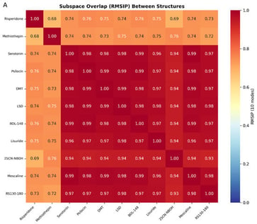
Background/Objectives: AI-based protein–ligand co-folding methods such as Boltz-2 are able to reproduce experimentally determined ligand binding poses at G protein-coupled receptors (GPCRs) with increasing accuracy and precision. As these models advance, they raise several natural questions: assuming the correct placement of a ligand, do current methods generate structural ensembles that are sufficient as a starting point for predictive analysis of receptor dynamics? Does the noise introduced between predictions render these unreliable, or can they be trusted to a degree comparable with experimental structures (which themselves carry inherent noise)? We investigate this at the serotonin 5-HT2A receptor (5-HT2AR), leveraging a rich panel of experimentally determined structures and prior benchmarking of Boltz-2 at this receptor. Methods: First, a reference dynamic landscape is established by performing anisotropic network model (ANM) analysis across 11 published cryo-EM and crystal structures trimmed to a uniform 236-residue analysis mask spanning the structured receptor (TM1-H8). We then ask whether Boltz-2 co-folding ensembles recapitulate these experimentally derived dynamics. Results: All nine Gq-coupled agonist structures converge to near-identical dynamic subspaces (mean RMSIP = 0.970 ± 0.019), discriminating activation state but not agonist identity, chemotype, or hallucinogenic status. Principal component analysis of the Boltz-2 ensemble yields moderate, highly uniform overlap with the experimental ANM modes (mean RMSIP = 0.599 ± 0.007), with the first two principal components capturing 56.9% of structural variance. Conclusions: Replicate predictions of the same ligand are highly self-consistent yet show no usable ligand-specific discrimination (within- versus between-ligand effect size is negligible). These findings suggest a quantitative benchmark for the precision required to derive elastic network dynamics from predicted structures and support the distinction between learned pose recapitulation and genuine biophysical prediction.
Receptors
9 October 2026
![GABA synthesis, uptake, degradation, and its receptors in insects. GABAergic neurons produce GABA from glutamate by glutamic acid decarboxylase 1 (GAD1). GABA is transported into synaptic vesicles by vesicular GABA transporter (VGAT). Postsynaptic neurons can express various ionotropic GABAA and metabotropic GABAB receptors. From the synaptic cleft, GABA is taken up by astrocytes via GABA transporter (GAT) to terminate signaling and recycled through the GABA shunt and the tricarboxylic acid cycle (TCA). The glutamine thus produced is returned to GABAergic neurons. Other abbreviations: GABAT: GABA transaminase; GS: glutamine synthetase; α-KG: α-ketoglutarate; SSA: succinic semialdehyde; SSADH: succinic semialdehyde dehydrogenase. Adapted from [1] with permission from the publisher.](https://mdpi-res.com/cdn-cgi/image/width=281%2Cheight=192/https://mdpi-res.com/receptors/receptors-05-00029/article_deploy/html/images/receptors-05-00029-g001-550.jpg)


