Next Issue
Volume 18, February
Previous Issue
Volume 17, December
 
 
ijms-logo

Journal Browser

Journal Browser

Int. J. Mol. Sci., Volume 18, Issue 1 (January 2017) – 226 articles

Cover Story (view full-size image): 
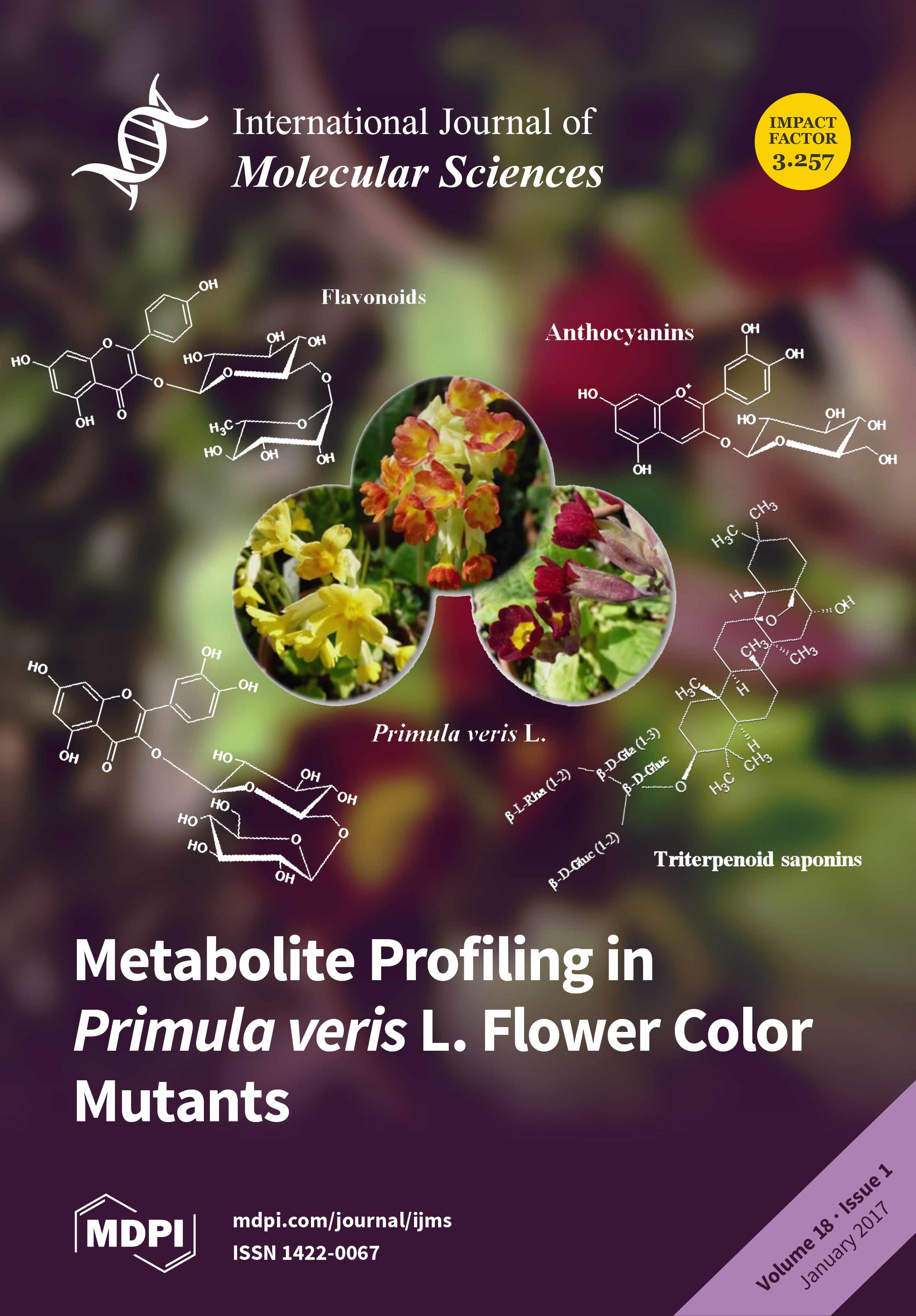
Besides yellow flowering wild-type plants of Primula veris, natural color mutants with red petals and intermediate orange flowers have recently been discovered, which were compared in the present study with regard to their metabolite profile. LC–MS analyses revealed the occurrence of triterpenoid saponins, predominantly in the roots of flavonoids and flavonoid glycosides; as well as four novel methylated flavonoid glycosides in the leaves and flowers of all three plant varieties. In contrast, five anthocyanins only occurred in the petals of red and orange color mutants. Cover image by L. Apel. View this paper.
  • Issues are regarded as officially published after their release is announced to the table of contents alert mailing list.
  • You may sign up for e-mail alerts to receive table of contents of newly released issues.
  • PDF is the official format for papers published in both, html and pdf forms. To view the papers in pdf format, click on the "PDF Full-text" link, and use the free Adobe Reader to open them.
Order results
Result details
Section
Select all
Export citation of selected articles as:
6998 KiB  
Article
Expression of Lipid Metabolism-Related Proteins Differs between Invasive Lobular Carcinoma and Invasive Ductal Carcinoma
by Yoon Jin Cha, Hye Min Kim and Ja Seung Koo
Int. J. Mol. Sci. 2017, 18(1), 232; https://doi.org/10.3390/ijms18010232 - 23 Jan 2017
Cited by 13 | Viewed by 7198
Abstract
We comparatively investigated the expression and clinical implications of lipid metabolism-related proteins in invasive lobular carcinoma (ILC) and invasive ductal carcinoma (IDC) of the breast. A total of 584 breast cancers (108 ILC and 476 IDC) were subjected to tissue microarray and immunohistochemical [...] Read more.
We comparatively investigated the expression and clinical implications of lipid metabolism-related proteins in invasive lobular carcinoma (ILC) and invasive ductal carcinoma (IDC) of the breast. A total of 584 breast cancers (108 ILC and 476 IDC) were subjected to tissue microarray and immunohistochemical analysis for lipid metabolism-related proteins including hormone-sensitive lipase (HSL), perilipin A, fatty acid binding protein (FABP)4, carnitine palmitoyltransferase (CPT)-1, acyl-CoA oxidase 1, and fatty acid synthetase (FASN). HSL, perilipin A, and FABP4 expression (all p < 0.001) differed significantly: HSL and FABP4 were more frequently present in ILC, whereas perilipin A was more frequently detected in IDC. Among all invasive cancers, HSL and FABP4 were highly expressed in luminal A-type ILC (p < 0.001) and perilipin A in luminal A-type IDC (p = 0.007). Among luminal B-type cancers, HSL and FABP4 were more highly expressed in ILC (p < 0.001). Univariate analysis found associations of shorter disease-free survival with CPT-1 positivity (p = 0.004) and acyl-CoA oxidase 1 positivity (p = 0.032) and of shorter overall survival with acyl-CoA oxidase 1 positivity (p = 0.027). In conclusion, ILC and IDC exhibited different immunohistochemical lipid metabolism-related protein expression profiles. Notably, ILC exhibited high HSL and FABP4 and low perilipin A expression. Full article
(This article belongs to the Section Molecular Pathology, Diagnostics, and Therapeutics)
Show Figures

Graphical abstract

4562 KiB  
Article
Evolutionary Conservation of pou5f3 Genomic Organization and Its Dynamic Distribution during Embryogenesis and in Adult Gonads in Japanese Flounder Paralichthys olivaceus
by Jinning Gao, Xubo Wang and Quanqi Zhang
Int. J. Mol. Sci. 2017, 18(1), 231; https://doi.org/10.3390/ijms18010231 - 23 Jan 2017
Cited by 12 | Viewed by 5143
Abstract
Octamer-binding transcription factor 4 (Oct4) is a member of POU (Pit-Oct-Unc) transcription factor family Class V that plays a crucial role in maintaining the pluripotency and self-renewal of stem cells. Though it has been deeply investigated in mammals, its lower vertebrate homologue, especially [...] Read more.
Octamer-binding transcription factor 4 (Oct4) is a member of POU (Pit-Oct-Unc) transcription factor family Class V that plays a crucial role in maintaining the pluripotency and self-renewal of stem cells. Though it has been deeply investigated in mammals, its lower vertebrate homologue, especially in the marine fish, is poorly studied. In this study, we isolated the full-length sequence of Paralichthys olivaceus pou5f3 (Popou5f3), and we found that it is homologous to mammalian Oct4. We identified two transcript variants with different lengths of 3′-untranslated regions (UTRs) generated by alternative polyadenylation (APA). Quantitative real-time RT-PCR (qRT-PCR), in situ hybridization (ISH) and immunohistochemistry (IHC) were implemented to characterize the spatial and temporal expression pattern of Popou5f3 during early development and in adult tissues. Our results show that Popou5f3 is maternally inherited, abundantly expressed at the blastula and early gastrula stages, then greatly diminishes at the end of gastrulation. It is hardly detectable from the heart-beating stage onward. We found that Popou5f3 expression is restricted to the adult gonads, and continuously expresses during oogenesis while its dynamics are downregulated during spermatogenesis. Additionally, numerous cis-regulatory elements (CRE) on both sides of the flanking regions show potential roles in regulating the expression of Popou5f3. Taken together, these findings could further our understanding of the functions and evolution of pou5f3 in lower vertebrates, and also provides fundamental information for stem cell tracing and genetic manipulation in Paralichthys olivaceus. Full article
(This article belongs to the Section Biochemistry)
Show Figures

Figure 1

3956 KiB  
Article
Growth Hormone (GH) and Rehabilitation Promoted Distal Innervation in a Child Affected by Caudal Regression Syndrome
by Jesús Devesa, Alba Alonso, Natalia López, José García, Carlos I. Puell, Tamara Pablos and Pablo Devesa
Int. J. Mol. Sci. 2017, 18(1), 230; https://doi.org/10.3390/ijms18010230 - 23 Jan 2017
Cited by 17 | Viewed by 8944
Abstract
Caudal regression syndrome (CRS) is a malformation occurring during the fetal period and mainly characterized by an incomplete development of the spinal cord (SC), which is often accompanied by other developmental anomalies. We studied a 9-month old child with CRS who presented interruption [...] Read more.
Caudal regression syndrome (CRS) is a malformation occurring during the fetal period and mainly characterized by an incomplete development of the spinal cord (SC), which is often accompanied by other developmental anomalies. We studied a 9-month old child with CRS who presented interruption of the SC at the L2–L3 level, sacral agenesis, a lack of innervation of the inferior limbs (flaccid paraplegia), and neurogenic bladder and bowel. Given the known positive effects of growth hormone (GH) on neural stem cells (NSCs), we treated him with GH and rehabilitation, trying to induce recovery from the aforementioned sequelae. The Gross Motor Function Test (GMFM)-88 test score was 12.31%. After a blood analysis, GH treatment (0.3 mg/day, 5 days/week, during 3 months and then 15 days without GH) and rehabilitation commenced. This protocol was followed for 5 years, the last GH dose being 1 mg/day. Blood analysis and physical exams were performed every 3 months initially and then every 6 months. Six months after commencing the treatment the GMFM-88 score increased to 39.48%. Responses to sensitive stimuli appeared in most of the territories explored; 18 months later sensitive innervation was complete and the patient moved all muscles over the knees and controlled his sphincters. Three years later he began to walk with crutches, there was plantar flexion, and the GMFM-88 score was 78.48%. In summary, GH plus rehabilitation may be useful for innervating distal areas below the level of the incomplete spinal cord in CRS. It is likely that GH acted on the ependymal SC NSCs, as the hormone does in the neurogenic niches of the brain, and rehabilitation helped to achieve practically full functionality. Full article
(This article belongs to the Special Issue Growth Hormone: Therapeutic Possibilities)
Show Figures

Graphical abstract

8093 KiB  
Article
Surface Modification of Titanium with BMP-2/GDF-5 by a Heparin Linker and Its Efficacy as a Dental Implant
by Dae Hyeok Yang, Sang Woong Moon and Deok-Won Lee
Int. J. Mol. Sci. 2017, 18(1), 229; https://doi.org/10.3390/ijms18010229 - 23 Jan 2017
Cited by 30 | Viewed by 6162
Abstract
In this study, we prepared human bone morphogenetic protein-2 (hBMP-2)/human growth and differentiation factor-5 (hGDF-5)-coated titanium (Ti) disc and screw types for controlled release of the growth factors (GFs). The two growth factors were coated onto Ti with a smooth surface using their [...] Read more.
In this study, we prepared human bone morphogenetic protein-2 (hBMP-2)/human growth and differentiation factor-5 (hGDF-5)-coated titanium (Ti) disc and screw types for controlled release of the growth factors (GFs). The two growth factors were coated onto Ti with a smooth surface using their specific interaction with heparin, because they have heparin binding sites in their molecular structures. Efficacy of the two growth factor-coated Ti for enhancement of bone formation and osseointegration was compared to pristine Ti, and hBMP-2- and hGDF-5-coated Ti in vivo. The surface chemical composition, surface morphology, and wettability characteristics of the metal samples were determined by X-ray photoelectron spectroscopy (XPS), scanning electron microscopy (SEM), and contact angle measurement, respectively. The initial burst of hBMP-2, hGDF-5, and their combination, occurred within one day of the release study, resulting in 12.5%, 4.5%, and 13.5%/3.2%, and then there was a sustained, even release of these two growth factors from the coated metal for 30 days. In vitro tests revealed that MC3T3-E1 cells cultured on the two growth factor-coated Ti had a higher proliferation rate and a higher activity for alkaline phosphatase (ALP), which led to a larger amount of calcium deposition and larger expressions of type I collagen (COL 1), ALP, and osteocalcin (OCN) mRNAs. In vivo animal tests using ten white New Zealand rabbits showed that the two growth factor-coated Ti enhanced bone formation and osseointegration at the interface between the implants and host bone. In addition, histological evaluation showed that bone remodeling, including bone formation by osteoblasts and bone resorption by osteoclasts, actively occurred between the two growth factor-coated Ti and host bone. Consequently, it is suggested that Ti surface modification with the combination of hBMP-2 and hGDF-5 for the two growth factor-coated Ti implants can improve the clinical properties of implants for orthopedic and dental applications. Full article
(This article belongs to the Section Materials Science)
Show Figures

Graphical abstract

2962 KiB  
Article
Molecular Basis for the Neutralization of Tumor Necrosis Factor α by Certolizumab Pegol in the Treatment of Inflammatory Autoimmune Diseases
by Jee Un Lee, Woori Shin, Ji Young Son, Ki-Young Yoo and Yong-Seok Heo
Int. J. Mol. Sci. 2017, 18(1), 228; https://doi.org/10.3390/ijms18010228 - 23 Jan 2017
Cited by 28 | Viewed by 8956
Abstract
Monoclonal antibodies against TNFα, including infliximab, adalimumab, golimumab, and certolizumab pegol, are widely used for the treatment of the inflammatory diseases such as rheumatoid arthritis and inflammatory bowel disease. Recently, the crystal structures of TNFα, in complex with the Fab fragments of infliximab [...] Read more.
Monoclonal antibodies against TNFα, including infliximab, adalimumab, golimumab, and certolizumab pegol, are widely used for the treatment of the inflammatory diseases such as rheumatoid arthritis and inflammatory bowel disease. Recently, the crystal structures of TNFα, in complex with the Fab fragments of infliximab and adalimumab, have revealed the molecular mechanisms of these antibody drugs. Here, we report the crystal structure of TNFα in complex with the Fab fragment of certolizumab pegol to clarify the precise antigen-antibody interactions and the structural basis for the neutralization of TNFα by this therapeutic antibody. The structural analysis and the mutagenesis study revealed that the epitope is limited to a single protomer of the TNFα trimer. Additionally, the DE loop and the GH loop of TNFα play critical roles in the interaction with certolizumab, suggesting that this drug exerts its effects by partially occupying the receptor binding site of TNFα. In addition, a conformational change of the DE loop was induced by certolizumab binding, thereby interrupting the TNFα-receptor interaction. A comprehensive comparison of the interactions of TNFα blockers with TNFα revealed the epitope diversity on the surface of TNFα, providing a better understanding of the molecular mechanism of TNFα blockers. The accumulation of these structural studies can provide a basis for the improvement of therapeutic antibodies against TNFα. Full article
Show Figures

Graphical abstract

1712 KiB  
Review
Prelysosomal Compartments in the Unconventional Secretion of Amyloidogenic Seeds
by Helena Borland and Frederik Vilhardt
Int. J. Mol. Sci. 2017, 18(1), 227; https://doi.org/10.3390/ijms18010227 - 23 Jan 2017
Cited by 22 | Viewed by 9806
Abstract
A mechanistic link between neuron-to-neuron transmission of secreted amyloid and propagation of protein malconformation cytopathology and disease has recently been uncovered in animal models. An enormous interest in the unconventional secretion of amyloids from neurons has followed. Amphisomes and late endosomes are the [...] Read more.
A mechanistic link between neuron-to-neuron transmission of secreted amyloid and propagation of protein malconformation cytopathology and disease has recently been uncovered in animal models. An enormous interest in the unconventional secretion of amyloids from neurons has followed. Amphisomes and late endosomes are the penultimate maturation products of the autophagosomal and endosomal pathways, respectively, and normally fuse with lysosomes for degradation. However, under conditions of perturbed membrane trafficking and/or lysosomal deficiency, prelysosomal compartments may instead fuse with the plasma membrane to release any contained amyloid. After a brief introduction to the endosomal and autophagosomal pathways, we discuss the evidence for autophagosomal secretion (exophagy) of amyloids, with a comparative emphasis on Aβ1–42 and α-synuclein, as luminal and cytosolic amyloids, respectively. The ESCRT-mediated import of cytosolic amyloid into late endosomal exosomes, a known vehicle of transmission of macromolecules between cells, is also reviewed. Finally, mechanisms of lysosomal dysfunction, deficiency, and exocytosis are exemplified in the context of genetically identified risk factors, mainly for Parkinson’s disease. Exocytosis of prelysosomal or lysosomal organelles is a last resort for clearance of cytotoxic material and alleviates cytopathy. However, they also represent a vehicle for the concentration, posttranslational modification, and secretion of amyloid seeds. Full article
(This article belongs to the Special Issue Unconventional Proteins and Membranes Traffic)
Show Figures

Graphical abstract

4051 KiB  
Article
The Long Non-Coding RNA RHPN1-AS1 Promotes Uveal Melanoma Progression
by Linna Lu, Xiaoyu Yu, Leilei Zhang, Xia Ding, Hui Pan, Xuyang Wen, Shiqiong Xu, Yue Xing, Jiayan Fan, Shengfang Ge, He Zhang, Renbing Jia and Xianqun Fan
Int. J. Mol. Sci. 2017, 18(1), 226; https://doi.org/10.3390/ijms18010226 - 23 Jan 2017
Cited by 57 | Viewed by 6472
Abstract
Increasing evidence suggests that aberrant long non-coding RNAs (lncRNAs) are significantly correlated with the pathogenesis, development and metastasis of cancers. RHPN1 antisense RNA 1 (RHPN1-AS1) is a 2030-bp transcript originating from human chromosome 8q24. However, the role of RHPN1-AS1 in uveal [...] Read more.
Increasing evidence suggests that aberrant long non-coding RNAs (lncRNAs) are significantly correlated with the pathogenesis, development and metastasis of cancers. RHPN1 antisense RNA 1 (RHPN1-AS1) is a 2030-bp transcript originating from human chromosome 8q24. However, the role of RHPN1-AS1 in uveal melanoma (UM) remains to be clarified. In this study, we aimed to elucidate the molecular function of RHPN1-AS1 in UM. The RNA levels of RHPN1-AS1 in UM cell lines were examined using the quantitative real-time polymerase chain reaction (qRT-PCR). Short interfering RNAs (siRNAs) were designed to quench RHPN1-AS1 expression, and UM cells stably expressing short hairpin (sh) RHPN1-AS1 were established. Next, the cell proliferation and migration abilities were determined using a colony formation assay and a transwell migration/invasion assay. A tumor xenograft model in nude mice was established to confirm the function of RHPN1-AS1 in vivo. RHPN1-AS1 was significantly upregulated in a number of UM cell lines compared with the normal human retinal pigment epithelium (RPE) cell line. RHPN1-AS1 knockdown significantly inhibited UM cell proliferation and migration in vitro and in vivo. Our data suggest that RHPN1-AS1 could be an oncoRNA in UM, which may serve as a candidate prognostic biomarker and target for new therapies in malignant UM. Full article
(This article belongs to the Collection Regulation by Non-coding RNAs)
Show Figures

Graphical abstract

9512 KiB  
Article
Composition of Lignin-to-Liquid Solvolysis Oils from Lignin Extracted in a Semi-Continuous Organosolv Process
by Camilla Løhre, Hilde Vik Halleraker and Tanja Barth
Int. J. Mol. Sci. 2017, 18(1), 225; https://doi.org/10.3390/ijms18010225 - 23 Jan 2017
Cited by 14 | Viewed by 6538
Abstract
The interest and on-going research on utilisation of lignin as feedstock for production of renewable and sustainable aromatics is expanding and shows great potential. This study investigates the applicability of semi-continuously organosolv extracted lignin in Lignin-to-Liquid (LtL) solvolysis, using formic acid as hydrogen [...] Read more.
The interest and on-going research on utilisation of lignin as feedstock for production of renewable and sustainable aromatics is expanding and shows great potential. This study investigates the applicability of semi-continuously organosolv extracted lignin in Lignin-to-Liquid (LtL) solvolysis, using formic acid as hydrogen donor and water as solvent under high temperature–high pressure (HTHP) conditions. The high purity of the organosolv lignin provides high conversion yields at up to 94% based on lignin mass input. The formic acid input is a dominating parameter in lignin conversion. Carbon balance calculations of LtL-solvolysis experiments also indicate that formic acid can give a net carbon contribution to the bio-oils, in addition to its property as hydrogenation agent. Compound specific quantification of the ten most abundant components in the LtL-oils describe up to 10% of the bio-oil composition, and reaction temperature is shown to be the dominating parameter for the structures present. The structural and quantitative results from this study identify components of considerable value in the LtL-oil, and support the position of this oil as a potentially important source of building blocks for the chemical and pharmaceutical industry. Full article
(This article belongs to the Special Issue The Lignin Challenge: Exploring Innovative Applications)
Show Figures

Figure 1

1992 KiB  
Article
mir-660-p53-mir-486 Network: A New Key Regulatory Pathway in Lung Tumorigenesis
by Cristina Borzi, Linda Calzolari, Giovanni Centonze, Massimo Milione, Gabriella Sozzi and Orazio Fortunato
Int. J. Mol. Sci. 2017, 18(1), 222; https://doi.org/10.3390/ijms18010222 - 23 Jan 2017
Cited by 24 | Viewed by 6386
Abstract
Lung cancer is the most frequent cause of cancer-related death worldwide, with limited therapeutic options and rapid development of drug resistance. MicroRNAs, a class of small non-coding RNAs that control different physiological processes, have been associated with cancer development, as either oncomiRNAs or [...] Read more.
Lung cancer is the most frequent cause of cancer-related death worldwide, with limited therapeutic options and rapid development of drug resistance. MicroRNAs, a class of small non-coding RNAs that control different physiological processes, have been associated with cancer development, as either oncomiRNAs or tumor-suppressor miRNAs. In the present study we investigated the interaction between mir-486-5p and mir-660-5p, two independent tumor-suppressor miRNAs, to assess their possible role and synergistic effect in lung cancer treatment. Our data show that mir-660-5p over-expression in A549 lung cancer cells induced a remarkable increase in mir-486-5p expression level and activity, detected as a reduction of its target gene, p85. mir-486-5p expression was confirmed by microRNA in situ hybridization. mir-660-5p modulated mir-486-5p through the silencing of Mouse Double Minute 2 (MDM2), one of its direct target, and then through p53 stimulation. This regulatory pathway was effective in A549, but not in H1299; therefore, only in the context of a functional p53 protein. Our findings support the conclusion that mir-486-5p is positively regulated by mir-660-5p in lung cancer cell lines, through the mir-660-MDM2-p53 pathway, making mir-660-5p even more interesting for its potential successful use in lung cancer therapy. Full article
(This article belongs to the Special Issue Cancer Epigenetics)
Show Figures

Graphical abstract

5226 KiB  
Article
Therapeutic Potential of Induced Neural Stem Cells for Parkinson’s Disease
by Dong-Hee Choi, Ji-Hye Kim, Sung Min Kim, Kyuree Kang, Dong Wook Han and Jongmin Lee
Int. J. Mol. Sci. 2017, 18(1), 224; https://doi.org/10.3390/ijms18010224 - 22 Jan 2017
Cited by 32 | Viewed by 7623
Abstract
Parkinson’s disease (PD) is a chronic, neurodegenerative disorder that results from the loss of cells in the substantia nigra (SN) which is located in the midbrain. However, no cure is available for PD. Recently, fibroblasts have been directly converted into induced neural stem [...] Read more.
Parkinson’s disease (PD) is a chronic, neurodegenerative disorder that results from the loss of cells in the substantia nigra (SN) which is located in the midbrain. However, no cure is available for PD. Recently, fibroblasts have been directly converted into induced neural stem cells (iNSCs) via the forced expression of specific transcription factors. Therapeutic potential of iNSC in PD has not been investigated yet. Here, we show that iNSCs directly converted from mouse fibroblasts enhanced functional recovery in an animal model of PD. The rotational behavior test was performed to assess recovery. Our results indicate that iNSC transplantation into the striatum of 6-hydroxydopamine (6-OHDA)-injected mice can significantly reduce apomorphine-induced rotational asymmetry. The engrafted iNSCs were able to survive in the striatum and migrated around the medial forebrain bundle and the SN pars compacta. Moreover, iNSCs differentiated into all neuronal lineages. In particular, the transplanted iNSCs that committed to the glial lineage were significantly increased in the striatum of 6-OHDA-injected mice. Engrafted iNSCs differentiated to dopaminergic (DA) neurons and migrated into the SN in the 6-OHDA lesion mice. Therefore, iNSC transplantation serves as a valuable tool to enhance the functional recovery in PD. Full article
(This article belongs to the Special Issue Neurological Injuries’ Monitoring, Tracking and Treatment 2016)
Show Figures

Figure 1

589 KiB  
Review
What Do We Know about Opioids and the Kidney?
by Mary Mallappallil, Jacob Sabu, Eli A. Friedman and Moro Salifu
Int. J. Mol. Sci. 2017, 18(1), 223; https://doi.org/10.3390/ijms18010223 - 22 Jan 2017
Cited by 55 | Viewed by 24143
Abstract
Evidence suggests a link between opioid use and kidney disease. This review summarizes the known renal manifestations of opioid use including its role in acute and chronic kidney injury. Both the direct and indirect effects of the drug, and the context which leads [...] Read more.
Evidence suggests a link between opioid use and kidney disease. This review summarizes the known renal manifestations of opioid use including its role in acute and chronic kidney injury. Both the direct and indirect effects of the drug, and the context which leads to the development of renal failure, are explored. While commonly used safely for pain control and anesthesia in those with kidney disease, the concerns with respect to side effects and toxicity of opioids are addressed. This is especially relevant with the worldwide increase in the use of opioids for medical and recreational use. Full article
(This article belongs to the Special Issue Nephrotoxicity)
Show Figures

Figure 1

2876 KiB  
Article
Talin Modulation by a Synthetic N-Acylurea Derivative Reduces Angiogenesis in Human Endothelial Cells
by I-Rang Lim, Hyung Joon Joo, Minseon Jeong, Jong-Ho Kim, Seung-Cheol Choi, Chungho Kim, Jong-Wha Jung and Soon Jun Hong
Int. J. Mol. Sci. 2017, 18(1), 221; https://doi.org/10.3390/ijms18010221 - 22 Jan 2017
Cited by 6 | Viewed by 5340
Abstract
Talin is a focal adhesion protein that activates integrins and recruits other focal adhesion proteins. Talin regulates the interactions between integrins and the extracellular matrix, which are critical for endothelial cells during angiogenesis. In this study, we successfully synthesized a novel talin modulator, [...] Read more.
Talin is a focal adhesion protein that activates integrins and recruits other focal adhesion proteins. Talin regulates the interactions between integrins and the extracellular matrix, which are critical for endothelial cells during angiogenesis. In this study, we successfully synthesized a novel talin modulator, N-((2-(1H-indol-3-yl)ethyl)carbamoyl)-2-(benzo[d][1,3]dioxol-5-yloxy)acetamide, referred to as KCH-1521. KCH-1521 was determined to bind talin and modulate downstream signaling molecules of talin. After 24 h of treatment, KCH-1521 changed the cell morphology of human umbilical vein endothelial cells (HUVECs) and reduced focal adhesion protein expression including vinculin and paxillin. Talin downstream signaling is regulated via focal adhesion kinase (FAK), kinase B (AKT), and extracellular signal-regulated kinase (ERK) pathways, however, treatment with KCH-1521 decreased phosphorylation of FAK, AKT, and ERK, leading to reduction of cell proliferation, survival, and angiogenesis. Interestingly, the expression of various angiogenic genes was significantly decreased after treatment with KCH-1521. Also, in vitro tube forming assay revealed that KCH-1521 reduced angiogenic networks in a time-dependent manner. To investigate the reversibility of its effects, KCH-1521 was removed after treatment. HUVECs recovered their morphology through rearrangement of the cytoskeleton and the expression of angiogenic genes was also recovered. By further optimization and in vivo studies of KCH-1521, a novel drug of talin modulation could be used to achieve therapeutic anti-angiogenesis for vascular diseases and cancers. Full article
(This article belongs to the Section Biochemistry)
Show Figures

Graphical abstract

2905 KiB  
Article
Decreased Sp1 Expression Mediates Downregulation of SHIP2 in Gastric Cancer Cells
by Yan Ye, Xue Yi Qian, Miao Miao Xiao, Yu Ling Shao, Li Mei Guo, Dong Ping Liao, Jie Da, Lin Jie Zhang and Jiegou Xu
Int. J. Mol. Sci. 2017, 18(1), 220; https://doi.org/10.3390/ijms18010220 - 22 Jan 2017
Cited by 7 | Viewed by 4432
Abstract
Past studies have shown that the Src homology 2-containing inositol 5-phosphatase 2 (SHIP2) is commonly downregulated in gastric cancer, which contributes to elevated activation of PI3K/Akt signaling, proliferation and tumorigenesis of gastric cancer cells. However, the mechanisms underlying the reduced expression of SHIP2 [...] Read more.
Past studies have shown that the Src homology 2-containing inositol 5-phosphatase 2 (SHIP2) is commonly downregulated in gastric cancer, which contributes to elevated activation of PI3K/Akt signaling, proliferation and tumorigenesis of gastric cancer cells. However, the mechanisms underlying the reduced expression of SHIP2 in gastric cancer remain unclear. While gene copy number variation analysis and exon sequencing indicated the absence of genomic alterations of SHIP2, bisulfite genomic sequencing (BGS) showed promoter hypomethylation of SHIP2 in gastric cancer cells. Analysis of transcriptional activity of SHIP2 promoter revealed Specificity protein 1 (Sp1) was responsible for the regulation of SHIP2 expression in gastric cancer cells. Furthermore, Sp1 expression, but not Sp3, was frequently downregulated in gastric cancer compared with normal gastric mucosa, which was associated with a paralleled reduction in SHIP2 levels in gastric cancer. Moreover, overexpression of Sp1 inhibited cell proliferation, induced apoptosis, suppressed cell motility and invasion in gastric cancer cells in vitro, which was, at least in part, due to transcriptional activation of SHIP2 mediated by Sp1, thereby inactivating Akt. Collectively, these results indicate that decreased expression of transcription factor Sp1 contributes to suppression of SHIP2 in gastric cancer cells. Full article
(This article belongs to the Section Biochemistry)
Show Figures

Figure 1

892 KiB  
Article
Transcriptome-Wide Identification and Prediction of miRNAs and Their Targets in Paris polyphylla var. yunnanensis by High-Throughput Sequencing Analysis
by Li-Zhen Ling, Shu-Dong Zhang, Fan Zhao, Jin-Long Yang, Wen-Hui Song, Shen-Min Guan, Xin-Shu Li, Zhuang-Jia Huang and Le Cheng
Int. J. Mol. Sci. 2017, 18(1), 219; https://doi.org/10.3390/ijms18010219 - 22 Jan 2017
Cited by 17 | Viewed by 5192
Abstract
Long dormancy period of seeds limits the large-scale artificial cultivation of the scarce Paris polyphylla var. yunnanensis, an important traditional Chinese medicine. Characterizing miRNAs and their targets is crucial to understanding the role of miRNAs during seed dormancy in this species. Considering [...] Read more.
Long dormancy period of seeds limits the large-scale artificial cultivation of the scarce Paris polyphylla var. yunnanensis, an important traditional Chinese medicine. Characterizing miRNAs and their targets is crucial to understanding the role of miRNAs during seed dormancy in this species. Considering the limited genome information of this species, we first sequenced and assembled the transcriptome data of dormant seeds and their seed coats as the reference genome. A total of 146,671 unigenes with an average length of 923 bp were identified and showed functional diversity based on different annotation methods. Two small RNA libraries from respective seeds and seed coats were sequenced and the combining data indicates that 263 conserved miRNAs belonging to at least 83 families and 768 novel miRNAs in 1174 transcripts were found. The annotations of the predicted putative targets of miRNAs suggest that these miRNAs were mainly involved in the cell, metabolism and genetic information processing by direct and indirect regulation patterns in dormant seeds of P. polyphylla var. yunnanensis. Therefore, we provide the first known miRNA profiles and their targets, which will assist with further study of the molecular mechanism of seed dormancy in P. polyphylla var. yunnanensis. Full article
(This article belongs to the Special Issue microRNA Regulation 2017)
Show Figures

Figure 1

1767 KiB  
Article
Affinin (Spilanthol), Isolated from Heliopsis longipes, Induces Vasodilation via Activation of Gasotransmitters and Prostacyclin Signaling Pathways
by Jesús Eduardo Castro-Ruiz, Alejandra Rojas-Molina, Francisco J. Luna-Vázquez, Fausto Rivero-Cruz, Teresa García-Gasca and César Ibarra-Alvarado
Int. J. Mol. Sci. 2017, 18(1), 218; https://doi.org/10.3390/ijms18010218 - 22 Jan 2017
Cited by 22 | Viewed by 8743
Abstract
Heliopsis longipes roots have been widely used in Mexican traditional medicine to relieve pain, mainly, toothaches. Previous studies have shown that affinin, the major alkamide of these roots, induces potent antinociceptive and anti-inflammatory activities. However, the effect of H. longipes root extracts and [...] Read more.
Heliopsis longipes roots have been widely used in Mexican traditional medicine to relieve pain, mainly, toothaches. Previous studies have shown that affinin, the major alkamide of these roots, induces potent antinociceptive and anti-inflammatory activities. However, the effect of H. longipes root extracts and affinin on the cardiovascular system have not been investigated so far. In the present study, we demonstrated that the dichloromethane and ethanolic extracts of H. longipes roots, and affinin, isolated from these roots, produce a concentration-dependent vasodilation of rat aorta. Affinin-induced vasorelaxation was partly dependent on the presence of endothelium and was significantly blocked in the presence of inhibitors of NO, H2S, and CO synthesis (NG-nitro-l-arginine methyl ester (l-NAME), dl-propargylglycine (PAG), and chromium mesoporphyrin (CrMP), respectively); K+ channel blockers (glibenclamide (Gli) and tetraethyl ammonium (TEA)), and guanylate cyclase and cyclooxygenase inhibitors (1H-[1,2,4]oxadiazolo[4,3-a]quinoxalin-1-one (ODQ) and indomethacin (INDO), respectively). Our results demonstrate, for the first time, that affinin induces vasodilation by mechanisms that involve gasotransmitters, and prostacyclin signaling pathways. These findings indicate that this natural alkamide has therapeutic potential in the treatment of cardiovascular diseases. Full article
(This article belongs to the Special Issue Biological Activity of Natural Secondary Metabolite Products)
Show Figures

Graphical abstract

915 KiB  
Article
In Vitro Biotransformation of Two Human CYP3A Probe Substrates and Their Inhibition during Early Zebrafish Development
by Evy Verbueken, Derek Alsop, Moayad A. Saad, Casper Pype, Els M. Van Peer, Christophe R. Casteleyn, Chris J. Van Ginneken, Joanna Wilson and Steven J. Van Cruchten
Int. J. Mol. Sci. 2017, 18(1), 217; https://doi.org/10.3390/ijms18010217 - 22 Jan 2017
Cited by 18 | Viewed by 5730
Abstract
At present, the zebrafish embryo is increasingly used as an alternative animal model to screen for developmental toxicity after exposure to xenobiotics. Since zebrafish embryos depend on their own drug-metabolizing capacity, knowledge of their intrinsic biotransformation is pivotal in order to correctly interpret [...] Read more.
At present, the zebrafish embryo is increasingly used as an alternative animal model to screen for developmental toxicity after exposure to xenobiotics. Since zebrafish embryos depend on their own drug-metabolizing capacity, knowledge of their intrinsic biotransformation is pivotal in order to correctly interpret the outcome of teratogenicity assays. Therefore, the aim of this in vitro study was to assess the activity of cytochrome P450 (CYP)—a group of drug-metabolizing enzymes—in microsomes from whole zebrafish embryos (ZEM) of 5, 24, 48, 72, 96 and 120 h post-fertilization (hpf) by means of a mammalian CYP substrate, i.e., benzyloxy-methyl-resorufin (BOMR). The same CYP activity assays were performed in adult zebrafish liver microsomes (ZLM) to serve as a reference for the embryos. In addition, activity assays with the human CYP3A4-specific Luciferin isopropyl acetal (Luciferin-IPA) as well as inhibition studies with ketoconazole and CYP3cide were carried out to identify CYP activity in ZLM. In the present study, biotransformation of BOMR was detected at 72 and 96 hpf; however, metabolite formation was low compared with ZLM. Furthermore, Luciferin-IPA was not metabolized by the zebrafish. In conclusion, the capacity of intrinsic biotransformation in zebrafish embryos appears to be lacking during a major part of organogenesis. Full article
(This article belongs to the Special Issue Zebrafish: A Model for Toxicological Research)
Show Figures

Graphical abstract

554 KiB  
Review
A Holistic Approach to Managing Microalgae for Biofuel Applications
by Pau Loke Show, Malcolm S. Y. Tang, Dillirani Nagarajan, Tau Chuan Ling, Chien-Wei Ooi and Jo-Shu Chang
Int. J. Mol. Sci. 2017, 18(1), 215; https://doi.org/10.3390/ijms18010215 - 22 Jan 2017
Cited by 110 | Viewed by 9764
Abstract
Microalgae contribute up to 60% of the oxygen content in the Earth’s atmosphere by absorbing carbon dioxide and releasing oxygen during photosynthesis. Microalgae are abundantly available in the natural environment, thanks to their ability to survive and grow rapidly under harsh and inhospitable [...] Read more.
Microalgae contribute up to 60% of the oxygen content in the Earth’s atmosphere by absorbing carbon dioxide and releasing oxygen during photosynthesis. Microalgae are abundantly available in the natural environment, thanks to their ability to survive and grow rapidly under harsh and inhospitable conditions. Microalgal cultivation is environmentally friendly because the microalgal biomass can be utilized for the productions of biofuels, food and feed supplements, pharmaceuticals, nutraceuticals, and cosmetics. The cultivation of microalgal also can complement approaches like carbon dioxide sequestration and bioremediation of wastewaters, thereby addressing the serious environmental concerns. This review focuses on the factors affecting microalgal cultures, techniques adapted to obtain high-density microalgal cultures in photobioreactors, and the conversion of microalgal biomass into biofuels. The applications of microalgae in carbon dioxide sequestration and phycoremediation of wastewater are also discussed. Full article
(This article belongs to the Special Issue Algae Based Bio-Renewable Energy for Sustainability)
Show Figures

Graphical abstract

6537 KiB  
Article
α-Tocopherol at Nanomolar Concentration Protects Cortical Neurons against Oxidative Stress
by Irina O. Zakharova, Tatiana V. Sokolova, Yulia A. Vlasova, Liubov V. Bayunova, Maria P. Rychkova and Natalia F. Avrova
Int. J. Mol. Sci. 2017, 18(1), 216; https://doi.org/10.3390/ijms18010216 - 21 Jan 2017
Cited by 26 | Viewed by 6344
Abstract
The aim of the present work is to study the mechanism of the α-tocopherol (α-T) protective action at nanomolar and micromolar concentrations against H2O2-induced brain cortical neuron death. The mechanism of α-T action on neurons at its nanomolar concentrations [...] Read more.
The aim of the present work is to study the mechanism of the α-tocopherol (α-T) protective action at nanomolar and micromolar concentrations against H2O2-induced brain cortical neuron death. The mechanism of α-T action on neurons at its nanomolar concentrations characteristic for brain extracellular space has not been practically studied yet. Preincubation with nanomolar and micromolar α-T for 18 h was found to increase the viability of cortical neurons exposed to H2O2; α-T effect was concentration-dependent in the nanomolar range. However, preincubation with nanomolar α-T for 30 min was not effective. Nanomolar and micromolar α-T decreased the reactive oxygen species accumulation induced in cortical neurons by the prooxidant. Using immunoblotting it was shown that preincubation with α-T at nanomolar and micromolar concentrations for 18 h prevented Akt inactivation and decreased PKCδ activation induced in cortical neurons by H2O2. α-T prevented the ERK1/2 sustained activation during 24 h caused by H2O2. α-T at nanomolar and micromolar concentrations prevented a great increase of the proapoptotic to antiapoptotic proteins (Bax/Bcl-2) ratio, elicited by neuron exposure to H2O2. The similar neuron protection mechanism by nanomolar and micromolar α-T suggests that a “more is better” approach to patients’ supplementation with vitamin E or α-T is not reasonable. Full article
(This article belongs to the Special Issue Neuroprotective Strategies 2016)
Show Figures

Graphical abstract

8658 KiB  
Article
Methylglyoxal-Mediated Stress Correlates with High Metabolic Activity and Promotes Tumor Growth in Colorectal Cancer
by Barbara Chiavarina, Marie-Julie Nokin, Justine Bellier, Florence Durieux, Noëlla Bletard, Félicie Sherer, Pierre Lovinfosse, Olivier Peulen, Laurine Verset, Romain Dehon, Pieter Demetter, Andrei Turtoi, Koji Uchida, Serge Goldman, Roland Hustinx, Philippe Delvenne, Vincent Castronovo and Akeila Bellahcène
Int. J. Mol. Sci. 2017, 18(1), 213; https://doi.org/10.3390/ijms18010213 - 21 Jan 2017
Cited by 48 | Viewed by 7704
Abstract
Cancer cells generally rely on aerobic glycolysis as a major source of energy. Methylglyoxal (MG), a dicarbonyl compound that is produced as a side product during glycolysis, is highly reactive and induces the formation of advanced glycation end-products that are implicated in several [...] Read more.
Cancer cells generally rely on aerobic glycolysis as a major source of energy. Methylglyoxal (MG), a dicarbonyl compound that is produced as a side product during glycolysis, is highly reactive and induces the formation of advanced glycation end-products that are implicated in several pathologies including cancer. All mammalian cells have an enzymatic defense against MG composed by glyoxalases GLO1 and GLO2 that converts MG to d-lactate. Colorectal cancer (CRC) is one of the most frequently occurring cancers with high morbidity and mortality. In this study, we used immunohistochemistry to examine the level of MG protein adducts, in a series of 102 CRC human tumors divided into four clinical stages. We consistently detected a high level of MG adducts and low GLO1 activity in high stage tumors compared to low stage ones suggesting a pro-tumor role for dicarbonyl stress. Accordingly, GLO1 depletion in CRC cells promoted tumor growth in vivo that was efficiently reversed using carnosine, a potent MG scavenger. Our study represents the first demonstration that MG adducts accumulation is a consistent feature of high stage CRC tumors. Our data point to MG production and detoxification levels as an important molecular link between exacerbated glycolytic activity and CRC progression. Full article
(This article belongs to the Special Issue Glyoxalase System)
Show Figures

Graphical abstract

1499 KiB  
Article
Modulation of the Unfolded Protein Response by Tauroursodeoxycholic Acid Counteracts Apoptotic Cell Death and Fibrosis in a Mouse Model for Secondary Biliary Liver Fibrosis
by Annelies Paridaens, Sarah Raevens, Lindsey Devisscher, Eliene Bogaerts, Xavier Verhelst, Anne Hoorens, Hans Van Vlierberghe, Leo A. Van Grunsven, Anja Geerts and Isabelle Colle
Int. J. Mol. Sci. 2017, 18(1), 214; https://doi.org/10.3390/ijms18010214 - 20 Jan 2017
Cited by 24 | Viewed by 5916
Abstract
The role of endoplasmic reticulum stress and the unfolded protein response (UPR) in cholestatic liver disease and fibrosis is not fully unraveled. Tauroursodeoxycholic acid (TUDCA), a hydrophilic bile acid, has been shown to reduce endoplasmic reticulum (ER) stress and counteract apoptosis in different [...] Read more.
The role of endoplasmic reticulum stress and the unfolded protein response (UPR) in cholestatic liver disease and fibrosis is not fully unraveled. Tauroursodeoxycholic acid (TUDCA), a hydrophilic bile acid, has been shown to reduce endoplasmic reticulum (ER) stress and counteract apoptosis in different pathologies. We aimed to investigate the therapeutic potential of TUDCA in experimental secondary biliary liver fibrosis in mice, induced by common bile duct ligation. The kinetics of the hepatic UPR and apoptosis during the development of biliary fibrosis was studied by measuring markers at six different timepoints post-surgery by qPCR and Western blot. Next, we investigated the therapeutic potential of TUDCA, 10 mg/kg/day in drinking water, on liver damage (AST/ALT levels) and fibrosis (Sirius red-staining), in both a preventive and therapeutic setting. Common bile duct ligation resulted in the increased protein expression of CCAAT/enhancer-binding protein homologous protein (CHOP) at all timepoints, along with upregulation of pro-apoptotic caspase 3 and 12, tumor necrosis factor receptor superfamily, member 1A (TNFRsf1a) and Fas-Associated protein with Death Domain (FADD) expression. Treatment with TUDCA led to a significant reduction of liver fibrosis, accompanied by a slight reduction of liver damage, decreased hepatic protein expression of CHOP and reduced gene and protein expression of pro-apoptotic markers. These data indicate that TUDCA exerts a beneficial effect on liver fibrosis in a model of cholestatic liver disease, and suggest that this effect might, at least in part, be attributed to decreased hepatic UPR signaling and apoptotic cell death. Full article
(This article belongs to the Special Issue Modulators of Endoplasmic Reticulum Stress 2016)
Show Figures

Figure 1

2825 KiB  
Article
F-Box Protein FBXO22 Mediates Polyubiquitination and Degradation of CD147 to Reverse Cisplatin Resistance of Tumor Cells
by Bo Wu, Zhen-Yu Liu, Jian Cui, Xiang-Min Yang, Lin Jing, Yang Zhou, Zhi-Nan Chen and Jian-Li Jiang
Int. J. Mol. Sci. 2017, 18(1), 212; https://doi.org/10.3390/ijms18010212 - 20 Jan 2017
Cited by 28 | Viewed by 5846
Abstract
Drug resistance remains a major clinical obstacle to successful treatment of cancer. As posttranslational modification is becoming widely recognized to affect the function of oncoproteins, targeting specific posttranslational protein modification provides an attractive strategy for anticancer drug development. CD147 is a transmembrane glycoprotein [...] Read more.
Drug resistance remains a major clinical obstacle to successful treatment of cancer. As posttranslational modification is becoming widely recognized to affect the function of oncoproteins, targeting specific posttranslational protein modification provides an attractive strategy for anticancer drug development. CD147 is a transmembrane glycoprotein contributing to chemo-resistance of cancer cells in a variety of human malignancies. Ubiquitination is an important posttranslational modification mediating protein degradation. Degradation of oncoproteins, CD147 included, emerges as an attractive alternative for tumor inhibition. However, the ubiquitination of CD147 remains elusive. Here in this study, we found that deletion of the CD147 intracellular domain (CD147-ICD) prolonged the half-life of CD147 in HEK293T cells, and we identified that CD147-ICD interacts with FBXO22 using mass spectrometry and Western blot. Then, we demonstrated that FBXO22 mediates the polyubiquitination and degradation of CD147 by recognizing CD147-ICD. While knocking down of FBXO22 prolonged the half-life of CD147 in HEK293T cells, we found that FBXO22 regulates CD147 protein turnover in SMMC-7721, Huh-7 and A549 cells. Moreover, we found that the low level of FBXO22 contributes to the accumulation of CD147 and thereafter the cisplatin resistance of A549/DDP cells. To conclude, our study demonstrated that FBXO22 mediated the polyubiquitination and degradation of CD147 by interacting with CD147-ICD, and CD147 polyubiquitination by FBXO22 reversed cisplatin resistance of tumor cells. Full article
(This article belongs to the Section Molecular Pathology, Diagnostics, and Therapeutics)
Show Figures

Graphical abstract

3173 KiB  
Article
Mechanism Governing Human Kappa-Opioid Receptor Expression under Desferrioxamine-Induced Hypoxic Mimic Condition in Neuronal NMB Cells
by Jennifer Babcock, Alberto Herrera, George Coricor, Christopher Karch, Alexander H. Liu, Aida Rivera-Gines and Jane L. Ko
Int. J. Mol. Sci. 2017, 18(1), 211; https://doi.org/10.3390/ijms18010211 - 20 Jan 2017
Cited by 5 | Viewed by 4850
Abstract
Cellular adaptation to hypoxia is a protective mechanism for neurons and relevant to cancer. Treatment with desferrioxamine (DFO) to induce hypoxia reduced the viability of human neuronal NMB cells. Surviving/attached cells exhibited profound increases of expression of the human kappa-opioid receptor (hKOR) and [...] Read more.
Cellular adaptation to hypoxia is a protective mechanism for neurons and relevant to cancer. Treatment with desferrioxamine (DFO) to induce hypoxia reduced the viability of human neuronal NMB cells. Surviving/attached cells exhibited profound increases of expression of the human kappa-opioid receptor (hKOR) and hypoxia inducible factor-1α (HIF-1α). The functional relationship between hKOR and HIF-1α was investigated using RT-PCR, Western blot, luciferase reporter, mutagenesis, siRNA and receptor-ligand binding assays. In surviving neurons, DFO increased HIF-1α expression and its amount in the nucleus. DFO also dramatically increased hKOR expression. Two (designated as HIFC and D) out of four potential HIF response elements of the hKOR gene (HIFA–D) synergistically mediated the DFO response. Mutation of both elements completely abolished the DFO-induced effect. The CD11 plasmid (containing HIFC and D with an 11 bp spacing) produced greater augmentation than that of the CD17 plasmid (HIFC and D with a 17 bp-spacing), suggesting that a proper topological interaction of these elements synergistically enhanced the promoter activity. HIF-1α siRNA knocked down the increase of endogenous HIF-1α messages and diminished the DFO-induced increase of hKOR expression. Increased hKOR expression resulted in the up-regulation of hKOR protein. In conclusion, the adaptation of neuronal hKOR under hypoxia was governed by HIF-1, revealing a new mechanism of hKOR regulation. Full article
(This article belongs to the Special Issue Adaptation to Chronic Hypoxia: The Last Word Has Not yet Been Said)
Show Figures

Figure 1

7722 KiB  
Review
The Potential of Targeting Ribosome Biogenesis in High-Grade Serous Ovarian Cancer
by Shunfei Yan, Daniel Frank, Jinbae Son, Katherine M. Hannan, Ross D. Hannan, Keefe T. Chan, Richard B. Pearson and Elaine Sanij
Int. J. Mol. Sci. 2017, 18(1), 210; https://doi.org/10.3390/ijms18010210 - 20 Jan 2017
Cited by 18 | Viewed by 8667
Abstract
Overall survival for patients with ovarian cancer (OC) has shown little improvement for decades meaning new therapeutic options are critical. OC comprises multiple histological subtypes, of which the most common and aggressive subtype is high-grade serous ovarian cancer (HGSOC). HGSOC is characterized by [...] Read more.
Overall survival for patients with ovarian cancer (OC) has shown little improvement for decades meaning new therapeutic options are critical. OC comprises multiple histological subtypes, of which the most common and aggressive subtype is high-grade serous ovarian cancer (HGSOC). HGSOC is characterized by genomic structural variations with relatively few recurrent somatic mutations or dominantly acting oncogenes that can be targeted for the development of novel therapies. However, deregulation of pathways controlling homologous recombination (HR) and ribosome biogenesis has been observed in a high proportion of HGSOC, raising the possibility that targeting these basic cellular processes may provide improved patient outcomes. The poly (ADP-ribose) polymerase (PARP) inhibitor olaparib has been approved to treat women with defects in HR due to germline BRCA mutations. Recent evidence demonstrated the efficacy of targeting ribosome biogenesis with the specific inhibitor of ribosomal RNA synthesis, CX-5461 in v-myc avian myelocytomatosis viral oncogene homolog (MYC)-driven haematological and prostate cancers. CX-5461 has now progressed to a phase I clinical trial in patients with haematological malignancies and phase I/II trial in breast cancer. Here we review the currently available targeted therapies for HGSOC and discuss the potential of targeting ribosome biogenesis as a novel therapeutic approach against HGSOC. Full article
(This article belongs to the Special Issue Gynecologic Oncology: From Molecular Mechanisms to Targeted Therapies)
Show Figures

Graphical abstract

2943 KiB  
Review
Mesenchymal Stem Cells from Adipose Tissue in Clinical Applications for Dermatological Indications and Skin Aging
by Meenakshi Gaur, Marek Dobke and Victoria V. Lunyak
Int. J. Mol. Sci. 2017, 18(1), 208; https://doi.org/10.3390/ijms18010208 - 20 Jan 2017
Cited by 123 | Viewed by 20153
Abstract
Operating at multiple levels of control, mesenchymal stem cells from adipose tissue (ADSCs) communicate with organ systems to adjust immune response, provide signals for differentiation, migration, enzymatic reactions, and to equilibrate the regenerative demands of balanced tissue homeostasis. The identification of the mechanisms [...] Read more.
Operating at multiple levels of control, mesenchymal stem cells from adipose tissue (ADSCs) communicate with organ systems to adjust immune response, provide signals for differentiation, migration, enzymatic reactions, and to equilibrate the regenerative demands of balanced tissue homeostasis. The identification of the mechanisms by which ADSCs accomplish these functions for dermatological rejuvenation and wound healing has great potential to identify novel targets for the treatment of disorders and combat aging. Herein, we review new insights into the role of adipose-derived stem cells in the maintenance of dermal and epidermal homeostasis, and recent advances in clinical applications of ADSCs related to dermatology. Full article
Show Figures

Graphical abstract

4741 KiB  
Article
Metabolic Response of Human Osteoarthritic Cartilage to Biochemically Characterized Collagen Hydrolysates
by Saskia Schadow, Viktor S. Simons, Guenter Lochnit, Jens Kordelle, Zuzana Gazova, Hans-Christian Siebert and Juergen Steinmeyer
Int. J. Mol. Sci. 2017, 18(1), 207; https://doi.org/10.3390/ijms18010207 - 20 Jan 2017
Cited by 28 | Viewed by 7379
Abstract
The most frequent disease of the locomotor system is osteoarthritis (OA), which, as a chronic joint disease, might benefit more from nutrition than acute illnesses. Collagen hydrolysates (CHs) are peptidic mixtures that are often used as nutraceuticals for OA. Three CHs were characterized [...] Read more.
The most frequent disease of the locomotor system is osteoarthritis (OA), which, as a chronic joint disease, might benefit more from nutrition than acute illnesses. Collagen hydrolysates (CHs) are peptidic mixtures that are often used as nutraceuticals for OA. Three CHs were characterized biochemically and pharmacologically. Our biophysical (MALDI-TOF-MS, NMR, AFM) and fluorescence assays revealed marked differences between CHs of fish (Peptan® F 5000, Peptan® F 2000) and porcine (Mobiforte®) origin with respect to the total number of peptides and common peptides between them. Using a novel dual radiolabeling procedure, no CH modulated collagen biosynthesis in human knee cartilage explants. Peptan® F 2000 enhanced the activities of the aggrecanase ADMATS4 and ADMATS5 in vitro without loss of proteoglycan from cartilage explants; the opposite effect was observed with Mobiforte®. Interleukin (IL)-6, matrix metalloproteinase (MMP)-1, -3 and -13 levels were elevated in explants that were treated with Mobiforte® and Peptan® F 5000, but not with Peptan® F 2000. In conclusion, the heterogeneous peptide composition and disparate pharmacological effects between CHs suggest that the effect of a CH preparation cannot be extrapolated to other formulations. Thus, the declaration of a CH as a safe and effective nutraceutical requires a thorough examination of its pleiotropic effects. Full article
(This article belongs to the Special Issue Apoptotic Chondrocytes and Osteoarthritis)
Show Figures

Graphical abstract

3669 KiB  
Article
Reprogramming Methods Do Not Affect Gene Expression Profile of Human Induced Pluripotent Stem Cells
by Marta Trevisan, Giovanna Desole, Giulia Costanzi, Enrico Lavezzo, Giorgio Palù and Luisa Barzon
Int. J. Mol. Sci. 2017, 18(1), 206; https://doi.org/10.3390/ijms18010206 - 20 Jan 2017
Cited by 21 | Viewed by 7122
Abstract
Induced pluripotent stem cells (iPSCs) are pluripotent cells derived from adult somatic cells. After the pioneering work by Yamanaka, who first generated iPSCs by retroviral transduction of four reprogramming factors, several alternative methods to obtain iPSCs have been developed in order to increase [...] Read more.
Induced pluripotent stem cells (iPSCs) are pluripotent cells derived from adult somatic cells. After the pioneering work by Yamanaka, who first generated iPSCs by retroviral transduction of four reprogramming factors, several alternative methods to obtain iPSCs have been developed in order to increase the yield and safety of the process. However, the question remains open on whether the different reprogramming methods can influence the pluripotency features of the derived lines. In this study, three different strategies, based on retroviral vectors, episomal vectors, and Sendai virus vectors, were applied to derive iPSCs from human fibroblasts. The reprogramming efficiency of the methods based on episomal and Sendai virus vectors was higher than that of the retroviral vector-based approach. All human iPSC clones derived with the different methods showed the typical features of pluripotent stem cells, including the expression of alkaline phosphatase and stemness maker genes, and could give rise to the three germ layer derivatives upon embryoid bodies assay. Microarray analysis confirmed the presence of typical stem cell gene expression profiles in all iPSC clones and did not identify any significant difference among reprogramming methods. In conclusion, the use of different reprogramming methods is equivalent and does not affect gene expression profile of the derived human iPSCs. Full article
(This article belongs to the Special Issue Advances in Cell Transplantation)
Show Figures

Graphical abstract

1699 KiB  
Review
Coordinated Actions of Glyoxalase and Antioxidant Defense Systems in Conferring Abiotic Stress Tolerance in Plants
by Mirza Hasanuzzaman, Kamrun Nahar, Md. Shahadat Hossain, Jubayer Al Mahmud, Anisur Rahman, Masashi Inafuku, Hirosuke Oku and Masayuki Fujita
Int. J. Mol. Sci. 2017, 18(1), 200; https://doi.org/10.3390/ijms18010200 - 20 Jan 2017
Cited by 191 | Viewed by 11774
Abstract
Being sessile organisms, plants are frequently exposed to various environmental stresses that cause several physiological disorders and even death. Oxidative stress is one of the common consequences of abiotic stress in plants, which is caused by excess generation of reactive oxygen species (ROS). [...] Read more.
Being sessile organisms, plants are frequently exposed to various environmental stresses that cause several physiological disorders and even death. Oxidative stress is one of the common consequences of abiotic stress in plants, which is caused by excess generation of reactive oxygen species (ROS). Sometimes ROS production exceeds the capacity of antioxidant defense systems, which leads to oxidative stress. In line with ROS, plants also produce a high amount of methylglyoxal (MG), which is an α-oxoaldehyde compound, highly reactive, cytotoxic, and produced via different enzymatic and non-enzymatic reactions. This MG can impair cells or cell components and can even destroy DNA or cause mutation. Under stress conditions, MG concentration in plants can be increased 2- to 6-fold compared with normal conditions depending on the plant species. However, plants have a system developed to detoxify this MG consisting of two major enzymes: glyoxalase I (Gly I) and glyoxalase II (Gly II), and hence known as the glyoxalase system. Recently, a novel glyoxalase enzyme, named glyoxalase III (Gly III), has been detected in plants, providing a shorter pathway for MG detoxification, which is also a signpost in the research of abiotic stress tolerance. Glutathione (GSH) acts as a co-factor for this system. Therefore, this system not only detoxifies MG but also plays a role in maintaining GSH homeostasis and subsequent ROS detoxification. Upregulation of both Gly I and Gly II as well as their overexpression in plant species showed enhanced tolerance to various abiotic stresses including salinity, drought, metal toxicity, and extreme temperature. In the past few decades, a considerable amount of reports have indicated that both antioxidant defense and glyoxalase systems have strong interactions in conferring abiotic stress tolerance in plants through the detoxification of ROS and MG. In this review, we will focus on the mechanisms of these interactions and the coordinated action of these systems towards stress tolerance. Full article
(This article belongs to the Special Issue Glyoxalase System)
Show Figures

Figure 1

5290 KiB  
Article
Tumor-Derived Tissue Factor Aberrantly Activates Complement and Facilitates Lung Tumor Progression via Recruitment of Myeloid-Derived Suppressor Cells
by Xiao Han, Haoran Zha, Fei Yang, Bo Guo and Bo Zhu
Int. J. Mol. Sci. 2017, 18(1), 22; https://doi.org/10.3390/ijms18010022 - 19 Jan 2017
Cited by 23 | Viewed by 5886
Abstract
The initiator of extrinsic coagulation, tissue factor (TF), and its non-coagulant isoform alternatively spliced TF (asTF) are closely associated with tumor development. In the tumor microenvironment, the role of TF-induced coagulation in tumor progression remains to be fully elucidated. Using TF-knockdown lung tumor [...] Read more.
The initiator of extrinsic coagulation, tissue factor (TF), and its non-coagulant isoform alternatively spliced TF (asTF) are closely associated with tumor development. In the tumor microenvironment, the role of TF-induced coagulation in tumor progression remains to be fully elucidated. Using TF-knockdown lung tumor cells, we showed that TF is the dominant component of procoagulant activity but is dispensable in the cellular biology of tumor cells. In a xenograft model, using immunohistochemical analysis and flow cytometry analysis of the tumor microenvironment, we demonstrated that TF-induced fibrin deposition, which is correlated with complement activation and myeloid-derived suppressor cell (MDSC) recruitment, is positively associated with tumor progression. C5aR antagonism blunted the effect of TF on tumor progression and decreased MDSC recruitment. In conclusion, our data suggested that in tumor microenvironment, TF-induced coagulation activated the complement system and subsequently recruited myeloid-derived suppressor cells to promote tumor growth, which brings new insights into the coagulation-induced complement activation within the tumor microenvironment during tumor progression. Full article
(This article belongs to the Special Issue Tumor Microenvironment and Metabolism)
Show Figures

Figure 1

1475 KiB  
Review
Post-Translational Modifications of the TAK1-TAB Complex
by Yusuke Hirata, Miki Takahashi, Tohru Morishita, Takuya Noguchi and Atsushi Matsuzawa
Int. J. Mol. Sci. 2017, 18(1), 205; https://doi.org/10.3390/ijms18010205 - 19 Jan 2017
Cited by 101 | Viewed by 16204
Abstract
Transforming growth factor-β (TGF-β)-activated kinase 1 (TAK1) is a member of the mitogen-activated protein kinase kinase kinase (MAPKKK) family that is activated by growth factors and cytokines such as TGF-β, IL-1β, and TNF-α, and mediates a wide range of biological processes through activation [...] Read more.
Transforming growth factor-β (TGF-β)-activated kinase 1 (TAK1) is a member of the mitogen-activated protein kinase kinase kinase (MAPKKK) family that is activated by growth factors and cytokines such as TGF-β, IL-1β, and TNF-α, and mediates a wide range of biological processes through activation of the nuclear factor-κB (NF-κB) and the mitogen-activated protein (MAP) kinase signaling pathways. It is well established that activation status of TAK1 is tightly regulated by forming a complex with its binding partners, TAK1-binding proteins (TAB1, TAB2, and TAB3). Interestingly, recent evidence indicates the importance of post-translational modifications (PTMs) of TAK1 and TABs in the regulation of TAK1 activation. To date, a number of PTMs of TAK1 and TABs have been revealed, and these PTMs appear to fine-tune and coordinate TAK1 activities depending on the cellular context. This review therefore focuses on recent advances in the understanding of the PTMs of the TAK1-TAB complex. Full article
(This article belongs to the Special Issue Kinase Signal Transduction)
Show Figures

Graphical abstract

3315 KiB  
Article
Competition between Methanogens and Acetogens in Biocathodes: A Comparison between Potentiostatic and Galvanostatic Control
by Sam D. Molenaar, Pradip Saha, Annemerel R. Mol, Tom H. J. A. Sleutels, Annemiek Ter Heijne and Cees J. N. Buisman
Int. J. Mol. Sci. 2017, 18(1), 204; https://doi.org/10.3390/ijms18010204 - 19 Jan 2017
Cited by 43 | Viewed by 6904
Abstract
Microbial electrosynthesis is a useful form of technology for the renewable production of organic commodities from biologically catalyzed reduction of CO2. However, for the technology to become applicable, process selectivity, stability and efficiency need strong improvement. Here we report on the [...] Read more.
Microbial electrosynthesis is a useful form of technology for the renewable production of organic commodities from biologically catalyzed reduction of CO2. However, for the technology to become applicable, process selectivity, stability and efficiency need strong improvement. Here we report on the effect of different electrochemical control modes (potentiostatic/galvanostatic) on both the start-up characteristics and steady-state performance of biocathodes using a non-enriched mixed-culture inoculum. Based on our results, it seems that kinetic differences exist between the two dominant functional microbial groups (i.e., homoacetogens and methanogens) and that by applying different current densities, these differences may be exploited to steer product selectivity and reactor performance. Full article
(This article belongs to the Special Issue Bioelectrochemical Systems)
Show Figures

Graphical abstract

Previous Issue
Next Issue
Back to TopTop