


Diagnostics 2021, 11(11), 2030; https://doi.org/10.3390/diagnostics11112030 - 3 Nov 2021
Cited by 13 | Viewed by 1736
Abstract
►
Show Figures
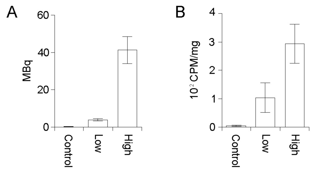
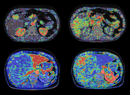
Background: The incidence of small intestinal (SI) and pancreatic neuroendocrine tumors (siNETs and pNETs) seems to have increased. The increased frequency of incidental findings might be a possible explanation. The study aimed to examine (1) changes in incidence and the stage at diagnosis
[...] Read more.
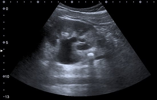
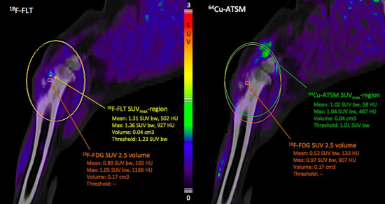
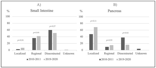
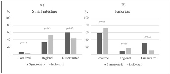
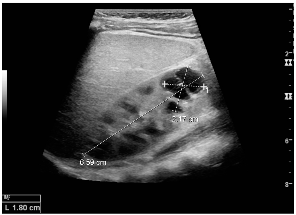
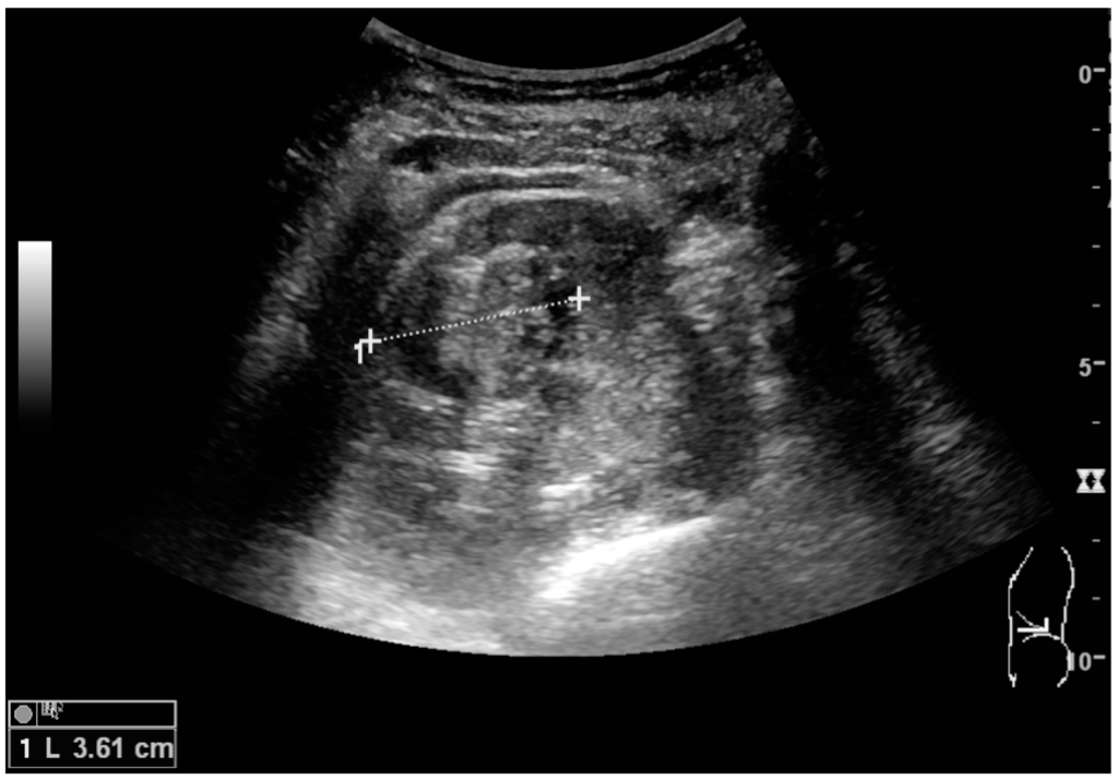
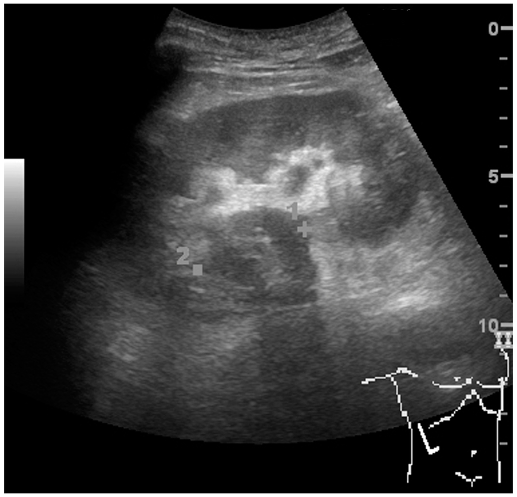
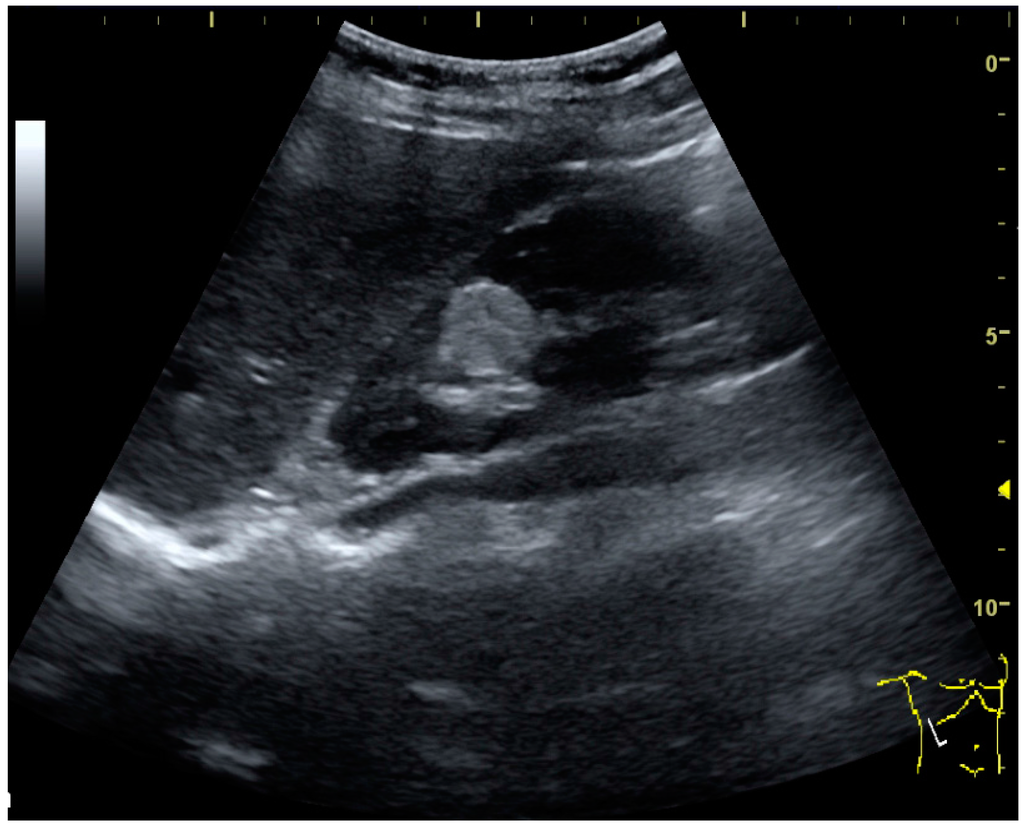
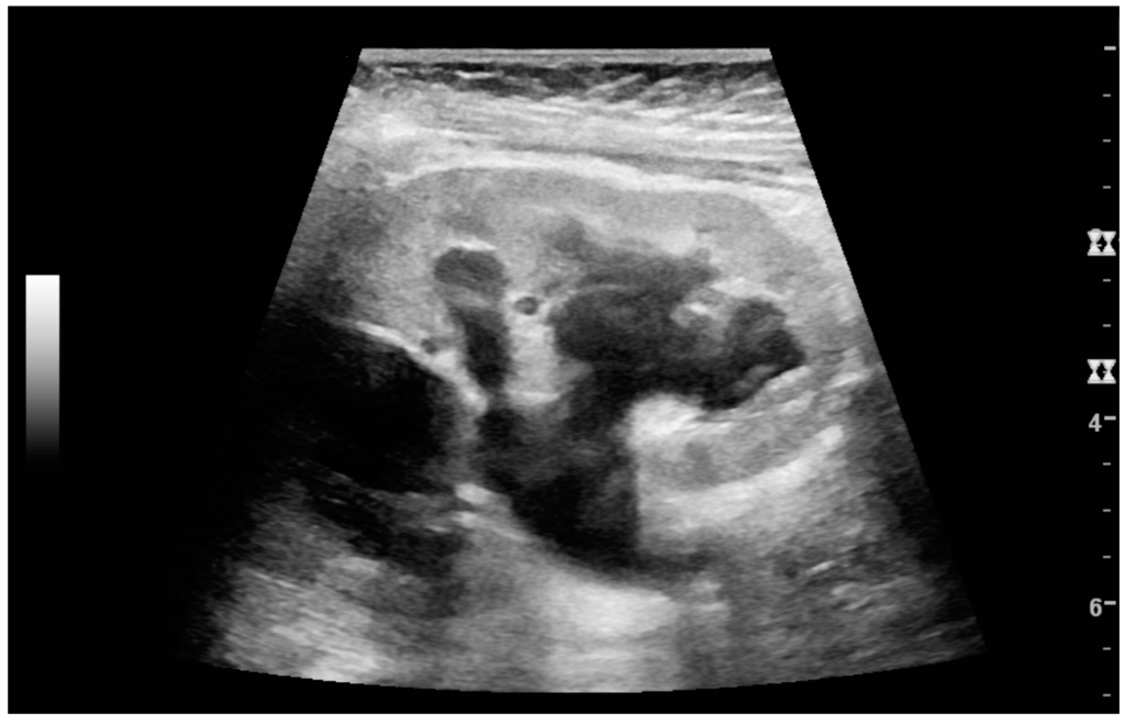
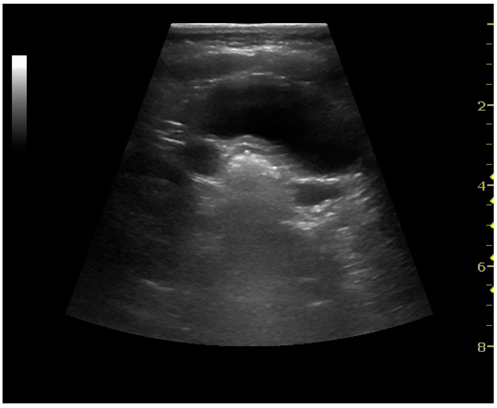
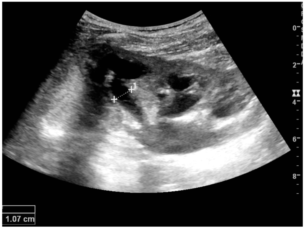
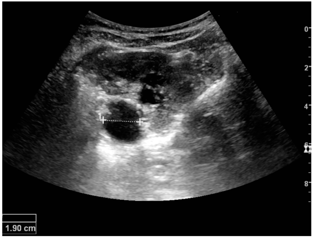
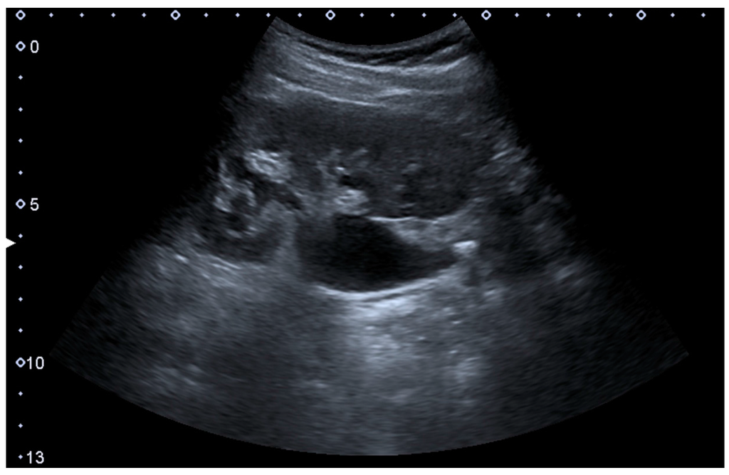
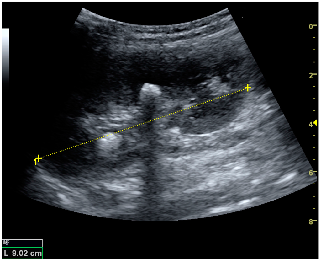
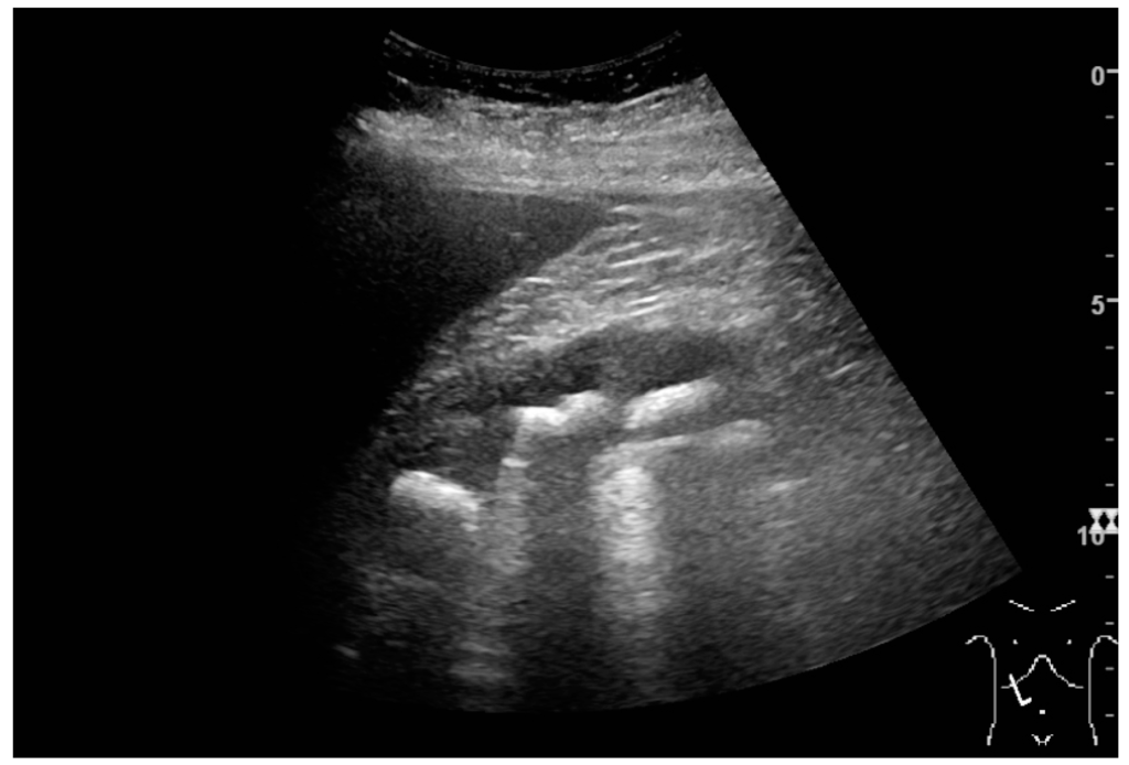
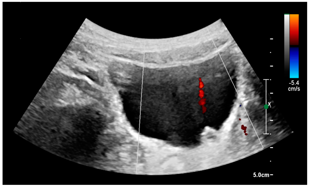
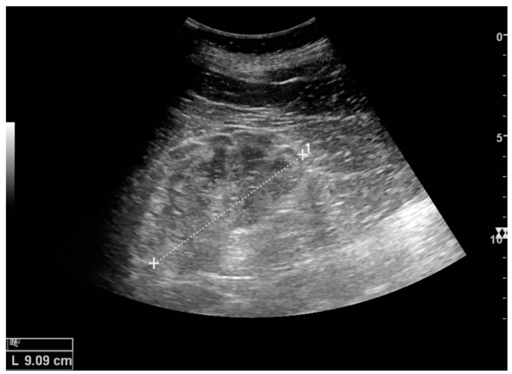
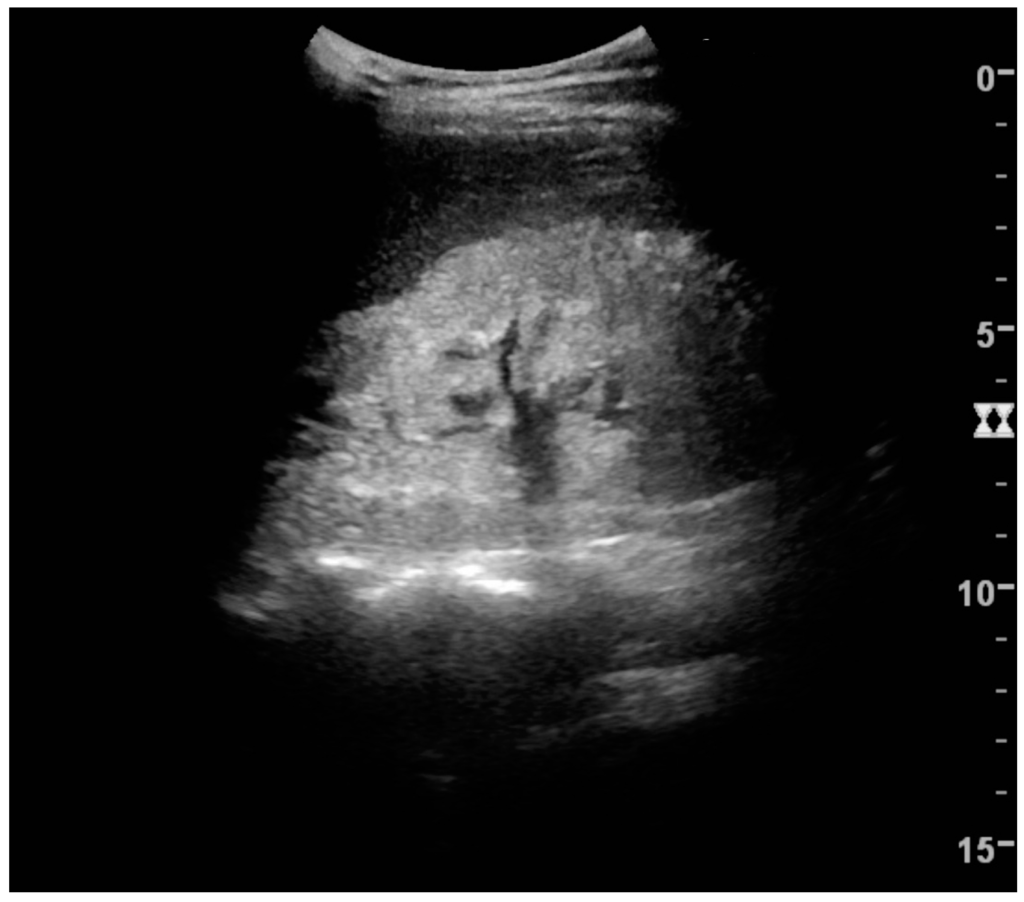
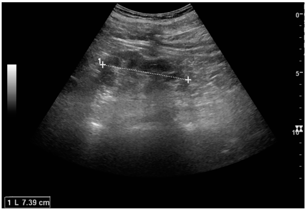
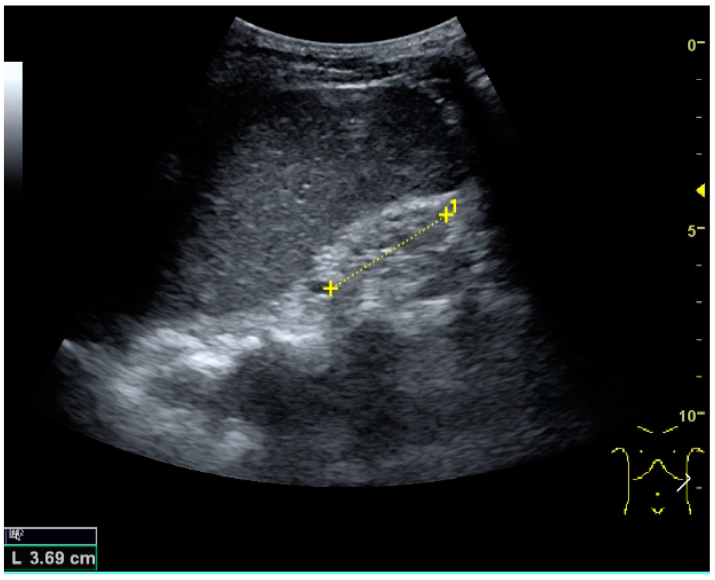
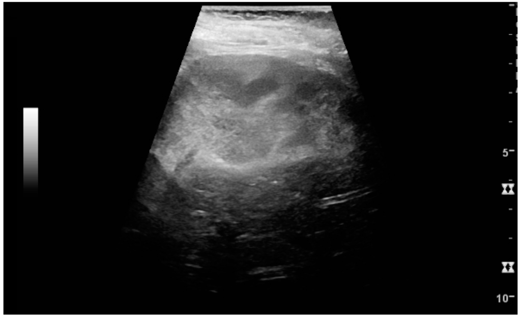
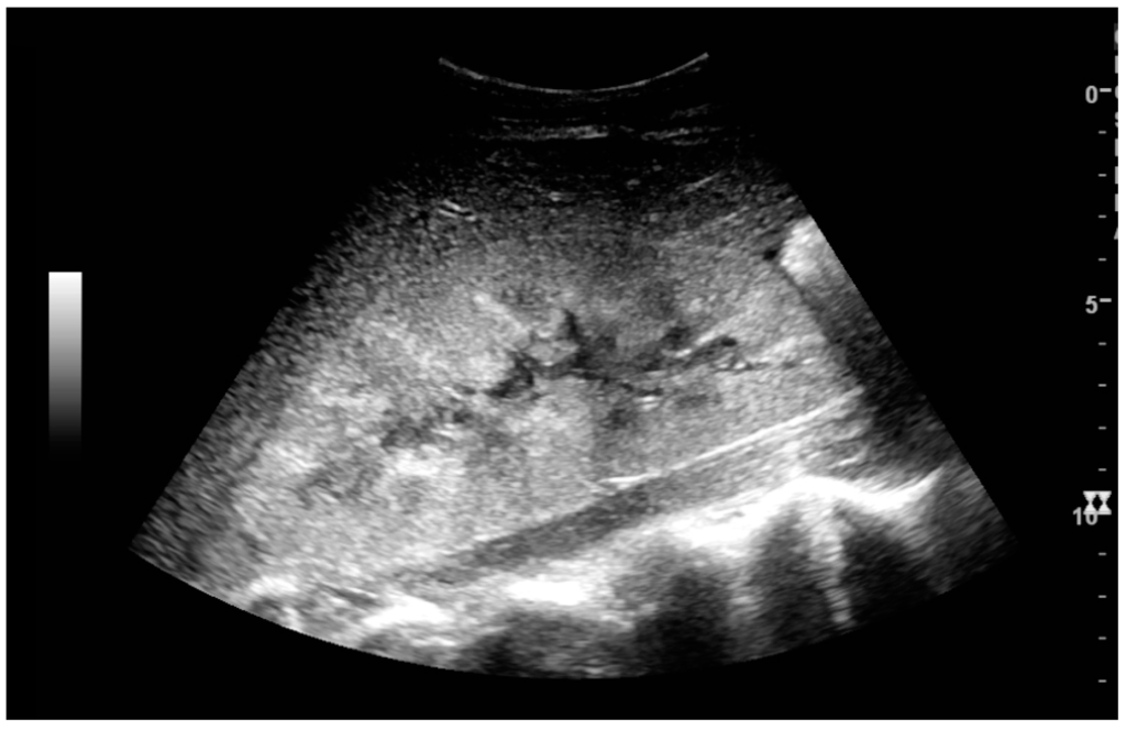
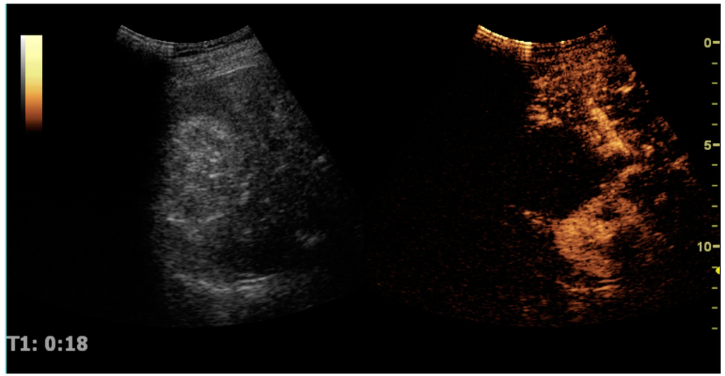
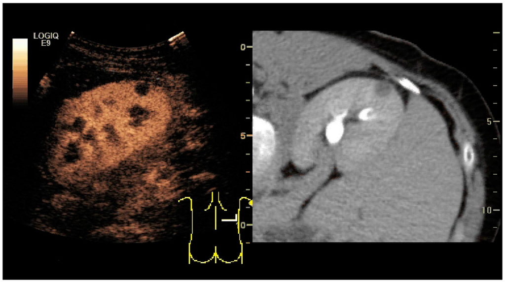
Background: The incidence of small intestinal (SI) and pancreatic neuroendocrine tumors (siNETs and pNETs) seems to have increased. The increased frequency of incidental findings might be a possible explanation. The study aimed to examine (1) changes in incidence and the stage at diagnosis (2010–2011 vs. 2019–2020), (2) changes in the initial indication for diagnostic workup and 3) the differences in stage between incidentally discovered vs. symptomatic disease during the entire study period. Methods: We performed a retrospective study, that includes consecutive siNET and pNET patients referred to the Copenhagen ENETS center of excellence in 2010–2011 and 2019–2020. Results: The annual incidence of siNET per 100,000 increased from 1.39 to 1.84, (p = 0.05). There was no change in the stage at diagnosis, and in both periods approximately 30% of patients were incidentally diagnosed (p = 0.62). Dissemination was found in 72/121 (60%) of symptomatic vs. 22/50 (44%) of incidentally discovered SI tumors in the entire cohort, (p = 0.06). The annual incidence of pNET increased from 0.42 to 1.39 per 100,000, (p < 0.001). The proportion of patients with disseminated disease decreased from 8/21 (38%) to 12/75 (16%), (p = 0.02) and the number of incidental findings increased from 4/21 (19%) to 43/75 (57%), (p = 0.002). More symptomatic patients had disseminated disease compared to patients with incidentally discovered tumors (15/49 (31%) vs. 5/47 (11%), (p = 0.01)). Conclusion: The incidence of siNET and pNETs increased over the past decade. For siNETs, the stage of disease and the distribution of symptomatic vs. incidentally discovered tumors were unchanged between the two periods. Patients with pNETs presented with more local and incidentally discovered tumors in the latter period. Patients with incidentally discovered siNETs had disseminated disease in 44% of the overall cases. The vast majority of incidentally found pNETs were localized.
Full article
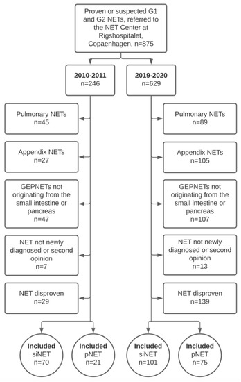
Figure 1


