


Diagnostics 2022, 12(3), 681; https://doi.org/10.3390/diagnostics12030681 - 10 Mar 2022
Cited by 6 | Viewed by 3433
Abstract
►
Show Figures
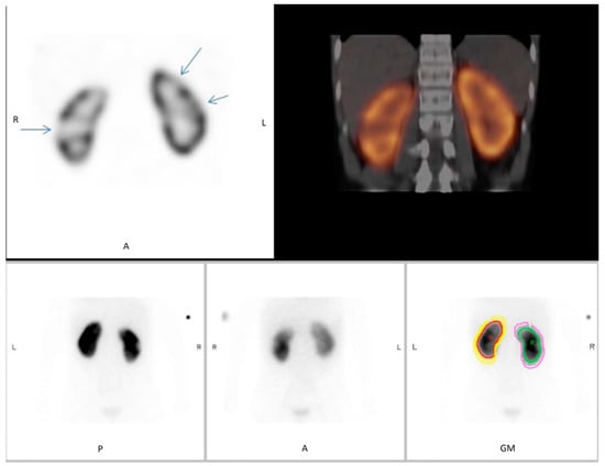
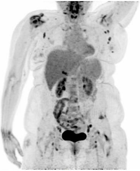
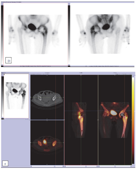
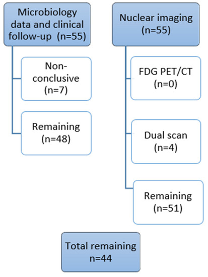
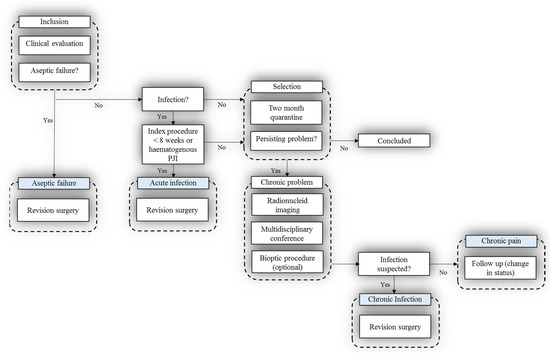
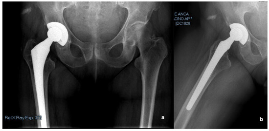
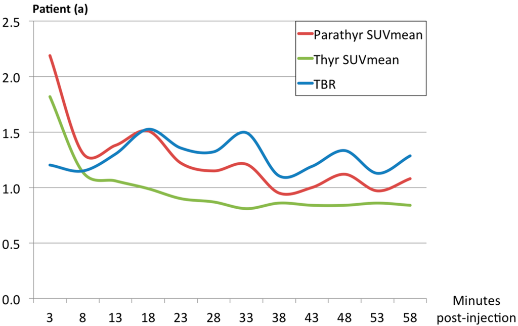
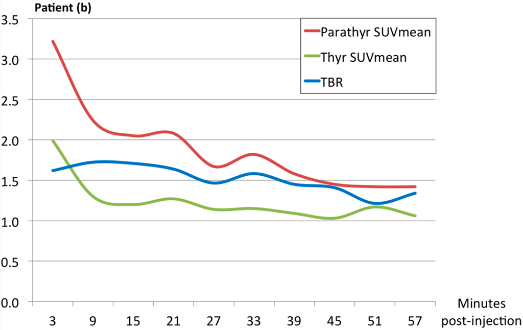
Background: The aim of this prospective study was to assess the diagnostic value of nuclear imaging with 18F-FDG PET/CT (FDG PET/CT), combined 111In-WBC/99mTc-Nanocoll, and 99mTc-HDP SPECT/CT (dual-isotope WBC/bone marrow scan) for patients with chronic problems related to knee
[...] Read more.
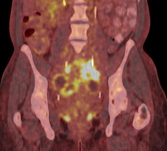
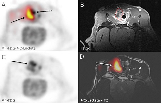
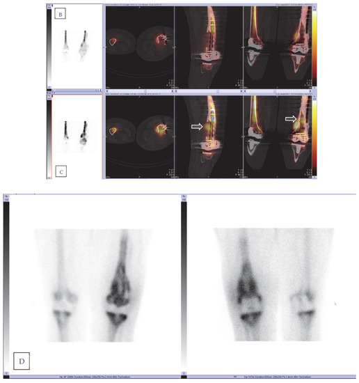
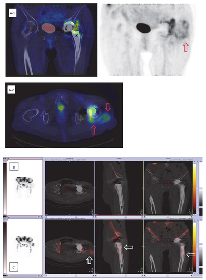
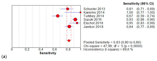
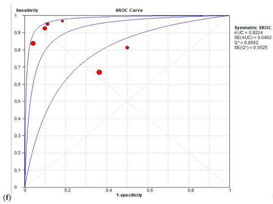
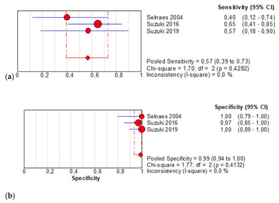
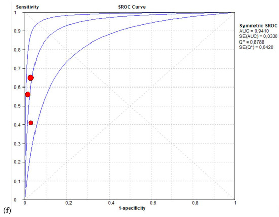
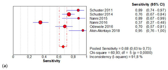
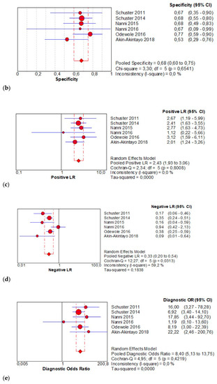
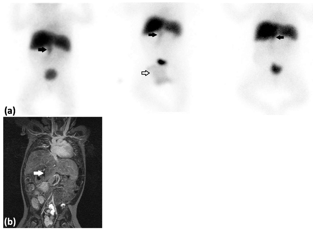
Background: The aim of this prospective study was to assess the diagnostic value of nuclear imaging with 18F-FDG PET/CT (FDG PET/CT), combined 111In-WBC/99mTc-Nanocoll, and 99mTc-HDP SPECT/CT (dual-isotope WBC/bone marrow scan) for patients with chronic problems related to knee or hip prostheses (TKA or THA) scheduled by a structured multidisciplinary algorithm. Materials and Methods: Fifty-five patients underwent imaging with 99mTc–HDP SPECT/CT (bone scan), dual-isotope WBC/bone marrow scan, and FDG PET/CT. The final diagnosis of prosthetic joint infection (PJI) and/or loosening was based on the intraoperative findings and microbiological culture results and the clinical follow-up. Results: The diagnostic performance of dual-isotope WBC/bone marrow SPECT/CT for PJI showed a sensitivity of 100% (CI 0.74–1.00), a specificity of 97% (CI 0.82–1.00), and an accuracy of 98% (CI 0.88–1.00); for PET/CT, the sensitivity, specificity, and accuracy were 100% (CI 0.74–1.00), 71% (CI 0.56–0.90), and 79% (CI 0.68–0.93), respectively. Conclusions: In a standardized prospectively scheduled patient group, the results showed highly specific performance of combined dual-isotope WBC/bone marrow SPECT/CT in confirming chronic PJI. FDG PET/CT has an appropriate accuracy, but the utility of its use in the clinical diagnostic algorithm of suspected PJI needs further evidence.
Full article
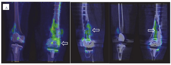
Figure 1


