Int. J. Mol. Sci. 2018, 19(7), 2043; https://doi.org/10.3390/ijms19072043 - 13 Jul 2018
Cited by 226 | Viewed by 21665
Abstract
The mammalian target of rapamycin, mTOR is the master regulator of a cell’s growth and metabolic state in response to nutrients, growth factors and many extracellular cues. Its dysregulation leads to a number of metabolic pathological conditions, including obesity and type 2 diabetes.
[...] Read more.
The mammalian target of rapamycin, mTOR is the master regulator of a cell’s growth and metabolic state in response to nutrients, growth factors and many extracellular cues. Its dysregulation leads to a number of metabolic pathological conditions, including obesity and type 2 diabetes. Here, we review recent findings on the role of mTOR in major metabolic organs, such as adipose tissues, liver, muscle, pancreas and brain. And their potentials as the mTOR related pharmacological targets will be also discussed.
Full article
(This article belongs to the Special Issue mTOR in Human Diseases)
►
Show Figures
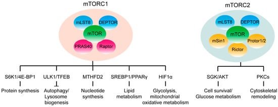
Figure 1

