by
Patrícia Sampaio Tavares Veras 1,2,* and Juliana Perrone Bezerra de Menezes 1
and Juliana Perrone Bezerra de Menezes 1
1
Laboratório de Patologia e Biointervenção, Instituto Gonçalo Moniz, FIOCRUZ, Salvador 40296-710, Brazil
2
Instituto Nacional de Ciência e Tecnologia para Doenças Tropicais (INCT-DT), Salvador 40110-160, Brazil
Int. J. Mol. Sci. 2016, 17(8), 1270; https://doi.org/10.3390/ijms17081270 - 19 Aug 2016
Cited by 33 | Viewed by 11081
Abstract
Leishmania is a protozoan parasite that causes a wide range of different clinical manifestations in mammalian hosts. It is a major public health risk on different continents and represents one of the most important neglected diseases. Due to the high toxicity of the
[...] Read more.
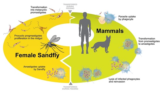
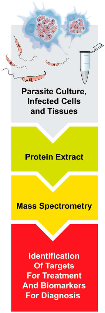
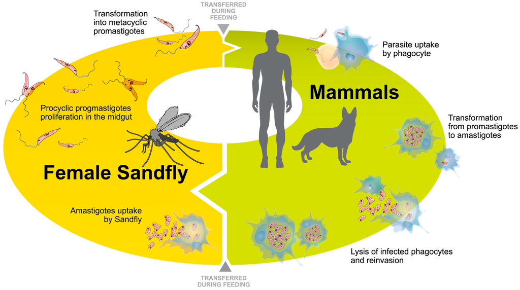
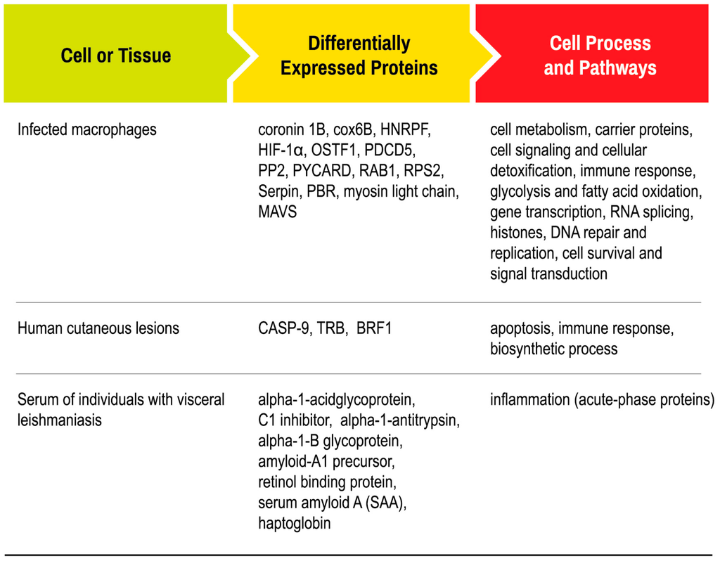
Leishmania is a protozoan parasite that causes a wide range of different clinical manifestations in mammalian hosts. It is a major public health risk on different continents and represents one of the most important neglected diseases. Due to the high toxicity of the drugs currently used, and in the light of increasing drug resistance, there is a critical need to develop new drugs and vaccines to control Leishmania infection. Over the past few years, proteomics has become an important tool to understand the underlying biology of Leishmania parasites and host interaction. The large-scale study of proteins, both in parasites and within the host in response to infection, can accelerate the discovery of new therapeutic targets. By studying the proteomes of host cells and tissues infected with Leishmania, as well as changes in protein profiles among promastigotes and amastigotes, scientists hope to better understand the biology involved in the parasite survival and the host-parasite interaction. This review demonstrates the feasibility of proteomics as an approach to identify new proteins involved in Leishmania differentiation and intracellular survival.
Full article
(This article belongs to the Special Issue Advances in Proteomic Research)
▼
Show Figures
Graphical abstract


