Pathogens 2020, 9(6), 439; https://doi.org/10.3390/pathogens9060439 - 3 Jun 2020
Cited by 22 | Viewed by 13840
Abstract
Autochthonous human and canine strongyloidiasis is reported in Europe but is unclear whether the transmission of infection still occurs. We report a previously unpublished human case in an Italian teen and perform a systematic review of literature on autochthonous human and canine strongyloidiasis
[...] Read more.
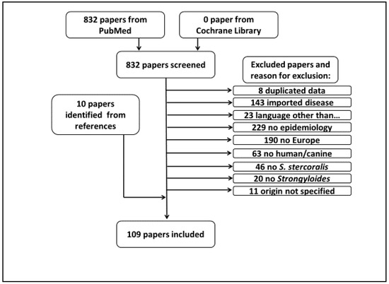
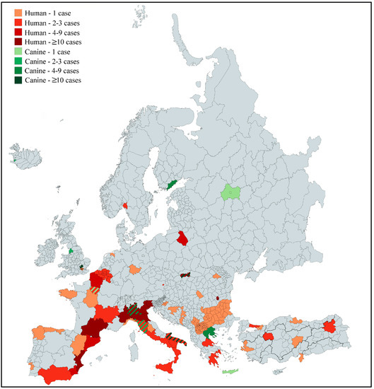
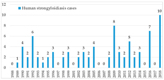
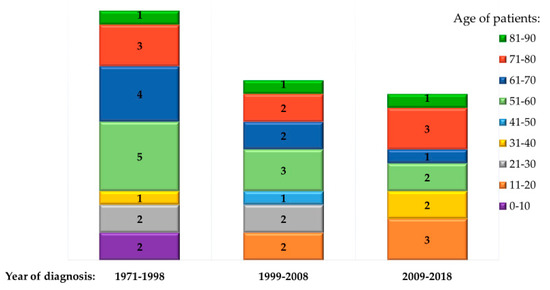
Autochthonous human and canine strongyloidiasis is reported in Europe but is unclear whether the transmission of infection still occurs. We report a previously unpublished human case in an Italian teen and perform a systematic review of literature on autochthonous human and canine strongyloidiasis in Europe to investigate the current dynamic of transmission. Overall, 109 papers published after 1987 were included and one previously unpublished Italian case was added. Eighty case reports were retrieved and 42 of them (52.5%) had severe strongyloidiasis. Most cases were diagnosed in Spain, Italy and France. The median age was 58, the most represented age group was 61–70 years, 11 patients were under 30, and 7 of them were diagnosed after 2000. Epidemiological studies on human strongyloidiasis showed prevalence ranging from 0.56% to 28%. Overall, agriculture work, mine work and walking barefoot were the most commonly reported risk factors for infection. Canine strongyloidiasis was reported mainly in Italy (68 cases), but a few cases occurred also in Iceland, Finland, England, Germany, France, Switzerland, Russia, Slovakia, Romania and Greece. Autochthonous strongyloidiasis is still reported in Europe and sporadic transmission still occurs. Health care professionals should be aware of this issue to identify infected subjects and avoid adverse outcomes, especially in immunosuppressed patients. Further investigations are needed to clarify the zoonotic transmission of this nematode.
Full article
(This article belongs to the Special Issue Prevalence of Strongyloidiasis and Schistosomiasis)
►
Show Figures
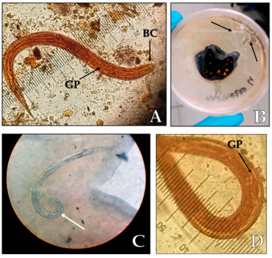
Figure 1


