

-
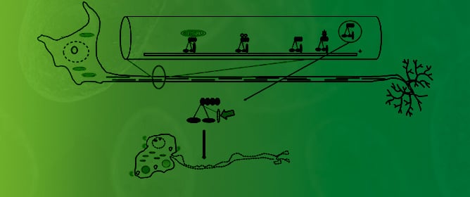
Human Mutant Dynactin Causes Aberrant Mitochondria, Motor Neuron Death, and ALS -
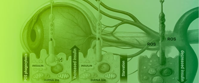
RPE-Derived Insulin: A Local Mechanism Preserving Retinal Metabolic Homeostasis -
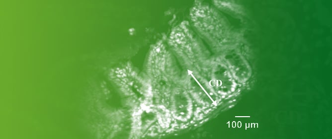
Kisspeptin-10 Effects on the Morphology of Intestinal and Islet Cells in Mice -
Neurofilament Light Chain as a Serum Biomarker in Anorexia Nervosa -
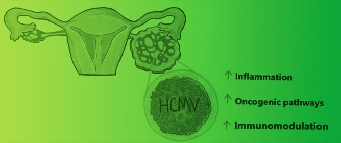
HCMV as an Oncomodulatory Virus in Ovarian Cancer Progression
Journal Description
Biomolecules
Biomolecules
is an international, peer-reviewed, open access journal on structures and functions of bioactive and biogenic substances, molecular mechanisms with biological and medical implications as well as biomaterials and their applications, published monthly online by MDPI.
- Open Access— free for readers, with article processing charges (APC) paid by authors or their institutions.
- High Visibility: indexed within Scopus, SCIE (Web of Science), PubMed, MEDLINE, PMC, Embase, CAPlus / SciFinder, and other databases.
- Journal Rank: JCR - Q1 (Biochemistry and Molecular Biology) / CiteScore - Q1 (Biochemistry)
- Rapid Publication: manuscripts are peer-reviewed and a first decision is provided to authors approximately 16.6 days after submission; acceptance to publication is undertaken in 3.3 days (median values for papers published in this journal in the first half of 2026).
- Recognition of Reviewers: reviewers who provide timely, thorough peer-review reports receive vouchers entitling them to a discount on the APC of their next publication in any MDPI journal, in appreciation of the work done.
- Sections: published in 15 topical sections.
- Testimonials: See what our editors and authors say about Biomolecules.
- Companion journal: Receptors.
Impact Factor:
5.6 (2025);
5-Year Impact Factor:
6.2 (2025)
Latest Articles
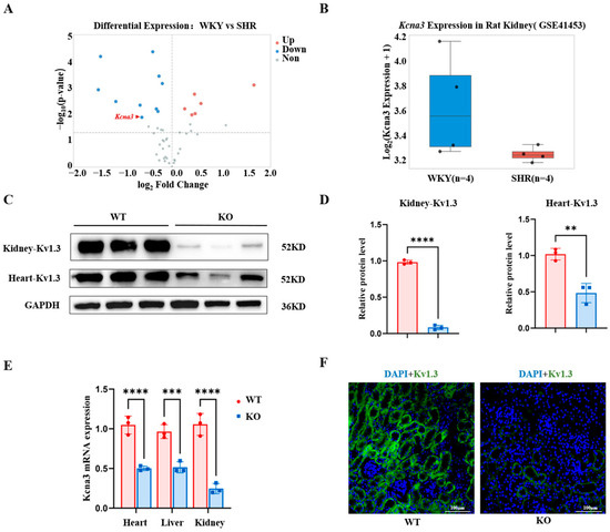
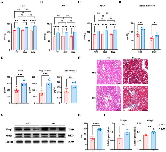
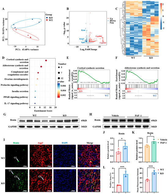
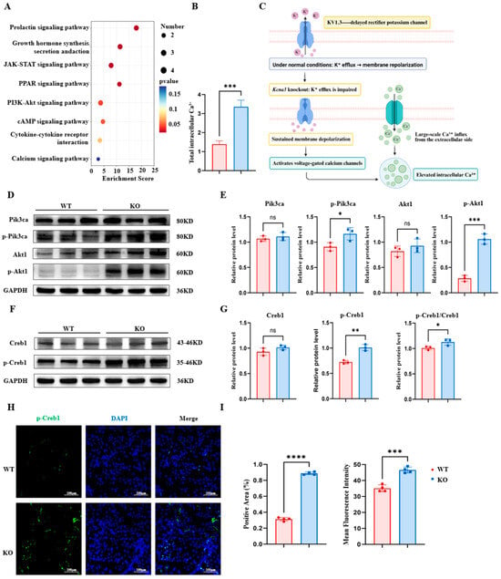
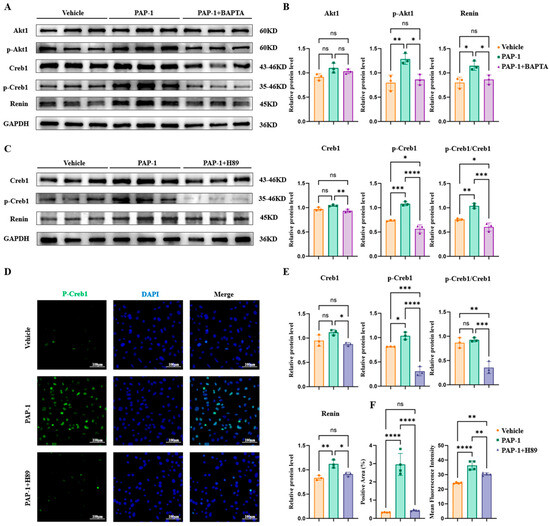
Kcna3 Deficiency Promotes Renin-Associated Hypertension Through Ca2+-Dependent AKT–PKA–CREB Signaling
Biomolecules 2026, 16(9), 1262; https://doi.org/10.3390/biom16091262 (registering DOI) - 31 Aug 2026
Abstract
►
Show Figures
Hypertension is an important risk factor for cardiovascular and renal diseases, yet the mechanisms linking ion channel dysfunction to hypertension remain poorly understood. The voltage-gated potassium channel Kv1.3 (encoded by Kcna3) regulates membrane potential, but its role in the pathogenesis of hypertension
[...] Read more.
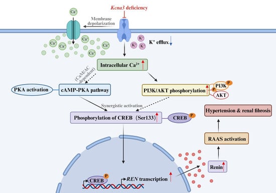
Hypertension is an important risk factor for cardiovascular and renal diseases, yet the mechanisms linking ion channel dysfunction to hypertension remain poorly understood. The voltage-gated potassium channel Kv1.3 (encoded by Kcna3) regulates membrane potential, but its role in the pathogenesis of hypertension remains unclear. In this study, we employed Kcna3 knockout (KO) mice, transcriptomic profiling, and pharmacological inhibition to investigate the role of Kv1.3. Kcna3-deficient mice showed increased blood pressure and renal fibrosis. Transcriptomic profiling showed activation of the renin–angiotensin–aldosterone system (RAAS), with increased renin expression in both Kcna3-deficient mice and Kv1.3 inhibitor-treated cells. Mechanistically, loss of Kv1.3 increased intracellular Ca2+ accumulation, activating the phosphoinositide 3-kinase (PI3K)-AKT and protein kinase A (PKA) pathways, leading to cAMP response element binding protein (CREB) phosphorylation and Renin upregulation. Pharmacological inhibition of Ca2+ signaling or PKA reduced CREB phosphorylation and Renin expression, confirming a causal signaling cascade. Thus, Kv1.3 links membrane excitability to RAAS activation via a Ca2+-dependent AKT–PKA–CREB signaling axis and represents a potential therapeutic target for hypertension and associated renal injury.
Full article
Open AccessArticle
Stroma-Dominant Colorectal Cancer Harbors CAF-Rich Spatial Architecture and EMT-Associated Cancer Cell Plasticity Detectable After Dissemination
by
Shuji Kitagawa, Naoki Mimura, Hironobu Kambara, Masaaki Hori, Erina Yamanishi, Ayako Ogo, Tatsushi Shiomi, Kazuhiko Yoshimatsu, Tomio Ueno and Shuya Yano
Biomolecules 2026, 16(9), 1261; https://doi.org/10.3390/biom16091261 (registering DOI) - 31 Aug 2026
Abstract
Colorectal cancers (CRCs) include stroma-dominant tumors with desmoplasia and differentiated, gland-forming tumors with little stroma. We asked whether this difference reflects stromal abundance alone or also involves a distinct cancer cell state. HEST-1K sections were classified as M-type (stroma-dominant; five patient/tissue units) or
[...] Read more.
Colorectal cancers (CRCs) include stroma-dominant tumors with desmoplasia and differentiated, gland-forming tumors with little stroma. We asked whether this difference reflects stromal abundance alone or also involves a distinct cancer cell state. HEST-1K sections were classified as M-type (stroma-dominant; five patient/tissue units) or D-type (differentiated and cancer cell-dominant; seven units). Prespecified EMT/pEMT, ECM/integrin, YAP/TAZ-TEAD, and DTP/persister gene sets were examined across spatial, bulk, and single-cell datasets and patient-derived malignant ascites cultures. M-type tumors contained broader CAF-rich compartments and higher activity of all four programs in EPCAM/KRT-high regions, including epithelial-dense tumor cores. Activity was greatest near CAF-rich areas. In GSE39582, EMT/pEMT, ECM/integrin, DTP/persister, and invasive epithelial programs were independently associated with recurrence. Ascites-derived cultures from stroma-dominant tumors showed higher ECM/integrin and survival programs. Independent institutional analyses supported the public data findings: focused real-time PCR showed higher MAPK/TGF-beta-related gene expression in M-type-derived than in D-type-derived ascites cells, and IHC showed stronger epithelial CD44v6 expression in M-type primary tumors and metastatic lymph node lesions. CAF co-culture increased VIM-promoter activity. These findings characterize stroma-dominant CRC as a spatially organized ecosystem in which a CAF-rich compartment is associated with stress-adapted cancer cell programs. Its poor prognosis may therefore involve not only stromal abundance but also a morphology-associated cancer cell state detectable after dissemination.
Full article
(This article belongs to the Special Issue Novel Insights into Epithelial Mesenchymal Transition (EMT) in Cancer)
Open AccessReview
Cannabinoids in Cancer: Molecular Mechanisms of Tumor Cell Death and Translational Opportunities
by
Alaa A. El Moghrabi, Ali Al Khatib, Israa Ahmad Cheikh, Charbel Al Hage, Dima Ismail, Mariam Zhour, Philip Mwesigwa and Nadine Darwiche
Biomolecules 2026, 16(9), 1260; https://doi.org/10.3390/biom16091260 (registering DOI) - 31 Aug 2026
Abstract
Cannabinoids are terpenophenolic compounds derived from Cannabis sativa L. that exert a broad range of biological and pharmacological activities. Increasing evidence highlights their potential as modulators of cancer progression specifically through the suppression of tumor cell growth, angiogenesis, and metastasis across multiple
[...] Read more.
Cannabinoids are terpenophenolic compounds derived from Cannabis sativa L. that exert a broad range of biological and pharmacological activities. Increasing evidence highlights their potential as modulators of cancer progression specifically through the suppression of tumor cell growth, angiogenesis, and metastasis across multiple tumor models. This review provides a comprehensive overview of the molecular mechanisms by which natural and synthetic cannabinoids induce regulated cancer cell death. Current evidence demonstrates that cannabinoids regulate multiple forms of cancer cell death, including apoptosis, autophagy-dependent cell death, necroptosis, ferroptosis, and parthanatos. These effects are mediated through complex and interconnected signaling pathways such as TRIB3/AKT/mTORC1, PI3K/AKT/mTOR, MAPK/ERK, NF-κB, ERK/JNK/p38-MAPK, and ceramide/Raf1/ERK/ROS. In addition to their direct antitumor effects, cannabinoids can enhance the efficacy of conventional anticancer therapies through the coordinated regulation of complementary cell death pathways. They also provide clinically relevant supportive benefits in palliative care, alleviating chemotherapy-induced nausea, cachexia, and mood or sleep disturbances. Collectively, these findings identify cannabinoids as promising anticancer agents and therapeutic adjuvants, predominantly in the preclinical setting. However, significant challenges remain regarding their safety, optimal dosing, formulation, and clinical efficacy. Further mechanistic studies, rigorous preclinical research, and well-designed clinical trials are required to establish the translation of cannabinoid-based therapies into precision oncology.
Full article
(This article belongs to the Section Molecular Biology)
Open AccessReview
Mechanism-Oriented Biomaterial Strategies for Bone Regeneration in BRONJ: From Pathological Barriers to Evidence-Matched Repair
by
Aiming Jiang, Juntong Liao, Yinyin Shi, Wenyan Song, Sisi Luo, Longjiang Li and Zhuoyuan Zhang
Biomolecules 2026, 16(9), 1259; https://doi.org/10.3390/biom16091259 (registering DOI) - 31 Aug 2026
Abstract
Bisphosphonate-related osteonecrosis of the jaw (BRONJ) remains a challenging complication of bisphosphonate therapy because jaw extraction sockets exposed to bisphosphonates represent impaired wound environments rather than ordinary bone defects. This narrative review summarizes clinical, cellular, animal, and biomaterial evidence on the mechanisms that
[...] Read more.
Bisphosphonate-related osteonecrosis of the jaw (BRONJ) remains a challenging complication of bisphosphonate therapy because jaw extraction sockets exposed to bisphosphonates represent impaired wound environments rather than ordinary bone defects. This narrative review summarizes clinical, cellular, animal, and biomaterial evidence on the mechanisms that limit BRONJ repair and discusses how these pathological barriers can inform local material design. Current evidence suggests that BRONJ repair is constrained by impaired osteoclast-mediated remodeling, osteocyte and osteoblast dysfunction, oxidative stress, unresolved inflammation, angiogenic insufficiency, microbial challenge, mucosal instability, and changes in bone material properties. Biomaterial strategies investigated to date include local delivery of regenerative factors, restoration of remodeling activity, extracellular vesicles, nucleic acid nanostructures, platelet-derived matrices, antibacterial and ion-releasing hydrogels, angiogenic or lymphangiogenic systems, and mechanically adaptive scaffolds. Most studies remain preclinical and are based on rodent extraction or mandibular defect models, and few establish a direct causal link between a specific material property and durable BRONJ resolution. Future materials should be judged not only by their ability to enhance bone formation, but also by whether they can re-establish a sealed, vascularized, immune-balanced, and remodeling-competent socket capable of sustained jawbone repair.
Full article
(This article belongs to the Special Issue Bone Tissue Morphology: From Pathophysiological Mechanisms to Regenerative Strategies)
Open AccessArticle
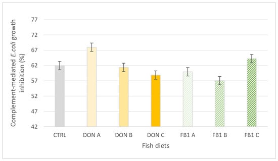
Deoxynivalenol and Fumonisin B1 in Gilthead Seabream Diets: Impact on Growth Performance, Hematology, Immunology, and Histopathology
by
Christina Papadouli, Sofia Vardali, Theodoros Karatzinos, Fotis Lykotrafitis, Myrto Maniaki, Panagiota Panagiotaki, George Rigos, Ioannis Nengas, Morgane Henry, Chrysanthi Nikoloudaki, Dimitra Kogiannou, Petros Chronopoulos and Eleni Golomazou
Biomolecules 2026, 16(9), 1258; https://doi.org/10.3390/biom16091258 (registering DOI) - 31 Aug 2026
Abstract
As aquafeed formulations include more plant-based materials, mycotoxin contamination is becoming relevant for fish farming. Deoxynivalenol (DON) and fumonisin B1 (FB1) frequently occur in fish feeds and may impair fish performance and health. This study evaluated the impact of DON and FB1 on
[...] Read more.
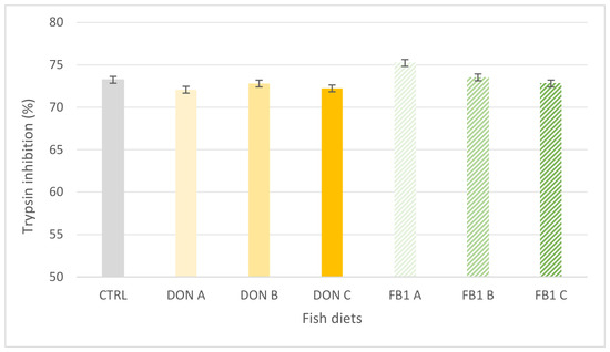
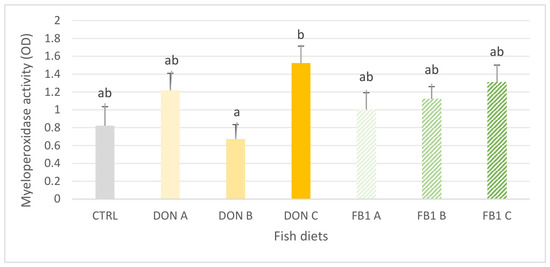
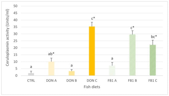
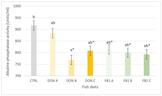
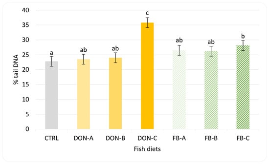
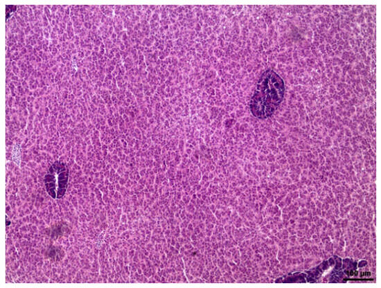
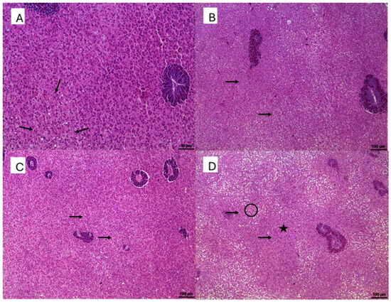
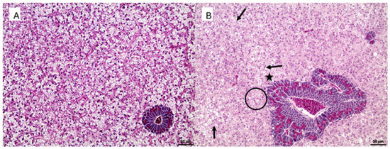
As aquafeed formulations include more plant-based materials, mycotoxin contamination is becoming relevant for fish farming. Deoxynivalenol (DON) and fumonisin B1 (FB1) frequently occur in fish feeds and may impair fish performance and health. This study evaluated the impact of DON and FB1 on growth and health parameters in gilthead seabream (Sparus aurata). Fish were fed ad libitum for seven weeks with six diets assessed in triplicate: DON A (300 ppb), DON B (2000 ppb), DON C (5000 ppb), FB1 A (5 ppb), FB1 B (10 ppb), FB1 C (40 ppb). The highest contamination levels significantly reduced feed intake, body weight, total length, and biomass, while body weight was also reduced in the lowest FB1 treatment. The feed conversion ratio increased in exposed fish, whereas the specific growth rate decreased in DON B, DON C, FB1 A, and FB1 C. Both mycotoxins altered hematological profiles, immune-related enzymatic activities, and liver morphology. DNA damage increased with exposure level and was greatest at the highest DON concentration. Overall, this study shows that short-term dietary exposure can impair seabream performance and health, supporting stricter mycotoxin monitoring in fish feeds and research on chronic exposure, recovery, and species-specific safety thresholds.
Full article
(This article belongs to the Special Issue Physiological Responses and Biomolecular Adaptations to Environmental and Biotic Stress in Aquatic Species)
►▼
Show Figures

Figure 1
Open AccessReview
Inter-Organelle Membrane Contact Sites as Physiological Regulatory Hubs in the Brain: From Neurons to Glial Cells
by
Yuchen Wu, Ginam Cho and Youngshin Lim
Biomolecules 2026, 16(9), 1257; https://doi.org/10.3390/biom16091257 (registering DOI) - 31 Aug 2026
Abstract
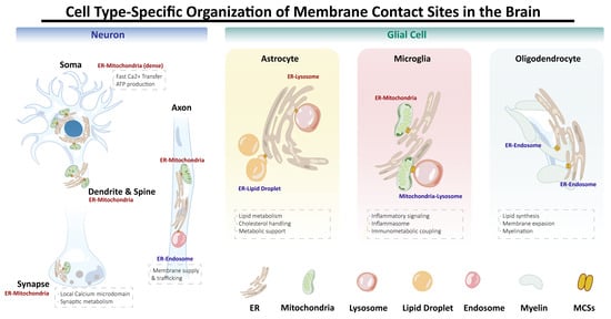
Inter-organelle membrane contact sites (MCSs) enable direct communication between organelles, and this communication is fundamental to cellular homeostasis, coordinated calcium (Ca2+) signaling, lipid metabolism, energy production, and stress responses. While MCSs are evolutionarily conserved, emerging evidence indicates that their organization and
[...] Read more.
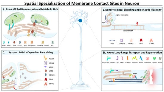
Inter-organelle membrane contact sites (MCSs) enable direct communication between organelles, and this communication is fundamental to cellular homeostasis, coordinated calcium (Ca2+) signaling, lipid metabolism, energy production, and stress responses. While MCSs are evolutionarily conserved, emerging evidence indicates that their organization and function are highly context dependent. In the brain, neurons and glial cells differ markedly in their physiological roles, morphologies, and metabolic demands, suggesting that inter-organelle contact networks might be specialized in a cell-type-dependent manner. Although much of the existing literature focuses on neurons, growing evidence indicates that these contact sites also play important roles in glial cells. Here, we review recent advances in our understanding of MCSs in the nervous system, focusing on cell-type-specific differences between neurons and glial cells. We highlight the spatial specialization of MCSs within neurons, emphasizing how subcellular localization shapes their functional output. Our analysis of the current literature suggests that neuronal MCSs are primarily optimized for rapid Ca2+ signaling and metabolic adaptation, whereas glial MCSs preferentially coordinate lipid metabolism, inflammatory signaling, and tissue homeostasis. We also review the context-dependent and disease-driven remodeling of MCSs in the brain, reflecting alterations in contact-site composition and function rather than simply increased or decreased organelle proximity. Furthermore, we discuss emerging therapeutic perspectives aimed at modulating inter-organelle communication in multiple neurological diseases and outline key unresolved questions and future directions necessary to elucidate how inter-organelle contact sites shape brain physiology and disease. Collectively, the evidence reviewed here indicates that MCSs serve as dynamic signaling platforms, with their specific physiological and pathological functions varying according to cell type, subcellular localization, and molecular composition.
Full article
(This article belongs to the Special Issue Tissue-Specific Organelle Dynamics)
►▼
Show Figures

Figure 1
Open AccessReview
Potential Role of Contact Pathway Factors in Catheter-Related Thrombosis: Emerging Evidence and Therapeutic Strategies
by
Mingyan Jin, Chunliang Liu, Song Lyu, Aoxue Li, Kesheng Dai and Jun Wan
Biomolecules 2026, 16(9), 1256; https://doi.org/10.3390/biom16091256 - 29 Aug 2026
Abstract
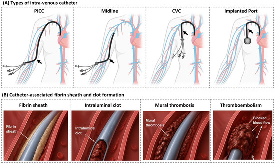
The catheter is among the most commonly used blood-contacting medical devices, but its use can induce surface-mediated coagulation activation, leading to catheter-related thrombosis (CRT). The occurrence of CRT causes venous thromboembolism and catheter malfunction, but current antithrombotic strategies have unsatisfactory efficacy and safety
[...] Read more.
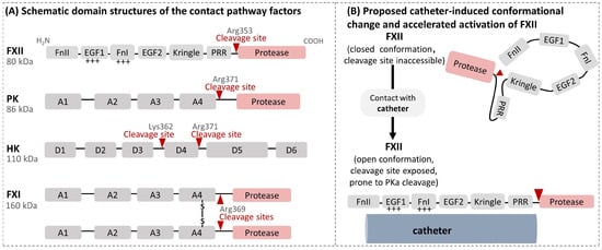
The catheter is among the most commonly used blood-contacting medical devices, but its use can induce surface-mediated coagulation activation, leading to catheter-related thrombosis (CRT). The occurrence of CRT causes venous thromboembolism and catheter malfunction, but current antithrombotic strategies have unsatisfactory efficacy and safety profiles. Here, we review recent advances in the understanding of the pathology of CRT, particularly the roles of the contact pathway factors, and promising novel therapeutic options. Recent studies using genetically modified animals, factor-deficient plasmas, specific inhibitors and purified systems demonstrated an important contribution of contact pathway factors XII and XI to catheter-related blood clotting. Accordingly, contact pathway inhibition has efficacy comparable to that of heparins in mitigating catheter-related coagulation or intraluminal occlusion in various in vitro and animal models, while having lower bleeding risk. Early human studies suggest potential thromboprotective effects of FXI inhibition in catheter placement and hemodialysis settings. However, inhibition of factors XII or XI may impair the defense against infection or disturb normal cardiac function, respectively. Larger human trials are needed to further confirm the efficacy and safety of these contact pathway inhibitors, and to explore whether low-dose combinations of contact pathway inhibitors with heparins are more effective for CRT protection.
Full article
(This article belongs to the Special Issue Molecular and Cellular Mechanisms in Anti-Thrombosis)
►▼
Show Figures

Figure 1
Open AccessArticle
Selective Redox Tuning Enables Potent Intracellular Reduction of Nicotinamide Cytosine Dinucleotide
by
Xiaojia Guo, Yanzhe Huang, Yinghan Hu, Lingyun Zhang and Zongbao K. Zhao
Biomolecules 2026, 16(9), 1255; https://doi.org/10.3390/biom16091255 - 29 Aug 2026
Abstract
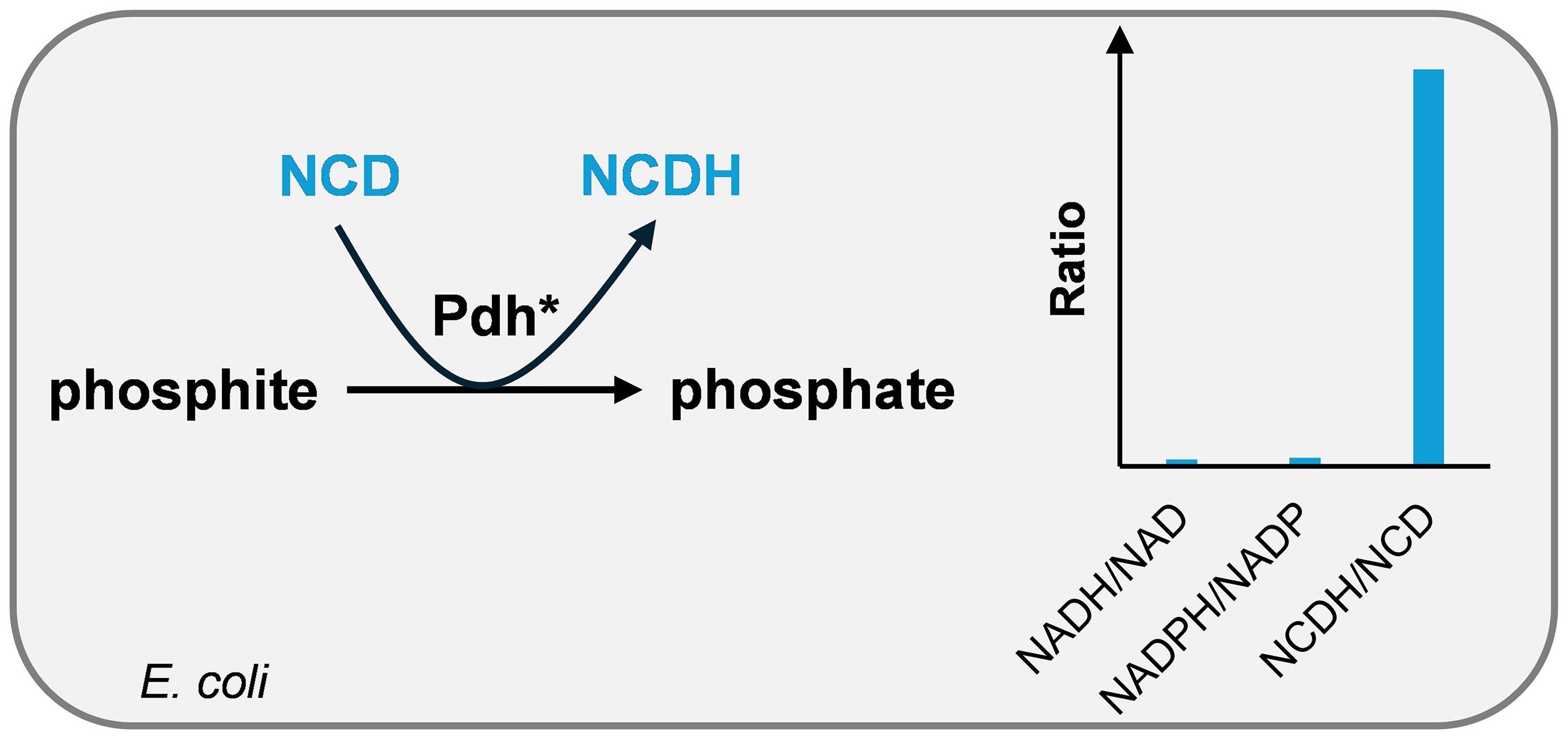
The ubiquitous nicotinamide adenine dinucleotide (NAD) engages in diverse biological processes, leading to non-selective energy transfer toward target synthetic reactions. To achieve selective energy transfer in complex biological systems, we previously constructed artificial systems mediated by the non-natural cofactor nicotinamide cytosine dinucleotide (NCD),
[...] Read more.
The ubiquitous nicotinamide adenine dinucleotide (NAD) engages in diverse biological processes, leading to non-selective energy transfer toward target synthetic reactions. To achieve selective energy transfer in complex biological systems, we previously constructed artificial systems mediated by the non-natural cofactor nicotinamide cytosine dinucleotide (NCD), which can be specifically recognized by engineered enzymes with minimal cross-talk with natural cofactors. For enhanced energy transfer and higher product yields, efficient conversion of NCD to NCDH is required to deliver reducing power in NCD-mediated biosynthetic pathways. Here, we established a comprehensive strategy for selective reduction in intracellular NCD. First, coupled enzymatic colorimetric assays with high specificity were validated for quantifying NAD, NADP, and NCD. With phosphite as the energy source, we selectively elevated the intracellular NCDH/NCD ratio with minimal perturbation to NADH/NAD and NADPH/NADP ratios in E. coli. To overcome the limitation of phosphite transmembrane transport, cell-free systems were constructed to confirm that phosphite could drive near-complete NCD reduction. Finally, cells were treated with polymyxin B, which promoted phosphite uptake and thereby enabled maximal reduction in intracellular NCD. An NCDH/NCD ratio of 47 was achieved, demonstrating that 98% of the intracellular NCD pool existed in the reduced form. This work demonstrates that NCD can function as an independent redox cofactor for selective regulation, providing viable strategies for artificial cofactor-driven biosynthesis.
Full article
(This article belongs to the Section Chemical Biology)
►▼
Show Figures

Graphical abstract
Open AccessReview
Beyond Lipid Lowering: A Narrative Review and Expert Perspective on Precision Cardiovascular Prevention in People with HIV After REPRIEVE
by
Pere Domingo and Paula Prieto
Biomolecules 2026, 16(9), 1254; https://doi.org/10.3390/biom16091254 - 29 Aug 2026
Abstract
The spectrum of diseases in individuals with human immunodeficiency virus (HIV) receiving successful antiretroviral therapy has evolved over time. In the past, they developed opportunistic infections and malignancies, whereas today, cardiovascular disease is among the most common causes of illness and premature death.
[...] Read more.
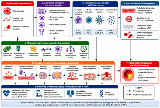
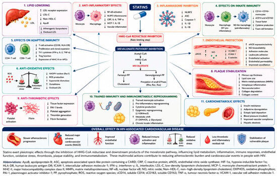
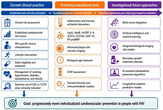
The spectrum of diseases in individuals with human immunodeficiency virus (HIV) receiving successful antiretroviral therapy has evolved over time. In the past, they developed opportunistic infections and malignancies, whereas today, cardiovascular disease is among the most common causes of illness and premature death. Traditional risk factors for atherosclerosis (hypertension, hyperlipidemia, smoking, diabetes, family history of heart disease) are more prevalent in people with HIV than in the general population. However, it is well established that HIV itself causes increased immune activation, chronic inflammation, vascular dysfunction, and a cluster of metabolic abnormalities that contribute to a faster-than-usual rate of biological aging and a higher risk of developing atherosclerosis, a risk not fully captured by current risk models. In the REPRIEVE study, treatment with pitavastatin was shown to reduce the rate of first cardiovascular events among individuals with HIV receiving antiretroviral therapy. Importantly, the beneficial effects of statins on atherosclerosis likely extend beyond lowering cholesterol to include effects on vascular function and on immune and metabolic systems altered by HIV. Even among individuals on statins, a considerable risk of cardiovascular disease remains. Here, We provide a narrative review of current evidence and an expert perspective on emerging approaches to residual cardiovascular risk after REPRIEVE. We review the current understanding of atherosclerosis pathogenesis in individuals with HIV, focusing on recent findings from the REPRIEVE trial. We outline current approaches to improving cardiovascular risk assessment across clinical, biological, and computational levels. We also examine a growing number of therapeutic options that address residual inflammation and atherogenic metabolic disturbance in individuals with HIV on long-term, effective antiretroviral therapy. Significantly, after REPRIEVE, we must move from prescribing statins to all individuals with HIV toward more individualized cardiovascular disease prevention strategies, integrating clinical information, a variety of biomarkers, imaging studies, and even molecular information to generate optimal individualized cardiovascular disease prevention regimens that reflect the complexity of this outcome in naturally diverse individuals.
Full article
(This article belongs to the Special Issue Molecular Mechanisms and Novel Treatments of Atherosclerosis)
►▼
Show Figures

Figure 1
Open AccessArticle
A Novel Cell-Based High-Throughput Screening Model for Inhibitors Targeting Influenza Virus Hemagglutinin–α-2,6-Sialic Acid Interaction
by
Keyu Guo, Xiaofang Chen, Chenyin Wang, Yaru Liu, Chao Liu, Yexiang Wu, Xiuyong Fan, Yanni Xu, Shuyi Si, Yongxin Zhang and Jing Zhang
Biomolecules 2026, 16(9), 1253; https://doi.org/10.3390/biom16091253 - 29 Aug 2026
Abstract
Rising drug resistance undermines current anti-influenza virus therapies. Although targeting the hemagglutinin (HA)–sialic acid receptor interaction is a promising strategy, progress is impeded by the lack of subtype-independent screening models. Herein, we established a fluorescence-based cell high-throughput model using fluorescein isothiocyanate-conjugated Sambucus Nigra
[...] Read more.
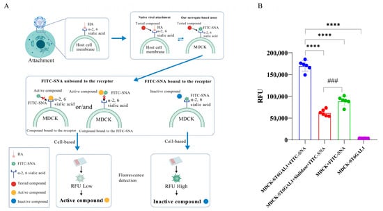
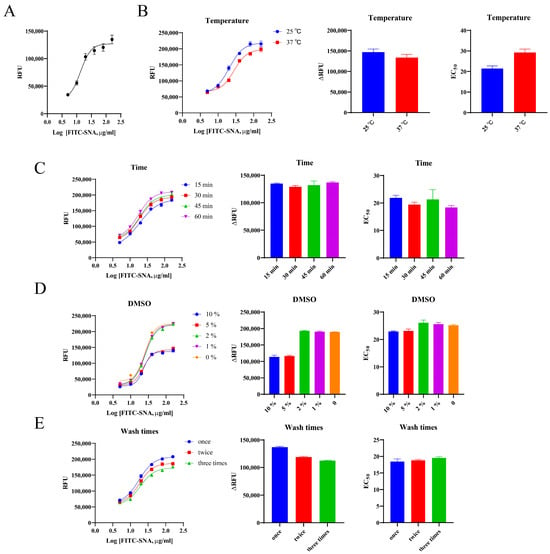
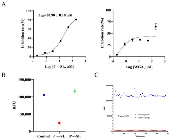
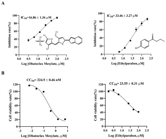
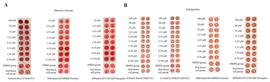
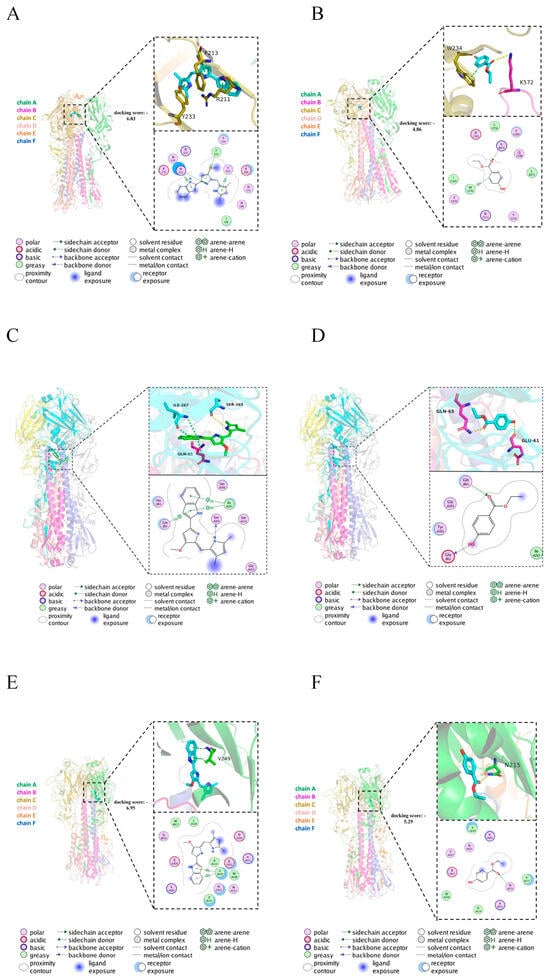
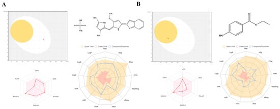
Rising drug resistance undermines current anti-influenza virus therapies. Although targeting the hemagglutinin (HA)–sialic acid receptor interaction is a promising strategy, progress is impeded by the lack of subtype-independent screening models. Herein, we established a fluorescence-based cell high-throughput model using fluorescein isothiocyanate-conjugated Sambucus Nigra Lectin (FITC-SNA) as a stable HA surrogate and α-2,6-sialyltransferase (ST6GAL1)-overexpressing MDCK cells to mimic the HA–receptor interface. This platform was designed to serve as an efficient primary screening tool to rapidly filter large compound libraries for potential binders to the receptor-binding interface. Screening 10,000 compounds identified Obatoclax Mesylate and Ethylparaben as primary hits. Both exhibited broad-spectrum HA inhibition activity, validating the model’s capability to identify compounds interfering with viral attachment. Further cellular antiviral assays revealed cytotoxicity for both compounds, resulting in low selectivity indexes (SIs), indicating that while these molecules effectively target the interaction site, they require substantial structural optimization for therapeutic use. Molecular docking confirmed their binding to type A H1N1, H3N2, and B/Victoria HA proteins, while ADMET predictions highlighted specific structural optimization needs to mitigate toxicity. In conclusion, this subtype-independent, highly specific high-throughput screening (HTS) model provides an efficient and reliable platform for early-stage influenza drug discovery and lead compound development.
Full article
(This article belongs to the Special Issue Design and Synthesis of Bioactive Compounds for Therapeutic Applications—2nd Edition)
►▼
Show Figures

Figure 1
Open AccessFeature PaperArticle
Uremic Serum Alters Gene Expression Profiles and Signaling Pathway Activity in Porcine Arterial Smooth Muscle Cells
by
Youyou Zheng, Kent A. Lee, Unimunkh Uriyanghai, Christine Wai, Mihaela Mocanu, Anthony Z. Yang, Huanjuan Su, Lianxia Li, Vinay A. Sudarsanam, John S. Poulton, Prabir Roy-Chaudhury and Gang Xi
Biomolecules 2026, 16(9), 1252; https://doi.org/10.3390/biom16091252 - 28 Aug 2026
Abstract
Uremic conditions are common in end-stage kidney disease (ESKD) patients. Accelerated vascular diseases in uremic patients lead to heart failure, stroke, and hypertension. To investigate the effects of uremia on porcine arterial smooth muscle cells (aSMCs), bulk RNA sequencing was used to identify
[...] Read more.
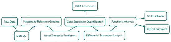
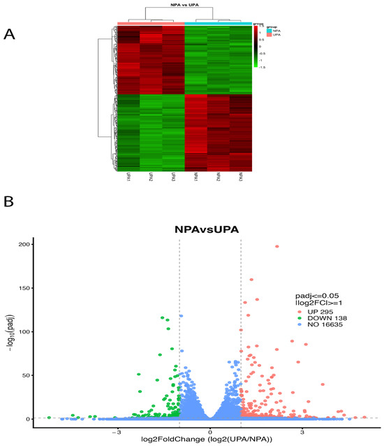
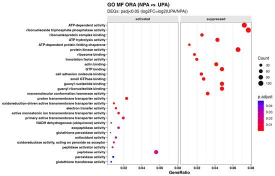
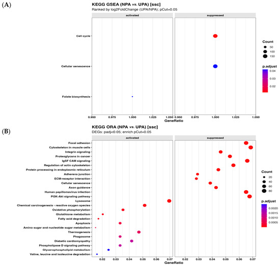
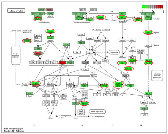
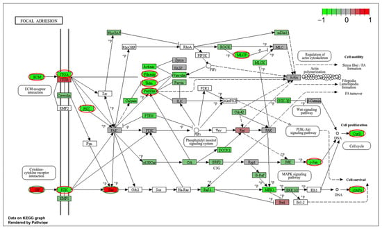
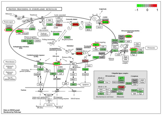
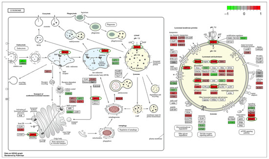
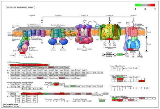
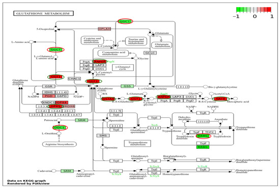
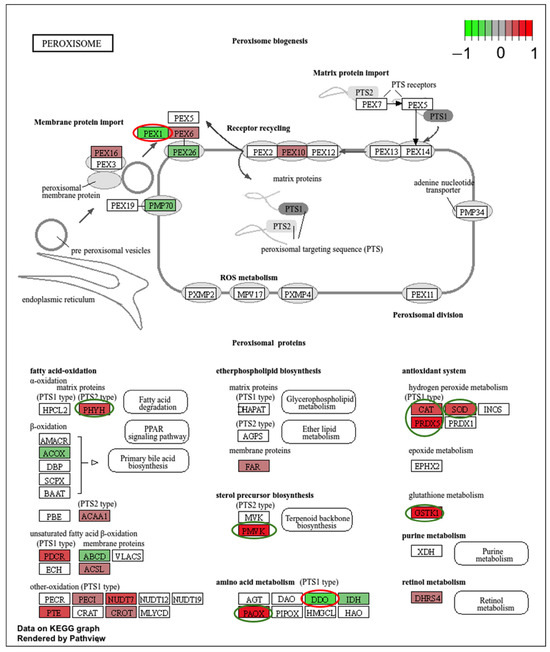
Uremic conditions are common in end-stage kidney disease (ESKD) patients. Accelerated vascular diseases in uremic patients lead to heart failure, stroke, and hypertension. To investigate the effects of uremia on porcine arterial smooth muscle cells (aSMCs), bulk RNA sequencing was used to identify uremia-induced alterations in signaling pathways of aSMCs that might explain the aggressive cardiovascular diseases seen in patients with chronic kidney disease (CKD) and ESKD. Bulk RNA sequencing was performed on porcine aSMCs cultured with serum from normal or uremic pigs. Differentially expressed gene (DEG) analysis revealed that 295 genes were upregulated and 138 genes were downregulated after uremic serum exposure. Gene Ontology molecular function analysis demonstrated that ATP-dependent activity, translation factor activity, and ATP-dependent protein folding chaperones were predicted to be negatively enriched after uremic serum exposure, while proton transmembrane transporter activity, antioxidant activity, and glutathione peroxidase activity were predicted to be positively enriched. Gene set enrichment analysis indicated that the cell cycle was predicted to be negatively enriched after uremic serum exposure in aSMCs. Overrepresentation analysis found that focal adhesion, protein processing in the endoplasmic reticulum (ER) and cell senescence were predicted to be negatively enriched, while lysosome, phagosome, apoptosis, and autophagy were predicted to be positively enriched after uremic serum exposure. This study suggests that the signaling pathways that regulate cellular redox homeostasis, the cellular waste disposal system, ER stress and autophagy are major signaling pathways involved in aSMCs’ responses to uremic serum exposure. These pathways may contribute to the severe arterial-specific clinical symptoms observed in CKD/ESKD patients, such as arterial stiffness, vascular calcification and cardiovascular disease.
Full article
(This article belongs to the Section Biomacromolecules: Proteins, Nucleic Acids and Carbohydrates)
►▼
Show Figures

Figure 1
Open AccessArticle
Breaking the Solubility-Permeability Tradeoff: Surfactant-Mediated Enhancement of Oral Etoposide Absorption
by
Noa Fine-Shamir, Avital Beig and Arik Dahan
Biomolecules 2026, 16(9), 1251; https://doi.org/10.3390/biom16091251 - 28 Aug 2026
Abstract
Developing effective oral formulations for poorly soluble anticancer drugs remains a major pharmaceutical challenge due to the combined limitations of solubility, permeability, and efflux transporter activity. In this work, we investigated the influence of the nonionic surfactants Cremophor EL, Pluronic P-85, and Pluronic
[...] Read more.
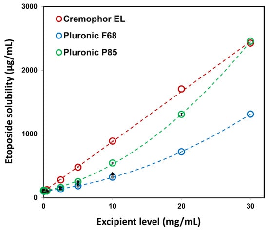
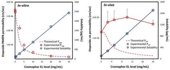
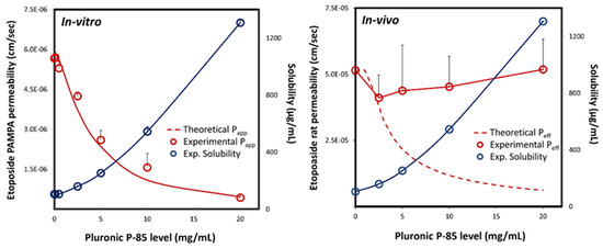
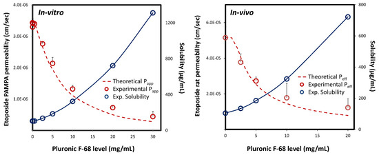
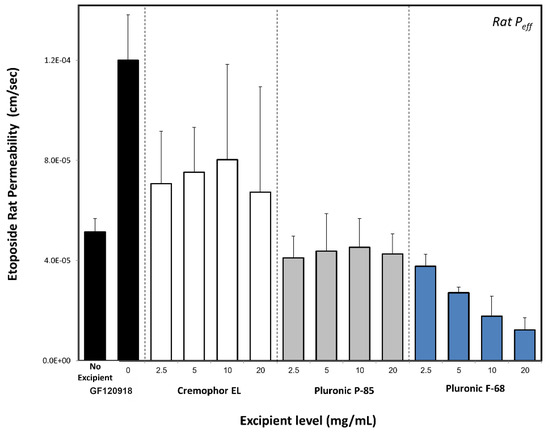
Developing effective oral formulations for poorly soluble anticancer drugs remains a major pharmaceutical challenge due to the combined limitations of solubility, permeability, and efflux transporter activity. In this work, we investigated the influence of the nonionic surfactants Cremophor EL, Pluronic P-85, and Pluronic F-68 on the solubility and intestinal permeability of the anticancer drug etoposide. All surfactants significantly increased etoposide aqueous solubility. While in vitro permeability across an artificial membrane demonstrated the expected solubility-permeability tradeoff, in vivo SPIP studies in rats revealed a distinctive solubility-permeability interplay for Cremophor EL and Pluronic P-85, which simultaneously enhanced solubility and permeability, likely through P-gp inhibition. In contrast, Pluronic F-68 exhibited the classical solubility-permeability tradeoff, consistent with its reported negligible P-gp inhibitory activity. Mechanistic analysis indicated that surfactant hydrophobicity and molecular weight critically influence P-gp inhibition via ATPase modulation. Surfactants with higher hydrophobicity and moderate molecular weight can integrate into the phospholipid bilayer, enabling direct interaction with P-gp and disruption of its ATPase function. These findings provide strategic insights for the rational design of oral formulations capable of overcoming the solubility-permeability tradeoff, improving the bioavailability of challenging anticancer drugs, and may facilitate the transition from intravenous to oral chemotherapy.
Full article
(This article belongs to the Section Molecular Medicine)
►▼
Show Figures

Figure 1
Open AccessReview
Construction and Applicability Scenarios of 3D Neurovascular Unit Models In Vitro
by
Baojian Yu, Zekai Shao, Zhuona Ni, Yuxin Gao, Ziyang Ding, Weifeng Jiang, Lin Li and Lisheng Chu
Biomolecules 2026, 16(9), 1250; https://doi.org/10.3390/biom16091250 - 28 Aug 2026
Abstract
The neurovascular unit (NVU) is composed of a diverse array of cells and an extracellular matrix (ECM). Neural cells and blood vessels are intricately interconnected, forming a cohesive whole. Specific cellular components and structures within the NVU play an indispensable role in maintaining
[...] Read more.
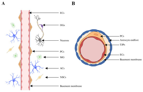
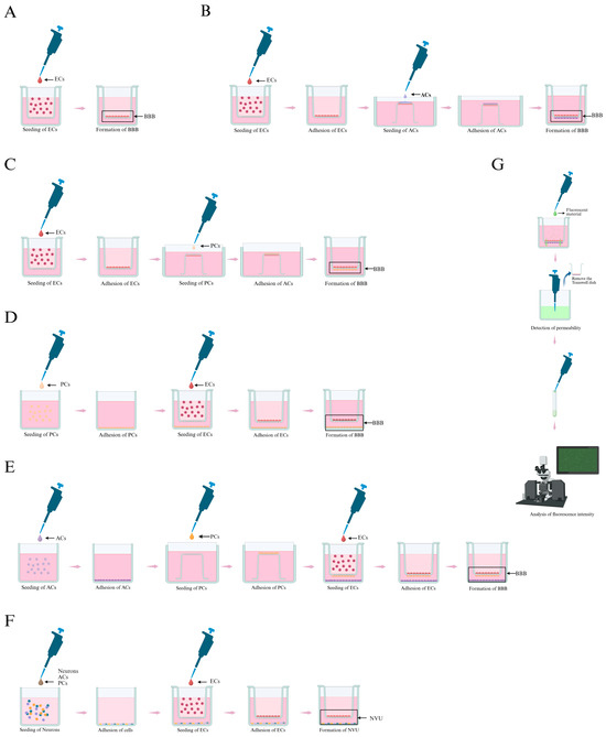
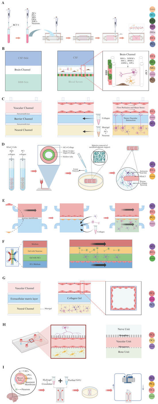
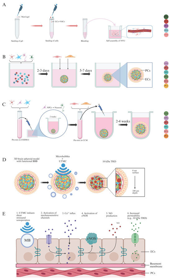
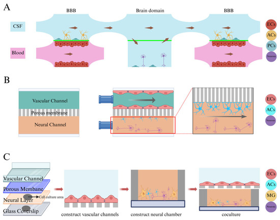
The neurovascular unit (NVU) is composed of a diverse array of cells and an extracellular matrix (ECM). Neural cells and blood vessels are intricately interconnected, forming a cohesive whole. Specific cellular components and structures within the NVU play an indispensable role in maintaining homeostasis of the central nervous system (CNS). With the advancement and maturation of cell co-culture technology, various three-dimensional (3D) NVU models continue to emerge, offering a more objective and comprehensive perspective for in vitro studies of CNS diseases. Specifically, these 3D NVU models include Transwell Chamber models, gel-PDNS-based 3D models, self-assembled NVU models and microfluidic NVU models, which reconstruct the complex NVU architecture to varying degrees. This review systematically summarizes multiple 3D construction strategies for in vitro NVU to overcome the limitations of conventional cellular tests or animal experiments, highlights the critical roles of biomimetic gel in recapitulating native cell-gel crosstalk, comparatively analyzes four major 3D NVU technical routes in terms of cellular composition, vascular morphology, barrier performance, and reproducibility, categorizes application scenarios of 3D NVU platforms oriented to practical research demands, including oxygen-glucose deprivation/reoxygenation (OGD/R) injury modeling, BBB permeability assay, CNS drug penetration screening, neuroinflammation and neurotoxicity evaluation, proposes practical principles for model selection under different experimental purposes, and concludes with current bottlenecks, including imperfect vascular network maturation and lack of unified evaluation criteria, together with future perspectives for standardized 3D NVU in vitro. By comparing the advantages and limitations of these approaches, we aim to clarify their optimal applicability for investigating specific pathological mechanisms and screening potential therapeutics.
Full article
(This article belongs to the Special Issue White Matter Injury and Neurovascular Dysfunction in Neurological Disorders)
►▼
Show Figures

Figure 1
Open AccessArticle
Germplasm Screening and Transcriptome Profiling Identify Phenylpropanoid Biosynthesis-Related PAL Candidate Genes Associated with Freezing Tolerance in Potato
by
Yuwei Ge, Qianqian Wang, Yuying Fu, Tingting Wang, Huajun Liao and Chongchong Yan
Biomolecules 2026, 16(9), 1249; https://doi.org/10.3390/biom16091249 - 28 Aug 2026
Abstract
Potato (Solanum tuberosum L.) seedlings are highly sensitive to freezing temperatures, which severely impairs growth and causes substantial losses in tuber yield and quality. This study aimed to screen freezing-tolerant potato germplasm accessions, identify candidate genes involved in the freezing response, and
[...] Read more.
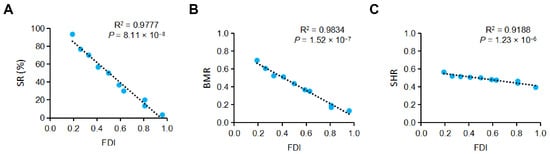
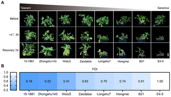
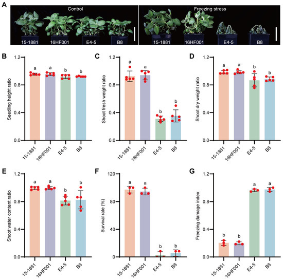
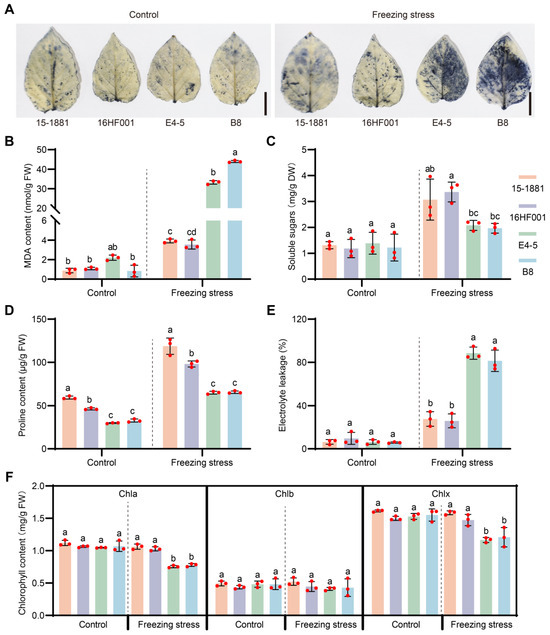
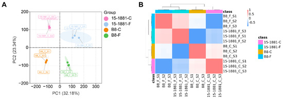
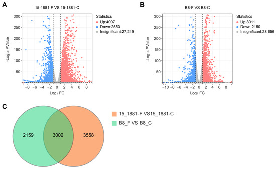
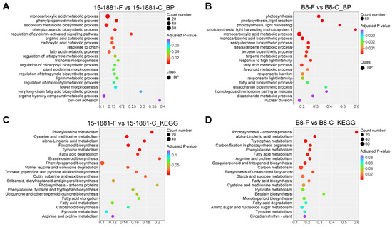
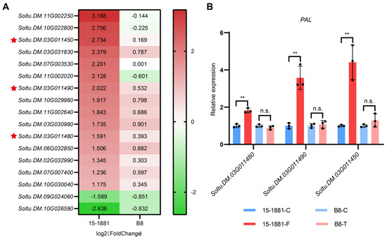
Potato (Solanum tuberosum L.) seedlings are highly sensitive to freezing temperatures, which severely impairs growth and causes substantial losses in tuber yield and quality. This study aimed to screen freezing-tolerant potato germplasm accessions, identify candidate genes involved in the freezing response, and provide elite parental materials and a theoretical reference for molecular breeding of freezing-tolerant cultivars. We evaluated seedling freezing tolerance of 73 potato accessions using the freezing damage index (FDI), and performed transcriptome sequencing on leaf samples from highly freezing-tolerant (HT) accession 15-1881 and highly freezing-susceptible (HS) accession B8 following freezing treatment. Substantial variation in freezing tolerance was observed across the germplasm panel, with 5 HT accessions and 12 HS accessions identified, showing marked phenotypic and physiological differences under freezing stress. Transcriptomic analysis detected 6560 differentially expressed genes (DEGs) in 15-1881 and 5161 DEGs in B8, with 3558 DEGs specific to 15-1881. The phenylpropanoid biosynthesis pathway exhibited noticeable expression divergence between 15-1881 and B8, harboring 17 15-1881-specific DEGs including three tandem phenylalanine ammonia-lyase (PAL) genes. The HT germplasm accessions obtained in this study provide breeding resources for freezing-tolerant potato improvement, and the PAL genes characterized here represent promising candidate genes associated with freezing response in potato.
Full article
(This article belongs to the Special Issue Molecular Genomics for Plant Stress Resilience and Improvement)
►▼
Show Figures

Figure 1
Open AccessArticle
Decreased Plasma IGF-1 Is Associated with Cortical Atrophy, but Not Concomitant Cerebrovascular Disease in Alzheimer’s Dementia
by
Amelia T. Y. Yam, Yuek Ling Chai, Saima Hilal, Cai Yuan, Vincent C. T. Mok, Narayanaswamy Venketasubramanian, Boon Yeow Tan, Ming Ann Sim, Mitchell K. P. Lai, Christopher P. Chen and Joyce R. Chong
Biomolecules 2026, 16(9), 1248; https://doi.org/10.3390/biom16091248 - 28 Aug 2026
Abstract
Dysregulated insulin signaling in the brain has been linked to cognitive impairment and dementia. Insulin-like growth factor 1 (IGF-1) is a peptide growth hormone crucial for neurogenesis and neuroprotection. Findings regarding potential involvement of IGF-1 in dementia have been conflicting, and the status
[...] Read more.
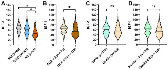
Dysregulated insulin signaling in the brain has been linked to cognitive impairment and dementia. Insulin-like growth factor 1 (IGF-1) is a peptide growth hormone crucial for neurogenesis and neuroprotection. Findings regarding potential involvement of IGF-1 in dementia have been conflicting, and the status of IGF-1 in clinical cohorts with Alzheimer’s disease (AD) and concomitant cerebrovascular disease (CeVD) burden is unknown. A Singapore-based memory clinic cohort consisting of 46 non-cognitively impaired (NCI), 101 with cognitive impairment, no dementia (CIND) and 81 AD dementia subjects underwent plasma IGF-1 measurements and neuroimaging assessments for association analyses of peripheral IGF-1 with regional brain volumes, as well as with neuroimaging CeVD markers (lacunes, cerebral microbleeds, white matter hyperintensities). Plasma IGF-1 levels were significantly lower in AD compared to NCI and CIND participants (both p < 0.001). Plasma IGF-1 was significantly associated with smaller hippocampal (p = 0.035), amygdala (p = 0.024), parietal lobe (p = 0.029), and frontal lobe (p = 0.002) volumes. In contrast, plasma IGF-1 did not associate with CeVD markers after covariate adjustments. Our findings suggest that plasma IGF-1 may be a blood-based biomarker for reduced brain volumes, while having no direct role in CeVD pathophysiology.
Full article
(This article belongs to the Section Molecular Biomarkers)
►▼
Show Figures

Figure 1
Open AccessReview
Active Human Transposable Elements: Long-Read Sequencing Technologies, Computational Analysis, and Implications for Human Disease
by
Dániel Vörösvácki, Nikolett Szakállas, Alexandra Kalmár, István Takács and Béla Molnár
Biomolecules 2026, 16(9), 1247; https://doi.org/10.3390/biom16091247 - 27 Aug 2026
Abstract
Transposable elements (TEs) account for nearly half of the human genome and shape chromatin organization, gene regulation, and genome evolution. However, their contributions to human physiology and disease remain incompletely understood. The most active elements in humans, LINE-1 (L1), Alu, and SVA, retain
[...] Read more.
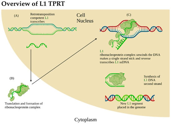
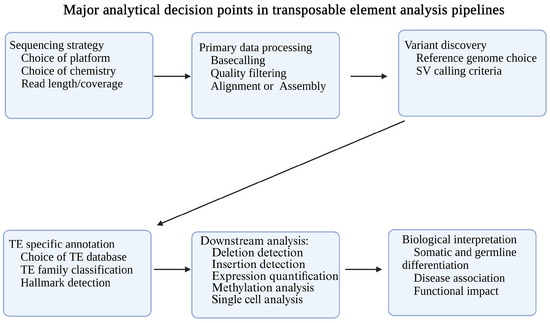
Transposable elements (TEs) account for nearly half of the human genome and shape chromatin organization, gene regulation, and genome evolution. However, their contributions to human physiology and disease remain incompletely understood. The most active elements in humans, LINE-1 (L1), Alu, and SVA, retain some copies with the ability to evade epigenetic repression and mobilize via target-primed reverse transcription (TPRT), whereas copies become inactive through various fragmentations and mutations. TE activity contributes to genomic instability and has been implicated in aging, cancer, neurological disorders, chromatin organization, and epigenetic regulation. Studying TE is challenging due to their repetitive and polymorphic nature. Recent advances in sequencing technologies and short- and long-read sequencing platforms, combined with specialized bioinformatic pipelines, currently enable more comprehensive characterization of TE insertions, deletions, expression, and epigenetic status. Computational approaches vary in sensitivity, specificity, and resource requirements, and their performance is influenced by sequencing modality, coverage, and the reference genome used. Assembly-based and read-based methods, as well as integrating methylation data or single-cell data, provide complementary insights into TE biology. This review summarizes the biology of active human TE, surveys state-of-the-art short- and long-read pipelines for TE analysis, and highlights their applications in studies of aging, cancer, and other complex diseases. We also provide practical guidance for selecting appropriate sequencing strategies and tools for TE-focused projects, and discuss emerging approaches and open questions in the field.
Full article
(This article belongs to the Section Bioinformatics and Systems Biology)
►▼
Show Figures

Figure 1
Open AccessReview
Advancing Epidermal Barrier Resilience in Atopic Dermatitis with Isosorbide Di-Fatty Acid Esters: From Disruption to Restoration
by
Ratan K. Chaudhuri and Raja K. Sivamani
Biomolecules 2026, 16(9), 1246; https://doi.org/10.3390/biom16091246 - 27 Aug 2026
Abstract
Atopic dermatitis (AD) is a chronic, relapsing inflammatory skin disease characterized by epidermal barrier dysfunction, immune dysregulation, microbial imbalance, and severe pruritus. Emerging evidence establishes that barrier disruption is a central pathogenic driver capable of initiating inflammatory signaling, neuroimmune activation, and chronic disease
[...] Read more.
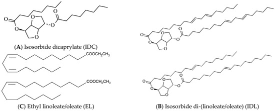
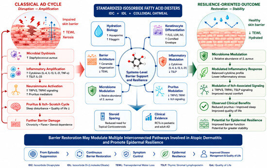
Atopic dermatitis (AD) is a chronic, relapsing inflammatory skin disease characterized by epidermal barrier dysfunction, immune dysregulation, microbial imbalance, and severe pruritus. Emerging evidence establishes that barrier disruption is a central pathogenic driver capable of initiating inflammatory signaling, neuroimmune activation, and chronic disease instability. This understanding has shifted therapeutic paradigms toward barrier-directed strategies aimed at restoring epidermal resilience. This narrative review evaluates the mechanistic and clinical evidence surrounding isosorbide fatty acid diester molecules—specifically isosorbide dicaprylate (IDC) and isosorbide di-(linoleate/oleate) (IDL)—as a barrier-first approach for AD management. Early in vitro and ex vivo investigations demonstrated that IDC significantly improves epidermal hydration, transepidermal water loss, and the expression of barrier-associated genes linked to epidermal integrity. Subsequent studies showed that IDL expands these effects through coordinated regulation of keratinocyte differentiation, lipid homeostasis, and inflammatory stress pathways. Furthermore, recent mechanistic data highlight synergistic anti-inflammatory and pruritus-modulating effects involving TRPA1-, TRPV3-, and TSLP-associated pathways, while preserving tissue integrity under cytokine-induced stress. Clinically, these findings are supported by randomized studies in pediatric and adult cohorts demonstrating significant reductions in pruritus, favorable Eczema Area and Severity Index (EASI) responses, decreased topical corticosteroid dependence, and a reduction in the relative abundance of Staphylococcus aureus. Collectively, these findings support a barrier-first therapeutic framework in which restoration of epidermal resilience may beneficially influence multiple interconnected pathways involved in atopic dermatitis.
Full article
(This article belongs to the Special Issue Bioactive Compounds in Dermatology)
►▼
Show Figures

Figure 1
Open AccessSystematic Review
Nanoparticle-Based Therapies for Myocardial Injury and Heart Failure: A Systematic Review and Translational Appraisal of Preclinical Evidence
by
Ayesha Jabeen, Ilaria Barison, Honoria Ocagli, Bruna Fata, Diego Perazzolo, Cristina Basso, Roberto Luisetto, Silvia Pozzo, Fabrizio Mancin, Enrico Grisan, Dario Gregori, Annalisa Angelini, Marny Fedrigo and Chiara Castellani
Biomolecules 2026, 16(9), 1245; https://doi.org/10.3390/biom16091245 - 27 Aug 2026
Abstract
Background: Heart failure remains a leading cause of morbidity and mortality, and current therapies rarely repair established myocardial damage. Nanoparticle-based interventions have been investigated across heterogeneous models of myocardial injury, remodeling, cardiomyopathy, and heart failure, but the distribution and translational maturity of this
[...] Read more.
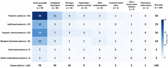
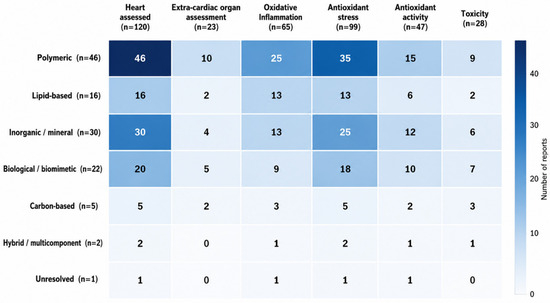
Background: Heart failure remains a leading cause of morbidity and mortality, and current therapies rarely repair established myocardial damage. Nanoparticle-based interventions have been investigated across heterogeneous models of myocardial injury, remodeling, cardiomyopathy, and heart failure, but the distribution and translational maturity of this evidence remain unclear. Methods: A systematic search of PubMed, Embase, Scopus, and Web of Science was conducted from database inception to June 2024. Eligible reports were mapped according to disease model, experimental system, carrier-level nanoparticle platform, payload, route, comparator, outcomes, biodistribution, safety assessment, and translational characteristics. Reports of non-therapeutic nanoparticle exposure were retained in a separate contextual safety/toxicology stratum and were not included in the therapeutic evidence-density map. Risk of bias was evaluated using design-appropriate tools. Results: Of 2640 records screened, 157 independent studies met the criteria: 140 in the main therapeutic/platform evidence map and 17 in a separate contextual safety/toxicology stratum. Within the main corpus, polymeric systems were the largest platform class (n = 50), followed by inorganic/mineral (n = 35), biological/biomimetic (n = 24), lipid-based (n = 23), carbon-based (n = 5), and hybrid/multicomponent systems (n = 3). Evidence was concentrated in acute myocardial injury (n = 76), while direct same-agent comparisons, long-term safety assessment, repeated dosing, quantitative biodistribution, and clinically aligned heart-failure models remained limited. Conclusions: The field demonstrates substantial formulation diversity and biological activity, but translation is constrained by fragmented characterization, sparse comparative evidence, and incomplete assessment of biological fate and safety.
Full article
(This article belongs to the Section Bio-Engineered Materials)
►▼
Show Figures

Figure 1
Open AccessArticle
Unveiling the Physicochemical Properties of Magnetic Nanoparticles as Solid Carriers for Laccase Immobilization Toward Different Reducing Substrates
by
Jessica Costa, Andrea Atrei, Juan José Valle-Delgado, Monika Österberg and Rebecca Pogni
Biomolecules 2026, 16(9), 1244; https://doi.org/10.3390/biom16091244 - 27 Aug 2026
Abstract
Laccases are multicopper oxidases capable of oxidizing a wide range of substrates using molecular oxygen as the terminal electron acceptor, producing water as the sole by-product. High-redox potential fungal laccases, such as those from Trametes versicolor, are particularly attractive for industrial and
[...] Read more.
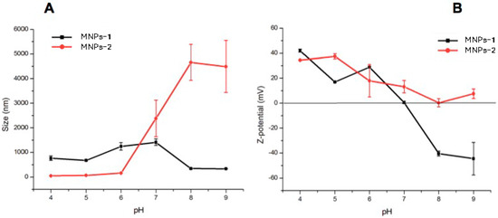
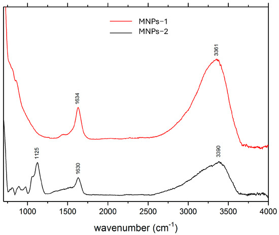
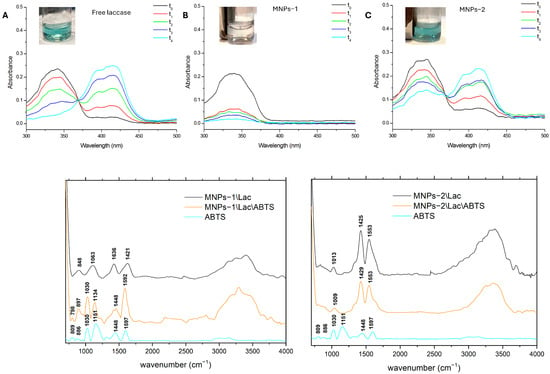
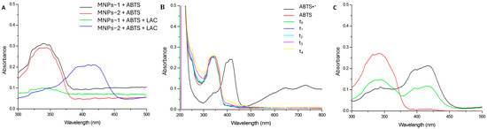
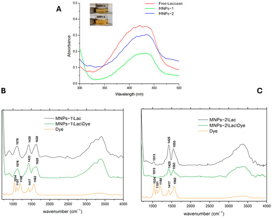
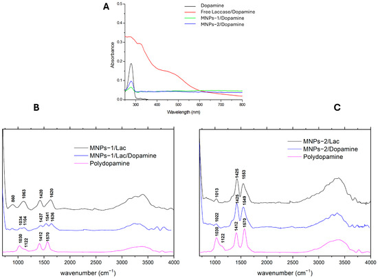
Laccases are multicopper oxidases capable of oxidizing a wide range of substrates using molecular oxygen as the terminal electron acceptor, producing water as the sole by-product. High-redox potential fungal laccases, such as those from Trametes versicolor, are particularly attractive for industrial and environmental applications, although their use is often limited by sensitivity to operational conditions. Enzyme immobilization represents an effective strategy to enhance laccase stability and reusability. In this work, magnetic nanoparticles (MNPs) were investigated as support for laccase immobilization due to their high surface area, biocompatibility, and ease of magnetic recovery. Two modified co-precipitation synthetic routes were systematically evaluated, and the size, morphology, and chemical composition of the products were characterized by microscopy, light scattering, and spectroscopic methods, while both adsorption and covalent immobilization strategies were explored. The MNP surface was found to be highly reactive toward radical species generated during laccase-catalyzed reactions, especially in the presence of small Fe2+ excess. While this can enhance the enzyme catalytic activity, it challenges the inertness of the support and promotes, in some cases, strong interactions between reaction products and the nanoparticle surface. These findings highlight a previously unexplored role of magnetic supports in laccase-based biocatalytic systems.
Full article
(This article belongs to the Special Issue Recent Advances in Laccases and Laccase-Based Bioproducts: 2nd Edition)
►▼
Show Figures

Figure 1
Open AccessArticle
Hibiscus Suspension Culture Extract Modulates Skin Metabolism and Cellular Pathways in Human Keratinocyte/Fibroblast Co-Cultures: A Proteomic Approach
by
Rachid Anane, Su Melser, Elodie Renouf, Rachid Ennamany and Jean-Michel Mérillon
Biomolecules 2026, 16(9), 1243; https://doi.org/10.3390/biom16091243 - 27 Aug 2026
Abstract
Hibiscus plant cell cultures were developed to produce a cosmetic ingredient with anti-ageing properties. UHPLC-DAD-MS analysis of Hibiscus plant cell cultures revealed a high content of polyphenols, particularly hydroxycinnamic acid derivatives, including caffeoyl and p-coumaroyl conjugates. The biological activity of a 50/50 mixture
[...] Read more.
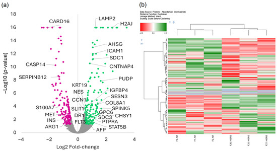
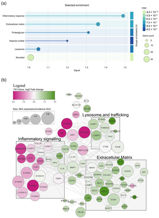
Hibiscus plant cell cultures were developed to produce a cosmetic ingredient with anti-ageing properties. UHPLC-DAD-MS analysis of Hibiscus plant cell cultures revealed a high content of polyphenols, particularly hydroxycinnamic acid derivatives, including caffeoyl and p-coumaroyl conjugates. The biological activity of a 50/50 mixture of extracts from Hibiscus syriacus and Hibiscus rosa-sinensis cells was investigated in a human keratinocyte/fibroblast co-culture model, which better reproduces the reciprocal epithelial–mesenchymal interactions between epidermal keratinocytes and dermal fibroblasts than monocultures, using quantitative data-independent acquisition (DIA) LC-MS/MS proteomics combined with functional enrichment and protein–protein interaction analyses. A total of 7062 proteins were identified, of which 280 were differentially expressed (107 upregulated and 171 downregulated) following hibiscus treatment. The proteomic profile suggested coordinated molecular reprogramming associated with extracellular matrix remodelling, tissue repair, hydration, and attenuation of inflammatory signalling. Functional enrichment analysis revealed coordinated modulation of extracellular matrix organization, glycosaminoglycan metabolism, lysosomal function, cell communication, and inflammatory signalling. Upregulation of extracellular matrix and adhesion proteins, including lumican, collagen VIII, fibulin-5, syndecans, and glypicans, suggested coordinated extracellular matrix remodelling that may promote skin firmness and elasticity, while the downregulation of inflammatory regulators, including CARD16 and S100 family proteins, suggested attenuation of innate inflammatory responses. Overall, these findings provide mechanistic insights into the biological activity of Hibiscus cell culture extracts and support their potential as cosmetic ingredients promoting skin homeostasis and healthy skin ageing.
Full article
(This article belongs to the Special Issue Plant Secondary Metabolism Engineering and Bioactive Compounds)
►▼
Show Figures

Figure 1
Journal Menu
► ▼ Journal Menu-
- Biomolecules Home
- Aims & Scope
- Editorial Board
- Reviewer Board
- Topical Advisory Panel
- Early Career Editorial Board
- Instructions for Authors
- Special Issues
- Topics
- Sections & Collections
- Article Processing Charge
- Indexing & Archiving
- Editor’s Choice Articles
- Most Cited & Viewed
- Journal Statistics
- Journal History
- Journal Awards
- Conferences
- Editorial Office
Journal Browser
► ▼ Journal BrowserHighly Accessed Articles
Latest Books
E-Mail Alert
News
Topics
Topic in
Biomolecules, Chemistry, IJMS, Molecules, Pharmaceuticals
Enzymes and Enzyme Inhibitors in Drug Research
Topic Editors: Athina Geronikaki, Cosimo D. Altomare, Maria Stefania SinicropiDeadline: 11 September 2026
Topic in
Biomedicines, IJMS, Sci. Pharm., Molecules, Future Pharmacology, Biomolecules
Natural Products and Drug Discovery—2nd Edition
Topic Editors: Sonia Piacente, Marta MenegazziDeadline: 30 September 2026
Topic in
Biomolecules, IJMS, Cancers, Proteomes
Extracellular Vesicles: Isolation, Characterization, Function, Application and Utility
Topic Editors: Suresh Mathivanan, Sai Vara Prasad ChittiDeadline: 30 October 2026
Topic in
Biomedicines, Future Pharmacology, Pharmacy, IJMS, Biomolecules, Genes
Prospects of Multi-Target Agonists in Metabolic and Epigenetic Medicine
Topic Editors: Riham Abouleisa, Yanming LiDeadline: 30 November 2026
Conferences
Special Issues
Special Issue in
Biomolecules
Exploring Thermophilic Enzymes: From Structural Foundations to Functional Enhancement
Guest Editor: Renjun GaoDeadline: 31 August 2026
Special Issue in
Biomolecules
Protein Arginine Methylation: Functions, Mechanisms, and Therapeutic Implications
Guest Editor: Yanzhong YangDeadline: 31 August 2026
Special Issue in
Biomolecules
Metabolic and Signal Transduction Pathways Controlling Platelet and Megakaryocyte Activity
Guest Editors: Ana Kasirer-Friede, Deepa GautamDeadline: 31 August 2026
Special Issue in
Biomolecules
State of the Art of Myology: 3rd Edition
Guest Editors: Guglielmo Sorci, Francesca RiuzziDeadline: 31 August 2026
Topical Collections
Topical Collection in
Biomolecules
Feature Papers in Bioinformatics and Systems Biology Section
Collection Editor: Lukasz Kurgan
Topical Collection in
Biomolecules
Cellular and Molecular Mechanisms Governing Stress Response in Plants
Collection Editors: Giovanna Serino, Daisuke Todaka
Topical Collection in
Biomolecules
Molecular Mechanisms of Obesity, Diabetes, Inflammation and Aging
Collection Editors: Yuxiang Sun, Susanne Talcott
Topical Collection in
Biomolecules
Feature Papers in Molecular Biomarkers
Collection Editor: Pietro Scicchitano