Next Issue
Volume 16, March
Previous Issue
Volume 16, January


Toxins, Volume 16, Issue 2 (February 2024) – 50 articles

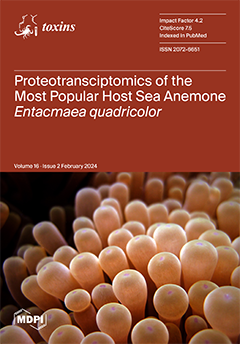
Cover Story (view full-size image): While the unique symbiotic relationship between anemonefishes and sea anemones is iconic, it is still not fully understood how anemonefishes can withstand and thrive within the venomous environment of their host sea anemone. In this study, we used a proteotranscriptomics approach to reveal the venom profile and found that 1251 different toxin transcripts were expressed in E. quadricolor tentacles (1.8% expression), whereas only 4% of proteins detected in venom were putative toxins (230, 14% expression). Thus, most proteins in milked venom do not appear to have a toxin function. This work raises the perils of defining a dominant venom phenotype in sea anemones based solely on transcriptomics data, as we found that the dominant venom phenotype varies depending on transcriptome and proteome abundance data. View this paper
  • Issues are regarded as officially published after their release is announced to the table of contents alert mailing list.
  • You may sign up for e-mail alerts to receive table of contents of newly released issues.
  • PDF is the official format for papers published in both, html and pdf forms. To view the papers in pdf format, click on the "PDF Full-text" link, and use the free Adobe Reader to open them.
Order results
Result details
Section
Select all
Export citation of selected articles as:
11 pages, 1352 KiB  
Article
Serum Phenylacetylglutamine among Potential Risk Factors for Arterial Stiffness Measuring by Carotid–Femoral Pulse Wave Velocity in Patients with Kidney Transplantation
by Hsiao-Hui Yang, Yen-Cheng Chen, Ching-Chun Ho and Bang-Gee Hsu
Toxins 2024, 16(2), 111; https://doi.org/10.3390/toxins16020111 - 18 Feb 2024
Viewed by 1123
Abstract
Phenylacetylglutamine (PAG), a gut microbiota metabolite, is associated with cardiovascular diseases. Arterial stiffness (AS), which is a marker of aging-associated vascular diseases, is an independent risk factor for cardiovascular morbidity and mortality. This study aimed to assess the correlation between serum PAG levels [...] Read more.
Phenylacetylglutamine (PAG), a gut microbiota metabolite, is associated with cardiovascular diseases. Arterial stiffness (AS), which is a marker of aging-associated vascular diseases, is an independent risk factor for cardiovascular morbidity and mortality. This study aimed to assess the correlation between serum PAG levels and AS in kidney transplantation (KT) patients, potentially uncovering new insights into the cardiovascular risks in this population. In this study, 100 KT patients were included. Carotid–femoral pulse wave velocity (cfPWV) was measured, and patients with cfPWV > 10 m/s were categorized as the AS group. Serum PAG levels were assessed using liquid chromatography–tandem mass spectrometry. Thirty KT patients (30.0%) exhibited AS, with higher percentages of diabetes mellitus, older age, and elevated levels of systolic blood pressure, serum fasting glucose, and PAG than the control group. After adjusting for factors significantly associated with AS by multivariate logistic regression analysis, serum PAG, age, fasting glucose levels, and systolic blood pressure were independent factors associated with AS. Furthermore, PAG levels had a negative correlation with the estimated glomerular filtration rate and a positive correlation with cfPWV values. Serum PAG levels are positively associated with cfPWV values and are a biomarker of AS in KT patients. Full article
(This article belongs to the Section Uremic Toxins)
Show Figures

Figure 1

18 pages, 3147 KiB  
Article
Cyanopeptolins and Anabaenopeptins Are the Dominant Cyanopeptides from Planktothrix Strains Collected in Canadian Lakes
by Catrina D. Earnshaw and David R. McMullin
Toxins 2024, 16(2), 110; https://doi.org/10.3390/toxins16020110 - 17 Feb 2024
Viewed by 1190
Abstract
Common bloom-forming cyanobacteria produce complex strain-specific mixtures of secondary metabolites. The beneficial and toxic properties of these metabolite mixtures have attracted both research and public health interest. The advancement of mass spectrometry-based platforms and metabolomics data processing has accelerated the identification of new [...] Read more.
Common bloom-forming cyanobacteria produce complex strain-specific mixtures of secondary metabolites. The beneficial and toxic properties of these metabolite mixtures have attracted both research and public health interest. The advancement of mass spectrometry-based platforms and metabolomics data processing has accelerated the identification of new metabolites and feature dereplication from microbial sources. The objective of this study was to use metabolomics data processing to decipher the intracellular cyanopeptide diversity of six Planktothrix strains collected from Canadian lakes. Data-dependent acquisition experiments were used to collect a non-targeted high-resolution mass spectrometry dataset. Principal component analysis and factor loadings were used to visualize cyanopeptide variation between strains and identified features contributing to the observed variation. GNPS molecular networking was subsequently used to show the diversity of cyanopeptides produced by the Planktothrix strains. Each strain produced a unique mixture of cyanopeptides, and a total of 225 cyanopeptides were detected. Planktothrix sp. CPCC 735 produced the most (n = 68) cyanopeptides, and P. rubescens CPCC 732 produced the fewest (n = 27). Microcystins and anabaenopeptins were detected from all strains. Cyanopeptolins, microviridins and aeruginosins were detected from five, four and two strains, respectively. Cyanopeptolin (n = 80) and anabaenopeptin (n = 61) diversity was the greatest, whereas microcystins (n = 21) were the least diverse. Interestingly, three of the P. rubescens strains had different cyanopeptide profiles, despite being collected from the same lake at the same time. This study highlights the diversity of cyanopeptides produced by Planktothrix and further hints at the underestimated cyanopeptide diversity from subpopulations of chemotypic cyanobacteria in freshwater lakes. Full article
(This article belongs to the Special Issue Toxins: 15th Anniversary)
Show Figures

Figure 1

14 pages, 2470 KiB  
Article
Optimizing Botulinum Toxin A Administration for Forehead Wrinkles: Introducing the Lines and Dots (LADs) Technique and a Predictive Dosage Model
by Kamal Alhallak
Toxins 2024, 16(2), 109; https://doi.org/10.3390/toxins16020109 - 17 Feb 2024
Viewed by 1807
Abstract
This study introduces the Lines and Dots (LADs) technique, a new approach for administering botulinum toxin type A (BoNT-A) in treating forehead wrinkles. (1) Background: BoNT-A application patterns in the forehead often rely solely on the anatomy of the frontalis muscle. The LADs [...] Read more.
This study introduces the Lines and Dots (LADs) technique, a new approach for administering botulinum toxin type A (BoNT-A) in treating forehead wrinkles. (1) Background: BoNT-A application patterns in the forehead often rely solely on the anatomy of the frontalis muscle. The LADs technique proposes a combination of anatomical features with nerve pathways. (2) Methods: The technique employed a grid system aligned with the supraorbital and supratrochlear nerve pathways and used an electronic acupuncture pen for validation. This study analyzed treatment outcomes for efficacy and safety and proposed a predictive model for BoNT-A dosage. (3) Results: LADs was associated with a high satisfaction rate and low side effect incidence. The predictive model followed BoNT-A Units=0.322×Muscle Pattern Code+1.282×Line Type Code+2.905×Severity Pre-Treatment+3.947. (4) Conclusions: The LADs technique offers an alternative approach to treating forehead wrinkles, optimizing efficacy while minimizing the BoNT-A dose required. Full article
(This article belongs to the Special Issue Clinical Application of Botulinum Toxin 2.0)
Show Figures

Figure 1

18 pages, 2038 KiB  
Article
Genome Sequence Analysis of Native Xenorhabdus Strains Isolated from Entomopathogenic Nematodes in Argentina
by Leopoldo Palma, Laureano Frizzo, Sebastian Kaiser, Colin Berry, Primitivo Caballero, Helge B. Bode and Eleodoro Eduardo Del Valle
Toxins 2024, 16(2), 108; https://doi.org/10.3390/toxins16020108 - 17 Feb 2024
Viewed by 1371
Abstract
Entomopathogenic nematodes from the genus Steinernema (Nematoda: Steinernematidae) are capable of causing the rapid killing of insect hosts, facilitated by their association with symbiotic Gram-negative bacteria in the genus Xenorhabdus (Enterobacterales: Morganellaceae), positioning them as interesting candidate tools for the control of insect [...] Read more.
Entomopathogenic nematodes from the genus Steinernema (Nematoda: Steinernematidae) are capable of causing the rapid killing of insect hosts, facilitated by their association with symbiotic Gram-negative bacteria in the genus Xenorhabdus (Enterobacterales: Morganellaceae), positioning them as interesting candidate tools for the control of insect pests. In spite of this, only a limited number of species from this bacterial genus have been identified from their nematode hosts and their insecticidal properties documented. This study aimed to perform the genome sequence analysis of fourteen Xenorhabdus strains that were isolated from Steinernema nematodes in Argentina. All of the strains were found to be able of killing 7th instar larvae of Galleria mellonella (L.) (Lepidoptera: Pyralidae). Their sequenced genomes harbour 110 putative insecticidal proteins including Tc, Txp, Mcf, Pra/Prb and App homologs, plus other virulence factors such as putative nematocidal proteins, chitinases and secondary metabolite gene clusters for the synthesis of different bioactive compounds. Maximum-likelihood phylogenetic analysis plus average nucleotide identity calculations strongly suggested that three strains should be considered novel species. The species name for strains PSL and Reich (same species according to % ANI) is proposed as Xenorhabdus littoralis sp. nov., whereas strain 12 is proposed as Xenorhabdus santafensis sp. nov. In this work, we present a dual insight into the biocidal potential and diversity of the Xenorhabdus genus, demonstrated by different numbers of putative insecticidal genes and biosynthetic gene clusters, along with a fresh exploration of the species within this genus. Full article
(This article belongs to the Section Bacterial Toxins)
Show Figures

Graphical abstract

16 pages, 1723 KiB  
Article
Microbial Composition of Water Kefir Grains and Their Application for the Detoxification of Aflatoxin B1
by Weidong Ouyang, Zhenlin Liao, Ximiao Yang, Xiao Zhang, Xiaoxuan Zhu, Qingping Zhong, Li Wang, Xiang Fang and Jie Wang
Toxins 2024, 16(2), 107; https://doi.org/10.3390/toxins16020107 - 15 Feb 2024
Viewed by 1125
Abstract
Water kefir grains (WKGs), the starter used to develop a traditional beverage named water kefir, consist of a symbiotic mixture of probiotics with diverse bioactivities, but little is known about their abilities to remove mycotoxins that have serious adverse effects on humans and [...] Read more.
Water kefir grains (WKGs), the starter used to develop a traditional beverage named water kefir, consist of a symbiotic mixture of probiotics with diverse bioactivities, but little is known about their abilities to remove mycotoxins that have serious adverse effects on humans and animals. This study investigated the ability of WKGs to remove aflatoxin B1 (AFB1), one of the most toxic mycotoxins, under different settings, and determined the mechanism of absorption mediated by WKGs and the effect of WKGs on the toxicity induced by AFB1 and the reduction in AFB1 in cow milk and tea soups. The results showed the WKGs used herein were dominated by Lactobacillus, Acetobacter, Phenylobacterium, Sediminibacterium, Saccharomyces, Issatchenkia, and Kodamaea. HPLC analysis demonstrated that the WKGs effectively removed AFB1 at concentrations ranging from 1 to 5 µg/mL, pH values ranging from 3 to 9, and temperatures ranging from 4 to 45 °C. Additionally, the removal of AFB1 mainly depended on absorption, which was consistent with the Freundlich and pseudo-second-order kinetic models. Moreover, only 49.63% of AFB1 was released from the AFB1-WKG complex after four washes when the release of AFB1 was non-detectable. Furthermore, WKG treatment caused a dramatic reduction in the mutagenicity induced by AFB1 according to an Ames test and reduced more than 54% of AFB1 in cow milk and three tea soups. These results suggested that WKGs can act as a potential bio-absorbent with a high binding ability to detoxify AFB1 in food and feed via a chemical action step and multi-binding sites of AFB1 absorption in a wide range of scenarios. Full article
(This article belongs to the Section Mycotoxins)
Show Figures

Graphical abstract

11 pages, 290 KiB  
Article
Effects of OnabotulinumtoxinA on Allodynia and Interictal Burden of Patients with Chronic Migraine
by Andreas A. Argyriou, Emmanouil V. Dermitzakis, Dimitrios Rikos, Georgia Xiromerisiou, Panagiotis Soldatos, Pantelis Litsardopoulos and Michail Vikelis
Toxins 2024, 16(2), 106; https://doi.org/10.3390/toxins16020106 - 15 Feb 2024
Cited by 2 | Viewed by 1110
Abstract
Background: We primarily aimed to ascertain whether treatment with OnabotulinumtoxinA (BoNTA) might influence the extent of the interictal burden and cutaneous allodynia in patients with chronic migraine (CM). Methods: Seventy CM patients, who received three consecutive cycles of BoNTA, were studied. The interictal [...] Read more.
Background: We primarily aimed to ascertain whether treatment with OnabotulinumtoxinA (BoNTA) might influence the extent of the interictal burden and cutaneous allodynia in patients with chronic migraine (CM). Methods: Seventy CM patients, who received three consecutive cycles of BoNTA, were studied. The interictal burden was assessed with the Migraine Interictal Burden Scale (MIBS-4), while cutaneous allodynia was examined with the Allodynia Symptom Checklist (ASC-12) together with PI-NRS VAS to obtain hair brushing scores, and then these were compared from baseline (T0) to the last efficacy evaluation follow-up (T1). Efficacy outcomes, mostly mean headache days (MHD) and “Headache Impact Test” scores, were also assessed between T0 and T1. Results: BONTA improved the interictal burden, with a decrease in MIBS-4 scoring by an average of −7 at T1, compared to baseline (p < 0.001). The percentage of patients with a moderate/severe interictal burden was substantially decreased. Likewise, BoNTA reduced the extent of cutaneous allodynia, with a significant reduction in both the ASC-12 (1 vs. 6; p < 0.001) and PI-NRS VAS (1 vs. 5; p < 0.001) to hair brushing median scores at T1, compared to baseline. Reduced MHD rates were significantly associated with a smaller interictal burden at T1. The efficacy of BoNTA, with a significant reduction in MHD and HIT-6 scores at T1 compared to T0, was re-confirmed. Conclusions: BoNTA resulted in a statistically significant reduction in the interictal burden and also improved cutaneous allodynia. The reduction in ictal burden was associated with the down-scaling of the interictal burden. Hence, BoNTA improved the full spectrum of migraine impairment by diminishing the clinical expression of central sensitization. Full article
(This article belongs to the Special Issue Botulinum Toxin and Migraine: Goals and Perspectives (Volume II))
17 pages, 2750 KiB  
Article
Adsorption of Zearalenone by Aureobasidium pullulans Autolyzed Biomass Preparation and Its Detoxification Properties in Cultures of Saccharomyces cerevisiae Yeast
by Anna Bzducha-Wróbel, Monika Janowicz, Marcin Bryła and Iga Grzesiuk
Toxins 2024, 16(2), 105; https://doi.org/10.3390/toxins16020105 - 15 Feb 2024
Viewed by 968
Abstract
Different preventive strategies are needed to minimize the intake risks of mycotoxins, including zearalenone (ZEN). The aim of this study was to determine the ZEN adsorption ability of an autolyzed biomass preparation of polymorphic yeast Aureobasidium pullulans A.p.-3. The evaluation of the antitoxic [...] Read more.
Different preventive strategies are needed to minimize the intake risks of mycotoxins, including zearalenone (ZEN). The aim of this study was to determine the ZEN adsorption ability of an autolyzed biomass preparation of polymorphic yeast Aureobasidium pullulans A.p.-3. The evaluation of the antitoxic properties of the preparation was also performed in relation to Saccharomyces cerevisiae yeast (ATCC 2366, ATCC 7090 and ATCC 9763) used as a model cell exposed to a toxic ZEN dose. The preparation at a dose of 5 mg/mL showed the adsorption of ZEN present in model systems at concentrations between 1 μg/mL to 100 μg/mL. The highest degree of adsorption was established for ZEN concentrations of 1 μg/mL and 5 μg/mL, becoming limited at higher doses of the toxin. Based on the Langmuir model of adsorption isotherms, the predicted maximum ZEN adsorption was approx. 190 µg/mL, regardless of pH. The growth of three strains of S. cerevisiae yeast cells in the medium with ZEN at concentrations within the range of 1.56 μg/mL–100 μg/mL was analyzed to determine the minimum inhibitory concentration. The growth of all tested strains was especially limited by high doses of ZEN, i.e., 50 and 100 μg/mL. The protective effect of the tested preparation was noted in relation to yeast cells exposed to toxic 100 μg/mL ZEN doses. The highest yeast cell growth (app. 36% percentage) was noted for a S. cerevisiae ATCC 9763 strain compared to the medium with ZEN but without preparation. More detailed tests determining the antitoxic mechanisms of the A. pullulans preparation are planned in the future, including cell culture bioassays and animal digestive tract models. Full article
(This article belongs to the Special Issue Mycotoxins: Decontamination and Adsorption)
Show Figures

Figure 1

13 pages, 1864 KiB  
Article
Immunological Cross-Reactivity and Preclinical Assessment of a Colombian Anticoral Antivenom against the Venoms of Three Micrurus Species
by Ariadna Rodríguez-Vargas, Adrián Marcelo Franco-Vásquez, Miguel Triana-Cerón, Shaha Noor Alam-Rojas, Derly C. Escobar-Wilches, Gerardo Corzo, Fernando Lazcano-Pérez, Roberto Arreguín-Espinosa and Francisco Ruiz-Gómez
Toxins 2024, 16(2), 104; https://doi.org/10.3390/toxins16020104 - 15 Feb 2024
Viewed by 1647
Abstract
Snakebite accident treatment requires the administration of antivenoms that provide efficacy and effectiveness against several snake venoms of the same genus or family. The low number of immunogenic components in venom mixtures that allow the production of antivenoms consequently gives them partial neutralization [...] Read more.
Snakebite accident treatment requires the administration of antivenoms that provide efficacy and effectiveness against several snake venoms of the same genus or family. The low number of immunogenic components in venom mixtures that allow the production of antivenoms consequently gives them partial neutralization and a suboptimal pharmacological response. This study evaluates the immunorecognition and neutralizing efficacy of the polyvalent anticoral antivenom from the Instituto Nacional de Salud (INS) of Colombia against the heterologous endemic venoms of Micrurus medemi, and M. sangilensis, and M. helleri by assessing immunoreactivity through affinity chromatography, ELISA, Western blot, and neutralization capability. Immunorecognition towards the venoms of M. medemi and M. sangilensis showed values of 62% and 68% of the protein composition according to the immunoaffinity matrix, respectively. The analysis by Western blot depicted the highest recognition patterns for M. medemi, followed by M. sangilensis, and finally by M. helleri. These findings suggest that the venom compositions are closely related and exhibit similar recognition by the antivenom. According to enzyme immunoassays, M. helleri requires a higher amount of antivenom to achieve recognition than the others. Besides reinforcing the evaluation of INS antivenom capability, this work recommends the use of M. helleri in the production of Colombian antisera. Full article
(This article belongs to the Special Issue Snake Venom: Toxicology and Associated Countermeasures)
Show Figures

Figure 1

12 pages, 2833 KiB  
Article
Long-Term Pulmonary Damage in Surviving Antitoxin-Treated Mice following a Lethal Ricin Intoxication
by Yoav Gal, Anita Sapoznikov, Shlomi Lazar, David Shoseyov, Moshe Aftalion, Hila Gutman, Yentl Evgy, Rellie Gez, Reinat Nevo and Reut Falach
Toxins 2024, 16(2), 103; https://doi.org/10.3390/toxins16020103 - 12 Feb 2024
Viewed by 1361
Abstract
Ricin, a highly potent plant-derived toxin, is considered a potential bioterrorism weapon due to its pronounced toxicity, high availability, and ease of preparation. Acute damage following pulmonary ricinosis is characterized by local cytokine storm, massive neutrophil infiltration, and edema formation, resulting in respiratory [...] Read more.
Ricin, a highly potent plant-derived toxin, is considered a potential bioterrorism weapon due to its pronounced toxicity, high availability, and ease of preparation. Acute damage following pulmonary ricinosis is characterized by local cytokine storm, massive neutrophil infiltration, and edema formation, resulting in respiratory insufficiency and death. A designated equine polyclonal antibody-based (antitoxin) treatment was developed in our laboratory and proved efficacious in alleviating lung injury and increasing survival rates. Although short-term pathogenesis was thoroughly characterized in antitoxin-treated mice, the long-term damage in surviving mice was never determined. In this study, long-term consequences of ricin intoxication were evaluated 30 days post-exposure in mice that survived antitoxin treatment. Significant pulmonary sequelae were demonstrated in surviving antitoxin-treated mice, as reflected by prominent histopathological changes, moderate fibrosis, increased lung hyperpermeability, and decreased lung compliance. The presented data highlight, for the first time to our knowledge, the possibility of long-term damage development in mice that survived lethal-dose pulmonary exposure to ricin due to antitoxin treatment. Full article
Show Figures

Figure 1

28 pages, 5258 KiB  
Article
The Influence of Long-Term Storage on the Epiphytic Microbiome of Postharvest Apples and on Penicillium expansum Occurrence and Patulin Accumulation
by Reem Al Riachy, Caroline Strub, Noël Durand, Vincent Chochois, Félicie Lopez-Lauri, Angélique Fontana and Sabine Schorr-Galindo
Toxins 2024, 16(2), 102; https://doi.org/10.3390/toxins16020102 - 12 Feb 2024
Viewed by 1216
Abstract
Patulin is a secondary metabolite primarily synthesized by the fungus Penicillium expansum, which is responsible for blue mold disease on apples. The latter are highly susceptible to fungal infection in the postharvest stages. Apples destined to produce compotes are processed throughout the [...] Read more.
Patulin is a secondary metabolite primarily synthesized by the fungus Penicillium expansum, which is responsible for blue mold disease on apples. The latter are highly susceptible to fungal infection in the postharvest stages. Apples destined to produce compotes are processed throughout the year, which implies that long periods of storage are required under controlled atmospheres. P. expansum is capable of infecting apples throughout the whole process, and patulin can be detected in the end-product. In the present study, 455 apples (organically and conventionally grown), destined to produce compotes, of the variety “Golden Delicious” were sampled at multiple postharvest steps. The apple samples were analyzed for their patulin content and P. expansum was quantified using real-time PCR. The patulin results showed no significant differences between the two cultivation techniques; however, two critical control points were identified: the long-term storage and the deck storage of apples at ambient temperature before transport. Additionally, alterations in the epiphytic microbiota of both fungi and bacteria throughout various steps were investigated through the application of a metabarcoding approach. The alpha and beta diversity analysis highlighted the effect of long-term storage, causing an increase in the bacterial and fungal diversity on apples, and showed significant differences in the microbial communities during the different postharvest steps. The different network analyses demonstrated intra-species relationships. Multiple pairs of fungal and bacterial competitive relationships were observed. Positive interactions were also observed between P. expansum and multiple fungal and bacterial species. These network analyses provide a basis for further fungal and bacterial interaction analyses for fruit disease biocontrol. Full article
(This article belongs to the Special Issue Toxins: 15th Anniversary)
Show Figures

Figure 1

14 pages, 1379 KiB  
Article
Fusarium sporotrichioides Produces Two HT-2-α-Glucosides on Rice
by Thomas Svoboda, Roman Labuda, Michael Sulyok, Rudolf Krska, Markus Bacher, Franz Berthiller and Gerhard Adam
Toxins 2024, 16(2), 99; https://doi.org/10.3390/toxins16020099 - 10 Feb 2024
Viewed by 1257
Abstract
Fusarium is a genus that mostly consists of plant pathogenic fungi which are able to produce a broad range of toxic secondary metabolites. In this study, we focus on a type A trichothecene-producing isolate (15-39) of Fusarium sporotrichioides from Lower Austria. We assessed [...] Read more.
Fusarium is a genus that mostly consists of plant pathogenic fungi which are able to produce a broad range of toxic secondary metabolites. In this study, we focus on a type A trichothecene-producing isolate (15-39) of Fusarium sporotrichioides from Lower Austria. We assessed the secondary metabolite profile and optimized the toxin production conditions on autoclaved rice and found that in addition to large amounts of T-2 and HT-2 toxins, this strain was able to produce HT-2-glucoside. The optimal conditions for the production of T-2 toxin, HT-2 toxin, and HT-2-glucoside on autoclaved rice were incubation at 12 °C under constant light for four weeks, darkness at 30 °C for two weeks, and constant light for three weeks at 20 °C, respectively. The HT-2-glucoside was purified, and the structure elucidation by NMR revealed a mixture of two alpha-glucosides, presumably HT-2-3-O-alpha-glucoside and HT-2-4-O-alpha-glucoside. The efforts to separate the two compounds by HPLC were unsuccessful. No hydrolysis was observed with two the alpha-glucosidases or with human salivary amylase and Saccharomyces cerevisiae maltase. We propose that the two HT-2-alpha-glucosides are not formed by a glucosyltransferase as they are in plants, but by a trans-glycosylating alpha-glucosidase expressed by the fungus on the starch-containing rice medium. Full article
(This article belongs to the Special Issue Metabolism and Toxicology of Mycotoxins and Their Masked Forms)
Show Figures

Figure 1

21 pages, 1138 KiB  
Review
Best Practice Guidelines for the Management of Patients with Post-Stroke Spasticity: A Modified Scoping Review
by Areerat Suputtitada, Supattana Chatromyen, Carl P. C. Chen and David M. Simpson
Toxins 2024, 16(2), 98; https://doi.org/10.3390/toxins16020098 - 10 Feb 2024
Viewed by 2207
Abstract
This article aims to provide a concise overview of the best available evidence for managing post-stroke spasticity. A modified scoping review, conducted following the PRISMA guidelines and the PRISMA Extension for Scoping Reviews (PRISMA-ScR), involved an intensive search on Medline and PubMed from [...] Read more.
This article aims to provide a concise overview of the best available evidence for managing post-stroke spasticity. A modified scoping review, conducted following the PRISMA guidelines and the PRISMA Extension for Scoping Reviews (PRISMA-ScR), involved an intensive search on Medline and PubMed from 1 January 2000 to 31 August 2023. The focus was placed on high-quality (GRADE A) medical, rehabilitation, and surgical interventions. In total, 32 treatments for post-stroke spasticity were identified. Two independent reviewers rigorously assessed studies, extracting data, and evaluating bias using GRADE criteria. Only interventions with GRADE A evidence were considered. The data included the study type, number of trials, participant characteristics, interventions, parameters, controls, outcomes, and limitations. The results revealed eleven treatments supported by GRADE A evidence, comprising 14 studies. Thirteen were systematic reviews and meta-analyses, and one was randomized control trial. The GRADE A treatments included stretching exercises, static stretching with positional orthosis, transcutaneous electrical nerve stimulation, extracorporeal shock wave therapy, peripheral magnetic stimulation, non-invasive brain stimulation, botulinum toxin A injection, dry needling, intrathecal baclofen, whole body vibration, and localized muscle vibration. In conclusion, this modified scoping review highlights the multimodal treatments supported by GRADE A evidence as being effective for improving functional recovery and quality of life in post-stroke spasticity. Further research and exploration of new therapeutic options are encouraged. Full article
Show Figures

Graphical abstract

5 pages, 199 KiB  
Editorial
Toxins, Pathogenicity, Anti-Toxins, a Bicentennial Contribution
by Michel R. Popoff and Daniel Ladant
Toxins 2024, 16(2), 97; https://doi.org/10.3390/toxins16020097 - 10 Feb 2024
Viewed by 1010
Abstract
The bicentenary of Louis Pasteur’s birth raises the opportunity to revisit the activity and influence of L [...] Full article
23 pages, 1717 KiB  
Review
From Toxin to Treatment: A Narrative Review on the Use of Botulinum Toxin for Autonomic Dysfunction
by Lucas Rempel, Raza N. Malik, Claire Shackleton, Martín Calderón-Juárez, Rahul Sachdeva and Andrei V. Krassioukov
Toxins 2024, 16(2), 96; https://doi.org/10.3390/toxins16020096 - 10 Feb 2024
Viewed by 2304
Abstract
Since its regulatory approval over a half-century ago, botulinum toxin has evolved from one of the most potent neurotoxins known to becoming routinely adopted in clinical practice. Botulinum toxin, a highly potent neurotoxin produced by Clostridium botulinum, can cause botulism illness, characterized by [...] Read more.
Since its regulatory approval over a half-century ago, botulinum toxin has evolved from one of the most potent neurotoxins known to becoming routinely adopted in clinical practice. Botulinum toxin, a highly potent neurotoxin produced by Clostridium botulinum, can cause botulism illness, characterized by widespread muscle weakness due to inhibition of acetylcholine transmission at neuromuscular junctions. The observation of botulinum toxin’s anticholinergic properties led to the investigation of its potential benefits for conditions with an underlying etiology of cholinergic transmission, including autonomic nervous system dysfunction. These conditions range from disorders of the integument to gastrointestinal and urinary systems. Several formulations of botulinum toxin have been developed and tested over time, significantly increasing the availability of this treatment for appropriate clinical use. Despite the accelerated and expanded use of botulinum toxin, there lacks an updated comprehensive review on its therapeutic use, particularly to treat autonomic dysfunction. This narrative review provides an overview of the effect of botulinum toxin in the treatment of autonomic dysfunction and summarizes the different formulations and dosages most widely studied, while highlighting reported outcomes and the occurrence of any adverse events. Full article
(This article belongs to the Special Issue Application of Botulinum Toxins in Diseases Treatment)
Show Figures

Figure 1

20 pages, 1353 KiB  
Review
Complexing Protein-Free Botulinum Neurotoxin A Formulations: Implications of Excipients for Immunogenicity
by Michael Uwe Martin, Juergen Frevert and Clifton Ming Tay
Toxins 2024, 16(2), 101; https://doi.org/10.3390/toxins16020101 - 10 Feb 2024
Viewed by 1858
Abstract
The formation of neutralizing antibodies is a growing concern in the use of botulinum neurotoxin A (BoNT/A) as it may result in secondary treatment failure. Differences in the immunogenicity of BoNT/A formulations have been attributed to the presence of pharmacologically unnecessary bacterial components. [...] Read more.
The formation of neutralizing antibodies is a growing concern in the use of botulinum neurotoxin A (BoNT/A) as it may result in secondary treatment failure. Differences in the immunogenicity of BoNT/A formulations have been attributed to the presence of pharmacologically unnecessary bacterial components. Reportedly, the rate of antibody-mediated secondary non-response is lowest in complexing protein-free (CF) IncobotulinumtoxinA (INCO). Here, the published data and literature on the composition and properties of the three commercially available CF-BoNT/A formulations, namely, INCO, Coretox® (CORE), and DaxibotulinumtoxinA (DAXI), are reviewed to elucidate the implications for their potential immunogenicity. While all three BoNT/A formulations are free of complexing proteins and contain the core BoNT/A molecule as the active pharmaceutical ingredient, they differ in their production protocols and excipients, which may affect their immunogenicity. INCO contains only two immunologically inconspicuous excipients, namely, human serum albumin and sucrose, and has demonstrated low immunogenicity in daily practice and clinical studies for more than ten years. DAXI contains four excipients, namely, L-histidine, trehalosedihydrate, polysorbate 20, and the highly charged RTP004 peptide, of which the latter two may increase the immunogenicity of BoNT/A by introducing neo-epitopes. In early clinical studies with DAXI, antibodies against BoNT/A and RTP004 were found at low frequencies; however, the follow-up period was critically short, with a maximum of three injections. CORE contains four excipients: L-methionine, sucrose, NaCl, and polysorbate 20. Presently, no data are available on the immunogenicity of CORE in human beings. It remains to be seen whether all three CF BoNT/A formulations demonstrate the same low immunogenicity in patients over a long period of time. Full article
(This article belongs to the Special Issue Immunogenicity of Botulinum Toxin)
Show Figures

Figure 1

13 pages, 1459 KiB  
Article
Effects of the Heterodimeric Neurotoxic Phospholipase A2 from the Venom of Vipera nikolskii on the Contractility of Rat Papillary Muscles and Thoracic Aortas
by Alexey Averin, Vladislav Starkov, Victor Tsetlin and Yuri Utkin
Toxins 2024, 16(2), 100; https://doi.org/10.3390/toxins16020100 - 10 Feb 2024
Viewed by 1402
Abstract
Phospholipases A2 (PLA2s) are a large family of snake toxins manifesting diverse biological effects, which are not always related to phospholipolytic activity. Snake venom PLA2s (svPLA2s) are extracellular proteins with a molecular mass of 13–14 kDa. [...] Read more.
Phospholipases A2 (PLA2s) are a large family of snake toxins manifesting diverse biological effects, which are not always related to phospholipolytic activity. Snake venom PLA2s (svPLA2s) are extracellular proteins with a molecular mass of 13–14 kDa. They are present in venoms in the form of monomers, dimers, and larger oligomers. The cardiovascular system is one of the multiple svPLA2 targets in prey organisms. The results obtained previously on the cardiovascular effects of monomeric svPLA2s were inconsistent, while the data on the dimeric svPLA2 crotoxin from the rattlesnake Crotalus durissus terrificus showed that it significantly reduced the contractile force of guinea pig hearts. Here, we studied the effects of the heterodimeric svPLA2 HDP-1 from the viper Vipera nikolskii on papillary muscle (PM) contractility and the tension of the aortic rings (ARs). HDP-1 is structurally different from crotoxin, and over a wide range of concentrations, it produced a long-term, stable, positive inotropic effect in PMs, which did not turn into contractures at the concentrations studied. This also distinguishes HDP-1 from the monomeric svPLA2s, which at high concentrations inhibited cardiac function. HDP-1, when acting on ARs preconstricted with 10 μM phenylephrine, induced a vasorelaxant effect, similar to some other svPLA2s. These are the first indications of the cardiac and vascular effects of true vipers’ heterodimeric svPLA2s. Full article
Show Figures

Figure 1

13 pages, 953 KiB  
Article
Fungal Species and Mycotoxins Associated with Maize Ear Rots Collected from the Eastern Cape in South Africa
by Jenna-Lee Price, Cobus Meyer Visagie, Hannalien Meyer and Neriman Yilmaz
Toxins 2024, 16(2), 95; https://doi.org/10.3390/toxins16020095 - 08 Feb 2024
Cited by 1 | Viewed by 1296
Abstract
Maize production in South Africa is concentrated in its central provinces. The Eastern Cape contributes less than 1% of total production, but is steadily increasing its production and has been identified as a priority region for future growth. In this study, we surveyed [...] Read more.
Maize production in South Africa is concentrated in its central provinces. The Eastern Cape contributes less than 1% of total production, but is steadily increasing its production and has been identified as a priority region for future growth. In this study, we surveyed ear rots at maize farms in the Eastern Cape, and mycotoxins were determined to be present in collected samples. Fungal isolations were made from mouldy ears and species identified using morphology and DNA sequences. Cladosporium, Diplodia, Fusarium and Gibberella ear rots were observed during field work, and of these, we collected 78 samples and isolated 83 fungal strains. Fusarium was identified from Fusarium ear rot (FER) and Gibberella ear rot (GER) and Stenocarpella from Diplodia ear rot (DER) samples, respectively. Using LC-MS/MS multi-mycotoxin analysis, it was revealed that 83% of the collected samples contained mycotoxins, and 17% contained no mycotoxins. Fifty percent of samples contained multiple mycotoxins (deoxynivalenol, 15-acetyl-deoxynivalenol, diplodiatoxin and zearalenone) and 33% contained a single mycotoxin. Fusarium verticillioides was not isolated and fumonisins not detected during this survey. This study revealed that ear rots in the Eastern Cape are caused by a wide range of species that may produce various mycotoxins. Full article
(This article belongs to the Special Issue Toxins: 15th Anniversary)
Show Figures

Figure 1

30 pages, 9946 KiB  
Review
Predatory and Defensive Strategies in Cone Snails
by Zahrmina Ratibou, Nicolas Inguimbert and Sébastien Dutertre
Toxins 2024, 16(2), 94; https://doi.org/10.3390/toxins16020094 - 07 Feb 2024
Viewed by 1595
Abstract
Cone snails are carnivorous marine animals that prey on fish (piscivorous), worms (vermivorous), or other mollusks (molluscivorous). They produce a complex venom mostly made of disulfide-rich conotoxins and conopeptides in a compartmentalized venom gland. The pharmacology of cone snail venom has been increasingly [...] Read more.
Cone snails are carnivorous marine animals that prey on fish (piscivorous), worms (vermivorous), or other mollusks (molluscivorous). They produce a complex venom mostly made of disulfide-rich conotoxins and conopeptides in a compartmentalized venom gland. The pharmacology of cone snail venom has been increasingly investigated over more than half a century. The rising interest in cone snails was initiated by the surprising high human lethality rate caused by the defensive stings of some species. Although a vast amount of information has been uncovered on their venom composition, pharmacological targets, and mode of action of conotoxins, the venom–ecology relationships are still poorly understood for many lineages. This is especially important given the relatively recent discovery that some species can use different venoms to achieve rapid prey capture and efficient deterrence of aggressors. Indeed, via an unknown mechanism, only a selected subset of conotoxins is injected depending on the intended purpose. Some of these remarkable venom variations have been characterized, often using a combination of mass spectrometry and transcriptomic methods. In this review, we present the current knowledge on such specific predatory and defensive venoms gathered from sixteen different cone snail species that belong to eight subgenera: Pionoconus, Chelyconus, Gastridium, Cylinder, Conus, Stephanoconus, Rhizoconus, and Vituliconus. Further studies are needed to help close the gap in our understanding of the evolved ecological roles of many cone snail venom peptides. Full article
Show Figures

Figure 1

15 pages, 2214 KiB  
Article
Biopreservative and Anti-Mycotoxigenic Potentials of Lactobacillus paracasei MG847589 and Its Bacteriocin in Soft White Cheese
by Mohamed G. Shehata, Tawfiq Alsulami, Nourhan M. Abd El-Aziz, Hagar S. Abd-Rabou, Sobhy A. El Sohaimy, Amira M. G. Darwish, Karolina Hoppe, Hatem S. Ali and Ahmed Noah Badr
Toxins 2024, 16(2), 93; https://doi.org/10.3390/toxins16020093 - 07 Feb 2024
Viewed by 1239
Abstract
Probiotics and their bacteriocins have increasingly attracted interest for their use as safe food preservatives. This study aimed to produce soft white cheese fortified with Lacticaseibacillus MG847589 (Lb. paracasei MG847589) and/or its bacteriocin; cheese with Lacticaseibacillus (CP), cheese with bacteriocin (CB), and [...] Read more.
Probiotics and their bacteriocins have increasingly attracted interest for their use as safe food preservatives. This study aimed to produce soft white cheese fortified with Lacticaseibacillus MG847589 (Lb. paracasei MG847589) and/or its bacteriocin; cheese with Lacticaseibacillus (CP), cheese with bacteriocin (CB), and cheese with both Lacticaseibacillus and bacteriocin (CPB) were compared to control cheese (CS) to evaluate their biopreservative and anti-mycotoxigenic potentials for prolonged shelf life and safe food applications. The effects of these fortifications on physiochemical, microbial, texture, microstructure, and sensory properties were studied. Fortification with Lacticaseibacillus (CP) increased acidity (0.61%) and microbial counts, which may make the microstructure porous, while CPB showed intact microstructure. The CPB showed the highest hardness value (3988.03 g), while the lowest was observed with CB (2525.73 g). Consequently, the sensory assessment reflected the panelists’ preference for CPB, which gained higher scores than the control (CS). Fortification with Lb. paracasei MG847589 and bacteriocin (CPB) showed inhibition effects against S. aureus from 6.52 log10 CFU/g at time zero to 2.10 log10 CFU/g at the end of storage, A. parasiticus (from 5.06 to 3.03 log10 CFU/g), and P. chrysogenum counts (from 5.11 to 2.86 log10 CFU/g). Additionally, CPB showed an anti-mycotoxigenic effect against aflatoxins AFB1 and AFM1, causing them to be decreased (69.63 ± 0.44% and 71.38 ± 0.75%, respectively). These potentials can extend shelf life and pave the way for more suggested food applications of safe food production by fortification with both Lb. paracasei MG847589 and its bacteriocin as biopreservatives and anti-mycotoxigenic. Full article
(This article belongs to the Special Issue Strategies to Prevent Mycotoxin Contamination of Food and Feed)
Show Figures

Figure 1

13 pages, 591 KiB  
Article
Toxic Effects of Bt-(Cry1Ab+Vip3Aa) Maize on Storage Pest Paralipsa gularis (Zeller)
by Shuang Chen, Wenhui Wang, Guodong Kang, Xianming Yang and Kongming Wu
Toxins 2024, 16(2), 92; https://doi.org/10.3390/toxins16020092 - 07 Feb 2024
Viewed by 1054
Abstract
Paralipsa gularis (Zeller) is a storage pest; however, in recent years it has evolved into a considerable maize pest during the late growth stage in the border region between China and other Southeast Asian countries. Bt transgenic insect-resistant maize is an effective measure [...] Read more.
Paralipsa gularis (Zeller) is a storage pest; however, in recent years it has evolved into a considerable maize pest during the late growth stage in the border region between China and other Southeast Asian countries. Bt transgenic insect-resistant maize is an effective measure in controlling a wide range of lepidopteran pests, but there is a lack of research on the toxic effects of storage pests. We tested the toxicity of Bt-Cry1Ab, Vip3Aa, and their complex proteins against P. gularis via bioassay and investigated the efficiency of Bt-(Cry1Ab+Vip3Aa) maize in controlling P. gularis during the late growth stage of maize in the period 2022–2023. The bioassay results show that the susceptibilities of P. gularis to the two Bt proteins and their complex proteins were significantly different. The LC50 values of DBNCry1Ab (“DBN9936” event), DBNVip3Aa (“DBN9501” event), DBN Cry1Ab+Vip3Aa (“DBN3601T” event), and Syngenta Cry1Ab+Vip3Aa (“Bt11” event × “MIR162” event) were 0.038 μg/g, 0.114 μg/g, 0.110 μg/g, and 0.147 μg/g, and the GIC50 values were 0.014 μg/g, 0.073 μg/g, 0.027 μg/g, and 0.026 μg/g, respectively. Determination of the expression content of the insecticidal protein in different tissues of Bt-(Cry1Ab+Vip3Aa) maize shows that the total Bt protein content in different tissues was in the following order: stalk > bract > cob > kernel. However, the bioassay results show that the mortalities of P. gularis feeding on Bt-(Cry1Ab+Vip3Aa) maize in different tissues at different growth stages were all above 93.00%. The field trial indicates that the occurrence density of larvae and plant damage rate for conventional maize were 422.10 individuals/100 plants and 94.40%, respectively, whereas no larvae were found on Bt-(Cry1Ab+Vip3Aa) maize. In summary, this study implies that Bt-(Cry1Ab+Vip3Aa) maize has a high potential for control of P. gularis, providing a new technical measure for the management of the pest. Full article
Show Figures

Figure 1

15 pages, 2525 KiB  
Article
Environmental Factors Impacting the Development of Toxic Cyanobacterial Proliferations in a Central Texas Reservoir
by Katherine A. Perri, Brent J. Bellinger, Matt P. Ashworth and Schonna R. Manning
Toxins 2024, 16(2), 91; https://doi.org/10.3390/toxins16020091 - 06 Feb 2024
Viewed by 1151
Abstract
Cyanobacterial harmful algal proliferations (cyanoHAPs) are increasingly associated with dog and livestock deaths when benthic mats break free of their substrate and float to the surface. Fatalities have been linked to neurotoxicosis from anatoxins, potent alkaloids produced by certain genera of filamentous cyanobacteria. [...] Read more.
Cyanobacterial harmful algal proliferations (cyanoHAPs) are increasingly associated with dog and livestock deaths when benthic mats break free of their substrate and float to the surface. Fatalities have been linked to neurotoxicosis from anatoxins, potent alkaloids produced by certain genera of filamentous cyanobacteria. After numerous reports of dog illnesses and deaths at a popular recreation site on Lady Bird Lake, Austin, Texas in late summer 2019, water and floating mat samples were collected from several sites along the reservoir. Water quality parameters were measured and mat samples were maintained for algal isolation and DNA identification. Samples were also analyzed for cyanobacterial toxins using LC-MS. Dihydroanatoxin-a was detected in mat materials from two of the four sites (0.6–133 ng/g wet weight) while water samples remained toxin-free over the course of the sampling period; no other cyanobacterial toxins were detected. DNA sequencing analysis of cyanobacterial isolates yielded a total of 11 genera, including Geitlerinema, Tyconema, Pseudanabaena, and Phormidium/Microcoleus, taxa known to produce anatoxins, including dihydroanatoxin, among other cyanotoxins. Analyses indicate that low daily upriver dam discharge, higher TP and NO3 concentrations, and day of the year were the main parameters associated with the presence of toxic floating cyanobacterial mats. Full article
(This article belongs to the Special Issue Analysis, Detection, and Effect of Algal Toxins in Fresh Waters)
Show Figures

Figure 1

13 pages, 1428 KiB  
Review
Trichothecenes and Fumonisins: Key Players in Fusarium–Cereal Ecosystem Interactions
by Alexandre Perochon and Fiona M. Doohan
Toxins 2024, 16(2), 90; https://doi.org/10.3390/toxins16020090 - 06 Feb 2024
Viewed by 1418
Abstract
Fusarium fungi produce a diverse array of mycotoxic metabolites during the pathogenesis of cereals. Some, such as the trichothecenes and fumonisins, are phytotoxic, acting as non-proteinaceous effectors that facilitate disease development in cereals. Over the last few decades, we have gained some depth [...] Read more.
Fusarium fungi produce a diverse array of mycotoxic metabolites during the pathogenesis of cereals. Some, such as the trichothecenes and fumonisins, are phytotoxic, acting as non-proteinaceous effectors that facilitate disease development in cereals. Over the last few decades, we have gained some depth of understanding as to how trichothecenes and fumonisins interact with plant cells and how plants deploy mycotoxin detoxification and resistance strategies to defend themselves against the producer fungi. The cereal-mycotoxin interaction is part of a co-evolutionary dance between Fusarium and cereals, as evidenced by a trichothecene-responsive, taxonomically restricted, cereal gene competing with a fungal effector protein and enhancing tolerance to the trichothecene and resistance to DON-producing F. graminearum. But the binary fungal–plant interaction is part of a bigger ecosystem wherein other microbes and insects have been shown to interact with fungal mycotoxins, directly or indirectly through host plants. We are only beginning to unravel the extent to which trichothecenes, fumonisins and other mycotoxins play a role in fungal-ecosystem interactions. We now have tools to determine how, when and where mycotoxins impact and are impacted by the microbiome and microfauna. As more mycotoxins are described, research into their individual and synergistic toxicity and their interactions with the crop ecosystem will give insights into how we can holistically breed for and cultivate healthy crops. Full article
Show Figures

Figure 1

12 pages, 2966 KiB  
Communication
Structural Assignment of the Product Ion Generated from a Natural Ciguatoxin-3C Congener, 51-Hydroxyciguatoxin-3C, and Discovery of Distinguishable Signals in Congeners Bearing the 51-Hydroxy Group
by Ryogo Ukai, Hideaki Uchida, Kouichi Sugaya, Jun-ichi Onose, Naomasa Oshiro, Takeshi Yasumoto and Naoki Abe
Toxins 2024, 16(2), 89; https://doi.org/10.3390/toxins16020089 - 06 Feb 2024
Viewed by 1023
Abstract
Ciguatoxins (CTXs) stand as the primary toxins causing ciguatera fish poisoning (CFP) and are essential compounds distinguished by their characteristic polycyclic ether structure. In a previous report, we identified the structures of product ions generated via homolytic fragmentation by assuming three charge sites [...] Read more.
Ciguatoxins (CTXs) stand as the primary toxins causing ciguatera fish poisoning (CFP) and are essential compounds distinguished by their characteristic polycyclic ether structure. In a previous report, we identified the structures of product ions generated via homolytic fragmentation by assuming three charge sites in the mass spectrometry (MS)/MS spectrum of ciguatoxin-3C (CTX3C) using LC-MS. This study aims to elucidate the homolytic fragmentation of a ciguatoxin-3C congener. We assigned detailed structures of the product ions in the MS/MS spectrum of a naturally occurring ciguatoxin-3C congener, 51-hydroxyciguatoxin-3C (51-hydoxyCTX3C), employing liquid chromatography/quadrupole time-of-flight mass spectrometry with an atmospheric pressure chemical ionization (APCI) source. The introduction of a hydroxy substituent on C51 induced different fragmentation pathways, including a novel cleavage mechanism of the M ring involving the elimination of 51-OH and the formation of enol ether. Consequently, new cleavage patterns generated product ions at m/z 979 (C55H79O15), 439 (C24H39O7), 149 (C10H13O), 135 (C9H11O), and 115 (C6H11O2). Additionally, characteristic product ions were observed at m/z 509 (C28H45O8), 491 (C28H43O7), 481 (C26H41O8), 463 (C26H39O7), 439 (C24H39O7), 421 (C24H37O6), 171 (C9H15O3), 153 (C9H13O2), 141 (C8H13O2), and 123 (C8H11O). Full article
(This article belongs to the Section Marine and Freshwater Toxins)
Show Figures

Graphical abstract

13 pages, 2155 KiB  
Article
N-Terminal α-Helices in Domain I of Bacillus thuringiensis Vip3Aa Play Crucial Roles in Disruption of Liposomal Membrane
by Ensi Shao, Hanye Huang, Jin Yuan, Yaqi Yan, Luru Ou, Xiankun Chen, Xiaohong Pan, Xiong Guan and Li Sha
Toxins 2024, 16(2), 88; https://doi.org/10.3390/toxins16020088 - 06 Feb 2024
Viewed by 1173
Abstract
Bacillus thuringiensis Vip3 toxins form a tetrameric structure crucial for their insecticidal activity. Each Vip3Aa monomer comprises five domains. Interaction of the first four α-helices in domain I with the target cellular membrane was proposed to be a key step before pore formation. [...] Read more.
Bacillus thuringiensis Vip3 toxins form a tetrameric structure crucial for their insecticidal activity. Each Vip3Aa monomer comprises five domains. Interaction of the first four α-helices in domain I with the target cellular membrane was proposed to be a key step before pore formation. In this study, four N-terminal α-helix-deleted truncations of Vip3Aa were produced and, it was found that they lost both liposome permeability and insecticidal activity against Spodoptera litura. To further probe the role of domain I in membrane permeation, the full-length domain I and the fragments of N-terminal α-helix-truncated domain I were fused to green fluorescent protein (GFP), respectively. Only the fusion carrying the full-length domain I exhibited permeability against artificial liposomes. In addition, seven Vip3Aa-Cry1Ac fusions were also constructed by combination of α-helices from Vip3Aa domains I and II with the domains II and III of Cry1Ac. Five of the seven combinations were determined to show membrane permeability in artificial liposomes. However, none of the Vip3Aa-Cry1Ac combinations exhibited insecticidal activity due to the significant reduction in proteolytic stability. These results indicated that the N-terminal helix α1 in the Vip3Aa domain I is essential for both insecticidal activity and liposome permeability and that domain I of Vip3Aa preserved a high liposome permeability independently from domains II–V. Full article
(This article belongs to the Special Issue Bacillus thuringiensis: A Broader View of Its Biocidal Activity)
Show Figures

Figure 1

30 pages, 910 KiB  
Review
Effect of Acrylamide and Mycotoxins in SH-SY5Y Cells: A Review
by Luna Bridgeman, Cristina Juan, Houda Berrada and Ana Juan-García
Toxins 2024, 16(2), 87; https://doi.org/10.3390/toxins16020087 - 06 Feb 2024
Viewed by 1374
Abstract
Thermal processes induce the formation of undesired toxic components, such as acrylamide (AA), which has been shown to induce brain toxicity in humans and classified as Group 2A by the International Agency of Research in Cancer (IARC), as well as some mycotoxins. AA [...] Read more.
Thermal processes induce the formation of undesired toxic components, such as acrylamide (AA), which has been shown to induce brain toxicity in humans and classified as Group 2A by the International Agency of Research in Cancer (IARC), as well as some mycotoxins. AA and mycotoxins’ toxicity is studied in several in vitro models, including the neuroblastoma cell line model SH-SY5Y cells. Both AA and mycotoxins occur together in the same food matrix cereal base (bread, pasta, potatoes, coffee roasting, etc.). Therefore, the goal of this review is to deepen the knowledge about the neurological effects that AA and mycotoxins can induce on the in vitro model SH-SY5Y and its mechanism of action (MoA) focusing on the experimental assays reported in publications of the last 10 years. The analysis of the latest publications shows that most of them are focused on cytotoxicity, apoptosis, and alteration in protein expression, while others are interested in oxidative stress, axonopathy, and the disruption of neurite outgrowth. While both AA and mycotoxins have been studied in SH-SY5Y cells separately, the mixture of them is starting to draw the interest of the scientific community. This highlights a new and interesting field to explore due to the findings reported in several publications that can be compared and the implications in human health that both could cause. In relation to the assays used, the most employed were the MTT, axonopathy, and qPCR assays. The concentration dose range studied was 0.1–10 mM for AA and 2 fM to 200 µM depending on the toxicity and time of exposure for mycotoxins. A healthy and varied diet allows the incorporation of a large family of bioactive compounds that can mitigate the toxic effects associated with contaminants present in food. Although this has been reported in some publications for mycotoxins, there is still a big gap for AA which evidences that more investigations are needed to better explore the risks for human health when exposed to AA and mycotoxins. Full article
Show Figures

Figure 1

13 pages, 3483 KiB  
Article
In Silico Analysis of Shiga Toxin-Producing Escherichia coli O157:H7 Strains from Presumptive Super- and Low-Shedder Cattle
by Emmanuel W. Bumunang, Vinicius S. Castro, Trevor Alexander, Rahat Zaheer, Tim A. McAllister, Le Luo Guan and Kim Stanford
Toxins 2024, 16(2), 86; https://doi.org/10.3390/toxins16020086 - 05 Feb 2024
Viewed by 1188
Abstract
Cattle are the primary reservoir for STEC O157, with some shedding >104 CFU/g in feces, a phenomenon known as super-shedding (SS). The mechanism(s) responsible for SS are not understood but have been attributed to the environment, host, and pathogen. This study aimed [...] Read more.
Cattle are the primary reservoir for STEC O157, with some shedding >104 CFU/g in feces, a phenomenon known as super-shedding (SS). The mechanism(s) responsible for SS are not understood but have been attributed to the environment, host, and pathogen. This study aimed to compare genetic characteristics of STEC O157 strains from cattle in the same commercial feedlot pens with SS or low-shedding (LS) status. Strains from SS (n = 35) and LS (n = 28) collected from 11 pens in three feedlots were analyzed for virulence genes, Shiga toxin-carrying bacteriophage insertion sites, and phylogenetic relationships. In silico analysis showed limited variation regarding virulence gene profiles. Stx-encoding prophage insertion sites mrlA and wrbA for stx1a and stx2a, respectively, were all occupied, but two isolates had fragments of the stx-carrying phage in mrlA and wrbA loci without stx1a and stx2a. All strains screened for lineage-specific polymorphism assay (LSPA-6) were 111111, lineage I. Of the isolates, 61 and 2 were clades 1 and 8, respectively. Phylogenetic analysis revealed that pens with more than one SS had multiple distantly related clusters of SS and LS isolates. Although virulence genes and lineage were largely similar within and across feedlots, multiple genetic origins of strains within a single feedlot pen illustrate challenges for on-farm control of STEC. Full article
(This article belongs to the Collection Shiga Toxins)
Show Figures

Graphical abstract

26 pages, 1225 KiB  
Article
Proteotransciptomics of the Most Popular Host Sea Anemone Entacmaea quadricolor Reveals Not All Toxin Genes Expressed by Tentacles Are Recruited into Its Venom Arsenal
by Cassie M. Hoepner, Zachary K. Stewart, Robert Qiao, Emily K. Fobert, Peter J. Prentis, Alex Colella, Tim Chataway, Karen Burke da Silva and Catherine A. Abbott
Toxins 2024, 16(2), 85; https://doi.org/10.3390/toxins16020085 - 05 Feb 2024
Viewed by 1553
Abstract
While the unique symbiotic relationship between anemonefishes and sea anemones is iconic, it is still not fully understood how anemonefishes can withstand and thrive within the venomous environment of their host sea anemone. In this study, we used a proteotranscriptomics approach to elucidate [...] Read more.
While the unique symbiotic relationship between anemonefishes and sea anemones is iconic, it is still not fully understood how anemonefishes can withstand and thrive within the venomous environment of their host sea anemone. In this study, we used a proteotranscriptomics approach to elucidate the proteinaceous toxin repertoire from the most common host sea anemone, Entacmaea quadricolor. Although 1251 different toxin or toxin-like RNA transcripts were expressed in E. quadricolor tentacles (0.05% of gene clusters, 1.8% of expression) and 5375 proteins were detected in milked venom, only 4% of proteins detected in venom were putative toxins (230), and they only represent on average 14% of the normalised protein expression in the milked venom samples. Thus, most proteins in milked venom do not appear to have a toxin function. This work raises the perils of defining a dominant venom phenotype based on transcriptomics data alone in sea anemones, as we found that the dominant venom phenotype differs between the transcriptome and proteome abundance data. E. quadricolor venom contains a mixture of toxin-like proteins of unknown and known function. A newly identified toxin protein family, Z3, rich in conserved cysteines of unknown function, was the most abundant at the RNA transcript and protein levels. The venom was also rich in toxins from the Protease S1, Kunitz-type and PLA2 toxin protein families and contains toxins from eight venom categories. Exploring the intricate venom toxin components in other host sea anemones will be crucial for improving our understanding of how anemonefish adapt to the venomous environment. Full article
(This article belongs to the Section Marine and Freshwater Toxins)
Show Figures

Graphical abstract

13 pages, 3878 KiB  
Article
Bee Venom Stimulates Growth Factor Release from Adipose-Derived Stem Cells to Promote Hair Growth
by Jung Hyun Kim, Tae Yoon Kim, Bonhyuk Goo and Yeoncheol Park
Toxins 2024, 16(2), 84; https://doi.org/10.3390/toxins16020084 - 04 Feb 2024
Cited by 1 | Viewed by 1597
Abstract
Limited evidence suggests that stimulating adipose-derived stem cells (ASCs) indirectly promotes hair growth. We examined whether bee venom (BV) activated ASCs and whether BV-induced hair growth was facilitated by enhanced growth factor release by ASCs. The induction of the telogen-to-anagen phase was studied [...] Read more.
Limited evidence suggests that stimulating adipose-derived stem cells (ASCs) indirectly promotes hair growth. We examined whether bee venom (BV) activated ASCs and whether BV-induced hair growth was facilitated by enhanced growth factor release by ASCs. The induction of the telogen-to-anagen phase was studied in mice. The underlying mechanism was investigated using organ cultures of mouse vibrissa hair follicles. When BV-treated ASCs were injected subcutaneously into mice, the telogen-to-anagen transition was accelerated and, by day 14, the hair weight increased. Quantitative polymerase chain reaction (qPCR) revealed that BV influenced the expression of several molecules, including growth factors, chemokines, channels, transcription factors, and enzymes. Western blot analysis was employed to verify the protein expression levels of extracellular-signal-regulated kinase (ERK) and phospho-ERK. Both the Boyden chamber experiment and scratch assay confirmed the upregulation of cell migration by BV. Additionally, ASCs secreted higher levels of growth factors after exposure to BV. Following BV therapy, the gene expression levels of alkaline phosphatase (ALP), fibroblast growth factor (FGF)-1 and 6, endothelial cell growth factor, and platelet-derived growth factor (PDGF)-C were upregulated. The findings of this study suggest that bee venom can potentially be utilized as an ASC-preconditioning agent for hair regeneration. Full article
(This article belongs to the Section Animal Venoms)
Show Figures

Figure 1

24 pages, 5459 KiB  
Article
Venom Composition of Neglected Bothropoid Snakes from the Amazon Rainforest: Ecological and Toxinological Implications
by Luciana A. Freitas-de-Sousa, Mônica Colombini, Vinicius C. Souza, Joanderson P. C. Silva, Ageane Mota-da-Silva, Marllus R. N. Almeida, Reginaldo A. Machado, Wirven L. Fonseca, Marco A. Sartim, Jacqueline Sachett, Solange M. T. Serrano, Inácio L. M. Junqueira-de-Azevedo, Felipe G. Grazziotin, Wuelton M. Monteiro, Paulo S. Bernarde and Ana M. Moura-da-Silva
Toxins 2024, 16(2), 83; https://doi.org/10.3390/toxins16020083 - 04 Feb 2024
Viewed by 1242
Abstract
Snake venoms have evolved in several families of Caenophidae, and their toxins have been assumed to be biochemical weapons with a role as a trophic adaptation. However, it remains unclear how venom contributes to the success of venomous species for adaptation to different [...] Read more.
Snake venoms have evolved in several families of Caenophidae, and their toxins have been assumed to be biochemical weapons with a role as a trophic adaptation. However, it remains unclear how venom contributes to the success of venomous species for adaptation to different environments. Here we compared the venoms from Bothrocophias hyoprora, Bothrops taeniatus, Bothrops bilineatus smaragdinus, Bothrops brazili, and Bothrops atrox collected in the Amazon Rainforest, aiming to understand the ecological and toxinological consequences of venom composition. Transcriptomic and proteomic analyses indicated that the venoms presented the same toxin groups characteristic from bothropoids, but with distinct isoforms with variable qualitative and quantitative abundances, contributing to distinct enzymatic and toxic effects. Despite the particularities of each venom, commercial Bothrops antivenom recognized the venom components and neutralized the lethality of all species. No clear features could be observed between venoms from arboreal and terrestrial habitats, nor in the dispersion of the species throughout the Amazon habitats, supporting the notion that venom composition may not shape the ecological or toxinological characteristics of these snake species and that other factors influence their foraging or dispersal in different ecological niches. Full article
(This article belongs to the Section Animal Venoms)
Show Figures

Figure 1

13 pages, 7510 KiB  
Article
Bioaccumulation of Microcystin-LR and Induced Physio-Biochemical Changes in Rice (Oryza sativa L.) at Vegetative Stage under Hydroponic Culture Conditions
by Jinlin Jiang, Yue Shi, Feng Tian, Tao Long, Xuzhi Li and Rongrong Ying
Toxins 2024, 16(2), 82; https://doi.org/10.3390/toxins16020082 - 04 Feb 2024
Viewed by 1244
Abstract
Irrigation with water containing a variety of microcystins (MCs) may pose a potential threat to the normal growth of agricultural plants. To investigate the phytotoxicity of MC-LR at environmental concentrations on rice (Oryza sativa L.), the characteristics of uptake and accumulation in [...] Read more.
Irrigation with water containing a variety of microcystins (MCs) may pose a potential threat to the normal growth of agricultural plants. To investigate the phytotoxicity of MC-LR at environmental concentrations on rice (Oryza sativa L.), the characteristics of uptake and accumulation in plant tissues, as well as a series of key physio-biochemical process changes in leaves of rice seedlings, were measured at concentrations of 0.10, 1.0, 10.0, and 50.0 μg·L−1 in hydroponic nutrient solutions for 7, 15, 20, and 34 days. Results showed that MC-LR could be detected in rice leaves and roots in exposure groups; however, a significant accumulation trend of MC-LR in plants (BCF > 1) was only found in the 0.10 μg·L−1 group. The time-course study revealed a biphasic response of O2•− levels in rice leaves to the exposure of MC-LR, which could be attributed to the combined effects of the antioxidant system and detoxification reaction in rice. Exposure to 1.0–50.0 μg·L−1 MC-LR resulted in significant depletion of GSH and MDA contents in rice leaves at later exposure times (15–34 days). Low MC-LR concentrations promoted nitric oxide synthase (NOS) activity, whereas high concentrations inhibited NOS activity during the later exposure times. The reduced sucrose synthase (SS) activities in rice exposed to MC-LR for 34 days indicated a decrease in the carbon accumulation ability of plants, and therefore may be directly related to the inhibition of plant growth under MC exposure. These findings indicate that the normal physiological status would be disrupted in terrestrial plants, even under exposure to low concentrations of MC-LR. Full article
Show Figures

Figure 1

Previous Issue
Next Issue
Back to TopTop