Antioxidants 2020, 9(1), 43; https://doi.org/10.3390/antiox9010043 - 4 Jan 2020
Cited by 146 | Viewed by 7484
Abstract
The heavy metal contamination in plant-soil environment has increased manifold recently. In order to reduce the harmful effects of metal stress in plants, the application of beneficial soil microbes is gaining much attention. In the present research, the role of Serratia marcescens BM1
[...] Read more.
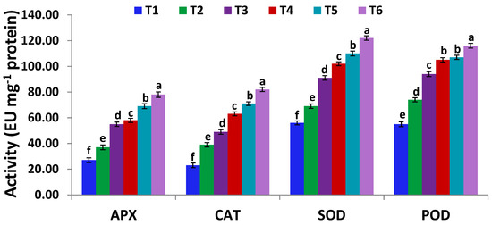
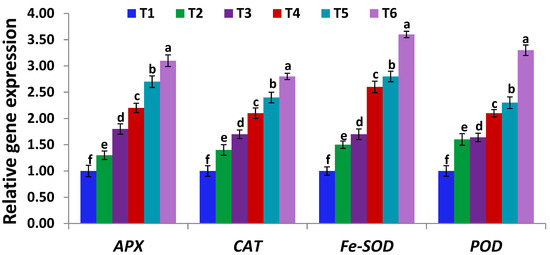
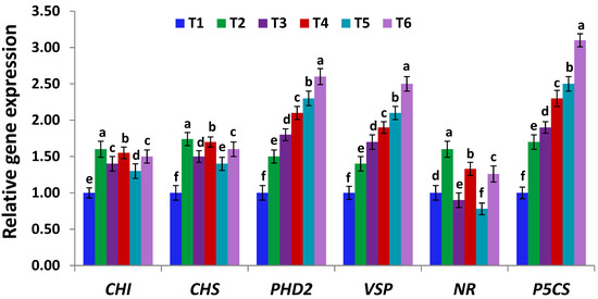
The heavy metal contamination in plant-soil environment has increased manifold recently. In order to reduce the harmful effects of metal stress in plants, the application of beneficial soil microbes is gaining much attention. In the present research, the role of Serratia marcescens BM1 in enhancing cadmium (Cd) stress tolerance and phytoremediation potential of soybean plants, was investigated. Exposure of soybean plants to two Cd doses (150 and 300 µM) significantly reduced plant growth, biomass, gas exchange attributes, nutrients uptake, antioxidant capacity, and the contents of chlorophyll, total phenolics, flavonoids, soluble sugars, and proteins. Additionally, Cd induced the stress levels of Cd, proline, glycine betaine, hydrogen peroxide, malondialdehyde, antioxidant enzymes (i.e., catalase, CAT; ascorbate peroxidase, APX; superoxide dismutase, SOD; peroxidise, POD), and the expression of stress-related genes (i.e., APX, CAT, Fe-SOD, POD, CHI, CHS, PHD2, VSO, NR, and P5CS) in soybean leaves. On the other hand, inoculation of Cd-stressed soybean plants with Serratia marcescens BM1 significantly enhanced the plant growth, biomass, gas exchange attributes, nutrients uptake, antioxidant capacity, and the contents of chlorophyll, total phenolics, flavonoids, soluble sugars, and proteins. Moreover, Serratia marcescens BM1 inoculation reduced the levels of cadmium and oxidative stress markers, but significantly induced the activities of antioxidant enzymes and the levels of osmolytes and stress-related genes expression in Cd-stressed plants. The application of 300 µM CdCl2 and Serratia marcescens triggered the highest expression levels of stress-related genes. Overall, this study suggests that inoculation of soybean plants with Serratia marcescens BM1 promotes phytoremediation potential and Cd stress tolerance by modulating the photosynthetic attributes, osmolytes biosynthesis, antioxidants machinery, and the expression of stress-related genes.
Full article
(This article belongs to the Special Issue Oxidative Stress in Plant)
►
Show Figures
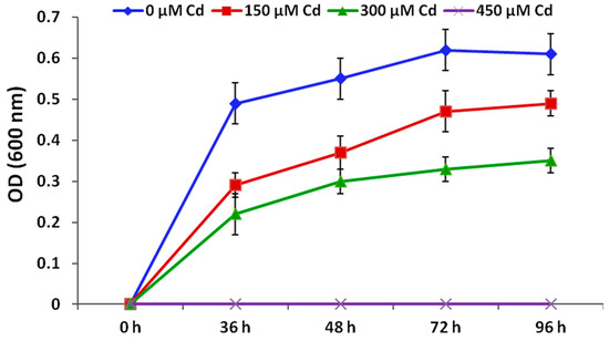
Figure 1


