Next Issue
Volume 6, August
Previous Issue
Volume 6, June


J. Clin. Med., Volume 6, Issue 7 (July 2017) – 12 articles

Cover Story (view full-size image): 
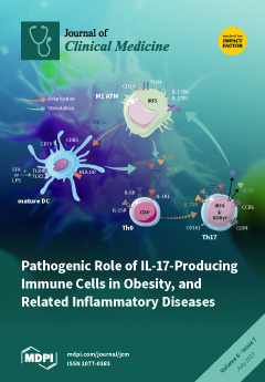
Pro-inflammatory M1 adipose tissue macrophages (ATM) promote IL-17-secreting CD4+ Th17 cells in obese adipose tissue (AT). Obese M1 ATM secrete high levels of pro-inflammatory cytokines, including IL-1β, IL-6, IL-23, and IL-15, which are involved in the promotion and maintenance of Th17 cells. Th17 cells can be either induced from naive IL-23R CD4+ T cells in response to IL-1β, IL-6 and IL-15 or resting IL-23R+ Th17 cells in response to pro-inflammatory cytokines plus IL-23. Interestingly, CD14+ M1 can induce maturation of infiltrating dendritic cells (DC) when stimulated by lipopolysaccharide (LPS) or free-fatty acids (FFA). Mature DC, can lead to Th17 cell skewing, through secretion of IL-1β, IL-6 and IL-23, in obese AT. View this paper.
  • Issues are regarded as officially published after their release is announced to the table of contents alert mailing list.
  • You may sign up for e-mail alerts to receive table of contents of newly released issues.
  • PDF is the official format for papers published in both, html and pdf forms. To view the papers in pdf format, click on the "PDF Full-text" link, and use the free Adobe Reader to open them.
Order results
Result details
Section
Select all
Export citation of selected articles as:
206 KiB  
Review
The Role of Th17 Cells in the Pathogenesis of Behcet’s Disease
by Yuki Nanke, Toru Yago and Shigeru Kotake
J. Clin. Med. 2017, 6(7), 74; https://doi.org/10.3390/jcm6070074 - 21 Jul 2017
Cited by 41 | Viewed by 5120
Abstract
Behcet’s disease (BD) is a polysymptomatic and recurrent systemic vasculitis with a chronic course and unknown cause. The pathogenesis of BD has not been fully elucidated; however, BD has been considered to be a typical Th1-mediated inflammatory disease, characterized by elevated levels of [...] Read more.
Behcet’s disease (BD) is a polysymptomatic and recurrent systemic vasculitis with a chronic course and unknown cause. The pathogenesis of BD has not been fully elucidated; however, BD has been considered to be a typical Th1-mediated inflammatory disease, characterized by elevated levels of Th1 cytokines such as IFN-γ, IL-2, and TNF-α. Recently, some studies reported that Th17-associated cytokines were increased in BD; thus, Th17 cells and the IL17/IL23 pathway may play important roles in the pathogenesis of BD. In this chapter, we focus on the pathogenic role of Th17 cells in BD. Full article
(This article belongs to the Special Issue Th17 Cell in Autoimmune and Inflammatory Diseases)
529 KiB  
Review
Th17 in Animal Models of Rheumatoid Arthritis
by Motomu Hashimoto
J. Clin. Med. 2017, 6(7), 73; https://doi.org/10.3390/jcm6070073 - 21 Jul 2017
Cited by 38 | Viewed by 8380
Abstract
IL-17-secreting helper CD4 T cells (Th17 cells) constitute a newly identified subset of helper CD4 T cells that play a key role in the development of rheumatoid arthritis (RA) in its animal models. Recently, several models of spontaneous RA, which elucidate the mechanism [...] Read more.
IL-17-secreting helper CD4 T cells (Th17 cells) constitute a newly identified subset of helper CD4 T cells that play a key role in the development of rheumatoid arthritis (RA) in its animal models. Recently, several models of spontaneous RA, which elucidate the mechanism of RA onset, have been discovered. These animal models shed new light on the role of Th17 in the development of autoimmune arthritis. Th17 cells coordinate inflammation and promote joint destruction, acting on various cells, including neutrophils, macrophages, synovial fibroblasts, and osteoclasts. Regulatory T cells cannot control Th17 cells under conditions of inflammation. In this review, the pathogenic role of Th17 cells in arthritis development, which was revealed by the recent animal models of RA, is discussed. Full article
(This article belongs to the Special Issue Th17 Cell in Autoimmune and Inflammatory Diseases)
Show Figures

Figure 1

244 KiB  
Article
Effects of Assault Type on Cognitive Behaviour Therapy for Coexisting Depression and Alcohol Misuse
by Kylie A. Bailey, Amanda L. Baker, Patrick McElduff, Mark A. Jones, Christopher Oldmeadow and David J. Kavanagh
J. Clin. Med. 2017, 6(7), 72; https://doi.org/10.3390/jcm6070072 - 21 Jul 2017
Viewed by 4501
Abstract
Although assault exposure is common in mental health and substance misusing populations, screening for assaults in treatment settings is frequently overlooked. This secondary analysis explored the effects of past sexual (SA) and physical (PA) assault on depression, alcohol misuse, global functioning and attrition [...] Read more.
Although assault exposure is common in mental health and substance misusing populations, screening for assaults in treatment settings is frequently overlooked. This secondary analysis explored the effects of past sexual (SA) and physical (PA) assault on depression, alcohol misuse, global functioning and attrition in the Depression and Alcohol Integrated and Single focussed Intervention (DAISI) project, whose participants (N = 278) received cognitive behaviour therapy (CBT) for their depression and/or alcohol misuse. Of the 278 DAISI participants, 220 consented to screening for past assault (either by a stranger or non-stranger) at baseline. Depression, alcohol, and global functioning assessments were administered at baseline and 3, 12, 24, and 36 months post baseline. A between-group analysis was used to assess differences between SA and No SA, and PA and No PA groupings, on adjusted mean treatment outcomes across all assessment periods. SA and PA participants had similar mean symptom reductions compared to No SA and No PA participants except for lower depression and global functioning change scores at the 12-month follow-up. People with coexisting depression and alcohol misuse reporting SA or PA can respond well to CBT for depression and alcohol misuse. However, follow-up is recommended in order to monitor fluctuations in outcomes. Full article
2384 KiB  
Review
Evidence of Oxidative Stress and Secondary Mitochondrial Dysfunction in Metabolic and Non-Metabolic Disorders
by Karolina M. Stepien, Robert Heaton, Scott Rankin, Alex Murphy, James Bentley, Darren Sexton and Iain P. Hargreaves
J. Clin. Med. 2017, 6(7), 71; https://doi.org/10.3390/jcm6070071 - 19 Jul 2017
Cited by 95 | Viewed by 11682
Abstract
Mitochondrial dysfunction and oxidative stress have been implicated in the pathogenesis of a number of diseases and conditions. Oxidative stress occurs once the antioxidant defenses of the body become overwhelmed and are no longer able to detoxify reactive oxygen species (ROS). The ROS [...] Read more.
Mitochondrial dysfunction and oxidative stress have been implicated in the pathogenesis of a number of diseases and conditions. Oxidative stress occurs once the antioxidant defenses of the body become overwhelmed and are no longer able to detoxify reactive oxygen species (ROS). The ROS can then go unchallenged and are able to cause oxidative damage to cellular lipids, DNA and proteins, which will eventually result in cellular and organ dysfunction. Although not always the primary cause of disease, mitochondrial dysfunction as a secondary consequence disease of pathophysiology can result in increased ROS generation together with an impairment in cellular energy status. Mitochondrial dysfunction may result from either free radical-induced oxidative damage or direct impairment by the toxic metabolites which accumulate in certain metabolic diseases. In view of the importance of cellular antioxidant status, a number of therapeutic strategies have been employed in disorders associated with oxidative stress with a view to neutralising the ROS and reactive nitrogen species implicated in disease pathophysiology. Although successful in some cases, these adjunct therapies have yet to be incorporated into the clinical management of patients. The purpose of this review is to highlight the emerging evidence of oxidative stress, secondary mitochondrial dysfunction and antioxidant treatment efficacy in metabolic and non-metabolic diseases in which there is a current interest in these parameters. Full article
Show Figures

Figure 1

581 KiB  
Review
Omega-3 Polyunsaturated Fatty Acids for the Treatment of IgA Nephropathy
by Junichi Hirahashi
J. Clin. Med. 2017, 6(7), 70; https://doi.org/10.3390/jcm6070070 - 19 Jul 2017
Cited by 15 | Viewed by 8910
Abstract
IgA nephropathy is a common disease that causes end-stage renal failure and requires renal replacement therapy. The main purpose of therapeutic intervention in this disease is not limited to improvement of prognosis and prevention of transition to end-stage renal failure, but also prevention [...] Read more.
IgA nephropathy is a common disease that causes end-stage renal failure and requires renal replacement therapy. The main purpose of therapeutic intervention in this disease is not limited to improvement of prognosis and prevention of transition to end-stage renal failure, but also prevention of the occurrence of cardiovascular lesions, which increases risk in patients with chronic kidney disease. Steroids and immunosuppressants have been widely used as remission induction therapies; however, the balance between their therapeutic benefits and detrimental side-effects are controversial. In this regard, it is critical to identify alternative therapies which would provide holistic life-long benefits. Currently, the potential of ω-3 fatty acids as anti-inflammatory and inflammation-convergent drugs—especially the remarkable progress of the multifunctional ω-3 polyunsaturated fatty acids (PUFAs)—has garnered attention. In this section, we outline the background and current status of ω-3 PUFA-based treatment in IgA nephropathy. Full article
(This article belongs to the Special Issue Molecular Biology of IgA Nephropathy)
Show Figures

Figure 1

823 KiB  
Review
An Evaluation of Ischaemic Preconditioning as a Method of Reducing Ischaemia Reperfusion Injury in Liver Surgery and Transplantation
by Francis P. Robertson, Barry J. Fuller and Brian R. Davidson
J. Clin. Med. 2017, 6(7), 69; https://doi.org/10.3390/jcm6070069 - 14 Jul 2017
Cited by 40 | Viewed by 6961
Abstract
Liver Ischaemia Reperfusion (IR) injury is a major cause of post-operative liver dysfunction, morbidity and mortality following liver resection surgery and transplantation. There are no proven therapies for IR injury in clinical practice and new approaches are required. Ischaemic Preconditioning (IPC) can be [...] Read more.
Liver Ischaemia Reperfusion (IR) injury is a major cause of post-operative liver dysfunction, morbidity and mortality following liver resection surgery and transplantation. There are no proven therapies for IR injury in clinical practice and new approaches are required. Ischaemic Preconditioning (IPC) can be applied in both a direct and remote fashion and has been shown to ameliorate IR injury in small animal models. Its translation into clinical practice has been difficult, primarily by a lack of knowledge regarding the dominant protective mechanisms that it employs. A review of all current studies would suggest that IPC/RIPC relies on creating a small tissue injury resulting in the release of adenosine and l-arginine which act through the Adenosine receptors and the haem-oxygenase and endothelial nitric oxide synthase systems to reduce hepatocyte necrosis and improve the hepatic microcirculation post reperfusion. The next key step is to determine how long the stimulus requires to precondition humans to allow sufficient injury to occur to release the potential mediators. This would open the door to a new therapeutic chapter in this field. Full article
Show Figures

Graphical abstract

982 KiB  
Review
Pathogenic Role of IL-17-Producing Immune Cells in Obesity, and Related Inflammatory Diseases
by Marwa Chehimi, Hubert Vidal and Assia Eljaafari
J. Clin. Med. 2017, 6(7), 68; https://doi.org/10.3390/jcm6070068 - 14 Jul 2017
Cited by 122 | Viewed by 13885
Abstract
Obesity is associated with low-grade chronic inflammation. Indeed, adipose tissues (AT) in obese individuals are the former site of progressive infiltration by pro-inflammatory immune cells, which together with increased inflammatory adipokine secretion induce adipocyte insulin resistance. IL-17-producing T (Th17) cells are part of [...] Read more.
Obesity is associated with low-grade chronic inflammation. Indeed, adipose tissues (AT) in obese individuals are the former site of progressive infiltration by pro-inflammatory immune cells, which together with increased inflammatory adipokine secretion induce adipocyte insulin resistance. IL-17-producing T (Th17) cells are part of obese AT infiltrating cells, and are likely to be promoted by adipose tissue-derived mesenchymal stem cells, as previously reported by our team. Whereas Th17 cell are physiologically implicated in the neutralization of fungal and bacterial pathogens through activation of neutrophils, they may also play a pivotal role in the onset and/or progression of chronic inflammatory diseases, or cancer, in which obesity is recognized as a risk factor. In this review, we will highlight the pathogenic role of IL-17A producing cells in the mechanisms leading to inflammation in obesity and to progression of obesity-related inflammatory diseases. Full article
(This article belongs to the Special Issue Th17 Cell in Autoimmune and Inflammatory Diseases)
Show Figures

Figure 1

496 KiB  
Review
The Plasticity of Th17 Cells in the Pathogenesis of Rheumatoid Arthritis
by Shigeru Kotake, Toru Yago, Tsuyoshi Kobashigawa and Yuki Nanke
J. Clin. Med. 2017, 6(7), 67; https://doi.org/10.3390/jcm6070067 - 10 Jul 2017
Cited by 70 | Viewed by 9700
Abstract
Helper T (Th) cells play an important role in the pathogenesis of autoimmune diseases, including rheumatoid arthritis (RA). It has been revealed that Th17 cells can shift to Th1 cells (i.e., “nonclassic Th1 cells”), which are reported to be more pathogenic than Th17 [...] Read more.
Helper T (Th) cells play an important role in the pathogenesis of autoimmune diseases, including rheumatoid arthritis (RA). It has been revealed that Th17 cells can shift to Th1 cells (i.e., “nonclassic Th1 cells”), which are reported to be more pathogenic than Th17 cells per se. Thus, the association of Th cells in the pathogenesis of autoimmune disease has become more complicated. We recently reported using peripheral blood from untreated and early-onset RA patients that the ratio of CD161+Th1 cells (i.e., Th17-derived Th1 cells to CD161+Th17 cells) is elevated and that levels of interferon-γ (IFNγ)+Th17 cells are inversely correlated with levels of anti-CCP antibodies. Here, we review the plasticity of Th17 cells in the pathogenesis of RA, suggesting possible implications for novel therapies. Full article
(This article belongs to the Special Issue Th17 Cell in Autoimmune and Inflammatory Diseases)
Show Figures

Figure 1

228 KiB  
Review
When Less Is Good, Is None Better? The Prognostic and Therapeutic Significance of Peri-Transplant Minimal Residual Disease Assessment in Pediatric Acute Lymphoblastic Leukemia
by Adam Lamble, Rachel Phelan and Michael Burke
J. Clin. Med. 2017, 6(7), 66; https://doi.org/10.3390/jcm6070066 - 07 Jul 2017
Cited by 9 | Viewed by 5163
Abstract
The measurement of minimal residual disease (MRD) in pediatric acute lymphoblastic leukemia (ALL) has become the most important prognostic tool of, and the backbone to, upfront risk stratification. While MRD assessment is the standard of care for assessing response and predicting outcomes for [...] Read more.
The measurement of minimal residual disease (MRD) in pediatric acute lymphoblastic leukemia (ALL) has become the most important prognostic tool of, and the backbone to, upfront risk stratification. While MRD assessment is the standard of care for assessing response and predicting outcomes for pediatric patients with ALL receiving chemotherapy, its use in allogeneic hematopoietic stem cell transplant (HSCT) has been less clearly defined. Herein, we discuss the importance of MRD assessment during the peri-HSCT period and its role in prognostication and management. Full article
(This article belongs to the Special Issue Role of Minimal Residual Disease Assessment in Hematological Cancers)
538 KiB  
Review
T Helper 17 Cells in Primary Sjögren’s Syndrome
by Kiyoshi Matsui and Hajime Sano
J. Clin. Med. 2017, 6(7), 65; https://doi.org/10.3390/jcm6070065 - 05 Jul 2017
Cited by 36 | Viewed by 8710
Abstract
Primary Sjögren’s syndrome is an autoimmune disease characterized by diffuse infiltration of lymphocytes into exocrine glands and other tissues. The infiltrating lymphocytes have been identified as subsets of B cells and T cells, including T helper 17 cells, T regulatory cells and follicular [...] Read more.
Primary Sjögren’s syndrome is an autoimmune disease characterized by diffuse infiltration of lymphocytes into exocrine glands and other tissues. The infiltrating lymphocytes have been identified as subsets of B cells and T cells, including T helper 17 cells, T regulatory cells and follicular helper T cells. The role of these cells in the development of the syndrome is now known, as is their impact on the production of proinflammatory cytokines such as IL-6, IL-17, IL-22 and IL-23. In particular, experimental animal models and patients suggest that a shift in Th17/Treg balance toward the proinflammatory Th17 axis exacerbates primary Sjögren’s syndrome and other autoimmune disorders. Nevertheless, the pathogenesis of the disorder is not yet fully elucidated. This review summarizes the recent advances in therapeutic control of the Treg/Th17 balance, as well as the efficacy of candidate therapeutics against primary Sjögren’s syndrome. Full article
(This article belongs to the Special Issue Th17 Cell in Autoimmune and Inflammatory Diseases)
Show Figures

Graphical abstract

300 KiB  
Review
Myopathology of Adult and Paediatric Mitochondrial Diseases
by Rahul Phadke
J. Clin. Med. 2017, 6(7), 64; https://doi.org/10.3390/jcm6070064 - 04 Jul 2017
Cited by 29 | Viewed by 7740
Abstract
Mitochondria are dynamic organelles ubiquitously present in nucleated eukaryotic cells, subserving multiple metabolic functions, including cellular ATP generation by oxidative phosphorylation (OXPHOS). The OXPHOS machinery comprises five transmembrane respiratory chain enzyme complexes (RC). Defective OXPHOS gives rise to mitochondrial diseases (mtD). The incredible [...] Read more.
Mitochondria are dynamic organelles ubiquitously present in nucleated eukaryotic cells, subserving multiple metabolic functions, including cellular ATP generation by oxidative phosphorylation (OXPHOS). The OXPHOS machinery comprises five transmembrane respiratory chain enzyme complexes (RC). Defective OXPHOS gives rise to mitochondrial diseases (mtD). The incredible phenotypic and genetic diversity of mtD can be attributed at least in part to the RC dual genetic control (nuclear DNA (nDNA) and mitochondrial DNA (mtDNA)) and the complex interaction between the two genomes. Despite the increasing use of next-generation-sequencing (NGS) and various omics platforms in unravelling novel mtD genes and pathomechanisms, current clinical practice for investigating mtD essentially involves a multipronged approach including clinical assessment, metabolic screening, imaging, pathological, biochemical and functional testing to guide molecular genetic analysis. This review addresses the broad muscle pathology landscape including genotype–phenotype correlations in adult and paediatric mtD, the role of immunodiagnostics in understanding some of the pathomechanisms underpinning the canonical features of mtD, and recent diagnostic advances in the field. Full article
1238 KiB  
Review
Pathophysiology of Hemophilic Arthropathy
by Daniela Melchiorre, Mirko Manetti and Marco Matucci-Cerinic
J. Clin. Med. 2017, 6(7), 63; https://doi.org/10.3390/jcm6070063 - 25 Jun 2017
Cited by 98 | Viewed by 10829
Abstract
Spontaneous joint bleeding and repeated hemarthroses lead to hemophilic arthropathy—a debilitating disease with a significant negative impact on mobility and quality of life. Iron, cytokines, and angiogenic growth factors play a pivotal role in the onset of the inflammatory process that involves the [...] Read more.
Spontaneous joint bleeding and repeated hemarthroses lead to hemophilic arthropathy—a debilitating disease with a significant negative impact on mobility and quality of life. Iron, cytokines, and angiogenic growth factors play a pivotal role in the onset of the inflammatory process that involves the synovial tissue, articular cartilage, and subchondral bone, with early damages and molecular changes determining the perpetuation of a chronic inflammatory condition. Synovitis is one of the earliest complications of hemarthrosis, and is characterized by synovial hypertrophy, migration of inflammatory cells, and a high degree of neo-angiogenesis with subsequent bleeding. The pathogenic mechanisms and molecular pathways by which blood in the joint cavity causes articular cartilage and subchondral bone destruction have yet to be fully elucidated. Both cytokines and matrix metalloproteinases and hydroxyl radicals may induce chondrocyte apoptosis. Members of the tumor necrosis factor receptor superfamily (such as the molecular triad: osteoprotegerin—OPG; receptor activator of nuclear factor κB—RANK; RANK ligand—RANKL) seem instead to play a major role in the inflammatory process. These pathogenic processes interact with each other and ultimately lead to a fibrotic joint and the disabling condition characteristic of hemophilic arthropathy. Full article
(This article belongs to the Special Issue Outstanding Advances in Hemophilia Therapies)
Show Figures

Figure 1

Previous Issue
Next Issue
Back to TopTop