J. Clin. Med. 2020, 9(1), 242; https://doi.org/10.3390/jcm9010242 - 16 Jan 2020
Cited by 16 | Viewed by 7793
Abstract
Various three-dimensional (3D) culture methods have been introduced to overcome the limitations of in vitro culture and mimic in vivo conditions. This study aimed to evaluate two microsphere-forming culture methods and a monolayer culture method. We evaluated cell morphology, viability, osteo-, adipo-, and
[...] Read more.
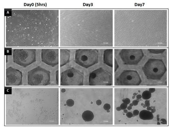
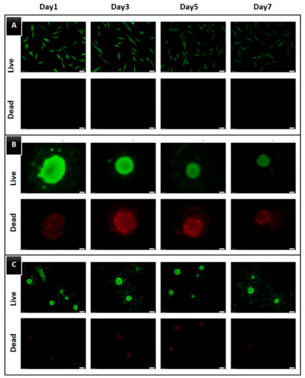
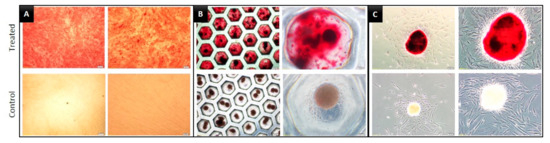
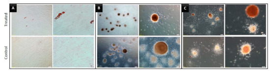
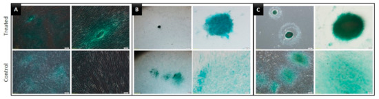
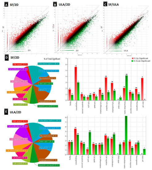
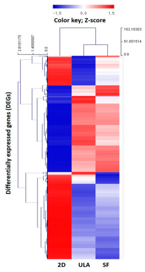
Various three-dimensional (3D) culture methods have been introduced to overcome the limitations of in vitro culture and mimic in vivo conditions. This study aimed to evaluate two microsphere-forming culture methods and a monolayer culture method. We evaluated cell morphology, viability, osteo-, adipo-, and chondrogenic differentiation potential of dental pulp stem cells (DPSCs) cultured in 3D culture plates: ultra-low attachment (ULA) and U-bottomed StemFit 3D (SF) plates, and a two-dimensional (2D) monolayer plate. RNA sequencing (RNA-seq) revealed differentially expressed gene (DEG) profiles of the DPSCs. In contrast to an increasing pattern in the 2D group, cell viability in 3D groups (ULA and SF) showed a decreasing pattern; however, high multilineage differentiation was observed in both the 3D groups. RNA-seq showed significantly overexpressed gene ontology categories including angiogenesis, cell migration, differentiation, and proliferation in the 3D groups. Hierarchical clustering analysis revealed a similar DEG regulation pattern between the 3D groups; however, a comparatively different DEG was observed between the 2D and 3D groups. Taken together, this study shows that DPSCs cultured in microsphere-forming plates present superior multilineage differentiation capacities and demonstrate higher DEG expression in regeneration-related gene categories compared to that in DPSCs cultured in a conventional monolayer plate.
Full article
(This article belongs to the Special Issue Innovations in Endodontic Dentistry)
►
Show Figures
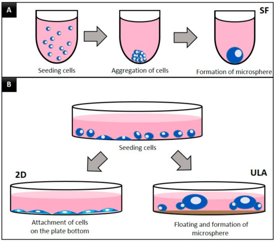
Figure 1


