24 pages, 4581 KiB  
Article
Transcriptome Changes in Glioma Cells Cultivated under Conditions of Neurosphere Formation
by Natalia S. Vasileva 1, Elena V. Kuligina 1, Maya A. Dymova 1, Yulya I. Savinovskaya 1, Nikita D. Zinchenko 1, Alisa B. Ageenko 1, Sergey V. Mishinov 2, Anton S. Dome 1, Grigory A. Stepanov 1, Vladimir A. Richter 1 and Dmitry V. Semenov 1,*
1 Institute of Chemical Biology and Fundamental Medicine, Siberian Branch, Russian Academy of Sciences, Lavrentyev Avenue 8, Novosibirsk 630090, Russia
2 Novosibirsk Research Institute of Traumatology and Orthopedics n.a. Ya.L. Tsivyan, Department of Neurosurgery, Frunze Street 17, Novosibirsk 630091, Russia
Cells 2022, 11(19), 3106; https://doi.org/10.3390/cells11193106 - 2 Oct 2022
Cited by 9 | Viewed by 3003
Abstract
Glioma is the most common and heterogeneous primary brain tumor. The development of a new relevant preclinical models is necessary. As research moves from cultures of adherent gliomas to a more relevant model, neurospheres, it is necessary to understand the changes that cells [...] Read more.
Glioma is the most common and heterogeneous primary brain tumor. The development of a new relevant preclinical models is necessary. As research moves from cultures of adherent gliomas to a more relevant model, neurospheres, it is necessary to understand the changes that cells undergo at the transcriptome level. In the present work, we used three patient-derived gliomas and two immortalized glioblastomas, while their cultivation was carried out under adherent culture and neurosphere (NS) conditions. When comparing the transcriptomes of monolayer (ML) and NS cell cultures, we used Enrichr genes sets enrichment analysis to describe transcription factors (TFs) and the pathways involved in the formation of glioma NS. It was observed that NS formation is accompanied by the activation of five common gliomas of TFs, SOX2, UBTF, NFE2L2, TCF3 and STAT3. The sets of transcripts controlled by TFs MYC and MAX were suppressed in NS. Upregulated genes are involved in the processes of the epithelial–mesenchymal transition, cancer stemness, invasion and migration of glioma cells. However, MYC/MAX-dependent downregulated genes are involved in translation, focal adhesion and apical junction. Furthermore, we found three EGFR and FGFR signaling feedback regulators common to all analyzed gliomas—SPRY4, ERRFI1, and RAB31—which can be used for creating new therapeutic strategies of suppressing the invasion and progression of gliomas. Full article
(This article belongs to the Section Cells of the Nervous System)
Show Figures

Figure 1

14 pages, 1939 KiB  
Article
Application of Lacunarity for Quantification of Single Molecule Localization Microscopy Images
by Bálint Barna H. Kovács 1,*, Dániel Varga 1, Dániel Sebők 2, Hajnalka Majoros 3,4, Róbert Polanek 5,6, Tibor Pankotai 3,4,7, Katalin Hideghéty 5,6, Ákos Kukovecz 2 and Miklós Erdélyi 1,*
1 Department of Optics and Quantum Electronics, University of Szeged, 6720 Szeged, Hungary
2 Department of Applied and Environmental Chemistry, University of Szeged, 6720 Szeged, Hungary
3 Institute of Pathology, Albert Szent-Györgyi Medical School, University of Szeged, 6725 Szeged, Hungary
4 Centre of Excellence for Interdisciplinary Research, Development and Innovation, University of Szeged, 6723 Szeged, Hungary
5 Biomedical Applications Group, ELI-ALPS Research Institute, ELI-HU Non-Profit Ltd., 6728 Szeged, Hungary
6 Department of Oncotherapy, University of Szeged, 6720 Szeged, Hungary
7 Genome Integrity and DNA Repair Group, Hungarian Centre of Excellence for Molecular Medicine (HCEMM), University of Szeged, 6728 Szeged, Hungary
Cells 2022, 11(19), 3105; https://doi.org/10.3390/cells11193105 - 2 Oct 2022
Cited by 4 | Viewed by 2488
Abstract
The quantitative analysis of datasets achieved by single molecule localization microscopy is vital for studying the structure of subcellular organizations. Cluster analysis has emerged as a multi-faceted tool in the structural analysis of localization datasets. However, the results it produces greatly depend on [...] Read more.
The quantitative analysis of datasets achieved by single molecule localization microscopy is vital for studying the structure of subcellular organizations. Cluster analysis has emerged as a multi-faceted tool in the structural analysis of localization datasets. However, the results it produces greatly depend on the set parameters, and the process can be computationally intensive. Here we present a new approach for structural analysis using lacunarity. Unlike cluster analysis, lacunarity can be calculated quickly while providing definitive information about the structure of the localizations. Using simulated data, we demonstrate how lacunarity results can be interpreted. We use these interpretations to compare our lacunarity analysis with our previous cluster analysis-based results in the field of DNA repair, showing the new algorithm’s efficiency. Full article
(This article belongs to the Special Issue Nanoscopy in Cell Biology)
Show Figures

Figure 1

19 pages, 2222 KiB  
Article
Prolonged Antiretroviral Treatment Induces Adipose Tissue Remodelling Associated with Mild Inflammation in SIV-Infected Macaques
by Aude Mausoléo 1, Anaelle Olivo 1, Delphine Desjardins 1, Asier Sáez-Cirión 2, Aurélie Barrail-Tran 3, Véronique Avettand-Fenoel 4, Nicolas Noël 5, Claire Lagathu 6, Véronique Béréziat 6, Roger Le Grand 1, Olivier Lambotte 5 and Christine Bourgeois 1,*
1 Université Paris-Saclay, Inserm (UMR 1184), CEA (IDMIT Department, IBFJ), Center for Immunology of Viral, Auto-Immune, Hematological and Bacterial Diseases (IMVA-HB), 92260 Fontenay-aux-Roses & 94276 Le Kremlin-Bicêtre, France
2 HIV Inflammation and Persistence, Institut Pasteur, Université Paris Cité, 75015 Paris, France
3 Université Paris-Saclay, AP-HP Hôpitaux Universitaires Paris Saclay, Service de Pharmacie, Bicêtre, Inserm (UMR 1184), CEA (IDMIT Department, IBFJ), Center for Immunology of Viral, Auto-Immune, Hematological and Bacterial Diseases (IMVA-HB), 94276 Le Kremlin Bicêtre, France
4 Université Paris Cité, INSERM, U1016, CNRS, UMR8104, Institut Cochin, APHP, Hôpital Cochin, Laboratoire de Virologie, 75014 Paris, France
5 Université Paris-Saclay, AP-HP Hôpitaux Universitaires Paris Saclay, Service de Médecine Interne, Bicêtre, (UMR 1184), CEA (IDMIT Department, IBFJ), Center for Immunology of Viral, Auto-Immune, Hematological and Bacterial Diseases (IMVA-HB), 94276 Le Kremlin Bicêtre, France
6 Inserm UMR_S938, Centre de Recherche Saint-Antoine, Institut Hospitalo-Universitaire de Cardio-Métabolisme et Nutrition (ICAN), Sorbonne Université, 75012 Paris, France
Cells 2022, 11(19), 3104; https://doi.org/10.3390/cells11193104 - 2 Oct 2022
Cited by 6 | Viewed by 3189
Abstract
During chronic SIV/HIV infection, adipose tissue (AT) is the target of both antiretroviral treatment (ART) and the virus. AT might subsequently contribute to the low-grade systemic inflammation observed in patients on ART. To evaluate the inflammatory profile of AT during chronic SIV/HIV infection, [...] Read more.
During chronic SIV/HIV infection, adipose tissue (AT) is the target of both antiretroviral treatment (ART) and the virus. AT might subsequently contribute to the low-grade systemic inflammation observed in patients on ART. To evaluate the inflammatory profile of AT during chronic SIV/HIV infection, we assayed subcutaneous and visceral abdominal AT from non-infected (SIV−, control), ART-naïve SIV-infected (SIV+) and ART-controlled SIV-infected (SIV+ART+) cynomolgus macaques for the mRNA expression of genes coding for factors related to inflammation. Significant differences were observed only when comparing the SIV+ART+ group with the SIV+ and/or SIV− groups. ART-treated infection impacted the metabolic fraction (with elevated expression of PPARγ and CEBPα), the extracellular matrix (with elevated expression of COL1A2 and HIF-1α), and the inflammatory profile. Both pro- and anti-inflammatory signatures were detected in AT, with greater mRNA expression of anti-inflammatory markers (adiponectin and CD163) and markers associated with inflammation (TNF-α, Mx1, CCL5 and CX3CL1). There were no intergroup differences in other markers (IL-6 and MCP-1). In conclusion, we observed marked differences in the immune and metabolic profiles of AT in the context of an ART-treated, chronic SIV infection; these differences were related more to ART than to SIV infection per se. Full article
(This article belongs to the Special Issue The Role of Adipose Tissue in Metabolic Diseases and Beyond)
Show Figures

Figure 1

15 pages, 2636 KiB  
Review
Reprogramming T-Cell Metabolism for Better Anti-Tumor Immunity
by Yu Ping 1, Chunyi Shen 1, Bo Huang 2,* and Yi Zhang 1,*
1 Biotherapy Center, The First Affiliated Hospital of Zhengzhou University, Zhengzhou 450052, China
2 Department of Immunology & National Key Laboratory of Medical Molecular Biology, Institute of Basic Medical Sciences, Chinese Academy of Medical Sciences (CAMS) & Peking Union Medical College, Beijing 100005, China
Cells 2022, 11(19), 3103; https://doi.org/10.3390/cells11193103 - 1 Oct 2022
Cited by 14 | Viewed by 6279
Abstract
T cells play central roles in the anti-tumor immunity, whose activation and differentiation are profoundly regulated by intrinsic metabolic reprogramming. Emerging evidence has revealed that metabolic processes of T cells are generally altered by tumor cells or tumor released factors, leading to crippled [...] Read more.
T cells play central roles in the anti-tumor immunity, whose activation and differentiation are profoundly regulated by intrinsic metabolic reprogramming. Emerging evidence has revealed that metabolic processes of T cells are generally altered by tumor cells or tumor released factors, leading to crippled anti-tumor immunity. Therefore, better understanding of T cell metabolic mechanism is crucial in developing the next generation of T cell-based anti-tumor immunotherapeutics. In this review, we discuss how metabolic pathways affect T cells to exert their anti-tumor effects and how to remodel the metabolic programs to improve T cell-mediated anti-tumor immune responses. We emphasize that glycolysis, carboxylic acid cycle, fatty acid oxidation, cholesterol metabolism, amino acid metabolism, and nucleotide metabolism work together to tune tumor-reactive T-cell activation and proliferation. Full article
Show Figures

Figure 1

21 pages, 3881 KiB  
Article
Exosomal Plasminogen Activator Inhibitor-1 Induces Ionizing Radiation-Adaptive Glioblastoma Cachexia
by Eunguk Shin 1, Hyunkoo Kang 1, Haksoo Lee 1, Sungmin Lee 1,†, Jaewan Jeon 2, Kimoon Seong 3, Hyesook Youn 4 and Buhyun Youn 1,5,*
1 Department of Integrated Biological Science, Pusan National University, Busan 46241, Korea
2 Department of Radiation Oncology, Haeundae Paik Hospital, Inje University College of Medicine, Busan 48108, Korea
3 Laboratory of Biological Dosimetry, National Radiation Emergency Medical Center (NREMC), Korea Institute of Radiological and Medical Sciences (KIRAMS), Seoul 01812, Korea
4 Department of Integrative Bioscience and Biotechnology, Sejong University, Seoul 05006, Korea
5 Department of Biological Sciences, Pusan National University, Busan 46241, Korea
Current affiliation: Institute of Bioinnovation Research, Kolon Life Science, Seoul 07793, Korea.
Cells 2022, 11(19), 3102; https://doi.org/10.3390/cells11193102 - 1 Oct 2022
Cited by 12 | Viewed by 2976
Abstract
Cancer cachexia is a muscle-wasting syndrome that leads to a severely compromised quality of life and increased mortality. A strong association between cachexia and poor prognosis has been demonstrated in intractable cancers, including glioblastoma (GBM). In the present study, it was demonstrated that [...] Read more.
Cancer cachexia is a muscle-wasting syndrome that leads to a severely compromised quality of life and increased mortality. A strong association between cachexia and poor prognosis has been demonstrated in intractable cancers, including glioblastoma (GBM). In the present study, it was demonstrated that ionizing radiation (IR), the first-line treatment for GBM, causes cancer cachexia by increasing the exosomal release of plasminogen activator inhibitor-1 (PAI-1) from glioblastoma cells. Exosomal PAI-1 delivered to the skeletal muscle is directly penetrated in the muscles and phosphorylates STAT3 to intensify muscle atrophy by activating muscle RING-finger protein-1 (MuRF1) and muscle atrophy F-box (Atrogin1); furthermore, it hampers muscle protein synthesis by inhibiting mTOR signaling. Additionally, pharmacological inhibition of PAI-1 by TM5441 inhibited muscle atrophy and rescued muscle protein synthesis, thereby providing survival benefits in a GBM orthotopic xenograft mouse model. In summary, our data delineated the role of PAI-1 in the induction of GBM cachexia associated with radiotherapy-treated GBM. Our data also indicated that targeting PAI-1 could serve as an attractive strategy for the management of GBM following radiotherapy, which would lead to a considerable improvement in the quality of life of GBM patients undergoing radiotherapy. Full article
Show Figures

Figure 1

56 pages, 10701 KiB  
Review
Advances in Concentration Gradient Generation Approaches in a Microfluidic Device for Toxicity Analysis
by Nicole M. E. Valle 1,2,†, Mariana P. Nucci 1,3,†, Arielly H. Alves 1,†, Luiz D. Rodrigues 1, Javier B. Mamani 1, Fernando A. Oliveira 1, Caique S. Lopes 2, Alexandre T. Lopes 4, Marcelo N. P. Carreño 4 and Lionel F. Gamarra 1,2,*
1 Hospital Israelita Albert Einstein, São Paulo 05652-000, Brazil
2 Pontifícia Universidade Católica de São Paulo, São Paulo 01303-050, Brazil
3 LIM44—Hospital das Clínicas da Faculdade Medicina da Universidade de São Paulo, São Paulo 05403-000, Brazil
4 Departamento de Engenharia de Sistema Eletrônicos, Escola Politécnica, Universidade de São Paulo, São Paulo 05508-010, Brazil
These authors contributed equally to this work.
Cells 2022, 11(19), 3101; https://doi.org/10.3390/cells11193101 - 1 Oct 2022
Cited by 11 | Viewed by 4326
Abstract
This systematic review aimed to analyze the development and functionality of microfluidic concentration gradient generators (CGGs) for toxicological evaluation of different biological organisms. We searched articles using the keywords: concentration gradient generator, toxicity, and microfluidic device. Only 33 of the 352 articles found [...] Read more.
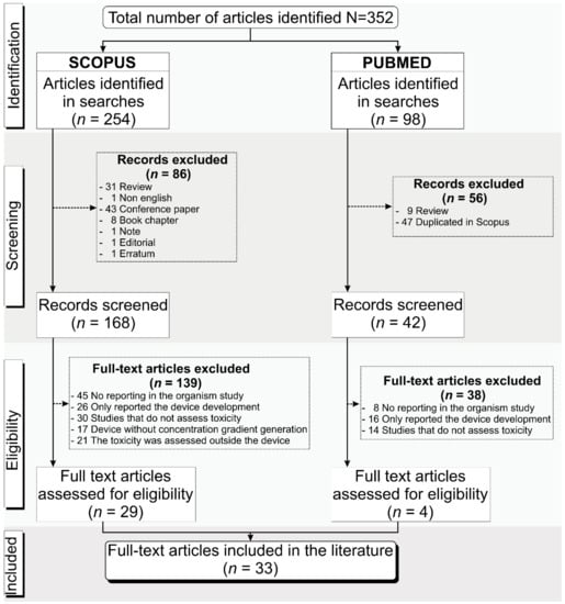
This systematic review aimed to analyze the development and functionality of microfluidic concentration gradient generators (CGGs) for toxicological evaluation of different biological organisms. We searched articles using the keywords: concentration gradient generator, toxicity, and microfluidic device. Only 33 of the 352 articles found were included and examined regarding the fabrication of the microdevices, the characteristics of the CGG, the biological model, and the desired results. The main fabrication method was soft lithography, using polydimethylsiloxane (PDMS) material (91%) and SU-8 as the mold (58.3%). New technologies were applied to minimize shear and bubble problems, reduce costs, and accelerate prototyping. The Christmas tree CGG design and its variations were the most reported in the studies, as well as the convective method of generation (61%). Biological models included bacteria and nematodes for antibiotic screening, microalgae for pollutant toxicity, tumor and normal cells for, primarily, chemotherapy screening, and Zebrafish embryos for drug and metal developmental toxicity. The toxic effects of each concentration generated were evaluated mostly with imaging and microscopy techniques. This study showed an advantage of CGGs over other techniques and their applicability for several biological models. Even with soft lithography, PDMS, and Christmas tree being more popular in their respective categories, current studies aim to apply new technologies and intricate architectures to improve testing effectiveness and reduce common microfluidics problems, allowing for high applicability of toxicity tests in different medical and environmental models. Full article
(This article belongs to the Special Issue In Vitro Model for Micro and Nano Technologies)
Show Figures

Figure 1

15 pages, 2116 KiB  
Article
Illustration of Gut–Thyroid Axis in Alcohol Use Disorder: Interplay of Gut Dysfunction, Pro-Inflammatory Responses, and Thyroid Function
by Manasa Sagaram 1,2, Amor J. Royer 1,2, Huirong Hu 2,3, Abhas Rajhans 2,4, Ranganathan Parthasarathy 1,2, Sathya Sridevi Krishnasamy 5, Sri Prakash Mokshagundam 5, Maiying Kong 3,6, Melanie L. Schwandt 7, Dipendra Parajuli 1,8, Matthew C. Cave 1,6,8 and Vatsalya Vatsalya 1,2,6,7,*
1 Division of Gastroenterology, Hepatology and Nutrition, Department of Medicine, University of Louisville, Louisville, KY 40202, USA
2 Clinical Laboratory for the Intervention Development of AUD and Organ Severity, University of Louisville, Louisville, KY 40202, USA
3 Department of Bioinformatics and Biostatistics, University of Louisville, Louisville, KY 40202, USA
4 Department of Neuroscience, University of California Los Angeles, Los Angeles, CA 90095, USA
5 Division of Endocrinology, Metabolism & Diabetes, University of Louisville, Louisville, KY 40202, USA
6 University of Louisville Alcohol Research Center, University of Louisville, Louisville, KY 40202, USA
7 National Institute on Alcohol Abuse and Alcoholism, Bethesda, MD 20892, USA
8 Robley Rex VA Medical Center, Louisville, KY 40206, USA
Cells 2022, 11(19), 3100; https://doi.org/10.3390/cells11193100 - 1 Oct 2022
Cited by 4 | Viewed by 3013
Abstract
(1) Background: Heavy and chronic alcohol drinking leads to altered gut dysfunction, coupled with a pro-inflammatory state. Thyroid-associated hormones and proteins may be dysregulated by heavy and chronic alcohol intake; however, the mechanism for altered gut-derived changes in thyroid function has not been [...] Read more.
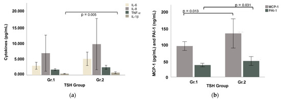
(1) Background: Heavy and chronic alcohol drinking leads to altered gut dysfunction, coupled with a pro-inflammatory state. Thyroid-associated hormones and proteins may be dysregulated by heavy and chronic alcohol intake; however, the mechanism for altered gut-derived changes in thyroid function has not been studied thus far. This study investigates the role of alcohol-induced gut dysfunction and pro-inflammatory cytokine profile in the thyroid function of patients with alcohol use disorder (AUD). (2) Methods: Male and female AUD patients (n = 44) were divided into Gr.1, patients with normal thyroid-stimulating hormone (TSH) levels (n = 28, 0.8 ≤ TSH ≤ 3 mIU/L); and Gr.2, patients with clinically elevated TSH levels (n = 16, TSH > 3 mIU/L). Demographics, drinking measures, comprehensive metabolic panels, and candidate thyroid markers (TSH, circulating triiodothyronine (T3), and free thyroxine (fT4)) were analyzed. Gut-dysfunction-associated markers (lipopolysaccharide (LPS), LPS-binding protein (LBP), and soluble LPS-induced pathogen-associated protein (sCD14)), and candidate pro-inflammatory cytokines (IL-1β, TNF-α, IL-6, IL-8, MCP-1, PAI-1) were also evaluated. (3) Results: Patients in both groups presented with a borderline overweight BMI category. Gr.2 reported numerically higher indices of chronic and heavy drinking patterns than Gr.1. The fT4 levels were elevated, while T3 was within normal limits in both groups. The gut dysfunction markers LBP and sCD14 were numerically elevated in Gr.2 vs. Gr.1, suggesting subtle ongoing changes. Candidate pro-inflammatory cytokines were significantly elevated in Gr.2, including IL-1 β, MCP-1, and PAI-1. Gr.2 showed a strong and statistically significant effect on the gut–immune–thyroid response (r = 0.896, 36 p = 0.002) on TSH levels in a multivariate regression model with LBP, sCD14, and PAI-1 levels as upstream variables in the gut–thyroid pathway. In addition, AUROC analysis demonstrated that many of the cytokines strongly predicted TSH in Gr.2, including IL-6 (area = 0.774, 39 p < 0.001) and TNF-α (area = 0.708, p = 0.017), among others. This was not observed in Gr.1. Gr.2 demonstrated elevated fT4, as well as TSH, which suggests that there was subclinical thyroiditis with underlying CNS dysfunction and a lack of a negative feedback loop. (4) Conclusions: These findings reveal the toxic effects of heavy and chronic drinking that play a pathological role in thyroid gland dysregulation by employing the gut–brain axis. These results also emphasize potential directions to carefully evaluate thyroid dysregulation in the overall medical management of AUD. Full article
(This article belongs to the Special Issue Gut Microbiota in Nutrition and Health)
Show Figures

Figure 1

27 pages, 1648 KiB  
Review
Effects of Noonan Syndrome-Germline Mutations on Mitochondria and Energy Metabolism
by Donald Bajia 1, Emanuela Bottani 2,* and Katarzyna Derwich 1,*
1 Department of Pediatric Oncology, Hematology and Transplantology, Poznan University of Medical Sciences, Ul. Fredry 10, 61701 Poznan, Poland
2 Department of Diagnostics and Public Health, Section of Pharmacology, University of Verona, Piazzale L. A. Scuro 10, 37134 Verona, Italy
Cells 2022, 11(19), 3099; https://doi.org/10.3390/cells11193099 - 1 Oct 2022
Cited by 6 | Viewed by 5709
Abstract
Noonan syndrome (NS) and related Noonan syndrome with multiple lentigines (NSML) contribute to the pathogenesis of human diseases in the RASopathy family. This family of genetic disorders constitute one of the largest groups of developmental disorders with variable penetrance and severity, associated with [...] Read more.
Noonan syndrome (NS) and related Noonan syndrome with multiple lentigines (NSML) contribute to the pathogenesis of human diseases in the RASopathy family. This family of genetic disorders constitute one of the largest groups of developmental disorders with variable penetrance and severity, associated with distinctive congenital disabilities, including facial features, cardiopathies, growth and skeletal abnormalities, developmental delay/mental retardation, and tumor predisposition. NS was first clinically described decades ago, and several genes have since been identified, providing a molecular foundation to understand their physiopathology and identify targets for therapeutic strategies. These genes encode proteins that participate in, or regulate, RAS/MAPK signalling. The RAS pathway regulates cellular metabolism by controlling mitochondrial homeostasis, dynamics, and energy production; however, little is known about the role of mitochondrial metabolism in NS and NSML. This manuscript comprehensively reviews the most frequently mutated genes responsible for NS and NSML, covering their role in the current knowledge of cellular signalling pathways, and focuses on the pathophysiological outcomes on mitochondria and energy metabolism. Full article
(This article belongs to the Special Issue Recent Advances in Metabolism and Oxidative Stress in Human Diseases)
Show Figures

Figure 1

14 pages, 694 KiB  
Review
RUNX Family in Hypoxic Microenvironment and Angiogenesis in Cancers
by You Mie Lee 1,2
1 Vessel-Organ Interaction Research Center, VOICE (MRC), Kyungpook National University, 80 Daehak-ro, Buk-gu, Daegu 41566, Korea
2 Lab of Molecular Pathophysiology, College of Pharmacy, Kyungpook National University, 80 Daehak-ro, Buk-gu, Daegu 41566, Korea
Cells 2022, 11(19), 3098; https://doi.org/10.3390/cells11193098 - 1 Oct 2022
Cited by 9 | Viewed by 2979
Abstract
The tumor microenvironment (TME) is broadly implicated in tumorigenesis, as tumor cells interact with surrounding cells to influence the development and progression of the tumor. Blood vessels are a major component of the TME and are attributed to the creation of a hypoxic [...] Read more.
The tumor microenvironment (TME) is broadly implicated in tumorigenesis, as tumor cells interact with surrounding cells to influence the development and progression of the tumor. Blood vessels are a major component of the TME and are attributed to the creation of a hypoxic microenvironment, which is a common feature of advanced cancers and inflamed premalignant tissues. Runt-related transcription factor (RUNX) proteins, a transcription factor family of developmental master regulators, are involved in vital cellular processes such as differentiation, proliferation, cell lineage specification, and apoptosis. Furthermore, the RUNX family is involved in the regulation of various oncogenic processes and signaling pathways as well as tumor suppressive functions, suggesting that the RUNX family plays a strategic role in tumorigenesis. In this review, we have discussed the relevant findings that describe the crosstalk of the RUNX family with the hypoxic TME and tumor angiogenesis or with their signaling molecules in cancer development and progression. Full article
(This article belongs to the Special Issue Roles of RUNX Family in Cancer)
Show Figures

Figure 1

13 pages, 1497 KiB  
Review
The Interplay between the Cellular Response to DNA Double-Strand Breaks and Estrogen
by Lia Yedidia-Aryeh and Michal Goldberg *
Department of Genetics, The Institute of Life Sciences, The Hebrew University of Jerusalem, Jerusalem 190401, Israel
Cells 2022, 11(19), 3097; https://doi.org/10.3390/cells11193097 - 1 Oct 2022
Cited by 4 | Viewed by 3117
Abstract
Cancer development is often connected to impaired DNA repair and DNA damage signaling pathways. The presence of DNA damage in cells activates DNA damage response, which is a complex cellular signaling network that includes DNA repair, activation of the cell cycle checkpoints, cellular [...] Read more.
Cancer development is often connected to impaired DNA repair and DNA damage signaling pathways. The presence of DNA damage in cells activates DNA damage response, which is a complex cellular signaling network that includes DNA repair, activation of the cell cycle checkpoints, cellular senescence, and apoptosis. DNA double-strand breaks (DSBs) are toxic lesions that are mainly repaired by the non-homologous end joining and homologous recombination repair (HRR) pathways. Estrogen-dependent cancers, like breast and ovarian cancers, are frequently associated with mutations in genes that play a role in HRR. The female sex hormone estrogen binds and activates the estrogen receptors (ERs), ERα, ERβ and G-protein-coupled ER 1 (GPER1). ERα drives proliferation, while ERβ inhibits cell growth. Estrogen regulates the transcription, stability and activity of numerus DDR factors and DDR factors in turn modulate ERα expression, stability and transcriptional activity. Additionally, estrogen stimulates DSB formation in cells as part of its metabolism and proliferative effect. In this review, we will present an overview on the crosstalk between estrogen and the cellular response to DSBs. We will discuss how estrogen regulates DSB signaling and repair, and how DDR factors modulate the expression, stability and activity of estrogen. We will also discuss how the regulation of HRR genes by estrogen promotes the development of estrogen-dependent cancers. Full article
(This article belongs to the Special Issue DNA Damage Response Regulation and Cancer)
Show Figures

Figure 1

16 pages, 2375 KiB  
Article
miR319-Regulated TCP3 Modulates Silique Development Associated with Seed Shattering in Brassicaceae
by Biting Cao 1,2,3,†, Hongfeng Wang 3,4,†, Jinjuan Bai 3, Xuan Wang 3, Xiaorong Li 3, Yanfeng Zhang 5, Suxin Yang 6, Yuke He 3,* and Xiang Yu 1,*
1 School of Life Sciences and Biotechnology, Shanghai Jiao Tong University, Shanghai 200240, China
2 Shanghai Key Lab of Protected Horticultural Technology, Horticultural Research Institute, Shanghai Academy of Agricultural Sciences, Shanghai 201106, China
3 National Key Laboratory of Plant Molecular Genetics, Center for Excellence in Molecular Plant Science, Shanghai Institute of Plant Physiology and Ecology, Chinese Academy of Sciences, Fenglin Road 300, Shanghai 200032, China
4 The Key Laboratory of Plant Development and Environmental Adaptation Biology, Ministry of Education, School of Life Science, Shandong University, Qingdao 266101, China
5 Hybrid Rape Research Center of Shaanxi Province, Yangling 712100, China
6 Key Laboratory of Soybean Molecular Design Breeding, Northeast Institute of Geography and Agroecology, Changchun 130102, China
These authors contribute equally to this work.
Cells 2022, 11(19), 3096; https://doi.org/10.3390/cells11193096 - 1 Oct 2022
Cited by 12 | Viewed by 2353
Abstract
Seed shattering is an undesirable trait that leads to crop yield loss. Improving silique resistance to shattering is critical for grain and oil crops. In this study, we found that miR319-targeted TEOSINTE BRANCHED 1, CYCLOIDEA, and PROLIFERATING CELL NUCLEAR ANTIGEN BINDING FACTOR (TCPs) [...] Read more.
Seed shattering is an undesirable trait that leads to crop yield loss. Improving silique resistance to shattering is critical for grain and oil crops. In this study, we found that miR319-targeted TEOSINTE BRANCHED 1, CYCLOIDEA, and PROLIFERATING CELL NUCLEAR ANTIGEN BINDING FACTOR (TCPs) inhibited the process of post-fertilized fruits (silique) elongation and dehiscence via regulation of FRUITFULL (FUL) expression in Arabidopsis thaliana and Brassica napus. AtMIR319a activation resulted in a longer silique with thickened and lignified replum, whereas overexpression of an miR319a-resistant version of AtTCP3 (mTCP3) led to a short silique with narrow and less lignified replum. Further genetic and expressional analysis suggested that FUL acted downstream of TCP3 to negatively regulate silique development. Moreover, hyper-activation of BnTCP3.A8, a B. napus homolog of AtTCP3, in rapeseed resulted in an enhanced silique resistance to shattering due to attenuated replum development. Taken together, our findings advance our knowledge of TCP-regulated silique development and provide a potential target for genetic manipulation to reduce silique shattering in Brassica crops. Full article
(This article belongs to the Section Plant, Algae and Fungi Cell Biology)
Show Figures

Graphical abstract

18 pages, 1647 KiB  
Review
Nose-to-Brain: The Next Step for Stem Cell and Biomaterial Therapy in Neurological Disorders
by Natalia Villar-Gómez 1,†, Doddy Denise Ojeda-Hernandez 1,†, Eneritz López-Muguruza 1, Silvia García-Flores 1, Natalia Bonel-García 1, María Soledad Benito-Martín 1, Belen Selma-Calvo 1, Alejandro Arturo Canales-Aguirre 2, Juan Carlos Mateos-Díaz 3, Paloma Montero-Escribano 4, Jordi A. Matias-Guiu 4, Jorge Matías-Guiu 1,4 and Ulises Gómez-Pinedo 1,*
1 Laboratory of Neurobiology, Institute of Neurosciences, IdISSC, Hospital Clínico San Carlos, Universidad Complutense de Madrid, 28040 Madrid, Spain
2 Preclinical Evaluation Unit, Medical and Pharmaceutical Biotechnology Unit, CIATEJ-CONACyT, Guadalajara 44270, Mexico
3 Department of Industrial Biotechnology, CIATEJ-CONACyT, Zapopan 45019, Mexico
4 Department of Neurology, Institute of Neurosciences, IdISSC, Hospital Clínico San Carlos, Universidad Complutense de Madrid, 28040 Madrid, Spain
These authors contributed equally to this work.
Cells 2022, 11(19), 3095; https://doi.org/10.3390/cells11193095 - 1 Oct 2022
Cited by 18 | Viewed by 5081
Abstract
Neurological disorders are a leading cause of morbidity worldwide, giving rise to a growing need to develop treatments to revert their symptoms. This review highlights the great potential of recent advances in cell therapy for the treatment of neurological disorders. Through the administration [...] Read more.
Neurological disorders are a leading cause of morbidity worldwide, giving rise to a growing need to develop treatments to revert their symptoms. This review highlights the great potential of recent advances in cell therapy for the treatment of neurological disorders. Through the administration of pluripotent or stem cells, this novel therapy may promote neuroprotection, neuroplasticity, and neuroregeneration in lesion areas. The review also addresses the administration of these therapeutic molecules by the intranasal route, a promising, non-conventional route that allows for direct access to the central nervous system without crossing the blood–brain barrier, avoiding potential adverse reactions and enabling the administration of large quantities of therapeutic molecules to the brain. Finally, we focus on the need to use biomaterials, which play an important role as nutrient carriers, scaffolds, and immune modulators in the administration of non-autologous cells. Little research has been conducted into the integration of biomaterials alongside intranasally administered cell therapy, a highly promising approach for the treatment of neurological disorders. Full article
(This article belongs to the Section Stem Cells)
Show Figures

Graphical abstract

20 pages, 3505 KiB  
Article
Combination of Paclitaxel and PXR Antagonist SPA70 Reverses Paclitaxel-Resistant Non-Small Cell Lung Cancer
by Xiaxia Niu 1, Ting Wu 1, Qishuang Yin 2, Xinsheng Gu 3, Gege Li 1, Changlong Zhou 1, Mei Ma 1, Li Su 1, Shu Tang 4, Yanan Tian 4,*, Ming Yang 2 and Hongmei Cui 1,*
1 Institute of Toxicology, School of Public Health, Lanzhou University, Lanzhou 730000, China
2 State Key Laboratory of Applied Organic Chemistry, College of Chemistry and Chemical Engineering, Lanzhou University, Lanzhou 730000, China
3 College of Basic Medical Sciences, Hubei University of Medicine, Shiyan 442000, China
4 Department of Veterinary Physiology and Pharmacology, Texas A&M University, College Station, TX 77843, USA
Cells 2022, 11(19), 3094; https://doi.org/10.3390/cells11193094 - 1 Oct 2022
Cited by 14 | Viewed by 3461
Abstract
Paclitaxel (PTX) is one of the most efficient drugs for late-stage non-small cell lung cancer (NSCLC) patients. However, most patients gradually develop resistance to PTX with long-term treatments. The identification of new strategies to reverse PTX resistance in NSCLC is crucially important for [...] Read more.
Paclitaxel (PTX) is one of the most efficient drugs for late-stage non-small cell lung cancer (NSCLC) patients. However, most patients gradually develop resistance to PTX with long-term treatments. The identification of new strategies to reverse PTX resistance in NSCLC is crucially important for the treatment. PTX is an agonist for the pregnane X receptor (PXR) which regulates PTX metabolism. Antagonizing PXR, therefore, may render the NSCLC more sensitive to the PTX treatment. In this study, we investigated the PXR antagonist SPA70 and its role in PTX treatment of NSCLC. In vitro, SPA70 and PTX synergistically inhibited cell growth, migration and invasion in both paclitaxel-sensitive and paclitaxel-resistant A549 and H460 lung cancer cells. Mechanistically, we found PTX and SPA70 cotreatment disassociated PXR from ABCB1 (MDR1, P-gp) promoter, thus inhibiting P-gp expression. Furthermore, the combination regimen synergistically enhanced the interaction between PXR and Tip60, which abrogated Tip60-mediated α-tubulin acetylation, leading to mitosis defect, S-phase arrest and necroptosis/apoptosis. Combination of PXT and SPA70 dramatically inhibited tumor growth in a paclitaxel-resistant A549/TR xenograft tumor model. Taken together, we showed that SPA70 reduced the paclitaxel resistance of NSCLC. The combination regimen of PTX and SPA70 could be potential novel candidates for the treatment of taxane-resistant lung cancer. Full article
Show Figures

Figure 1

35 pages, 5199 KiB  
Review
Biochemical Pathways of Cellular Mechanosensing/Mechanotransduction and Their Role in Neurodegenerative Diseases Pathogenesis
by Ilaria Tortorella 1, Chiara Argentati 1, Carla Emiliani 1,2, Francesco Morena 1,* and Sabata Martino 1,2,*
1 Department of Chemistry, Biology and Biotechnology, University of Perugia, Via del Giochetto, 06123 Perugia, Italy
2 Centro di Eccellenza CEMIN (Materiali Innovativi Nanostrutturali per Applicazioni Chimica Fisiche e Biomediche), University of Perugia, 06123 Perugia, Italy
Cells 2022, 11(19), 3093; https://doi.org/10.3390/cells11193093 - 1 Oct 2022
Cited by 26 | Viewed by 7667
Abstract
In this review, we shed light on recent advances regarding the characterization of biochemical pathways of cellular mechanosensing and mechanotransduction with particular attention to their role in neurodegenerative disease pathogenesis. While the mechanistic components of these pathways are mostly uncovered today, the crosstalk [...] Read more.
In this review, we shed light on recent advances regarding the characterization of biochemical pathways of cellular mechanosensing and mechanotransduction with particular attention to their role in neurodegenerative disease pathogenesis. While the mechanistic components of these pathways are mostly uncovered today, the crosstalk between mechanical forces and soluble intracellular signaling is still not fully elucidated. Here, we recapitulate the general concepts of mechanobiology and the mechanisms that govern the mechanosensing and mechanotransduction processes, and we examine the crosstalk between mechanical stimuli and intracellular biochemical response, highlighting their effect on cellular organelles’ homeostasis and dysfunction. In particular, we discuss the current knowledge about the translation of mechanosignaling into biochemical signaling, focusing on those diseases that encompass metabolic accumulation of mutant proteins and have as primary characteristics the formation of pathological intracellular aggregates, such as Alzheimer’s Disease, Huntington’s Disease, Amyotrophic Lateral Sclerosis and Parkinson’s Disease. Overall, recent findings elucidate how mechanosensing and mechanotransduction pathways may be crucial to understand the pathogenic mechanisms underlying neurodegenerative diseases and emphasize the importance of these pathways for identifying potential therapeutic targets. Full article
(This article belongs to the Special Issue Mechanosensation and Mechanotransduction in Brain Cells)
Show Figures

Graphical abstract

18 pages, 2327 KiB  
Article
Antagonism of CGRP Receptor: Central and Peripheral Mechanisms and Mediators in an Animal Model of Chronic Migraine
by Rosaria Greco 1,*, Chiara Demartini 1,2, Miriam Francavilla 1, Anna Maria Zanaboni 1,2 and Cristina Tassorelli 1,2
1 Unit of Translational Neurovascular Research, IRCCS Mondino Foundation, 27100 Pavia, Italy
2 Department of Brain and Behavioral Sciences, University of Pavia, 27100 Pavia, Italy
Cells 2022, 11(19), 3092; https://doi.org/10.3390/cells11193092 - 30 Sep 2022
Cited by 34 | Viewed by 5238
Abstract
Calcitonin-gene-related peptide (CGRP) plays a key role in migraine pathophysiology and more specifically in the mechanisms underlying peripheral and central sensitization. Here, we explored the interaction of CGRP with other pain mediators relevant for neuronal sensitization in an animal model of chronic migraine. [...] Read more.
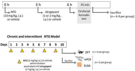
Calcitonin-gene-related peptide (CGRP) plays a key role in migraine pathophysiology and more specifically in the mechanisms underlying peripheral and central sensitization. Here, we explored the interaction of CGRP with other pain mediators relevant for neuronal sensitization in an animal model of chronic migraine. Male Sprague-Dawley rats were exposed to nitroglycerin (NTG, 5 mg/kg, i.p.) or vehicle co-administered with the CGRP receptor antagonist olcegepant (2 mg/kg i.p.), or its vehicle, every other day over a 9-day period. Twenty-four hours after the last injection of NTG (or vehicle), behavioral test and ex vivo analysis were performed. Olcegepant attenuated NTG-induced trigeminal hyperalgesia in the second phase of the orofacial formalin test. Interestingly, it also reduced gene expression and protein levels of CGRP, pro-inflammatory cytokines, inflammatory-associated miRNAs (miR-155-5p, miR-382-5p, and miR-34a-5p), and transient receptor potential ankyrin channels in the medulla–pons area, cervical spinal cord, and trigeminal ganglia. Similarly, olcegepant reduced the NTG-induced increase in CGRP and inflammatory cytokines in serum. The findings show that the activation of the CGRP pathway in a migraine animal model was associated to the persistent activation of inflammatory pathways, which was paralleled by a condition of hyperalgesia. These molecular events are relevant for informing us about the mechanisms underlying chronic migraine. Full article
(This article belongs to the Special Issue Migraine Neuroscience: From Experimental Models to Target Therapy)
Show Figures

Figure 1