Cells 2021, 10(2), 376; https://doi.org/10.3390/cells10020376 - 12 Feb 2021
Cited by 17 | Viewed by 6777
Abstract
The identification of the Mas-related G-protein-coupled receptors (Mrgpr) as targets of diverse stimuli of mast cells (MCs), including neuropeptides and pseudo-allergy causing drugs, has placed these receptors at a prime position in MC research. However, the species-dependent diversity of these receptors raises the
[...] Read more.
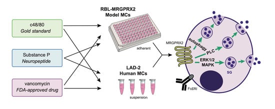
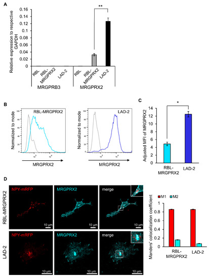
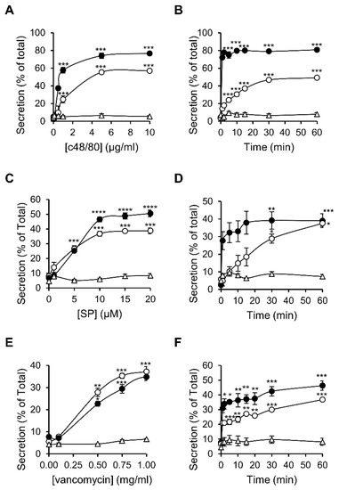
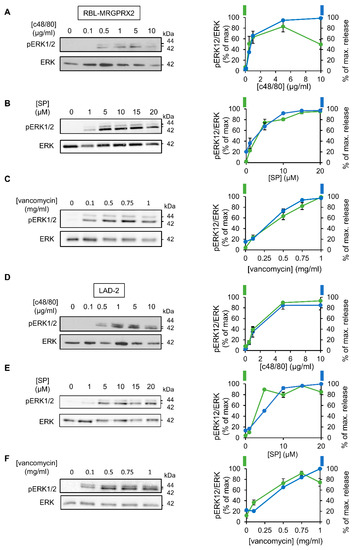
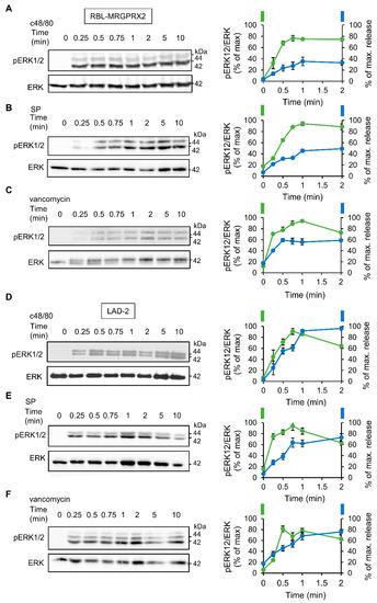
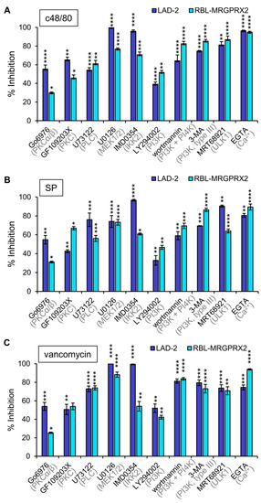
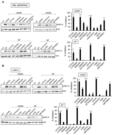
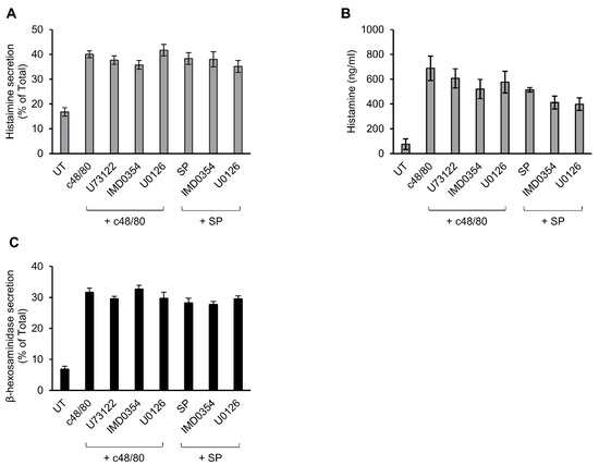
The identification of the Mas-related G-protein-coupled receptors (Mrgpr) as targets of diverse stimuli of mast cells (MCs), including neuropeptides and pseudo-allergy causing drugs, has placed these receptors at a prime position in MC research. However, the species-dependent diversity of these receptors raises the need for an adequate model for investigating the human MRGPRX2 receptor. RBL-2H3 cells, stably transfected with MRGPRX2 (RBL-MRGPRX2), are increasingly used for this purpose. Therefore, we investigated whether ectopically expressed MRGPRX2, in rat MCs, recapitulates its authentic signaling. To this purpose, we performed a broad comparative study of the responses of human LAD-2 MCs that express MRGPRX2 endogenously, and RBL-MRGPRX2 cells to compound 48/80, substance P and vancomycin, three proto-type ligands of MRGPRX2. We demonstrate that both models share similar dose–response relationships, kinetics and sensitivities to a wide range of signaling targeting drugs. Therefore, our results indicate that ectopically expressed MRGPRX2 preserves the signaling pathways employed to evoke human MC degranulation, which we show to rely on ERK1/2 MAP kinases, phospholipase C (PLC) and autophagy-related signaling. Importantly, we also show that the underlying mechanisms of MRGPRX2-triggered MC degranulation in either LAD-2 or RBL-MRGPRX2 cells are different from those elicited by its rodent orthologs.
Full article
(This article belongs to the Special Issue Mast Cell Development, Activation and Contribution to Health and Disease)
►
Show Figures
Graphical abstract


