22 pages, 2803 KB  
Review
Hot and Cold Tumors: Is Endoglin (CD105) a Potential Target for Vessel Normalization?
by Claudia Ollauri-Ibáñez, Blanca Ayuso-Íñigo and Miguel Pericacho
Cancers 2021, 13(7), 1552; https://doi.org/10.3390/cancers13071552 - 28 Mar 2021
Cited by 34 | Viewed by 10023
Abstract
Tumors are complex masses formed by malignant but also by normal cells. The interaction between these cells via cytokines, chemokines, growth factors, and enzymes that remodel the extracellular matrix (ECM) constitutes the tumor microenvironment (TME). This TME can be determinant in the prognosis [...] Read more.
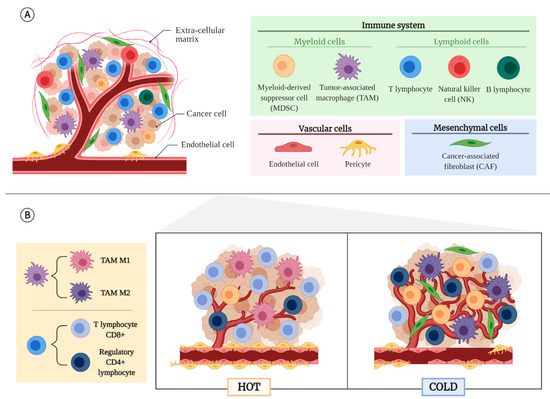
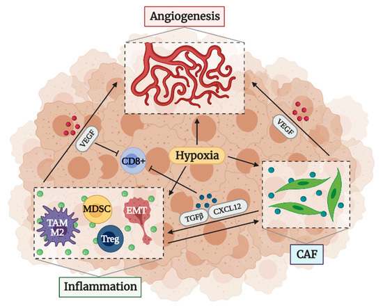
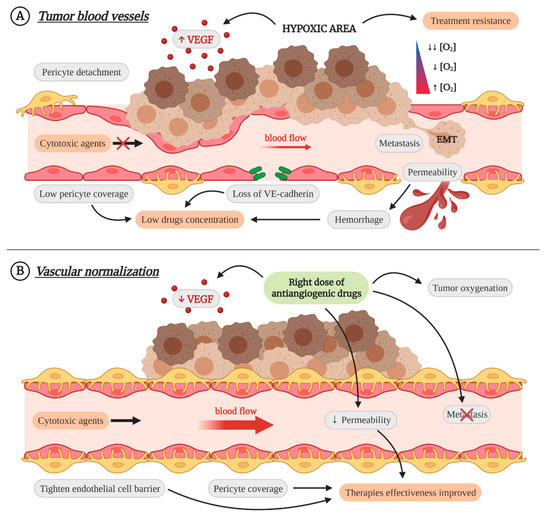
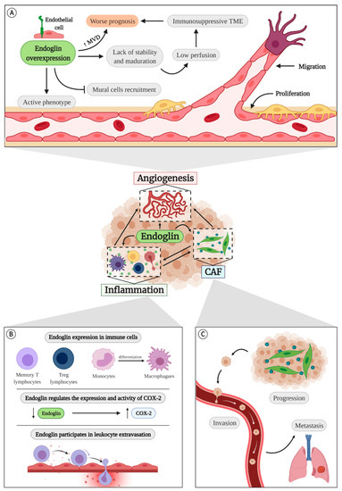
Tumors are complex masses formed by malignant but also by normal cells. The interaction between these cells via cytokines, chemokines, growth factors, and enzymes that remodel the extracellular matrix (ECM) constitutes the tumor microenvironment (TME). This TME can be determinant in the prognosis and the response to some treatments such as immunotherapy. Depending on their TME, two types of tumors can be defined: hot tumors, characterized by an immunosupportive TME and a good response to immunotherapy; and cold tumors, which respond poorly to this therapy and are characterized by an immunosuppressive TME. A therapeutic strategy that has been shown to be useful for the conversion of cold tumors into hot tumors is vascular normalization. In this review we propose that endoglin (CD105) may be a useful target of this strategy since it is involved in the three main processes involved in the generation of the TME: angiogenesis, inflammation, and cancer-associated fibroblast (CAF) accumulation. Moreover, the analysis of endoglin expression in tumors, which is already used in the clinic to study the microvascular density and that is associated with worse prognosis, could be used to predict a patient’s response to immunotherapy. Full article
(This article belongs to the Special Issue Targeting Blood Vessel Components to Treat Cancer)
Show Figures

Figure 1
14 pages, 4096 KB  
Article
Combination Assessment of Diffusion-Weighted Imaging and T2-Weighted Imaging Is Acceptable for the Differential Diagnosis of Lung Cancer from Benign Pulmonary Nodules and Masses
by Katsuo Usuda, Masahito Ishikawa, Shun Iwai, Yoshihito Iijima, Nozomu Motono, Munetaka Matoba, Mariko Doai, Keiya Hirata and Hidetaka Uramoto
Cancers 2021, 13(7), 1551; https://doi.org/10.3390/cancers13071551 - 28 Mar 2021
Cited by 17 | Viewed by 3627
Abstract
The purpose of this study is to determine whether the combination assessment of DWI and T2-weighted imaging (T2WI) improves the diagnostic ability for differential diagnosis of lung cancer from benign pulmonary nodules and masses (BPNMs). The optimal cut-off value (OCV) for differential diagnosis [...] Read more.
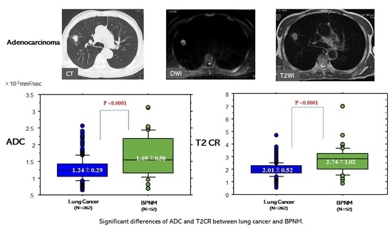
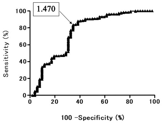
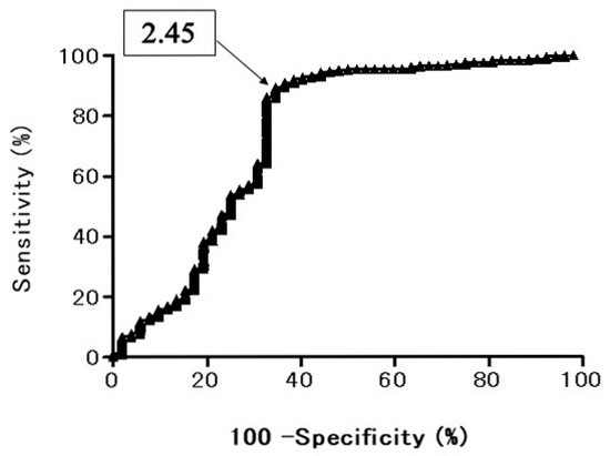
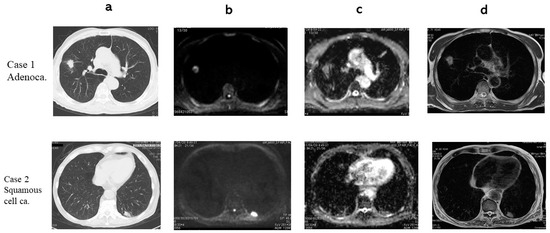
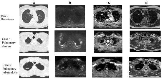
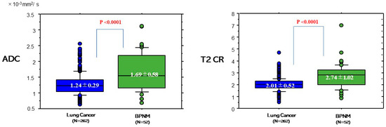
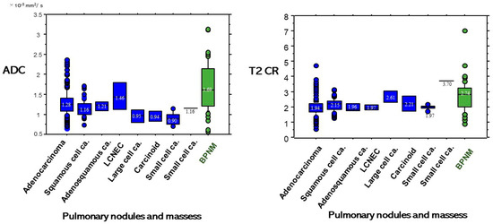
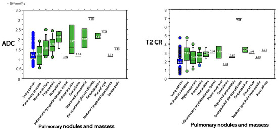
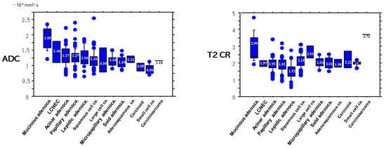
The purpose of this study is to determine whether the combination assessment of DWI and T2-weighted imaging (T2WI) improves the diagnostic ability for differential diagnosis of lung cancer from benign pulmonary nodules and masses (BPNMs). The optimal cut-off value (OCV) for differential diagnosis was set at 1.470 × 10⁻³ mm2/s for apparent diffusion coefficient (ADC), and at 2.45 for T2 contrast ratio (T2 CR). The ADC (1.24 ± 0.29 × 10⁻³ mm2/s) of lung cancer was significantly lower than that (1.69 ± 0.58 × 10⁻³ mm2/s) of BPNM. The T2 CR (2.01 ± 0.52) of lung cancer was significantly lower than that (2.74 ± 1.02) of BPNM. As using the OCV for ADC, the sensitivity was 83.9% (220/262), the specificity 63.4% (33/52), and the accuracy 80.6% (253/314). As using the OCV for T2 CR, the sensitivity was 89.7% (235/262), the specificity 61.5% (32/52), and the accuracy 85.0% (267/314). In 212 PNMs which were judged to be malignant by both DWI and T2WI, 203 PNMs (95.8%) were lung cancers. In 33 PNMs which were judged to be benign by both DWI and T2WI, 23 PNMs (69.7%) were BPNMs. The combined assessment of DWI and T2WI could judge PNMs more precisely and would be acceptable for differential diagnosis of PNMs. Full article
(This article belongs to the Special Issue New Challenges in Cancer Imaging)
Show Figures

Graphical abstract

14 pages, 1736 KB  
Article
Combing MRI Perfusion and 18F-FDG PET/CT Metabolic Biomarkers Helps Predict Survival in Advanced Nasopharyngeal Carcinoma: A Prospective Multimodal Imaging Study
by Sheng-Chieh Chan, Chih-Hua Yeh, Joseph Tung-Chieh Chang, Kai-Ping Chang, Jen-Hung Wang and Shu-Hang Ng
Cancers 2021, 13(7), 1550; https://doi.org/10.3390/cancers13071550 - 28 Mar 2021
Cited by 23 | Viewed by 3612
Abstract
We prospectively investigated the prognostic value of imaging parameters for nasopharyngeal carcinoma (NPC) using dynamic contrast-enhanced MRI (DCE-MRI), diffusion-weighted imaging (DWI), and 2-deoxy-2-[fluorine-18]fluoro-D-glucose positron emission tomography (18F-FDG PET)/computed tomography (CT). Patients with stage III–IVb NPC underwent F-FDG PET/CT, DCE-MRI, and DWI [...] Read more.
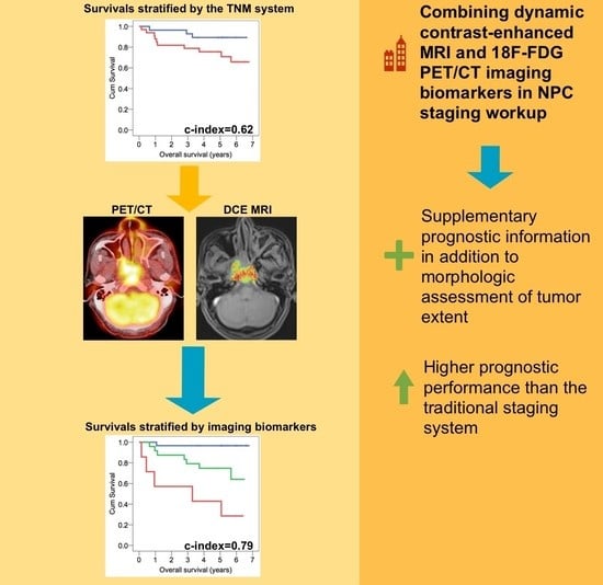
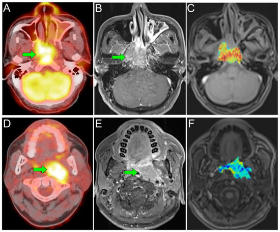
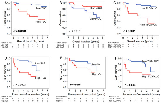
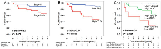
We prospectively investigated the prognostic value of imaging parameters for nasopharyngeal carcinoma (NPC) using dynamic contrast-enhanced MRI (DCE-MRI), diffusion-weighted imaging (DWI), and 2-deoxy-2-[fluorine-18]fluoro-D-glucose positron emission tomography (18F-FDG PET)/computed tomography (CT). Patients with stage III–IVb NPC underwent F-FDG PET/CT, DCE-MRI, and DWI before treatment. Kaplan-Meier and Cox-regression analyses were used to assess associations of PET and MRI imaging biomarkers with overall survival (OS) and recurrence-free survival (RFS). We used independent prognosticators to establish prognostic models; model performance was examined using Harrell’s concordance index (c-index). Sixty-one patients were available for analysis, as 13 patients died and 20 experienced recurrence. Total lesion glycolysis (TLG) (p = 0.002) from PET/CT and the initial area under the curve (iAUC) (p = 0.036) from DCE-MRI were identified as independent prognosticators of OS; Epstein-Barr virus (EBV) DNA (p = 0.027), the extracellular volume fraction (Ve) (p = 0.027) from DCE-MRI, and TLG/iAUC (p = 0.025) were significant predictors of RFS. The c-indices of the prognostic models incorporating TLG + iAUC in predicting OS and incorporating EBV DNA + Ve + TLG/iAUC in predicting RFS were 0.79 and 0.76, respectively. These were significantly higher than the corresponding c-indices of the TNM staging system (p = 0.047 and 0.025, respectively); they were also higher than those of models with only MRI or PET biomarkers. In conclusion, the combination of pretreatment DCE-MRI and 18F-FDG PET/CT imaging biomarkers helps survival prediction in advanced NPC. Integrating MRI perfusion with PET metabolism and plasma EBV information may aid clinicians in planning the optimal personalized management strategy. Full article
(This article belongs to the Section Cancer Biomarkers)
Show Figures

Graphical abstract

12 pages, 1182 KB  
Article
Early Clinical Outcomes of Intensity Modulated Radiation Therapy/Intensity Modulated Proton Therapy Combination in Comparison with Intensity Modulated Radiation Therapy Alone in Oropharynx Cancer Patients
by Han Gyul Yoon, Yong Chan Ahn, Dongryul Oh, Jae Myoung Noh, Seung Gyu Park, Heerim Nam, Sang Gyu Ju, Dongyeol Kwon and Seyjoon Park
Cancers 2021, 13(7), 1549; https://doi.org/10.3390/cancers13071549 - 27 Mar 2021
Cited by 15 | Viewed by 3883
Abstract
Purpose: To report the early clinical outcomes of combining intensity-modulated radiation therapy (IMRT) and intensity-modulated proton therapy (IMPT) in comparison with IMRT alone in treating oropharynx cancer (OPC) patients. Materials and Methods: The medical records of 148 OPC patients who underwent definitive radiotherapy [...] Read more.
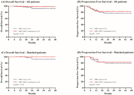
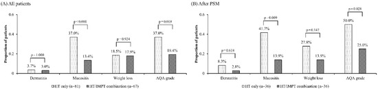
Purpose: To report the early clinical outcomes of combining intensity-modulated radiation therapy (IMRT) and intensity-modulated proton therapy (IMPT) in comparison with IMRT alone in treating oropharynx cancer (OPC) patients. Materials and Methods: The medical records of 148 OPC patients who underwent definitive radiotherapy (RT) with concurrent systemic therapy, from January 2016 till December 2019 at Samsung Medical Center, were retrospectively reviewed. During the 5.5 weeks’ RT course, the initial 16 (or 18) fractions were delivered by IMRT in all patients, and the subsequent 12 (or 10) fractions were either by IMRT in 81 patients (IMRT only) or by IMPT in 67 (IMRT/IMPT combination), respectively, based on comparison of adaptive re-plan profiles and availability of equipment. Propensity-score matching (PSM) was done on 76 patients (38 from each group) for comparative analyses. Results: With the median follow-up of 24.7 months, there was no significant difference in overall survival and progression free survival between groups, both before and after PSM. Before PSM, the IMRT/IMPT combination group experienced grade ≥ 3 acute toxicities less frequently: mucositis in 37.0% and 13.4% (p < 0.001); and analgesic quantification algorithm (AQA) in 37.0% and 19.4% (p = 0.019), respectively. The same trends were observed after PSM: mucositis in 39.5% and 15.8% (p = 0.021); and AQA in 47.4% and 21.1% (p = 0.016), respectively. In multivariate logistic regression, grade ≥ 3 mucositis was significantly less frequent in the IMRT/IMPT combination group, both before and after PSM (p = 0.027 and 0.024, respectively). AQA score ≥ 3 was also less frequent in the IMRT/IMPT combination group, both before and after PSM (p = 0.085 and 0.018, respectively). Conclusions: In treating the OPC patients, with comparable early oncologic outcomes, more favorable acute toxicity profiles were achieved following IMRT/IMPT combination than IMRT alone. Full article
(This article belongs to the Special Issue Proton Therapy For Cancers)
Show Figures

Figure 1
33 pages, 19920 KB  
Article
Overexpression of Human Syndecan-1 Protects against the Diethylnitrosamine-Induced Hepatocarcinogenesis in Mice
by Andrea Reszegi, Katalin Karászi, Gábor Tóth, Kristóf Rada, Lóránd Váncza, Lilla Turiák, Zsuzsa Schaff, András Kiss, László Szilák, Gábor Szabó, Gábor Petővári, Anna Sebestyén, Katalin Dezső, Eszter Regős, Péter Tátrai, Kornélia Baghy and Ilona Kovalszky
Cancers 2021, 13(7), 1548; https://doi.org/10.3390/cancers13071548 - 27 Mar 2021
Cited by 9 | Viewed by 5119
Abstract
Although syndecan-1 (SDC1) is known to be dysregulated in various cancer types, its implication in tumorigenesis is poorly understood. Its effect may be detrimental or protective depending on the type of cancer. Our previous data suggest that SDC1 is protective against hepatocarcinogenesis. To [...] Read more.
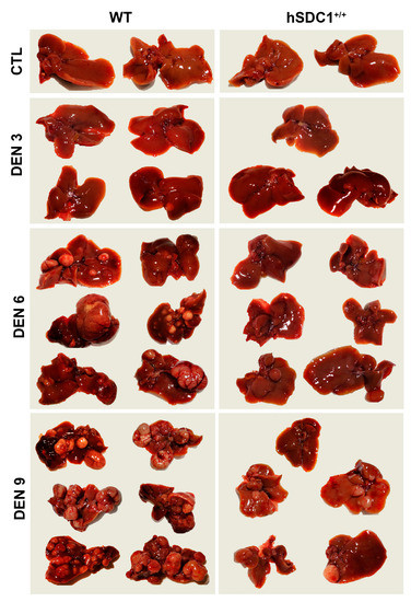
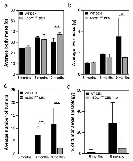
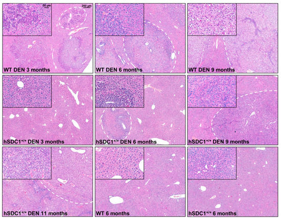
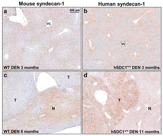
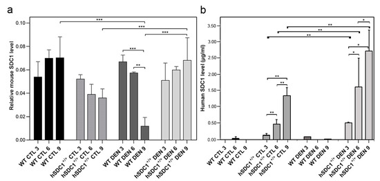
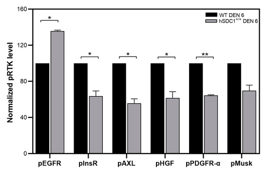
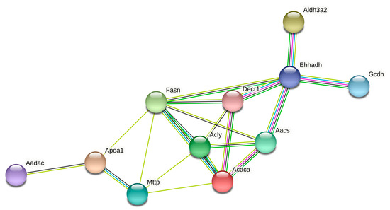
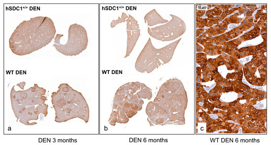
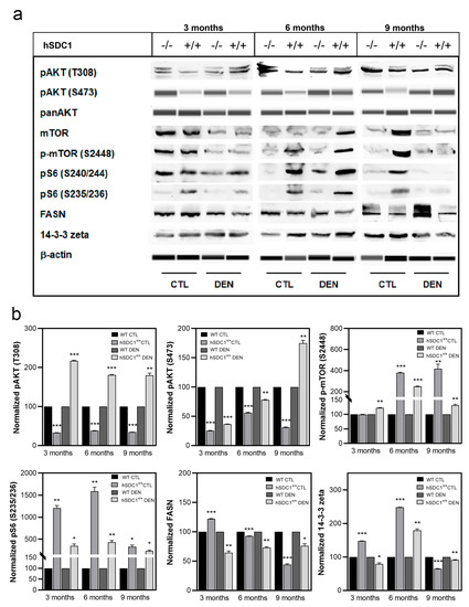
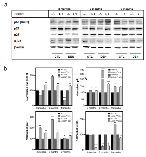
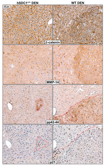
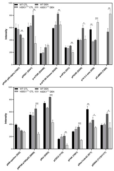
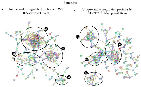
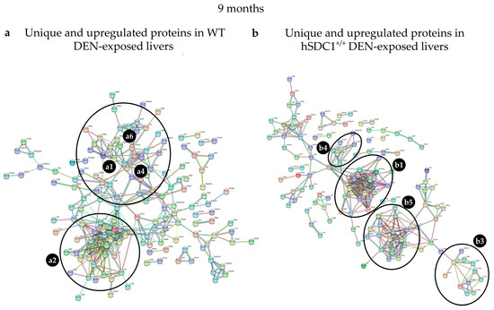
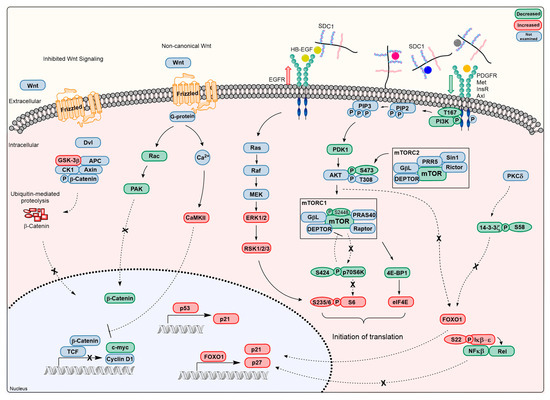
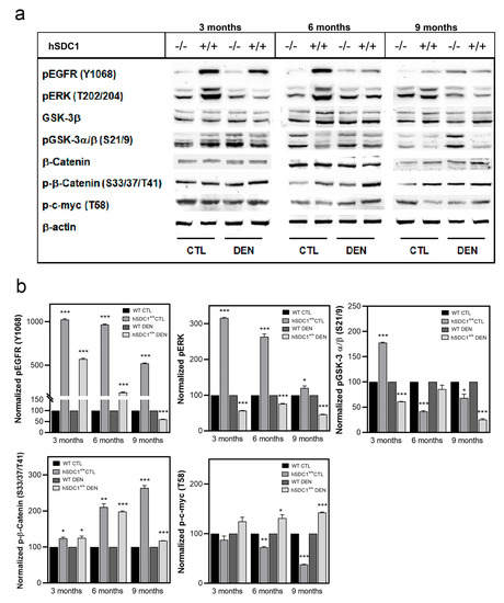
Although syndecan-1 (SDC1) is known to be dysregulated in various cancer types, its implication in tumorigenesis is poorly understood. Its effect may be detrimental or protective depending on the type of cancer. Our previous data suggest that SDC1 is protective against hepatocarcinogenesis. To further verify this notion, human SDC1 transgenic (hSDC1+/+) mice were generated that expressed hSDC1 specifically in the liver under the control of the albumin promoter. Hepatocarcinogenesis was induced by a single dose of diethylnitrosamine (DEN) at an age of 15 days after birth, which resulted in tumors without cirrhosis in wild-type and hSDC1+/+ mice. At the experimental endpoint, livers were examined macroscopically and histologically, as well as by immunohistochemistry, Western blot, receptor tyrosine kinase array, phosphoprotein array, and proteomic analysis. Liver-specific overexpression of hSDC1 resulted in an approximately six month delay in tumor formation via the promotion of SDC1 shedding, downregulation of lipid metabolism, inhibition of the mTOR and the β-catenin pathways, and activation of the Foxo1 and p53 transcription factors that lead to the upregulation of the cell cycle inhibitors p21 and p27. Furthermore, both of them are implicated in the regulation of intermediary metabolism. Proteomic analysis showed enhanced lipid metabolism, activation of motor proteins, and loss of mitochondrial electron transport proteins as promoters of cancer in wild-type tumors, inhibited in the hSDC1+/+ livers. These complex mechanisms mimic the characteristics of nonalcoholic steatohepatitis (NASH) induced human liver cancer successfully delayed by syndecan-1. Full article
(This article belongs to the Collection Matrix Effectors and Cancer)
Show Figures

Figure 1
18 pages, 6941 KB  
Review
Pineal Gland Tumors: A Review
by Gaia Favero, Francesca Bonomini and Rita Rezzani
Cancers 2021, 13(7), 1547; https://doi.org/10.3390/cancers13071547 - 27 Mar 2021
Cited by 68 | Viewed by 18707
Abstract
The pineal gland is a small, pinecone-shaped endocrine gland that participates in the biological rhythm regulation of vertebrates. The recognized major product of the pineal gland is melatonin—a multifunctional endogenous indoleamine. Accumulating evidence suggests that the pineal gland is important for preserving ideal [...] Read more.
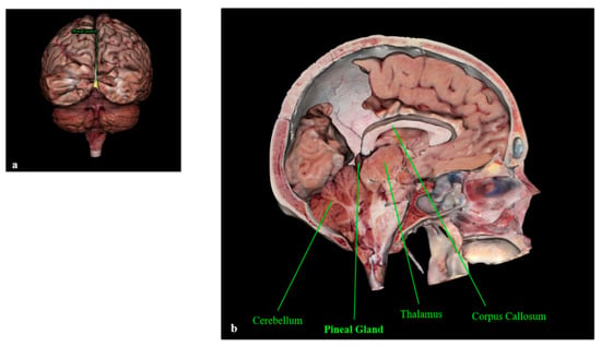
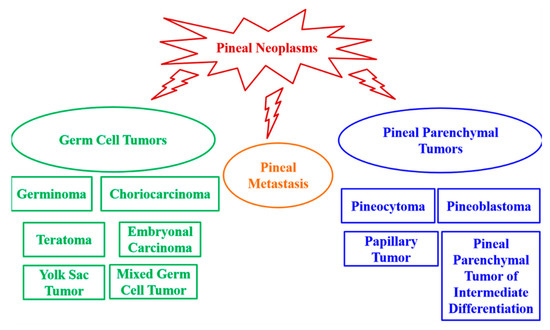
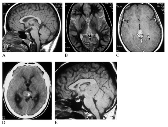
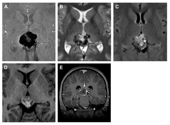
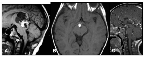
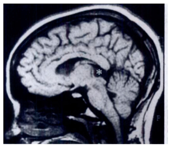
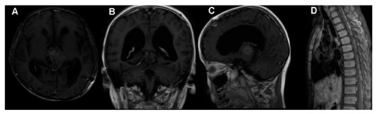
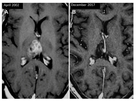
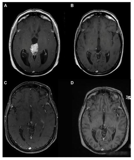
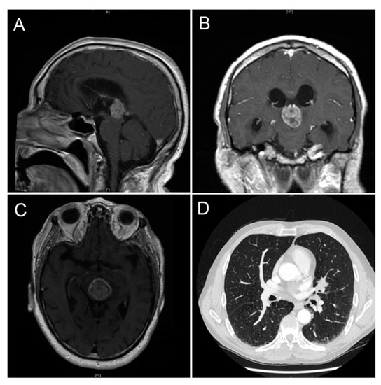
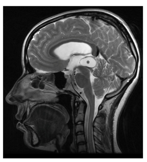
The pineal gland is a small, pinecone-shaped endocrine gland that participates in the biological rhythm regulation of vertebrates. The recognized major product of the pineal gland is melatonin—a multifunctional endogenous indoleamine. Accumulating evidence suggests that the pineal gland is important for preserving ideal health conditions in vertebrate. Tumors of the pineal region account for approximately 3–11% of pediatric brain neoplasms but fewer than 1% of brain neoplasms in adults. It is fundamental to expand advanced imaging techniques together with both clinical and laboratory knowledge, to help to differentiate among pineal neoplasms and thus facilitate accurate primary diagnoses and proper therapeutic interventions. In this review, we report the gross anatomy of the pineal gland and its functional significance and discuss the clinical relevance of pineal gland tumors, underlining the importance of identifying the leading causes of pineal region masses. Full article
(This article belongs to the Section Tumor Microenvironment)
Show Figures

Figure 1
16 pages, 3444 KB  
Article
Pulsed-Focused Ultrasound Slows B16 Melanoma and 4T1 Breast Tumor Growth through Differential Tumor Microenvironmental Changes
by Gadi Cohen, Parwathy Chandran, Rebecca M. Lorsung, Omer Aydin, Lauren E. Tomlinson, Robert B. Rosenblatt, Scott R. Burks and Joseph A. Frank
Cancers 2021, 13(7), 1546; https://doi.org/10.3390/cancers13071546 - 27 Mar 2021
Cited by 20 | Viewed by 5113
Abstract
Focused ultrasound (FUS) has shown promise as a non-invasive treatment modality for solid malignancies. FUS targeting to tumors has been shown to initiate pro-inflammatory immune responses within the tumor microenvironment. Pulsed FUS (pFUS) can alter the expression of cytokines, chemokines, trophic factors, cell [...] Read more.
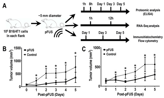
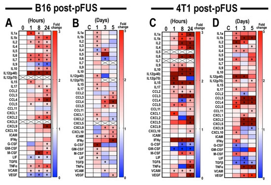
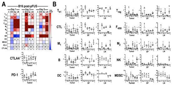
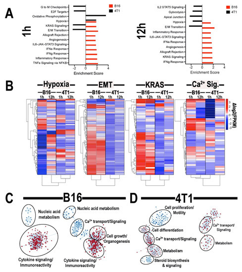
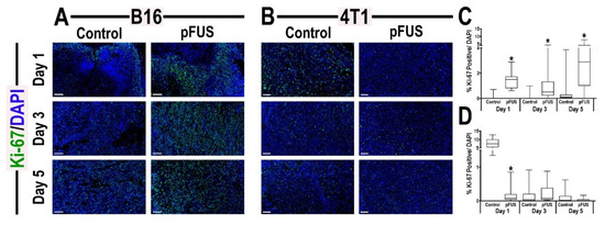
Focused ultrasound (FUS) has shown promise as a non-invasive treatment modality for solid malignancies. FUS targeting to tumors has been shown to initiate pro-inflammatory immune responses within the tumor microenvironment. Pulsed FUS (pFUS) can alter the expression of cytokines, chemokines, trophic factors, cell adhesion molecules, and immune cell phenotypes within tissues. Here, we investigated the molecular and immune cell effects of pFUS on murine B16 melanoma and 4T1 breast cancer flank tumors. Temporal changes following sonication were evaluated by proteomics, RNA-seq, flow-cytometry, and histological analyses. Proteomic profiling revealed molecular changes occurring over 24 h post-pFUS that were consistent with a shift toward inflamed tumor microenvironment. Over 5 days post-pFUS, tumor growth rates were significantly decreased while flow cytometric analysis revealed differences in the temporal migration of immune cells. Transcriptomic analyses following sonication identified differences in gene expression patterns between the two tumor types. Histological analyses further demonstrated reduction of proliferation marker, Ki-67 in 4T1, but not in B16 tumors, and activated cleaved-caspase 3 for apoptosis remained elevated up to 3 days post-pFUS in both tumor types. This study revealed diverse biological mechanisms following pFUS treatment and supports its use as a possible adjuvant to ablative tumor treatment to elicit enhanced anti-tumor responses and slow tumor growth. Full article
Show Figures

Figure 1
13 pages, 2661 KB  
Article
Impact of Goal Directed Therapy in Head and Neck Oncological Surgery with Microsurgical Reconstruction: Free Flap Viability and Complications
by Blanca Tapia, Elena Garrido, Jose Luis Cebrian, Jose Luis Del Castillo, Javier Gonzalez, Itsaso Losantos and Fernando Gilsanz
Cancers 2021, 13(7), 1545; https://doi.org/10.3390/cancers13071545 - 27 Mar 2021
Cited by 20 | Viewed by 3909
Abstract
(1) Background: Surgical outcomes in free flap reconstruction of head and neck defects in cancer patients have improved steadily in recent years; however, correct anaesthesia management is also important. The aim of this study has been to show whether goal directed therapy can [...] Read more.
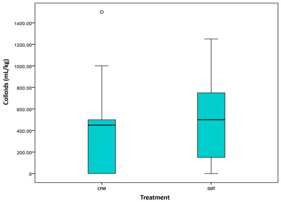
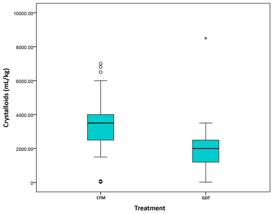
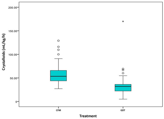
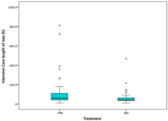
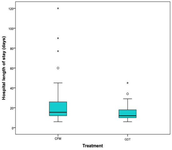
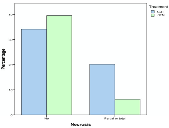
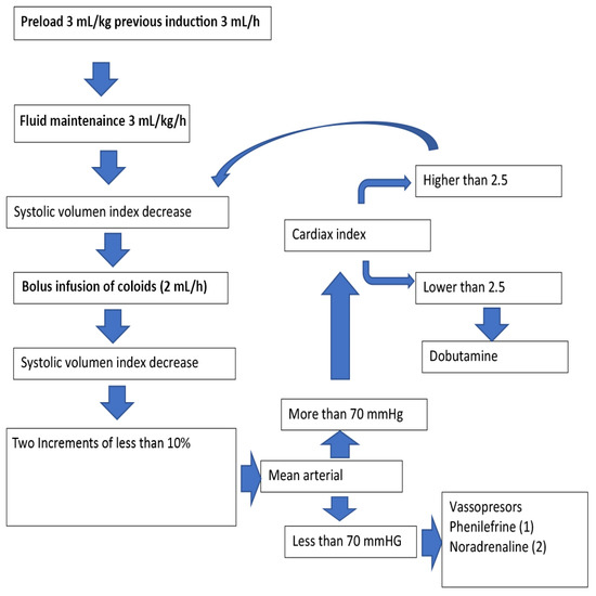
(1) Background: Surgical outcomes in free flap reconstruction of head and neck defects in cancer patients have improved steadily in recent years; however, correct anaesthesia management is also important. The aim of this study has been to show whether goal directed therapy can improve flap viability and morbidity and mortality in surgical patients. (2) Methods: we performed an observational case control study to analyse the impact of introducing a semi invasive device (Flo Trac®) during anaesthesia management to optimize fluid management. Patients were divided into two groups: one received goal directed therapy (GDT group) and the other conventional fluid management (CFM group). Our objective was to compare surgical outcomes, complications, fluid management, and length of stay between groups. (3) Results: We recruited 140 patients. There were no differences between groups in terms of demographic data. Statistically significant differences were observed in colloid infusion (GDT 53.1% vs. CFM 74.1%, p = 0.023) and also in intraoperative and postoperative infusion of crystalloids (CFM 5.72 (4.2, 6.98) vs. GDT 3.04 (2.29, 4.11), p < 0.001), which reached statistical significance. Vasopressor infusion in the operating room (CFM 25.5% vs. GDT 74.5%, p < 0.001) and during the first postoperative 24h (CFM 40.6% vs. GDT 75%, p > 0.001) also differed. Differences were also found in length of stay in the intensive care unit (hours: CFM 58.5 (40, 110) vs. GDT 40.5 (36, 64.5), p = 0.005) and in the hospital (days: CFM 15.5 (12, 26) vs. GDT 12 (10, 19), p = 0.009). We found differences in free flap necrosis rate (CMF 37.1% vs. GDT 13.6%, p = 0.003). One-year survival did not differ between groups (CFM 95.6% vs. GDT 86.8%, p = 0.08). (4) Conclusions: Goal directed therapy in oncological head and neck surgery improves outcomes in free flap reconstruction and also reduces length of stay in the hospital and intensive care unit, with their corresponding costs. It also appears to reduce morbidity, although these differences were not significant. Our results have shown that optimizing intraoperative fluid therapy improves postoperative morbidity and mortality. Full article
(This article belongs to the Special Issue Surgical Treatment of Head and Neck Squamous Cell Carcinomas (HNSCC))
Show Figures

Figure 1
8 pages, 490 KB  
Communication
Prognostic Evidence of the miRNA-Based Ovarian Cancer Signature MiROvaR in Independent Datasets
by Loris De Cecco, Marina Bagnoli, Paolo Chiodini, Sandro Pignata and Delia Mezzanzanica
Cancers 2021, 13(7), 1544; https://doi.org/10.3390/cancers13071544 - 27 Mar 2021
Cited by 7 | Viewed by 2556
Abstract
Epithelial ovarian cancer (EOC) remains the second most common cause of gynecological cancer deaths. To improve patients’ outcomes, we still need reliable biomarkers of early relapse, of which external independent validation is a crucial process. Our previously established prognostic signature, MiROvaR, including 35 [...] Read more.
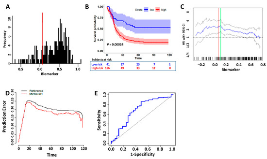
Epithelial ovarian cancer (EOC) remains the second most common cause of gynecological cancer deaths. To improve patients’ outcomes, we still need reliable biomarkers of early relapse, of which external independent validation is a crucial process. Our previously established prognostic signature, MiROvaR, including 35 microRNAs (miRNA) able to stratify EOC patients for their risk of relapse, was challenged on a new independent cohort of 197 EOC patients included in the Pelvic Mass Study whose miRNA profile was made publically available, thus resulting in the only accessible database aside from the EOC TCGA collection. Following accurate data matrix adjustment to account for the use of different miRNA platforms, MiROvaR confirmed its ability to discriminate early relapsing patients. The model’s original cutoff separated 156 (79.2%) high- and 41 (20.8%) low-risk patients with median progression free survival (PFS) of 16.3 months and not yet reached (NYR), respectively (hazard ratio (HR): 2.42–95% Confidence Interval (CI) 1.49–3.93; Log-rank p = 0.00024). The MiROvaR predictive accuracy (area under the curve (AUC) = 0.68; 95% Cl 0.57–0.79) confirms its prognostic value. This external validation in a totally independently collected, handled and profiled EOC cohort suggests that MiROvaR is a strong and reliable biomarker of EOC early relapse, warranting prospective validation. Full article
(This article belongs to the Special Issue Preclinical and Translational Research in Gynecological Cancers)
Show Figures

Figure 1
22 pages, 1326 KB  
Review
Apoptosis-Inducing TNF Superfamily Ligands for Cancer Therapy
by Olivia A. Diaz Arguello and Hidde J. Haisma
Cancers 2021, 13(7), 1543; https://doi.org/10.3390/cancers13071543 - 27 Mar 2021
Cited by 65 | Viewed by 6304
Abstract
Cancer is a complex disease with apoptosis evasion as one of its hallmarks; therefore, apoptosis induction in transformed cells seems a promising approach as a cancer treatment. TNF apoptosis-inducing ligands, which are naturally present in the body and possess tumoricidal activity, are attractive [...] Read more.
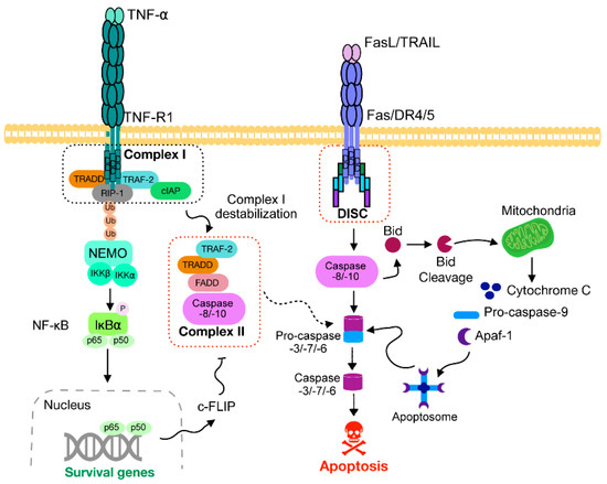
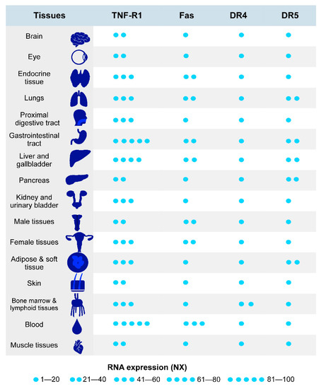
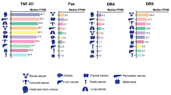
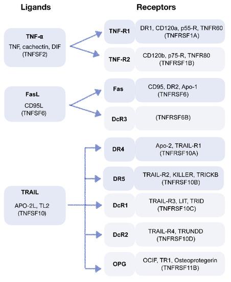
Cancer is a complex disease with apoptosis evasion as one of its hallmarks; therefore, apoptosis induction in transformed cells seems a promising approach as a cancer treatment. TNF apoptosis-inducing ligands, which are naturally present in the body and possess tumoricidal activity, are attractive candidates. The most studied proteins are TNF-α, FasL, and TNF-related apoptosis-inducing ligand (TRAIL). Over the years, different recombinant TNF family-derived apoptosis-inducing ligands and agonists have been designed. Their stability, specificity, and half-life have been improved because most of the TNF ligands have the disadvantages of having a short half-life and affinity to more than one receptor. Here, we review the outlook on apoptosis-inducing ligands as cancer treatments in diverse preclinical and clinical stages and summarize strategies of overcoming their natural limitations to improve their effectiveness. Full article
(This article belongs to the Section Cancer Therapy)
Show Figures

Graphical abstract

11 pages, 762 KB  
Article
Increased Visceral Adipose Tissue and Hyperinsulinemia Raise the Risk for Recurrence of Non-B Non-C Hepatocellular Carcinoma after Curative Treatment
by Kenji Imai, Koji Takai, Takao Miwa, Toshihide Maeda, Tatsunori Hanai, Makoto Shiraki, Atsushi Suetsugu and Masahito Shimizu
Cancers 2021, 13(7), 1542; https://doi.org/10.3390/cancers13071542 - 26 Mar 2021
Cited by 13 | Viewed by 3342
Abstract
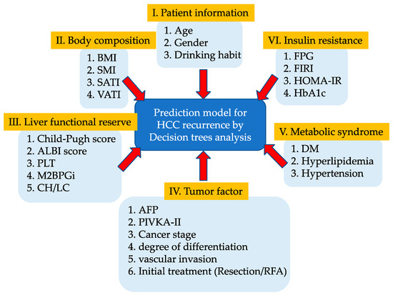
We investigated the factors affecting recurrence-free survival in patients with non-B non-C hepatocellular carcinoma (HCC) who received curative treatment. Decision-tree analysis was performed in 72 curative cases of non-B non-C HCC to extract the risk factors for recurrence. The reliability of the extracted [...] Read more.
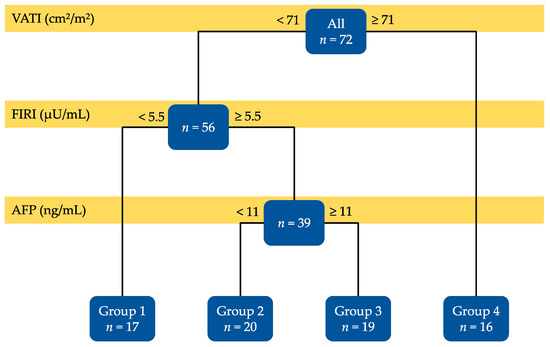
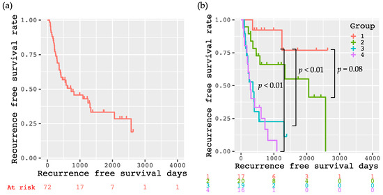
We investigated the factors affecting recurrence-free survival in patients with non-B non-C hepatocellular carcinoma (HCC) who received curative treatment. Decision-tree analysis was performed in 72 curative cases of non-B non-C HCC to extract the risk factors for recurrence. The reliability of the extracted risk factors was evaluated using the Kaplan–Meier method and the Cox proportional hazards model. The decision-tree analysis extracted three factors—visceral adipose tissue (VAT) index (VATI; <71 and ≥71 cm2/m2), which was the cross-sectional areas of VAT on the computed tomographic image at the umbilical level, normalized by the square of the height, fasting immunoreactive insulin (FIRI; <5.5 and ≥5.5 µU/mL), and alpha-fetoprotein (AFP; <11 and ≥11 ng/mL). The Cox proportional hazards model showed that VATI (hazard ratio (HR): 2.556, 95% confidence interval (CI): 1.191–5.486, p = 0.016), FIRI (HR: 3.149, 95% CI: 1.156–8.575, p = 0.025), and AFP (HR: 3.362, 95% CI: 1.550–7.288, p = 0.002) were all independent risk factors for HCC recurrence. Non-B non-C HCC patients with a higher VATI (≥71 cm2/m2) or higher FIRI (≥5.5 µU/mL) and AFP (≥11 ng/mL) if VATI was <71 cm2/m2 are prone to recurrence after curative treatment. Full article
(This article belongs to the Special Issue Translational Studies of Obesity-Associated Hepatocellular Cancer)
Show Figures

Figure 1
17 pages, 858 KB  
Review
Time to Move to the Single-Cell Level: Applications of Single-Cell Multi-Omics to Hematological Malignancies and Waldenström’s Macroglobulinemia—A Particularly Heterogeneous Lymphoma
by Ramón García-Sanz and Cristina Jiménez
Cancers 2021, 13(7), 1541; https://doi.org/10.3390/cancers13071541 - 26 Mar 2021
Cited by 12 | Viewed by 4094
Abstract
Single-cell sequencing techniques have become a powerful tool for characterizing intra-tumor heterogeneity, which has been reflected in the increasing number of studies carried out and reported. We have rigorously reviewed and compiled the information about these techniques inasmuch as they are relative to [...] Read more.
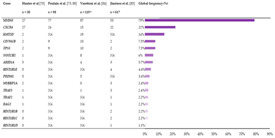
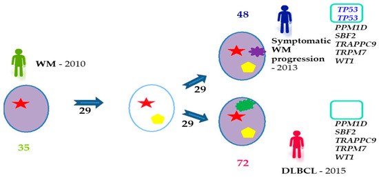
Single-cell sequencing techniques have become a powerful tool for characterizing intra-tumor heterogeneity, which has been reflected in the increasing number of studies carried out and reported. We have rigorously reviewed and compiled the information about these techniques inasmuch as they are relative to the area of hematology to provide a practical view of their potential applications. Studies show how single-cell multi-omics can overcome the limitations of bulk sequencing and be applied at all stages of tumor development, giving insights into the origin and pathogenesis of the tumors, the clonal architecture and evolution, or the mechanisms of therapy resistance. Information at the single-cell level may help resolve questions related to intra-tumor heterogeneity that have not been previously explained by other techniques. With that in mind, we review the existing knowledge about a heterogeneous lymphoma called Waldenström’s macroglobulinemia and discuss how single-cell studies may help elucidate the underlying causes of this heterogeneity. Full article
(This article belongs to the Special Issue Systems Biology and Intra-tumor Heterogeneity)
Show Figures

Figure 1
16 pages, 9242 KB  
Article
Prospective Validation of Indocyanine Green Lymphangiography Staging of Breast Cancer-Related Lymphedema
by Mads Gustaf Jørgensen, Navid Mohamadpour Toyserkani, Frederik Christopher Gulmark Hansen, Jørn Bo Thomsen and Jens Ahm Sørensen
Cancers 2021, 13(7), 1540; https://doi.org/10.3390/cancers13071540 - 26 Mar 2021
Cited by 33 | Viewed by 4245
Abstract
Indocyanine green lymphangiography (ICG-L) allows real-time investigation of lymphatics. Plastic surgeons performing lymphatic reconstruction use the ICG-L for patient selection and stratification using the MD Anderson (MDA) and the Arm Dermal Backflow (ADB) grading systems. However, the applicability of ICG-L in evaluating breast [...] Read more.
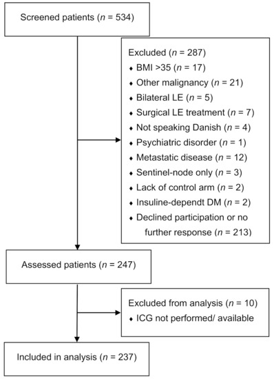
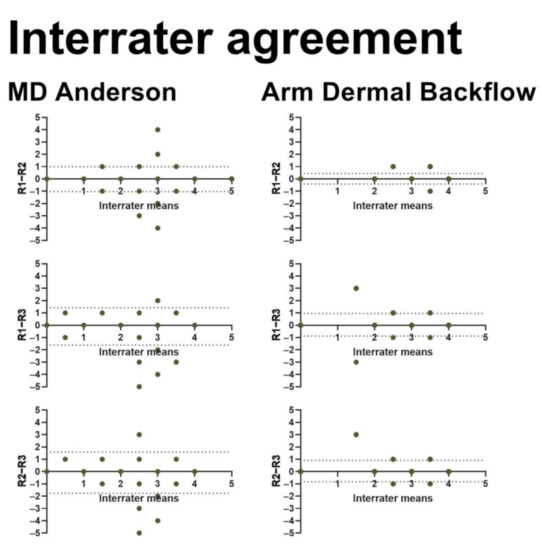
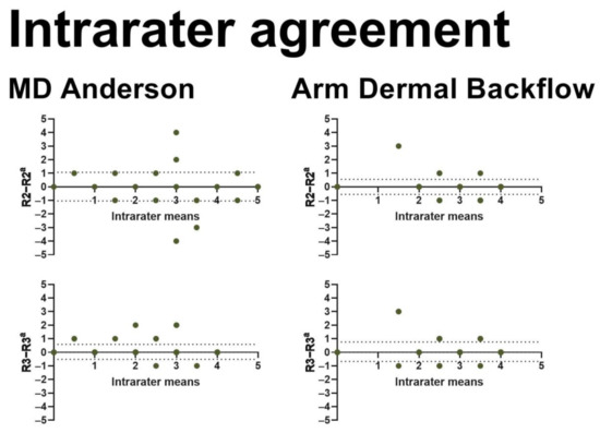
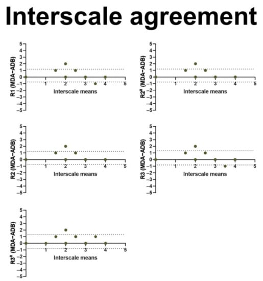
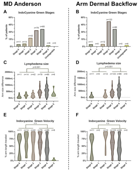
Indocyanine green lymphangiography (ICG-L) allows real-time investigation of lymphatics. Plastic surgeons performing lymphatic reconstruction use the ICG-L for patient selection and stratification using the MD Anderson (MDA) and the Arm Dermal Backflow (ADB) grading systems. However, the applicability of ICG-L in evaluating breast cancer-related lymphedema (BCRL) is sparse and not well established. This study comprehensively examines the usability of ICG-L in the assessment of BCRL. We prospectively performed ICG-L in 237 BCRL patients between January 2019 and February 2020. The aim of this study was to assess the interrater and intrarater agreement and interscale consensus of ratings made using the MDA and ADB scales. Three independent raters performed a total of 2607 ICG-L assessments. The ICG-L stage for each grading system was correlated to the lymphedema volume to assess the agreement between the ICG-L stage and clinical severity. The interrater agreement was near perfect for the MDA scale (kappa 0.82–0.90) and the ADB scale (kappa 0.80–0.91). Similarly, we found a near-perfect intrarater agreement for the MDA scale (kappa 0.84–0.94) and the ADB scale (kappa 0.88–0.89). The agreement between the MDA and the ADB scales was substantial (kappa 0.65–0.68); however, the ADB scale systematically overestimated lower ICG-L stages compared to the MDA scale. The volume of lymphedema correlated slightly with MDA stage (Spearmans rho = 0.44, p < 0.001) and ADB stage (rs = 0.35, p < 0.001). No serious adverse events occurred. The staging of BCRL with ICG-L is reliable, safe, and provides unique disease information unobtainable with clinical measurements alone. The MDA scale seems to provide better disease stratification compared to the ADB scale. Full article
(This article belongs to the Special Issue Cancer Related Lymphedema)
Show Figures

Figure 1
14 pages, 1252 KB  
Systematic Review
The Evolving Role of Marked Lymph Node Biopsy (MLNB) and Targeted Axillary Dissection (TAD) after Neoadjuvant Chemotherapy (NACT) for Node-Positive Breast Cancer: Systematic Review and Pooled Analysis
by Parinita K. Swarnkar, Salim Tayeh, Michael J. Michell and Kefah Mokbel
Cancers 2021, 13(7), 1539; https://doi.org/10.3390/cancers13071539 - 26 Mar 2021
Cited by 83 | Viewed by 7270
Abstract
Targeted axillary dissection (TAD) is a new axillary staging technique that consists of the surgical removal of biopsy-proven positive axillary nodes, which are marked (marked lymph node biopsy (MLNB)) prior to neoadjuvant chemotherapy (NACT) in addition to the sentinel lymph node biopsy (SLNB). [...] Read more.
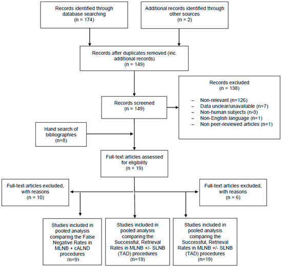
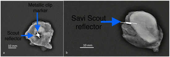
Targeted axillary dissection (TAD) is a new axillary staging technique that consists of the surgical removal of biopsy-proven positive axillary nodes, which are marked (marked lymph node biopsy (MLNB)) prior to neoadjuvant chemotherapy (NACT) in addition to the sentinel lymph node biopsy (SLNB). In a meta-analysis of more than 3000 patients, we previously reported a false-negative rate (FNR) of 13% using the SLNB alone in this setting. The aim of this systematic review and pooled analysis is to determine the FNR of MLNB alone and TAD (MLNB plus SLNB) compared with the gold standard of complete axillary lymph node dissection (cALND). The PubMed, Cochrane and Google Scholar databases were searched using MeSH-relevant terms and free words. A total of 9 studies of 366 patients that met the inclusion criteria evaluating the FNR of MLNB alone were included in the pooled analysis, yielding a pooled FNR of 6.28% (95% CI: 3.98–9.43). In 13 studies spanning 521 patients, the addition of SLNB to MLNB (TAD) was associated with a FNR of 5.18% (95% CI: 3.41–7.54), which was not significantly different from that of MLNB alone (p = 0.48). Data regarding the oncological safety of this approach were lacking. In a separate analysis of all published studies reporting successful identification and surgical retrieval of the MLN, we calculated a pooled success rate of 90.0% (95% CI: 85.1–95.1). The present pooled analysis demonstrates that the FNR associated with MLNB alone or combined with SLNB is acceptably low and both approaches are highly accurate in staging the axilla in patients with node-positive breast cancer after NACT. The SLNB adds minimal new information and therefore can be safely omitted from TAD. Further research to confirm the oncological safety of this de-escalation approach of axillary surgery is required. MLNB alone and TAD are associated with acceptably low FNRs and represent valid alternatives to cALND in patients with node-positive breast cancer after excellent response to NACT. Full article
Show Figures

Figure 1
27 pages, 1465 KB  
Review
Mechanisms of Resistance to PI3K Inhibitors in Cancer: Adaptive Responses, Drug Tolerance and Cellular Plasticity
by Sarah Christine Elisabeth Wright, Natali Vasilevski, Violeta Serra, Jordi Rodon and Pieter Johan Adam Eichhorn
Cancers 2021, 13(7), 1538; https://doi.org/10.3390/cancers13071538 - 26 Mar 2021
Cited by 85 | Viewed by 10811
Abstract
The phosphatidylinositol-3-kinase (PI3K) pathway plays a central role in the regulation of several signalling cascades which regulate biological processes such as cellular growth, survival, proliferation, motility and angiogenesis. The hyperactivation of this pathway is linked to tumour progression and is one of the [...] Read more.
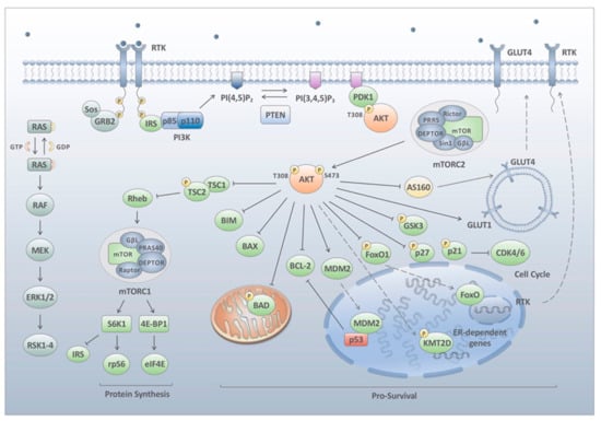
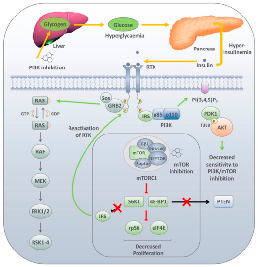
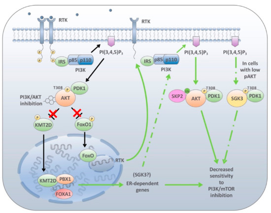
The phosphatidylinositol-3-kinase (PI3K) pathway plays a central role in the regulation of several signalling cascades which regulate biological processes such as cellular growth, survival, proliferation, motility and angiogenesis. The hyperactivation of this pathway is linked to tumour progression and is one of the most common events in human cancers. Additionally, aberrant activation of the PI3K pathway has been demonstrated to limit the effectiveness of a number of anti-tumour agents paving the way for the development and implementation of PI3K inhibitors in the clinic. However, the overall effectiveness of these compounds has been greatly limited by inadequate target engagement due to reactivation of the pathway by compensatory mechanisms. Herein, we review the common adaptive responses that lead to reactivation of the PI3K pathway, therapy resistance and potential strategies to overcome these mechanisms of resistance. Furthermore, we highlight the potential role in changes in cellular plasticity and PI3K inhibitor resistance. Full article
(This article belongs to the Special Issue Cellular Plasticity and the Untapped Therapeutic Potential in Cancer)
Show Figures

Figure 1