1
Department of Cardiology, West China Hospital of Sichuan University, Chengdu 610041, China
2
Key Laboratory of Obstetric & Gynecologic and Pediatric Diseases and Birth Defects of Ministry of Education, West China Second University Hospital, Sichuan University, Chengdu 610041, China
3
Laboratory of Molecular Translational Medicine, West China Institute of Women and Children's Health, West China Second University Hospital, Sichuan University, Chengdu 610041, China
4
Department of Forensic Pathology, Sichuan University, Chengdu 610041, China
†
These authors contributed equally to this work.
Int. J. Mol. Sci. 2015, 16(9), 22299-22318; https://doi.org/10.3390/ijms160922299 - 15 Sep 2015
Cited by 10 | Viewed by 6197
Abstract
Nicotinamide phosphoribosyltransferase (NAMPT) has crucial roles for myocardial development, cardiomyocyte energy metabolism and cell death/survival by regulating NAD+-dependent sirtuin-1 (SIRT1) deacetylase. This study aimed to determine if the single nucleotide polymorphisms (SNPs) of the NAMPT gene may affect the susceptibility and
[...] Read more.
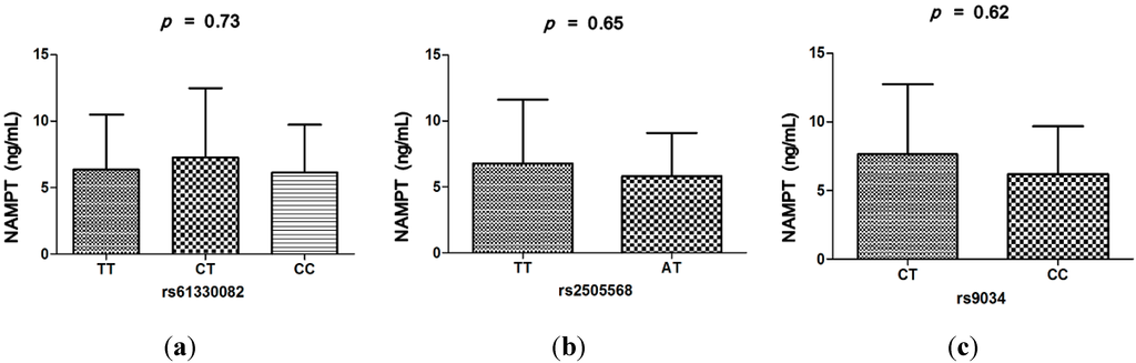
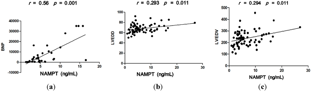
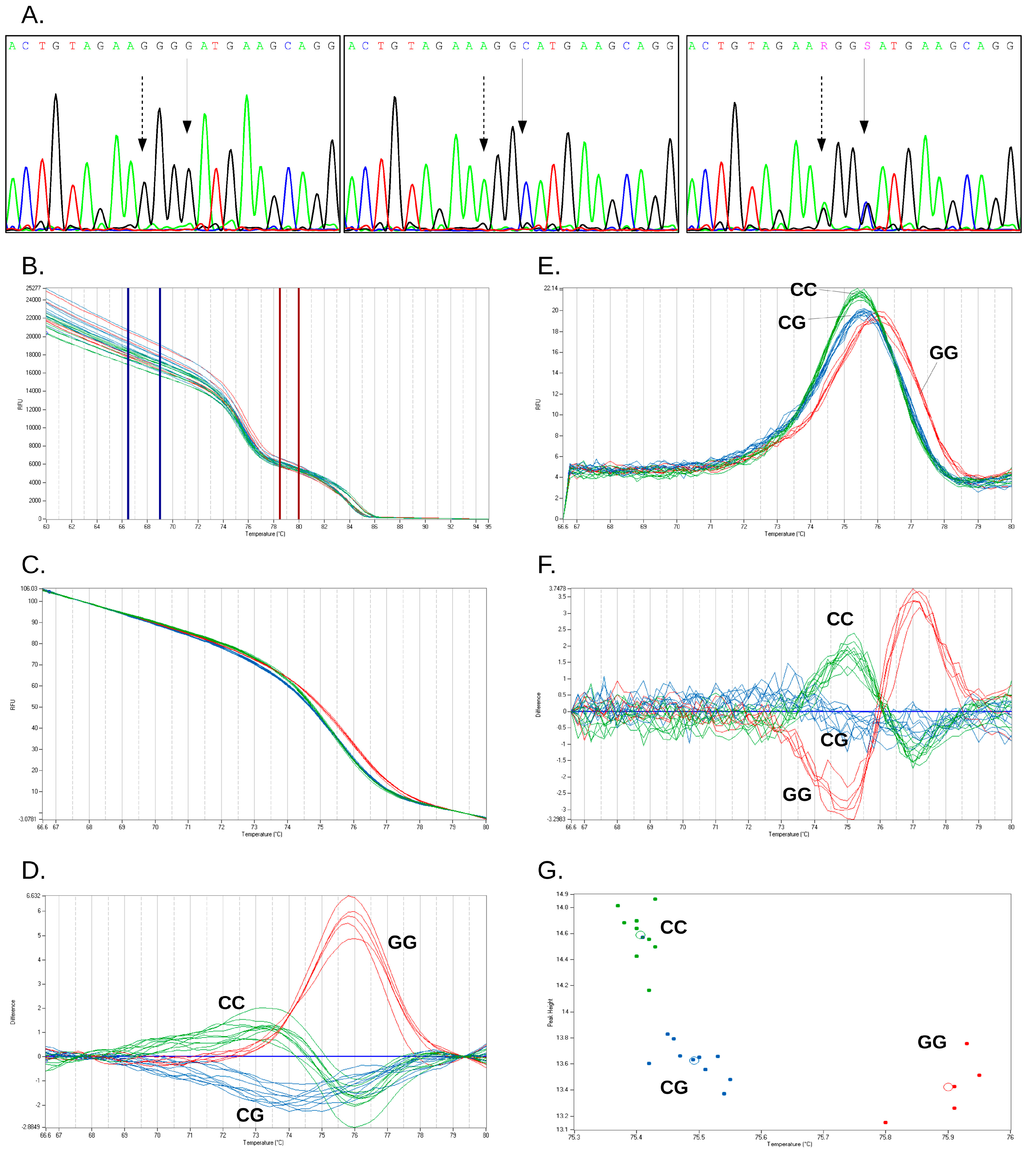
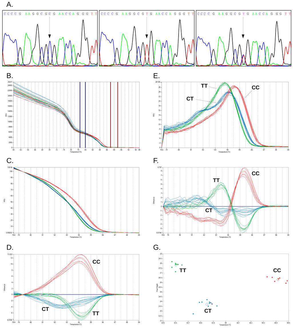
Nicotinamide phosphoribosyltransferase (NAMPT) has crucial roles for myocardial development, cardiomyocyte energy metabolism and cell death/survival by regulating NAD+-dependent sirtuin-1 (SIRT1) deacetylase. This study aimed to determine if the single nucleotide polymorphisms (SNPs) of the NAMPT gene may affect the susceptibility and prognosis for patients with dilated cardiomyopathy (DCM) and to describe the association of serum NAMPT levels with clinical features of DCM. Three SNPs (rs61330082, rs2505568, and rs9034) were analyzed by the polymerase chain reaction-restriction fragment length polymorphism method in a case-control study of 394 DCM patients and 395 controls from China. Serum NAMPT levels were measured by enzyme-linked immunosorbent assay kits. The homozygote for the minor allele at rs2505568 and rs9034 could not be detected in this study. Rs9034 T allele and CT genotype were associated with increased DCM risk (OR: 1.63, 95% CI = 1.16–2.27, p = 0.005 and OR: 1.72, 95% CI = 1.20–2.50, p = 0.0027, respectively). Nominally significant decreased DCM risk was found to be associated with the A allele and AT genotype of rs2505568 (OR: 0.48, 95% CI = 0.35–0.67, p < 0.0001 and OR: 0.44, 95% CI = 0.31–0.62, p < 0.0001, respectively), but it should be interpreted with caution because of Hardy-Weinberg disequilibrium in the control group. Of five haplotypes constructed, TAC (rs61330082-rs2505568-rs9034) was a protective haplotype to DCM (OR: 0.22, 95% CI = 0.13–0.39, p = 1.84 × 10⁻⁸). The Cox multivariate survival analysis indicated that the rs9034 CT genotype (hazard ratio (HR): 0.59, 95% CI = 0.37–0.96, p = 0.03) was an independently multivariate predictor for longer overall survival in DCM patients. Serum NAMPT levels were significantly higher in the DCM group than controls (p < 0.0001) and gradually increased with the increase of New York Heart Association grade in DCM patients. However, there was a lack of association of the three SNPs with serum NAMPT levels. Spearman correlation test revealed that the NAMPT level was positively associated with brain natriuretic peptide (r = 0.56, p = 0.001), left ventricular end-diastolic diameter (r = 0.293, p = 0.011) and left ventricular end-diastolic volume (r = 0.294, p = 0.011). Our study suggested that NAMPT may play an important role in the development of DCM.
Full article
(This article belongs to the Special Issue Human Single Nucleotide Polymorphisms and Disease Diagnostics)
▼
Show Figures
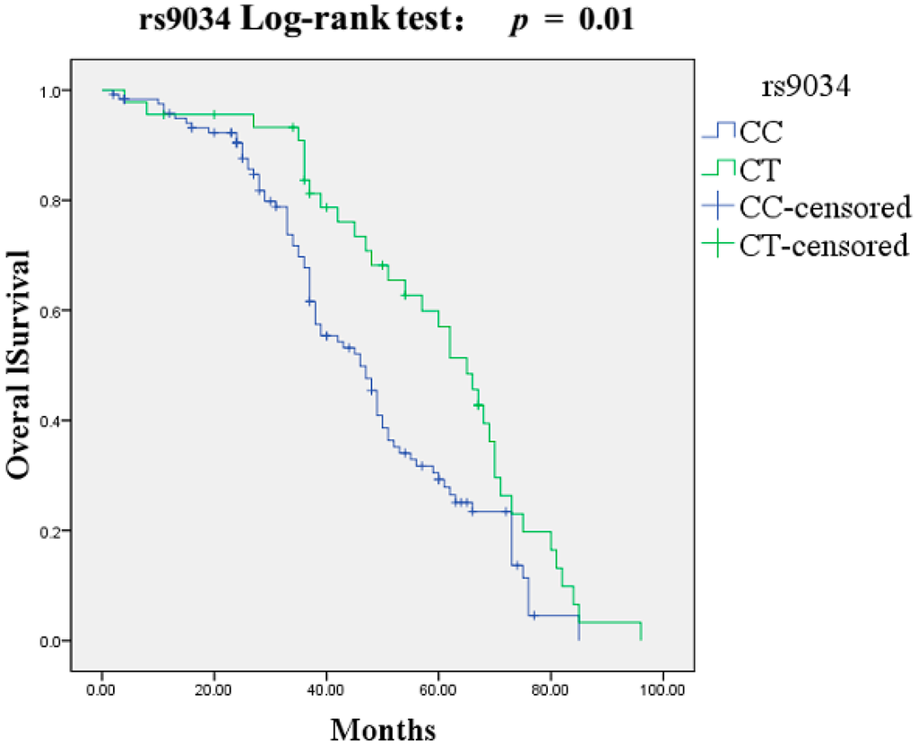
Figure 1


