


Cancers 2024, 16(9), 1706; https://doi.org/10.3390/cancers16091706 (registering DOI) - 27 Apr 2024
Abstract
FAM46C is a well-established tumour suppressor with a role that is not completely defined or universally accepted. Although FAM46C expression is down-modulated in several tumours, significant mutations in the FAM46C gene are only found in multiple myeloma (MM). Consequently, its tumour suppressor activity
[...] Read more.
FAM46C is a well-established tumour suppressor with a role that is not completely defined or universally accepted. Although FAM46C expression is down-modulated in several tumours, significant mutations in the FAM46C gene are only found in multiple myeloma (MM). Consequently, its tumour suppressor activity has primarily been studied in the MM context. However, emerging evidence suggests that FAM46C is involved also in other cancer types, namely colorectal, prostate and gastric cancer and squamous cell and hepatocellular carcinoma, where FAM46C expression was found to be significantly reduced in tumoural versus non-tumoural tissues and where FAM46C was shown to possess anti-proliferative properties. Accordingly, FAM46C was recently proposed to function as a pan-cancer prognostic marker, bringing FAM46C under the spotlight and attracting growing interest from the scientific community in the pathways modulated by FAM46C and in its mechanistic activity. Here, we will provide the first comprehensive review regarding FAM46C by covering (1) the intracellular pathways regulated by FAM46C, namely the MAPK/ERK, PI3K/AKT, β-catenin and TGF-β/SMAD pathways; (2) the models regarding its mode of action, specifically the poly(A) polymerase, intracellular trafficking modulator and inhibitor of centriole duplication models, focusing on connections and interdependencies; (3) the regulation of FAM46C expression in different environments by interferons, IL-4, TLR engagement or transcriptional modulators; and, lastly, (4) how FAM46C expression levels associate with increased/decreased tumour cell sensitivity to anticancer agents, such as bortezomib, dexamethasone, lenalidomide, pomalidomide, doxorubicin, melphalan, SK1-I, docetaxel and norcantharidin.
Full article
(This article belongs to the Special Issue Unique Perspectives in Cancer Signaling)
►
Show Figures
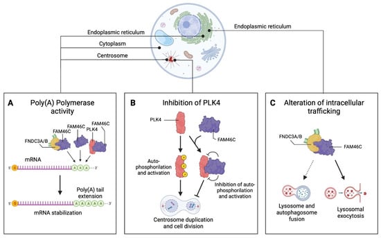
Figure 1


