Biotelemetric Monitoring of Brain Neurochemistry in Conscious Rats Using Microsensors and Biosensors

 , ,
, , 

Abstract
:1. Introduction
2. Results and Discussion
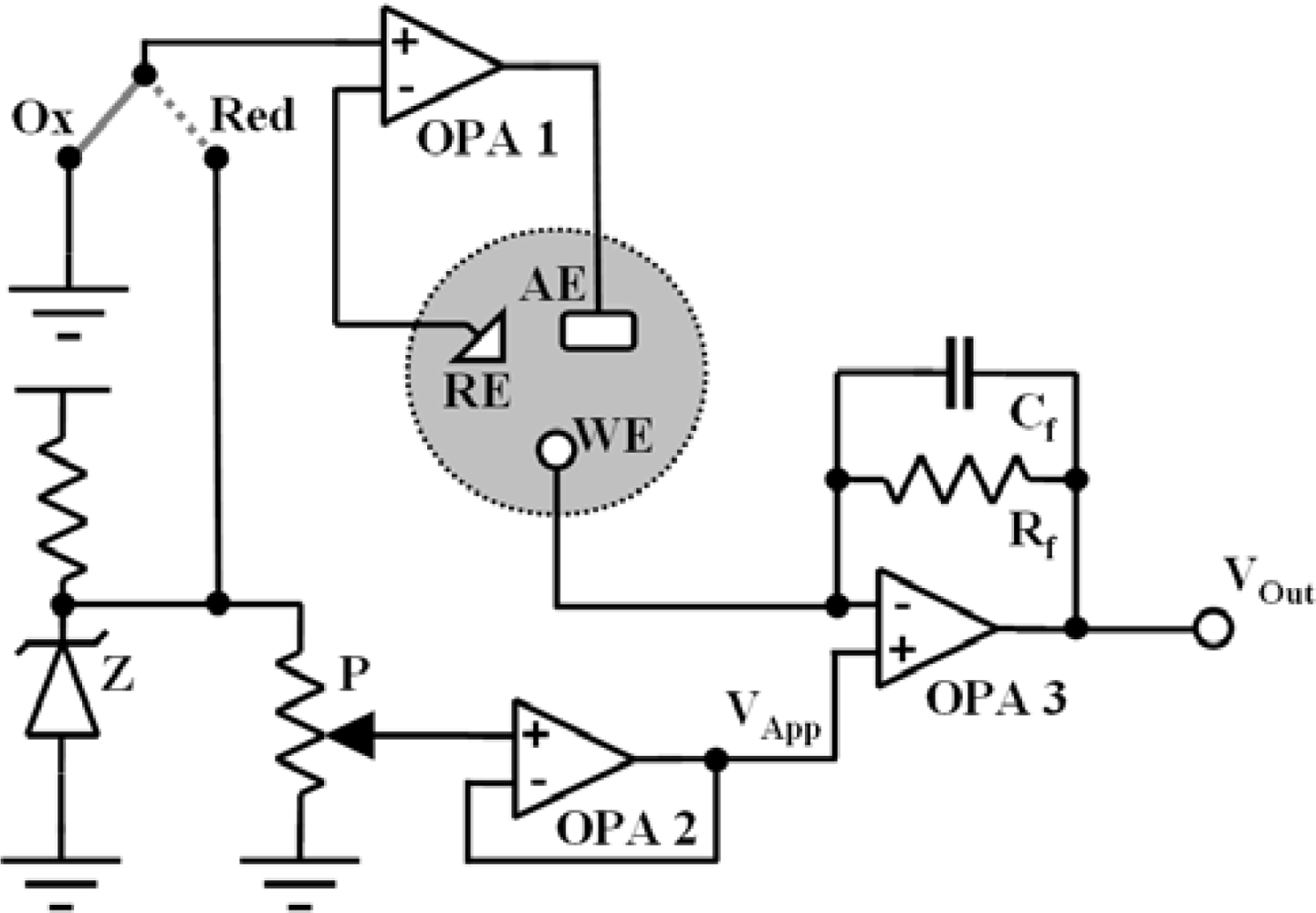
2.1. Biotelemetric device test and calibration
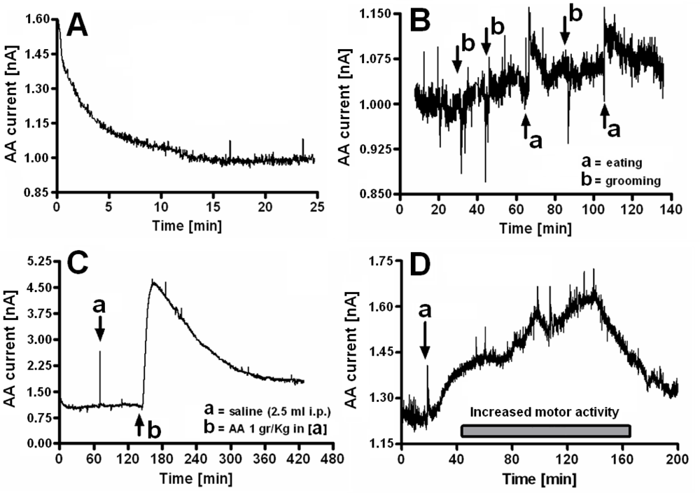
2.2. In-vitro calibration of ascorbic acid microsensor and in-vivo results
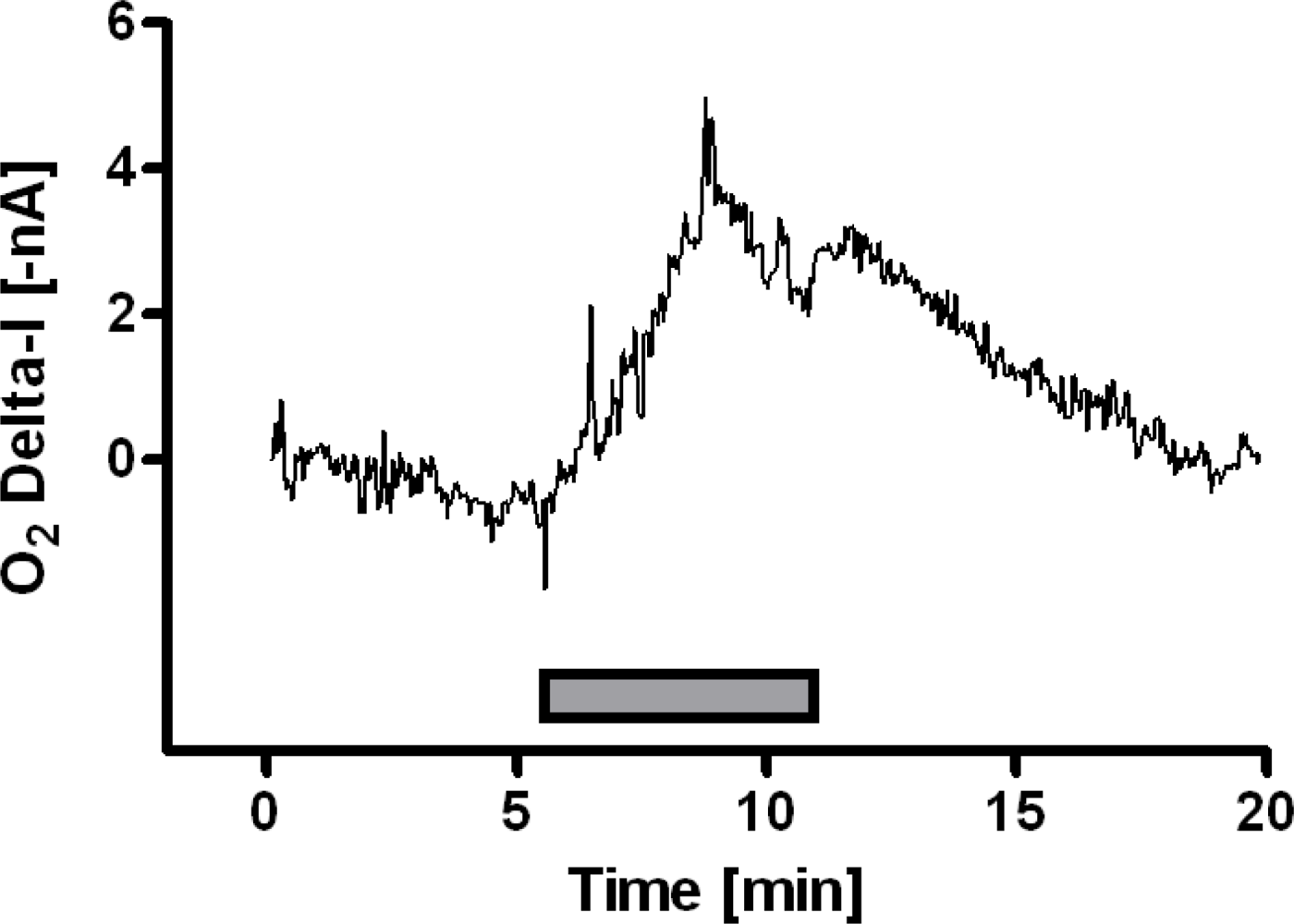
2.3. In-vitro calibration of oxygen microsensor and in-vivo results
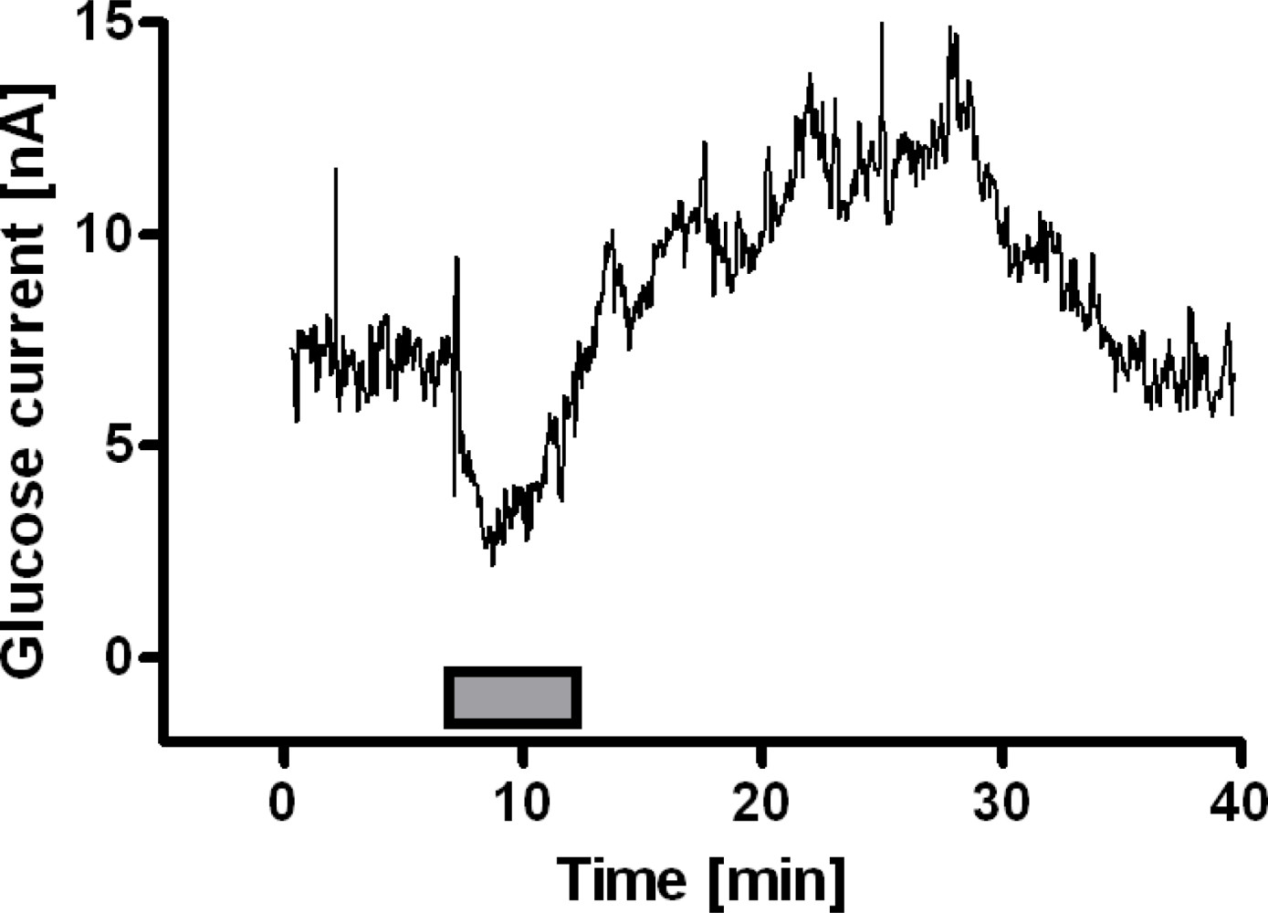
2.4. In-vitro calibration of glucose biosensor and in-vivo results
3. Experimental Section
3.1. Reagents, solutions and electronic parts
3.2. Biotelemetric device
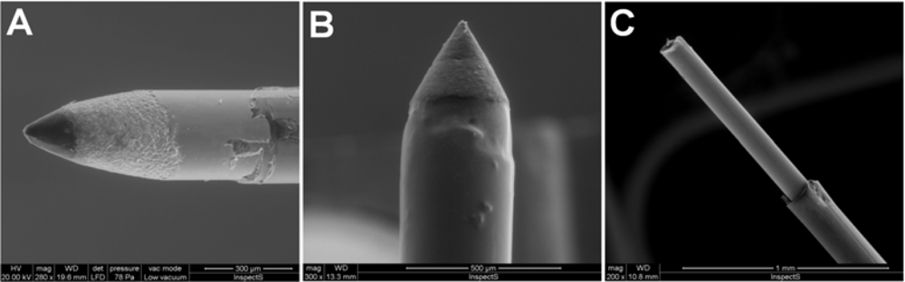
3.3. Preparation and calibration of microsensors and biosensors
3.4. Animals, stereotaxic surgery and in-vivo experimental procedures
3.5. Hystology
3.6. Statistical analysis
4. Conclusions
Acknowledgments
References
- Emerit, J.; Edeas, M. Neurodegenerative diseases and oxidative stress. Eur. Neuropsychopharmacol 2005, 15, S100–S101. [Google Scholar]
- Andersen, J.K. Oxidative stress in neurodegeneration: cause or consequence? Nat. Med 2004, 10, S18–S25. [Google Scholar]
- Serra, P.A.; Sciola, L.; Delogu, M.R.; Spano, A.; Monaco, G.; Miele, E.; Rocchitta, G.; Miele, M.; Migheli, R.; Desole, M.S. The neurotoxin 1-methyl-4-phenyl-1,2,3,6-tetrahydropyridine induces apoptosis in mouse nigrostriatal glia. Relevance to nigral neuronal death and striatal neurochemical changes. J Biol Chem 2002, 277, 34451–34461. [Google Scholar]
- Hediger, M.A. New view at C. Nat. Med 2002, 8, 445–446. [Google Scholar]
- O’Neill, R.D.; Fillenz, M.; Sundstrom, L.; Rawlins, J.N.P. Voltammetrically monitored brain ascorbate as an index of excitatory amino acid release in the unrestrained rat. Neurosci. Lett 1984, 52, 227–233. [Google Scholar]
- Rice, M.E. Ascorbate regulation and its neuroprotective role in the brain. Trends Neurosci 2000, 23, 209–216. [Google Scholar]
- Watanabe, Y.; Hyllbrant, B.B.; Langstrom, B. Tracing oxygen metabolism by use of positron emitter Oxygen-15. Biochem. Biophys. Res. Commun 1997, 231, 131–134. [Google Scholar]
- Bazzu, G.; Puggioni, G.G.; Dedola, S.; Calia, G.; Rocchitta, G.; Migheli, R.; Desole, M.S.; Lowry, J.P.; O'Neill, R.D.; Serra, P.A. Real-time monitoring of brain tissue oxygen using a miniaturized biotelemetric device implanted in freely-moving rats. Anal. Chem 2009. [Google Scholar] [CrossRef]
- Fillenz, M. The role of lactate in brain metabolism. Neurochem. Int 2005, 47, 413–417. [Google Scholar]
- Aubert, A.; Costalat, R.; Magistretti, P.J.; Pellerin, L. Brain lactate kinetics: modeling evidence for neuronal lactate uptake upon activation. Proc. Natl. Acad. Sci. USA 2005, 102, 16448–16453. [Google Scholar]
- Lowry, J.P.; Miele, M.; O’Neill, R.D.; Boutelle, M.G.; Fillenz, M. An amperometric glucose oxidase/poly(o-phenylenediamine) biosensor for monitoring brain extracellular glucose: In vivo characterisation in the striatum striatum of freely-moving rats. J. Neurosci. Methods 1998, 79, 65–74. [Google Scholar]
- Magistretti, P.J.; Pellerin, L.; Rothman, D.L.; Shulman, R.G. Energy on demand. Science 1999, 283, 496–497. [Google Scholar]
- Pfeiffer, D.; Schubert, F.; Wollenberger, U.; Scheller, F.W. Electrochemical sensors: Enzyme electrodes and field effect transistors. In Handbook of Chemical and Biological Sensors; Taylor, R.F., Schultz, J.S., Eds.; IOP Publishing Ltd: Bristol, UK, 1996; pp. 435–458. [Google Scholar]
- Wilson, R.; Turner, A.P.F. Glucose oxidase: an ideal enzyme. Biosens. Bioelectron 1992, 7, 165–185. [Google Scholar]
- Zhou, R.J; Hao, Z.Q. The present status and development of biotelemetry. Zhongguo Yi Liao Qi Xie Za Zhi 2002, 26, 212–214. [Google Scholar]
- FCC (Federal Communication Commission). Commission’s Rules to Create a Wireless Medical Telemetry Service; FCC: Washington, DC, USA, 2000; No. FCC 00-211,; pp. 1–24. [Google Scholar]
- Leuher, D.C. Overview of biomedical telemetry techniques. Eng. Med. Biol 1983, 3, 17–24. [Google Scholar]
- Serra, P.; Hebel, M.; Rocchitta, G.; Tate, R. Biotelemetry NET for neurochemical biosensor and microsensor applications: design, construction and validation. In Telemetry: Research, Technology and Applications; Barculo, D., Daniels, J., Eds.; Nova Science Publishers Inc: Hauppauge, NY, USA, 2009. [Google Scholar]
- Hebel, M.; Serra, P.A. Development of a parallel-computing embedded telemetry system for voltammetric microsensor and biosensor applications. In Sensors for Environment, Health and Security: Advanced Materials and Technologies; Baraton, M.I., Ed.; Springer: Dordrecht, The Netherlands, 2008; pp. 229–238. [Google Scholar]
- Rocchitta, G.; Migheli, R.; Dedola, S.; Calia, G.; Desole, M.S.; Miele, E.; Lowry, J.P.; O’Neill, R.D.; Serra, P.A. Development of a distributed, fully automated, bidirectional telemetry system for amperometric microsensor and biosensor applications. Sens. Actuat. B 2007, 126, 700–709. [Google Scholar]
- Serra, P.A.; Rocchitta, G.; Bazzu, G.; Manca, A.; Puggioni, G.M.; Lowry, J.P.; O’Neill, R.D. Design and construction of a low cost single-supply embedded telemetry system for amperometric biosensor applications. Sens. Actuat B 2007, 122, 118–126. [Google Scholar]
- Wu, L.; Zhang, X.; Ju, H. Amperometric glucose sensor based on catalytic reduction of dissolved oxygen at soluble carbon nanofiber. Biosens. Bioelectron 2007, 23, 479–484. [Google Scholar]
- Morita, H.; Abe, C.; Awazu, C.; Tanaka, K. Long-term hypergravity induces plastic alterations in vestibulo-cardiovascular reflex in conscious rats. Neurosc. Lett 2007, 412, 201–205. [Google Scholar]
- Leon, L.R.; Walker, L.D.; DuBose, D.A.; Stephenson, L.A. Biotelemetry transmitter implantation in rodents: impact on growth and circadian rhythms. Am. J. Physiol. Regul. Integr. Comp. Physiol 2004, 286, R967–R974. [Google Scholar]
- Miele, M.; Fillenz, M. In vivo determination of extracellular brain ascorbate. J. Neurosci. Methods 1996, 70, 15–19. [Google Scholar]
- Miele, M.; Mura, M.A.; Enrico, P.; Esposito, G.; Serra, P.A.; Migheli, R.; Zangani, D.; Miele, E.; Desole, M.S. On the mechanism of d-amphetamine-induced changes in glutamate, ascorbic acid and uric acid release in the striatum of freely-moving rats. Br. J. Pharmacol 2000, 129, 582–588. [Google Scholar]
- Hediger, M.A. Transporters for vitamin C keep vitamin concentrations optimal in the body. A new mouse knockout of one transporter reveals previously unknown requirements for the vitamin. Nat. Med 2002, 8, 514–517. [Google Scholar]
- Lowry, J.P.; Boutelle, M.G.; Fillenz, M. Measurement of brain tissue oxygen at a carbon paste electrode can serve as an index of increases in regional cerebral blood flow. J. Neurosci. Methods 1997, 71, 177–182. [Google Scholar]
- Bolger, F.B.; Lowry, J.P. Brain tissue oxygen: In vivo monitoring with carbon paste electrodes. Sensors 2005, 5, 473–487. [Google Scholar]
- Lowry, J.P.; Fillenz, M. Real-time monitoring of brain energy metabolism in vivo using microelectrochemical sensors: the effects of anesthesia. Bioelectrochemistry 2001, 54, 39–47. [Google Scholar]
- Lowry, J.P.; Fillenz, M. Evidence for uncoupling of oxygen and glucose utilization during neuronal activation in rat striatum. J. Physiol 1997, 498, 497–501. [Google Scholar]
- Dixon, B.M.; Lowry, J.P.; O’Neill, R.D. Characterization in vitro and in vivo of the oxygen dependence of an enzyme/polymer biosensor for monitoring brain glucose. J. Neurosci. Methods 2002, 119, 135–142. [Google Scholar]
- Fillenz, M.; Lowry, J.P.; Boutelle, M.G.; Fray, A.E. The role of astrocytes and noradrenaline in neuronal glucose metabolism. Acta Physiol. Scand 1999, 167, 275–284. [Google Scholar]
- Migheli, R.; Puggioni, G.; Dedola, S.; Rocchitta, G.; Calia, G.; Bazzu, G.; Esposito, G.; Lowry, J.P.; O’Neill, R.D.; Desole, M.S.; Miele, E.; Serra, P.A. Novel integrated microdialysis–amperometric system for in vitro detection of dopamine secreted from PC12 cells: design, construction, and validation. Anal. Biochem 2008, 380, 323–330. [Google Scholar]
- Duff, A.; O’Neill, R.D. Effect of probe size on the concentration of brain extracellular uric acid monitored with carbon paste electrodes. J. Neurochem 1994, 62, 1496–1502. [Google Scholar]
- Fumero, B.; Guadalupe, T.; Valladares, F.; Mora, F.; O'Neill, R.D.; Mas, M.; Gonzalez-Mora, J.L. Fixed versus removable microdialysis probes for in vivo neurochemical analysis: implications for behavioral studies. J. Neurochem 1994, 63, 1407–1415. [Google Scholar]
- Ryan, M.R.; Lowry, J.P.; O'Neill, R.D. Biosensor for neurotransmitter L-glutamic acid designed for efficient use of L-glutamate oxidase and effective rejection of interference. Analyst 1997, 122, 1419–1424. [Google Scholar]
- Wolfensohn, S.; Lloyd, M. Handbook of Laboratory Animal Management and Welfare, 3rd Ed ed; Blackwell Publishing: Cornwall, ON, Canada, 2003. [Google Scholar]
- Paxinos, G.; Watson, C. The Rat Brain in Stereotaxic Coordinates, 6th Ed ed; Academic Press: San Diego, CA, USA, 2007. [Google Scholar]
- Lowry, J.P.; Ryan, M.R.; O’Neill, R.D. Behaviourally induced changes in extracellular levels of brain glutamate monitored at 1 s resolution with an implanted biosensor. Anal. Commun 1998, 35, 87–89. [Google Scholar]


| Interference | AA microsensor (n = 4) | O2 microsensor (n = 4) | Glucose biosensor (n = 6) |
|---|---|---|---|
| AA (500 μM) | 3.65 ± 0.4 nA | N.D. | 0.73 ± 0.2 nA |
| DOPAC (10 μM) | 31 ± 6 pA | N.D. | 22 ± 5 pA |
| UA (10 μM) | 16 ± 7 pA | N.D. | 27 ± 9 pA |
| DA (1 μM) | N.D. | N.D. | 55 ± 11 pA |
© 2009 by the authors; licensee MDPI, Basel, Switzerland This article is an open access article distributed under the terms and conditions of the Creative Commons Attribution license (http://creativecommons.org/licenses/by/3.0/).
Share and Cite
Calia, G.; Rocchitta, G.; Migheli, R.; Puggioni, G.; Spissu, Y.; Bazzu, G.; Mazzarello, V.; Lowry, J.P.; O’Neill, R.D.; Desole, M.S.; et al. Biotelemetric Monitoring of Brain Neurochemistry in Conscious Rats Using Microsensors and Biosensors. Sensors 2009, 9, 2511-2523. https://doi.org/10.3390/s90402511
Calia G, Rocchitta G, Migheli R, Puggioni G, Spissu Y, Bazzu G, Mazzarello V, Lowry JP, O’Neill RD, Desole MS, et al. Biotelemetric Monitoring of Brain Neurochemistry in Conscious Rats Using Microsensors and Biosensors. Sensors. 2009; 9(4):2511-2523. https://doi.org/10.3390/s90402511
Chicago/Turabian StyleCalia, Giammario, Gaia Rocchitta, Rossana Migheli, Giulia Puggioni, Ylenia Spissu, Gianfranco Bazzu, Vittorio Mazzarello, John P. Lowry, Robert D. O’Neill, Maria S. Desole, and et al. 2009. "Biotelemetric Monitoring of Brain Neurochemistry in Conscious Rats Using Microsensors and Biosensors" Sensors 9, no. 4: 2511-2523. https://doi.org/10.3390/s90402511