Article

Synthesis and In Vitro Antiproliferative Activity of Novel Phenyl Ring-Substituted 5-Alkyl-12(H)-quino[3,4-b][1,4]benzothiazine Derivatives

1
Department of Organic Chemistry, School of Pharmacy with the Division of Laboratory Medicine in Sosnowiec, Medical University of Silesia in Katowice, Jagiellońska 4, 41-200 Sosnowiec, Poland
2
Department of Cell Biology, School of Pharmacy with the Division of Laboratory Medicine in Sosnowiec, Medical University of Silesia in Katowice, Jedności 9, 41-200 Sosnowiec, Poland
3
Center for Translational Research and Molecular Biology of Cancer, Maria Skłodowska-Curie Memorial Cancer Center and Institute of Oncology, Wybrzeże AK 15, 44-101 Gliwice, Poland
*
Author to whom correspondence should be addressed.
Molecules 2016, 21(11), 1455; https://doi.org/10.3390/molecules21111455
Submission received: 5 October 2016 / Revised: 20 October 2016 / Accepted: 24 October 2016 / Published: 4 November 2016
(This article belongs to the Section Medicinal Chemistry)

Abstract

:
A novel series of tetracyclic quinobenzothiazine derivatives was synthetized. Compounds containing a substituent (hydroxyl, methyl, phenyl, piperidyl, or piperazinyl) in positions 9 and 11 were obtained by cyclization of suitable 4-aminoquinolinium-3-thiolates. Quinobenzothiazine 10-O-substituted derivatives were obtained by alkylating the hydroxyl group in position 10 of the parent (quinobenzothiazine) system. Antiproliferative activity of the synthesized compounds was studied using cultured neoplastic cells (MDA-MB-231, SNB-19, and C-32 cell lines). Four selected compounds were investigated in more detail for cytotoxicity and antiproliferative effect. Transcriptional activity of genes regulating cell cycle (TP53), apoptosis (BAX, BCL-2), as well as proliferation (H3) were assessed. Finally, the ability of the selected compounds to bind DNA was checked in the presence of ethidium bromide.

Graphical Abstract

1. Introduction

Initial attempts of using phenothiazine derivatives as antimalarial agents go back to 1891 when Guttman and Ehrlich demonstrated chemotherapeutic effectiveness of methylene blue. Extensive search for antimalarial agents was undertaken later, starting with modification of the methylene blue structure via substitution of the N-methyl group with alkylaminoalkyl moieties [1]. Studies concerning antihistamine activity led to phenothiazine derivatives that contain alkylaminoalkyl substituents at the thiazine nitrogen atom. Compounds containing such a structural fragment are important neuroleptic agents. Their main representative, and a reference compound, is chlorpromazine [2]. Phenothiazines are typical examples of how modification of a basic structural fragment can affect directional activity and drug strength. Search for new therapeutics has been conducted more and more often by modifying the main structural fragment of known drugs. Since the advent of chlorpromazine, the quest for new phenothiazine derivatives has yielded several thousand compounds with novel properties and applications. Exchanging benzene rings for nitrogen-containing heterocycles has led to a series of novel azaphenothiazine derivatives featuring tri-, tetra- and pentacyclic systems [3,4,5]. Structural modifications of phenothiazine and azaphenothiazine have been achieved by introducing substituents and functional moieties mainly at the thiazine nitrogen or, less often, into the benzene ring or nitrogen-containing heterocycles. The compounds reported so far exhibit neuroleptic, antimalarial, immunopotentiating, antibacterial, antiviral, antifungal, antiproliferative, or antitumor activities and can mediate the reversal of multidrug resistance [6,7,8,9,10,11,12,13]. In our earlier papers we reported a novel method of synthesizing the 1,4-thiazine ring which proceeds via the substitution of a hydrogen atom with a thiolate sulfur atom. The obtained compounds have demonstrated promising anticancer properties [14,15,16,17,18]. Herein, we report on the synthesis of novel tetracyclic phenothiazine derivatives containing various substituents (aliphatic, aromatic, or heterocyclic) in positions 9, 10, and 11 of the quinobenzothiazine system. Using selected neoplastic cell lines, we demonstrate the anticancer activity of these compounds and present results shedding light on their underlying mechanism of action.

2. Results and Discussion

2.1. Chemistry

Cyclization of diphenylamines and azinylphenylamines in the presence of sulfur and its compounds, as well as cyclization of diphenyl or azinylphenyl sulfides are commonly used methods for the synthesis of phenothiazine and azaphenothiazine systems [2]. Our method of obtaining quinobenzothiazine tetracyclic derivatives is based on cyclization of betaine systems featuring 1-alkyl-4-(arylamino)quinolinium-3-thiolates structure via nucleophilic substitution of the hydrogen or halogen atom in the phenyl ring by a thiolate-derived sulfur atom. Reactions occur with high yield even at room temperature. They permit modification of the quinothiazine system by introducing substituents or functional groups into the benzene ring; this remains difficult when using other methods of synthesis [14,15,16]. Substrates which are suitable for obtaining 1-alkyl-4-arylaminoquinoline-3-thiolates are 5,12-(dialkyl)thioquinantrene salts 1 [19] as well as 1-alkyl-4-arylamino-3-(acylthio)quinolinium salts [20]. In this report we show synthesis of azaphenothiazine derivatives containing different types of aliphatic, aromatic and heterocyclic substituents in positions 9, 10 and 11 of the quinobenzothiazine system. The course of reaction between bis-chloride 1 with 2-, 3-, or 4-methoxyaniline, 4-piperidinylaniline, 4-piperazinylaniline, and 2-hydroxy-4-phenylaniline (Scheme 1) is dependent on the presence of oxygen in the reaction mixture.
When carried out at room temperature with pyridine as a solvent and under conditions disallowing oxygen access to the reaction milieu. These reactions led to 1-methyl-4-(arylamine)quinolinium-3-thiolates 2af. Betaines 2ae underwent cyclization in the presence of a hydrogen chloride donor and atmospheric oxygen to appropriate quinobenzothiazine chlorides 3ae (procedure A). Compounds 3ae can also be obtained directly from bis-chloride 1 using suitable amines and a carrying one-pot type reaction in the presence of atmospheric oxygen and without separating intermediate products 2ae (procedure B).
The reaction of 1,4-thiazine ring formation during cyclization of quinolinium-3-thiolate 2f obtained by reacting bis salt 1 with 3-methoxyaniline can occur via hydrogen atom substitution in positions 2 or 6 of the phenyl ring. 1H-NMR analysis has demonstrated the reaction product to be the mixture of isomers 3f and 3g (which form at a ca. 1:1 quantitative ratio) resulting from hydrogen atom substitution in both positions of the phenyl ring (Scheme 2). Compounds 3f and 3g obtained using the above method cannot be separated to purity.
In order to obtain quinobenzothiazine derivatives containing alkoxy and aminoalkoxy substituents in the position 10 of the quinobenzothiazine ring, we used 10-hydroxy-5-methyl-12(H)-quino[3,4-b][1,4]benzothiazinium chloride 3h. Its synthesis was reported earlier [16]. Alkalization of the aqueous solution of compound 3h with 5% NaHCO3 solution led to the elimination of hydrogen chloride and formation of quinobenzothiazine derivative 4 with quantitative yield. Alkylation of compound 4 in anhydrous 1,4-dioxane and in the presence of sodium hydroxide led to the corresponding 10-O-substituted quinobenzothiazine derivatives 5af (Scheme 3).
The present study was aimed at obtaining novel quinobenzothiazine derivatives that would contain various types of substituents in the phenyl ring of the quinobenzothiazine system and at examining their biological properties. Compounds 5af are totally insoluble in water; this difficulty was omitted by transforming them into corresponding hydrochlorides 3f, 3im which are very soluble in water (Scheme 4).
In the experimental part we have provided 1H-NMR data for all newly obtained compounds. A number of them are not very soluble in solvents commonly used in NMR. For the chosen derivatives from each group of compounds (obtained via different reaction paths) we provide a full assignment of 13C-NMR signals (based on two-dimensional spectra analysis) which fully corroborate their structures.

2.2. Biological Activity and Antineoplastic Properties

The anticancer effect of neuroleptic phenothiazines containing alkylaminoalkyl substituents at the thiazine nitrogen atom was observed early. Novel phenothiazine derivatives and their antiproliferative effects and mechanisms have been the subject of numerous reports. Their results suggest that antiproliferative action results from an interaction between such derivatives and proteins involved in inducing apoptosis, as well as from DNA intercalating properties inducing its fragmentation in cancer cells. The examined quinobenzothiazine salts have planar structural fragments which may facilitate intercalation into the DNA helix, similar to that exhibited by anthracycline antibiotics. The presence of an intercalating factor (substituent or functional group capable of forming hydrogen bonds with purine and pyrimidine bases) increases stability of the DNA-drug complex and may result in inhibited cell proliferation. Synthesis of novel phenothiazine derivatives with anticancer properties was, until now, accomplished via modification of the basic structural fragment, mainly by introducing substituents at the thiazine nitrogen atom. In the case of quinobenzothiazine derivatives described earlier, the greatest antiproliferative activity was demonstrated by compounds with an amine group in the benzene ring [16]. In this report, we describe novel quinobenzothiazine derivatives containing various types of substituents in positions 9, 10, and 11 including heterocyclic amine systems at different distances from the quinobenzothiazine system. It could be expected that the presence of additional amine basic centers might augment antiproliferative activity of the investigated compounds due to the stabilization of compound-DNA complexes following formation of additional hydrogen bonds with the DNA helix. The synthesized compounds were tested using three cancer cell lines: MDA-MB-231 (breast adenocarcinoma), SNB-19 (glioblastoma), and C-32 (amelanotic melanoma).

2.2.1. Cell Viability Studies

To assess the effect of the synthesized compounds on viability of cultured cells (dependent on cell number and metabolic activity), a WST-1 (a Water Soluble Tetrazolium salt) test was used. IC50 values for cell cultures exposed to the examined compounds for 72 h are shown in Table 1. All of the analyzed compounds inhibit growth of cultured cells (IC50 range 0.5–24.5 μM) when compared to control. The weakest antiproliferative activity was shown by compounds devoid of substituents with additional nitrogen atoms. Compounds 3d and 3e were the most active; they contain, respectively, piperidinyl and piperazinyl substituents linked directly to the ring in position 9. These are followed by derivatives 3k, 3l, and 3m, featuring alkoxy substituents with nitrogen-containing heterocyclic rings (pyrrolidine, piperidine, and morpholine) in position 10. No significant effect towards the tested cell lines was observed in terms of the distance between amine basic centers and the quinobenzothiazine system Four of the compounds (3c, 3e, 3f, and 3k) featuring different types of substituents were examined in more detail with respect to their effect on cultured cell lines; this was based on the assessment of crystal violet binding to DNA (CVDE, Crystal Violet Dye Elution test) as well as on the quantitation of dead cells based on mitochondrial dehydrogenase released into the culture medium (lactate dehydrogenase test—LDH). The four examined compounds showed activity on all three cancer cell lines studied. The derivatives substantially lower the number of cells in the culture (as compared to control); the effect is seen at 0.5 μg/mL and higher (Figure 1). This was not accompanied, however, by elevated lactate dehydrogenase levels which would otherwise point to high cytotoxicity of the examined compounds towards the type of cultured cells examined. The results of WST-1, CVDE, and LDH tests suggest, on the other hand, a blockade to cell division.

2.2.2. Effect of Compounds on the Transcriptional Activity of H3, BCL-2, BAX, and TP53

We examined the effect of the tested compounds upon transcriptional activity of genes encoding a proliferation marker (H3 histone), two mitochondrial apoptosis pathway-involved proteins (BCL-2 and BAX), and a cell cycle regulator (p53) following a 24 h exposure of cultured cells to the tested compounds (0.5 µg/mL). H3 histone gene expression analysis corroborated the observed effect of a numerical decrease of cultured cells following the inhibition of proliferation. The results point to limited proliferative activity of cells under the experimental conditions tested (see H3 histone mRNA in Figure 2).
Increased copy numbers of mRNA encoding P53 protein suggest a commitment of exposed cells to stepped-up regulatory processes. The ratio of mRNA copies (proapoptotic BAX)to antiapoptotic BCL-2 which was maintained at constant levels, suggests that, despite elevated expression of TP53, the loss in cell number observed in cultures exposed to the tested compounds does not result from apoptosis but is caused by some other process (e.g., inability of cells to proliferate).

2.2.3. DNA Binding of the Examined Compounds

Ethidium bromide (EtBr) is a DNA intercalator. Upon UV exposure of DNA-bound EtBr, its fluorescence increases. Incubation (1 h) of the selected novel derivatives with genomic DNA (at 5/1, 1/1, and 1/5 w/w) followed by the subsequent addition of EtBr, showed that the two derivatives 3e (two highest concentrations: 3e/DNA 5/1, 1/1 w/w) and 3k (highest concentration: 3k/DNA 5/1, w/w) can bind to DNA in amounts making DNA intercalation with EtBr impossible (Figure 3). The effect observed for 3e and DNA at 1/1 compound/DNA weight ratio does not occur in the case of cisplatin and DNA.

3. Materials and Methods

3.1. Chemistry

Melting points are uncorrected. NMR spectra were recorded using a Bruker Ascend 600 spectrometer (Bruker, Billerica, MS, USA). To assign the structures, the following 2D experiments were employed: 1H-13C gradient selected HSQC (Heteronuclear Single Quantum Coherence) and HMBC (Heteronuclear Multiple Bond Coherence) sequences. Standard experimental conditions and standard Bruker program were used. The 1H- and 13C-NMR spectral data are given relative to the TMS signal at 0.0 ppm. EI MS spectra were recorded using an LKB GC MS 20091 spectrometer at 75 eV (LKB, Bromma, Sweden).

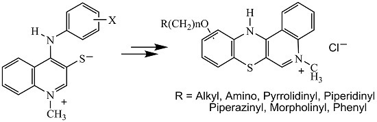
3.1.1. Synthesis of 1-Methyl-4-(arylamino)quinolinium-3-thiolates 2

Argon was passed through the suspension of bis-chloride (1) (0.419 g, 1 mmol) in dry pyridine (15 mL) at room temperature over 15 min. Amine (2.5 mmol) was added and the reaction mixture was bubbled with argon for a further 15 min. The mixture was then stirred at room temperature for 7 days. The solid product was filtered off and washed with dry ether. The raw product was purified through recrystallization from ethanol.
1-Methyl-4-((4-methoxy)phenylamino)quinolinium-3-thiolate (2a). Yield: 54%; m.p. 173 °C; 1H-NMR (DMSOd-6, 600 MHz) δ (ppm): 3.77 (s, 3H, OCH3), 4.16 (s, 3H, NCH3), 6.89–6.98 (m, 2H, Harom), 7.06–7.13 (m, 2H, Harom), 7.20–7.28 (m, 1H, H6quinolinyl), 7.42–7.48 (m, 1H, H8quinolinyl), 7.55–7.62 (m, 1H, H7quinolinyl), 7.88–7.94 (m, 1H, H5quinolinyl), 8.70 (s, 1H, H2quinolinyl), 9.99 (s, 1H, NH); 13C-NMR (DMSOd-6, 150.9 MHz) δ (ppm): 42.26 (NCH3), 55.79 (OCH3), 115.20 (C2′, C6′), 118.90 (C5), 124.54 (C4a), 124.65 (C8), 124.90 (C6), 124.99 (C3′, C5′), 129.57 (C7), 134.71 (C3), 135.74 (C8a), 145.00 (C2), 151.16 (C4′), 153.43 (C4), 157.47 (C1′); EI-MS (70eV) (m/z): 296 (M+, 100%); Anal. calcd. for C17H16N2OS: C, 68.89; H, 5.44; N, 9.45; S, 10.82. Found: C, 68.81; H, 5.35; N, 9.40; S, 8.78.
1-Methyl-4-((2-methoxy)phenylamino)quinolinium-3-thiolate (2b). Yield: 58%; m.p. 172–174 °C; 1H-NMR (DMSOd-6, 600 MHz) δ (ppm): 3.77 (s, 3H, OCH3), 4.19 (s, 3H, NCH3), 6.84–6.93 (m, 2H, Harom), 7.12–7.22 (m, 2H, Harom), 7.25–7.31 (m, 1H, H6quinolinyl), 7.45–7.49 (m, 1H, H8quinolinyl), 7.57–7.63 (m, 1H, H7quinolinyl), 7.89–7.94 (m, 1H, H5quinolinyl), 8.73 (s, 1H, H2quinolinyl), 9.86 (s, 1H, NH); EI-MS (70 eV) (m/z): 296 (M+, 100%); Anal. calcd. for C17H16N2OS: C, 68.89; H, 5.44; N, 9.45; S, 10.82. Found: C, 8.78; H, 5.39; N, 9.38; S, 8.75.
1-Methyl-4-((2-hydroxy-4-phenyl)phenylamino)quinolinium-3-thiolate (2c). Yield: 37%; m.p. 198 °C; 1H-NMR (DMSOd-6, 600 MHz) δ (ppm): 4.19 (s, 3H, NCH3), 7.03–7.08 (m, 1H, Harom), 7.15–7.18 (m, 1H, Harom), 7.22–7.26 (m, 1H, Harom), 7.28–7.37 (m, 3H, Harom), 7.37–7.40 (m, 1H, Harom), 7.40–7.48 (m, 2H, Harom), 7.60–7.67 (m, 1H, H7quinolinyl), 7.67–7.73 (m, 1H, H8quinolinyl), 7.93–7.98 (m, 1H, H5quinolinyl), 8.74 (s, 1H, H2quinolinyl), 9.97 (s, 1H, NH), 10.17 (s, 1H, OH), EI-MS (70 eV) (m/z): 358 (M+, 100%); Anal. calcd. for C22H18N2OS: C, 73.72; H, 5.06; N, 7.81; S, 8.94. Found: C, 73.67; H, 4.97; N, 7.76; S 8.91.
1-Methyl-4-(4-(N-piperidinyl)phenylamino)quinolinium-3-thiolate (2d). Yield: 84%; m.p. 208 °C; 1H-NMR (DMSOd-6, 600 MHz) δ (ppm): 1.45–1.80 (m, 6H, Hpiperidinyl), 3.10–3.30 (m, 4H, Hpiperidinyl), 4.15 (s, 3H NCH3), 7.03–7.08 (m, 4H, Harom), 7.25–7.38 (m, 1H, Harom), 7.55–7.74 (m, 1H, Harom), 7.92–7.98 (m, 1H, Harom), 8.69 (s, 1H, H2quinolinyl), 10.08 (s, 1H, NH); EI-MS (70 eV) (m/z): 349 (M+, 100%); Anal. calcd. for C21H23N3S: C, 72.17; H, 6.63; N, 12.02; S, 9.17. Found: C, 72.15; H, 6.54; N, 11.95; S, 9.13.
1-Methyl-4-(4-(N-piperazinyl)phenylamino)quinolinium-3-thiolate (2e). Yield: 86%; m.p. 137 °C; 1H-NMR (DMSOd-6, 600 MHz) δ (ppm): 3.05–3.35 (m, 8H, Hpiperazinyl), 4.17 (s, 3H, NCH3), 4.69 (s, 1H, NHpiperazinyl), 6.96–7.02 (m, 2H, Harom), 7.06–7.10 (m, 2H, Harom), 7.25–7.30 (m, 1H, Harom), 7.51–7.55 (m, 1H, Harom), 7.58–7.64 (m, 1H, Harom), 7.90–7.96 (m, 1H, Harom), 8.70 (s, 1H, H2quinolinyl), 10.01 (s, 1H, NH); EI-MS (70 eV) (m/z): 350 (M+, 100%); Anal. calcd. for C20H22N4S: C, 68.54; H, 6.33; N, 15.99; S, 9.15. Found: C, 68.49; H, 6.25; N, 15.90; S, 9.10.
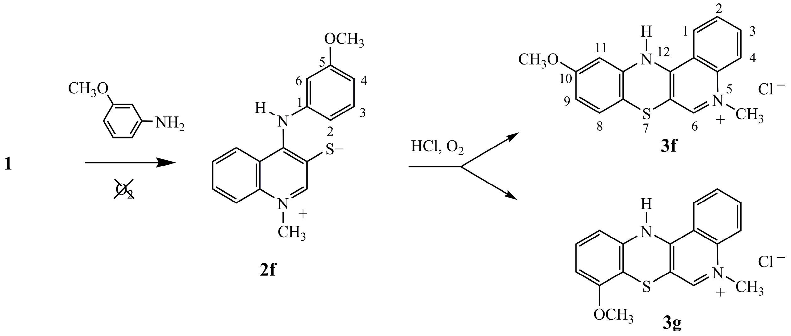
1-Methyl-4-(3-methoxyphenylamino)quinolinium-3-thiolate (2f). Yield: 66%; m.p. 181 °C; 1H-NMR (DMSOd-6, 600 MHz) δ (ppm): 3.69 (s, 3H, OCH3), 4.20 (s, 3H, NCH3), 6.58–6.63 (m, 1H, Harom), 6.69–6.77 (m, 1H, Harom), 7.20–7.26 (m, 1H, H4arom), 7.29–7.35 (m, 1H, H6quinolinyl), 7.49–7.54 (m, 1H, H8quinolinyl), 7.58–7.64 (m, 1H, H7quinolinyl), 7.94–7.99 (m, 1H, H5quinolinyl), 8.80 (s, 1H, H2quinolinyl), 9.91 (s, 1H, NH); EI-MS (70 eV) (m/z): 296 (M+, 100%); Anal. calcd for C17H16N2OS: C 68.89; H 5.44; N 9.45; S 10.82. Found: C, 68.79; H, 5.36; N, 9.40; S, 10.75.

3.1.2. Synthesis of 5-Methyl-12(H)-quino[3,4-b][1,4]benzothiazinium Chloride 3

Procedure (A): Aniline hydrochloride (0.155 g, 1.2 mmol) was added to the mixture of 3-thiolate (2) (1 mmol) in 10 mL of dry pyridine and the whole was mixed at 70 °C for 12 h. After cooling it down to room temperature, the formed precipitate was filtered off and washed with ether. The raw product was recrystallized from ethanol.
Procedure (B): Amine (2.5 mmol) was added to the mixture of bis-chloride (1) (0.419 g, 1 mmol) in 10 mL of dry pyridine and the whole was mixed at 70 °C for 12 h. The mixture was cooled down to room temperature and the formed precipitate was filtered off and washed with ether. The raw product was purified through recrystallization from ethanol.
9-Methoxy-5-methyl-12(H)-quino[3,4-b][1,4]benzothiazinium chloride (3a). Yield: Procedure (A) 68%, Procedure (B) 72%; 1H-NMR (DMSOd-6, 600 MHz) δ (ppm): 3.73 (s, 3H, OCH3), 4.19 (s, 3H, NCH3), 6.70–6.78 (m, 2H, H10, H11), 7.36–7.44 (m, 1H, H8), 7.76–7.84 (m, 1H, H2), 7.97–8.08 (m, 2H, H3, H4), 8.50–8.63 (m, 1H, H1), 8.84 (s, 1H, H6), 10.93 (s, 1H, NH); Anal. calcd. for C17H15ClN2OS: C, 61.72; H, 4.57; N, 8.47; S, 9.69. Found C, 61.63; H, 4.51; N, 8.41; S, 9.66.
11-Methoxy-5-methyl-12(H)-quino[3,4-b][1,4]benzothiazinium chloride (3b). Yield: Procedure (A) 65%, Procedure (B) 74%; 1H-NMR (DMSOd-6, 600 MHz) δ (ppm): 3.93 (s, 3H, OCH3), 4.18 (s, 3H, NCH3), 6.68–6.77 (d, 3J = 9 Hz, 1H, H8), 6.96–7.04 (d, 3J = 7.8 Hz, H10), 7.08–7.16 (d.d, 3J = 9 Hz, 3J = 7.8 Hz, H9), 7.82–7.90 (s, 1H, H2). 8.03–8.16 (m, 2H, H3, H4), 8.50–8.58 (m, 1H, H1), 8.80 (s, 1H, H6), 9.90 (s, 1H, NH); 13C-NMR (DMSOd-6, 150.9 MHz) δ (ppm): 43.28 (NCH3), 56.94 (OCH3), 107.54 (C6a), 112.28 (C12b), 116.28 (C8), 118.83 (C12a), 119.28 (C11), 119.32 (C10), 123.88 (C2), 125.40 (C11a), 128.10 (C9), 128.47 (C1), 134.99 (C3), 139.11 (C6), 144,20 (C4a), 148.90 (C7a), 152.03 (C4); Anal. calcd. for C17H15ClN2OS: C, 61.72; H, 4.57; N, 8.47, S, 9.69. Found: C, 6.66; H, 4.49; N, 8.45; S, 9.64.
11-Hydroxy-9-phenyl-5-methyl-12(H)-quino[3,4-b][1,4]benzothiazinium chloride (3c). Yield: Procedure (A) 59%, Procedure (B) 65%; 1H-NMR (DMSOd-6, 600 MHz) δ (ppm): 4.06 (s, 3H, NCH3), 6.93–7.02 (m, 2H, Harom), 7.30–7.35 (m, 2H, Harom), 7.38–7.44 (m, 1H, Harom), 7.44–7.51 (m, 2H, Harom), 7.74–7.85 (m, 1H, Harom), 7.98–8.07 (m, 2H, Harom), 8.60–8.65 (m, 2H, Harom), 10.10 (s, 1H, NH), 11.18 (s, 1H, OH); Anal. calcd. for C22H17ClN2OS: C, 67.25; H, 4.36; N, 7.13; S, 8.16. Found: C, 67.18; H, 4.30; N, 7.04; S 8.10.
9-(N-piperidinyl)-5-methyl-12(H)-quino[3,4-b][1,4]benzothiazinium chloride (3d). Yield: Procedure (A) 68%, Procedure (B) 77%; 1H-NMR (DMSOd-6, 600 MHz) δ (ppm): 1.43–1.70 (m, 6H, Hpiperidinyl), 3.08–3.20 (m, 4H, Hpiperidinyl). 4.05 (s, 3H, NCH3), 6.56–6.62 (d, 4J = 2.4 Hz, 1H, H8), 6.64–6.69 (d.d, 3J = 9 Hz, 4J = 2.4 Hz, 1H, H10), 7.44–7.50 (d, 3J = 9 Hz, 1H, H11), 6.70–6.75 (m, 1H, H2), 7.74–7.79 (m, 2H, H3, H4), 8.50 (s, 1H, H6), 9.01–9.06 (m, 1H, H1), 11.22 (s, 1H, NH). Anal. calcd. for C21H22ClN3S: C, 65.70; H, 5.78; N, 10.94; S, 8.35. Found C, 65.64; H, 5.73; N, 10.89; S, 8.32.
9-(N-piperazinyl)-5-methyl-12(H)-quino[3,4-b][1,4]benzothiazinium chloride (3e). Yield: Procedure (A) 66%, Procedure (B) 74%; 1H-NMR (DMSOd-6, 600 MHz) δ (ppm): 3.28–3.32 (m, 4H, Hpiperazinyl), 3.34–3.39 (m, 4H, Hpiperazinyl), 4.11 (s, 3H, NCH3) 6.65–6.69 (d, 4J = 3 Hz, 1H, H8), 6.72–6.77 (d, 3J = 9 Hz, 1H, H11), 6.77–6.82 (d.d, 3J = 9 Hz, 4J = 3 Hz, 1H, H10), 7.75–7.83 (m, 1H, Harom), 7.97–8.04 (m, 2H, Harom), 8.26 (s, 1H, H6), 8.48–8.53 (m, 1H, Harom); Anal. calcd. for C20H21ClN4S: C, 62.41; H, 5.50; N, 14.56; S, 8.33. Found C, 62.32; H, 5.44; N, 14.47; S, 8.28.

3.1.3. Synthesis of 10-Hydroxy-5-methyl-5(H)-quino[3,4-b][1,4]benzothiazine 4

Quinobenzothiazinium chloride (3h) (0.317 g, 1 mmol) was dissolved in 20 mL water (50 °C), the resulting solution was filtered and alkalized while mixing by using a 5% aqueous NaHCO3 solution (10 mL). The obtained solid product was filtered off and air-dried. Finally, the crude product was purified by recrystallization from ethanol.
10-Hydroxy-5-methyl-5(H)-quino[3,4-b][1,4]benzothiazine (4). Yield: 100%; m.p. 107–110 °C; 1H-NMR (CD3ODd-4, 600 MHz) δ (ppm): 3.90 (s, 3H, CH3), 6.42–6.47 (d.d, 3J = 8.4 Hz, 4J = 2.4 Hz, 1H, H9), 6.58 (s, 1H, H6), 6.61–6.67 (m, 1H, Harom), 7.53–7.61 (m, 1H, Harom), 7.66–7.74 (m, 2H, Harom), 7.78–7.86 (m, 1H, Harom), 8.34–8.39 (m, 1H, Harom); EI-MS (70 eV) (m/z): 280 (M+, 100%); Anal. calcd. for C16H12N2OS: C, 68.55; H, 4.31; N, 9.99; S, 11.44. Found: C, 68.53; H, 4.26; N, 9.94; S, 11.42.

3.1.4. Synthesis of 10-Alkoxy-5-methyl-5(H)-quino[3,4-b][1,4]benzothiazine 5

Anhydrous 1,4-dioxane (15 mL) was mixed with quinobenzothiazine (4) (0.28 g, 1 mmol) and sodium hydroxide (0.2 g, 5 mmol) and refluxed with mixing for 2 h. An alkylating agent (alkyl iodides or aminoalkyl chlorides) (1.3 mmol) was added stepwise and the mixture was refluxed for a subsequent 2 h. After cooling down to room temperature, the reaction mixture was poured into 50 mL of water and extracted with 15 mL chloroform. The resulting solution was dried over anhydrous calcium chloride and evaporated under vacuum. The dry residue was purified by chromatography using a silica gel-filled column and chloroform-ethanol (10:1 v/v) as eluent.
10-Methoxy-5-methyl-5(H)-quino[3,4-b][1,4]benzothiazine (5a). Yield: 30%; m.p. 268–270 °C; 1H-NMR (DMSOd-6, 600 MHz) δ (ppm): 3.51 (s, 3H, NCH3), 3.68 (s, 3H, OCH3), 6.38–6.44 (m, 2H, Harom), 6.56–6.60 (m, 1H, Harom), 7.07 (s, 1H, H6), 7.21–7.26 (m, 1H, Harom), 7.27–7.30 (m, 1H, Harom), 7.52–7.57 (m, 1H, Harom), 8.18–8.22 (m, 1H, Harom); 13C-NMR (DMSOd-6, 150.9 MHz) δ (ppm): 40.51 (NCH3), 55.46 (OCH3), 103.64 (12b), 111.09 (7a), 111.33(C9 or C11), 112.53 (C9 or C11), 115.89 (C4), 121.29 (6a), 123.96 (C2), 125.02 (C1), 126.01 (C8), 131.98 (3), 133.14 (C6), 140.75 (4a), 146.30 (C11a), 154.13 (C12a), 159.46 (C10); EI-MS (70 eV) (m/z): 294 (M+, 100%); Anal. calcd. for C17H14N2OS: C, 69.36; H, 4.79; N, 9.52; S, 10.89. Found: C, 69.29; H, 4.75; N, 9.48; S, 10.84.
10-Butyloxy-5-methyl-5(H)-quino[3,4-b][1,4]benzothiazine (5b). Yield: 37%; m.p. 280–282 °C; 1H-NMR (CDCl3, 600 MHz) δ (ppm): 2.58 (m, 3H, CH3), 2.70–2.80 (m, 2H, CH2), 3.38–3.50 (m, 2H, CH2), 3.76 (s, 3H, NCH3), 4.03–4.11 (t, J = 4.8 Hz, 2H, OCH2), 6.31–6.45 (m, 2H, Harom), 6.52–6.54 (m, 1H, Harom), 6.54–6.63 (m, 1H, Harom), 6.95–7.03 (m, 1H, Harom), 7.18–7.25 (m, 1H, Harom), 7.40–7.50 (m, 1H, Harom), 8.30–8.45 (m, 1H, Harom); EI-MS (70 eV) (m/z): 336 (M+, 100%); Anal. calcd. for C20H20N2OS: C, 71.40; H, 5.99; N, 8.33; S, 9.53. Found: C, 71.30; H, 5.94; N, 8.28; S, 9.49.
10-(3-(N,N-dimethylamino)propyl)oxy-5-methyl-5(H)-quino[3,4-b][1,4]benzothiazine (5c). Yield: 49%; m.p. 82–83 °C; 1H-NMR (DMSOd-6, 600 MHz) δ (ppm): 2.10–2.20 (t, 3J = 6 Hz, 2H, NCH2), 2.76 (s, 3H, NCH3), 2.77 (s, 3H, NCH3), 3.11–3.25 (m, 2H, CH2), 3.95–4.05 (t, 3J = 6 Hz, 2H, OCH2), 4.15 (s, 3H, NCH3), 6.65–6.75 (d.d, 3J = 8.4 Hz, 4J = 2.4 Hz, 1H, H9), 6.92–7.00 (d, 3J = 8.4 Hz, 1H, H8), 7.45–7.50 (d, 4J = 2.4 Hz, 1H, H11), 7.75–7.84 (m, 1H, Harom), 7.95–8.12 (m, 2H, Harom), 8.69 (s, 1H, H6), 9.15–9.27 (m, 1H, Harom); EI-MS (70 eV) (m/z): 365 (M+, 100%); Anal. calcd. for C21H23N3OS: C, 69.01; H, 6.34; N, 11.50; S, 8.77. Found: C, 68.96; H, 6.30; N, 11.47; S, 8.75.
10-(2-(N-pyrrolidinyl)ethyl)oxy-5-methyl-5(H)-quino[3,4-b][1,4]benzothiazine (5d). Yield: 46%; m.p. 148–150 °C; 1H-NMR (DMSOd-6, 600 MHz) δ (ppm): 1.62–1.76 (m, 4H, Hpyrrolidinyl), 2.52–2.57 (m, 4H, Hpyrrolidinyl), 2.73 (t, J = 6 Hz, 2H, NCH2), 3.51 (s, 3H, NCH3), 3.98 (t, J = 6 Hz, 2H, OCH2), 6.37–6.44 (m, 2H, Harom), 6.55–6.59 (m, 1H, Harom), 7.07 (s, 1H, H6), 7.22–7.25 (m, 1H, Harom), 7.25–7.30 (m, 1H, Harom), 7.52–7.57 (m, 1H, Harom), 8.17–8.24 (m, 1H, Harom); EI-MS (70 eV) (m/z): 377 (M+, 73%); Anal. calcd. for: C22H23N3OS: C, 70.00; H, 6.14; N, 11.13; S, 8.49. Found: C, 69.96; H, 6.08; N, 11.09; S, 8.48.
10-(2-(N-piperidinyl)ethyl)oxy-5-methyl-5(H)-quino[3,4-b][1,4]benzothiazine (5e). Yield: 67%; m.p. 70–73 °C; 1H-NMR (DMSOd-6, 600 MHz) δ (ppm): 1.30–1.40 (m, 2H, Hpiperidinyl), 1.40–1.53 (m, 4H, Hpiperidinyl), 2.35–2.45 (m, 4H, Hpiperidinyl), 2.54–2.61 (t, J = 6 Hz, 2H, NCH2), 3.51 (s, 3H, NCH3), 3.93–4.01 (t, 3J = 6 Hz, 2H, OCH2), 6.35–6.40 (m, 2H, Harom), 6.52–6.59 (m, 1H, Harom), 7.06 (s, 1H, H6), 7.15–7.32 (m, 2H, Harom), 7.49–7.55 (m, 1H, Harom), 8.15–8.22 (m, 1H, Harom); EI-MS (70 eV) (m/z): 392 (M+, 100%); Anal. calcd. for C23H25N3OS: C, 70.56; H, 6.44; N, 10.73; S, 8.19. Found: C, 70.48; H, 6.49; N, 10.70; S, 8.17.
10-(2-(N-morpholinyl)ethyl)oxy-5-methyl-5(H)-quino[3,4-b][1,4]benzothiazine (5f). Yield: 15%; m.p. 196–198 °C; 1H-NMR (DMSOd6, 600 MHz) δ (ppm): 2.38–2.45 (m, 4H, Hmorpholinyl), 2.57–2.65 (t, J = 5.4 Hz, 2H, NCH2), 3.51 (s, 2H, NCH3), 3.53–3.62 (m, 4H, Hmorpholinyl), 3.92–4.05 (t, J = 5.4 Hz, 2H, OCH2), 6.38–6.44 (m, 2H, Harom), 6.55–6.59 (m, 1H, Harom), 7.07 (s, 1H, H6), 7.20–7.32 (m, 2H, Harom), 7.48–7.57 (m, 1H, Harom), 8.18–8.24 (m, 1H, Harom). EI-MS (70 eV) (m/z): 394 (M+, 100%); Anal. calcd. for C22H23N3O2S: C, 67.15; H, 5.89; N, 10.68; S, 8.15. Found: C, 67.10; H, 5.83; N, 10.62; S 8.12.

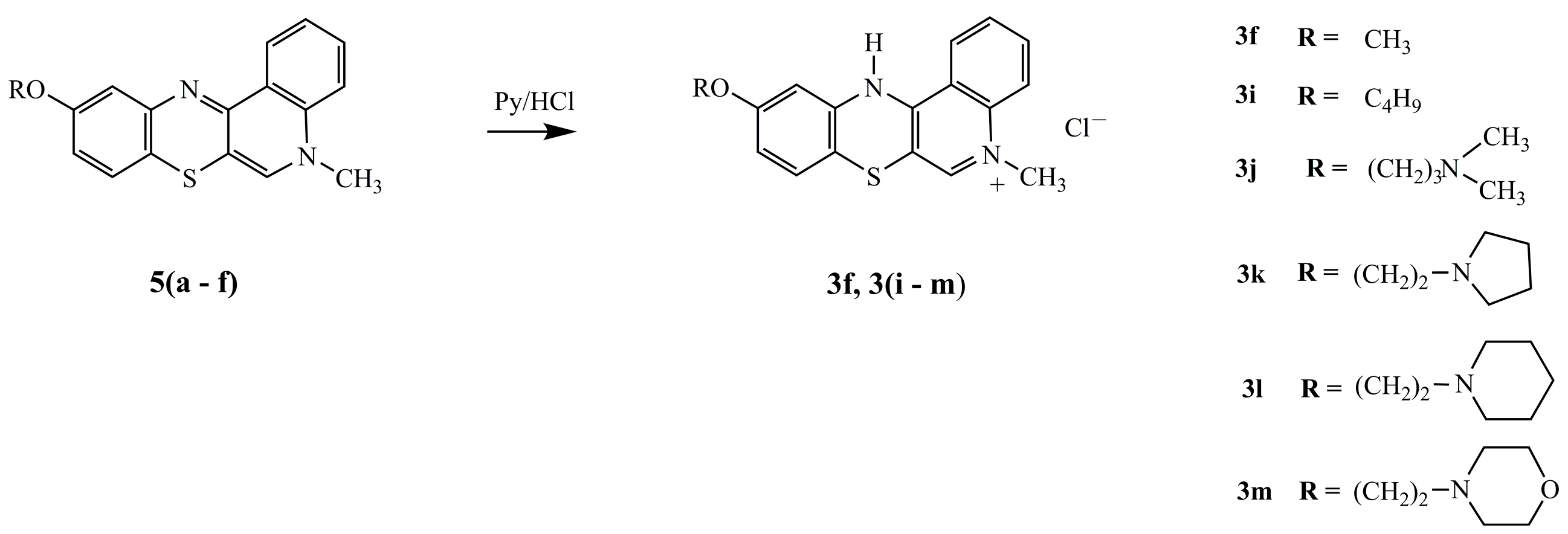
3.1.5. Synthesis of 12(H)-quino[3,4-b][1,4]benzothiazinium Chloride 3f, 3im

Quinobenzothiazine (5) (1 mmol) was dissolved in 15 mL anhydrous ethanol. Pyridine hydrochloride (1 mmol) was added and the reaction mix was refluxed for 2 h. Solvents were evaporated under vacuum and the dried remaining residue was purified using a chromatography column filled with aluminum oxide and chloroform:ethanol (10:1 v/v) as eluent.
10-Methoxy-5-methyl-12(H)-quino[3,4-b][1,4]benzothiazinium chloride (3f).Yield: 87%; 1H-NMR (DMSOd-6, 600 MHz) δ (ppm): 3.75 (s, 3H, NCH3), 4.15 (s, 3H, OCH3), 6.68–6.72 (m, 1H, Harom), 6.98–7.02 (m, 1H, Harom), 7.24–7.26 (d, 4J = 3 Hz, H11), 7.83–7.88 (m, 1H, Harom), 8.01–8.12 (m, 2H, Harom), 8.68 (s, 1H, H6), 8.92–8.98 (m, 1H, Harom), 10.97 (s, 1H, NH). Anal. calcd. for C17H15ClN2OS: C, 61.72; H, 4.57; N, 8.47; S, 9.69. Found: C, 61.65; H, 4.51; N, 8.42; S, 9.66.
10-Butyloxy-5-methyl-12(H)-quino[3,4-b][1,4]benzothiazinium chloride (3i). Yield 82%; 1H-NMR (DMSOd-6, 600 MHZ) δ (ppm): 3.15–3.25 (m, 3H, CH3), 3.71–3.85 (m, 2H, CH2), 3.95–4.02 (m, 2H, CH2), 4.15 (s, 3H, NCH3), 4.34–4.43 (t, J = 4.2 Hz, 2H, OCH2), 6.75–6.80 (d.d, 3J = 8.4 Hz, 4J = 2.4 Hz, 1H, H9), 7.03–7.08 (d, 3J = 8.4 Hz, 1H, H8), 7.25–7.31 (d, 4J =2.4 Hz, 1H, H11), 7.82–7.89 (m, 1H, Harom), 8.04–8.12 (m, 2H, Harom), 8.69 (s, 1H, H6), 8.91–8.97 (m, 1H, Harom), 11.11 (s, 1H, NH). Anal. calcd. for C20H21ClN2OS: C, 64.42; H, 5.68; N, 7.51; S, 8.60. Found: C, 64.38; H, 5.63; N, 7.75; S 8.57.
10-(3-(N,N-Dimethylamino)propyl)oxy-5-methyl-12(H)-quino[3,4-b][1,4]benzothiazinium chloride (3j). Yield: 80%; 1H-NMR (DMSOd-6, 600 MHz) δ (ppm): 2.16 (t, J = 6 Hz, 2H, NCH2), 2.77 (s, 3H, NCH3), 2.78 (s, 3H, NCH3), 3.19 (m, 2H, CH2), 4.04 (t, J = 6 Hz, 2H, OCH2), 4.15 (s, 3H, NCH3), 6.67–6.73 (d.d, 3J = 8.4 Hz, 4J = 2.4 Hz, 1H, H9), 6.98–7.02 (d, 3J = 8.4 Hz, 1H, H8), 7.48–7.53 (d, 4J = 2.4 Hz, 1H, H11), 7.78–7.85 (m, 1H, Harom), 8.02–8.11 (m, 2H, Harom), 8.69 (s, 1H, H6), 9.20–9.25 (m, 1H, Harom), 10.73–10.80 (m, 1H, Harom), 11.39 (s, 1H, NH); 13C-NMR (DMSOd-6, 150.9 MHz) δ (ppm): 24.16 (NCH2), 42.49 (N(CH3)2), 43.11 (NCH3), 54.30 (CH2), 65.72 (OCH2), 106.67 (C6a), 106.96 (C11), 107.40 (7a), 113.22 (C9), 116.17 (11a), 119.16 (C4), 124.88 (C1), 127.92 (C8), 128.23 (C12a), 134.85 (C2), 138.05 (C3), 139.10 (C4a), 143.69 (C6), 151.87 C12a), 158.92 (C10); Anal. calcd. for C21H24ClN3OS: C, 62.75; H, 6.02; N, 10.45; S, 7.98. Found: C, 62.71; H, 5.96; N, 10.40; S, 7.94.
10-(2-(N-Pyrrolidinyl)ethyl)oxy-5-methyl-12(H)-quino[3,4-b][1,4]benzothiazinium chloride (3k). Yield: 83%; 1H-NMR (DMSOd-6, 600 MHz) δ (ppm): 1.80–1.90 (m, 2H, Hpyrrolidinyl), 1.90–2.05 (m, 2H, Hpyrrolidinyl), 3.02–3.12 (m, 2H, NCH2), 3.51–3.62 (m, 4H, Hpyrrolidinyl), 4.11 (s, 3H, NCH3), 4.30–4.35 (t, J = 4.8 Hz, 2H, OCH2), 6.80–6.85 (m, 2H, Harom), 7.50–7.56 (m, 1H, Harom), 7.78–7.84 (m, 1H, Harom), 8.02–8.07 (m, 1H, Harom), 8.61 (s, 1H, H6), 8.95–9.00 (m, 1H, Harom), 10.95–11.04 (m, 1H, Harom), 11.13 (s, 1H, NH); Anal. calcd. for C22H24ClN3OS: C, 63.83; H, 5.84; N, 10.15; S, 7.74. Found: C, 63.86; H, 5.78; N, 10.11; S, 7.70.
10-(2-(N-Piperidinyl)ethyl)oxy-5-methyl-12(H)-quino[3,4-b][1,4]benzothiazinium chloride (3l). Yield: 86%; 1H-NMR (DMSOd-6, 600 MHz) δ (ppm): 1.32–1.45 (m, 2H, Hpiperydyl), 1.70–1.90 (m, 4H, Hpiperydyl), 2.95–3.10 (t, J = 4.8 Hz, 2H, NCH2), 3.45–3.50 (m, 4H, Hpiperydyl), 4.15 (s, 3H, NCH3), 4.38–4.43 (t, J = 4.8 Hz, 2H, OCH2), 6.73–6.78 (d.d, 3J = 9 Hz, 4J = 3 Hz, 1H, H9), 7.01–7.05 (d, 3J = 9 Hz, 1H, H8), 7.32–7.34 (d, 3J = 3 Hz, 1H, H11), 7.81–7.88 (m, 1H, Harom), 8.01–8.10 (m, 2H, Harom), 8.69 (s, 1H, H6), 8.98–9.04 (m, 1H, Harom), 10.46 (s, 1H, NH). Anal. calcd. for C23H26ClN3OS: C, 64.55; H, 6.12; N, 9.82; S, 7.49. Found: C, 64.50; H, 6.06; N, 9.78; S, 7.45.
10-(2-(N-morpholinyl)ethyl)oxy-5-methyl-12(H)-quino[3,4-b][1,4]benzothiazinium chloride (3m). Yield: 100%; 1H-NMR (DMSOd-6, 600 MHz) δ (ppm): 3.12–3.33 (m, 4H, Hmorpholinyl), 3.85–4.02 (m, 6H, NCH2, 4Hmorpholinyl), 4.16 (s, 3H, NCH3), 4.38–4.45 (t, J = 4.2 Hz, OCH2), 6.75–6.78 (d.d, 3J = 9 Hz, 4J = 2.4 Hz, H9), 7.02–7.06 (d, 3J = 9 Hz, 1H, H8), 7.48–7.53 (d, 4J = 2.4 Hz, 1H, H11), 7.78–7.68 (m, 1H, Harom), 8.03–8.12 (m, 2H, Harom), 8.71 (s, 1H, H6), 9.16–9.20 (m, 1H, Harom), 11.38 (s, 1H, NH). Anal. calcd. for C22H24ClN3OS: C, 61.46; H, 5.63; N, 9.77; S, 7.46. Found: C, 61.42; H, 5.59; N, 7.73; S, 7.44.

3.2. Biological Assays

3.2.1. Cell Culture

Biological activity of the examined compounds was assessed in vitrousing three cultured cell lines: (1) MDA-MB-231 invasive breast ductal carcinoma (ATCC, Rockville, MD, USA); (2) SNB-19 glioblastoma (DSMZ, Braunschweig, Germany); (3) C-32 amelanotic melanoma (ATCC, Rockville, MD, USA). Cell cultures were maintained using DMEM (Lonza, Basel, Switzerland) supplemented with 10% fetal bovine serum (FBS) (Biological Industries Cromwell, CT, USA) and penicillin (10,000 U/mL)—streptomycin (10 mg/mL) mix (Lonza, Basel, Switzerland).

3.2.2. Effect of Compounds on Number and Viability of Cells

Cells were cultured using 96 well plates (Nunc Thermo Fisher Scientific, Waltham, MA, USA). Cells were seeded (5 × 10³ cells/well) and incubated for 24 h in a standard incubator (37 °C, 5% CO2, relative humidity 95%). Next, medium was replaced with fresh aliquot containing an examined compound (0.1; 0.5; 1; 5; 10; 50; 100 µg/mL) and cells were further incubated for 72 h. Upon conclusion of incubation, a CVDE test (Aniara, West Chester, OH, USA) using crystal violet was performed to assess the relative number of cells in the culture. A WST-1 test (Roche) was performed to examine the metabolic activity of cells and test for the presence of LDH in the medium (Roche Diagnostics GmbH, Mannheim, Germany), allowing for the assessment of the relative number of dead cells (i.e., cytotoxicity of the examined compound). UVM340 microplate readers (BIOGENET, Józefów, Poland) were used to read absorbance values.

CVDE Test

AKCV96.1200 kit (Aniara) was used according to the manufacturer’s instructions. The test is based on cell membrane penetration by crystal violet and its ultimate interaction with DNA. Excess dye was washed off, cells were lysed, and absorbance measurements were performed for test and control samples at λ = 540 nm.

WST-1 Test

WST-1 colorimetric assay for cell proliferation (Roche Diagnostics GmbH, Mannheim, Germany, reagent kit cat. 11644807001) is based on the viable cells’ ability to cleave the bright red-colored stable tetrazolium salt WST-1 to dark red soluble formazan. This bioreduction occurs under the influence of mitochondrial dehydrogenases (depends mostly on production of NAD(P)H in viable cells). The amount of formazan dye formed correlates directly with the number of metabolically active cells in the culture and is measured by absorbance (λ = 450 nm) following 1 h incubation of cells with the reagent.

LDH Test

Lactate dehydrogenase (LDH) (Roche Diagnostics GmbH, Mannheim, Germany, reagent kit cat. 11644793001) is a cytosolic enzyme released into the culture medium following cell membrane damage. It can be used to assess the degree of toxicity of the examined substance. Relative number of dead cells (vs. control) in culture was determined using the LDH cytotoxicity detection kit (Roche Diagnostics GmbH, Mannheim Germany) according to the manufacturer’s protocol. Aliquots (100 μL) of the prepared reagent were added to media transferred (fresh plate) from wells with growing cells. Absorbance measurements (λ = 490 nm) of the samples were performed after 1 h.

3.2.3. Transcriptional Activity of H3, BCL-2, BAX and TP53 Genes

Transcriptional activity of the following genes was assessed: H3/encoding histone H3, a proliferation marker/BCL-2 and BAX/encoding BCL-2 and BAX, respectively, two apoptosis-related mitochondrial proteins/ and TP53/encoding P53, a cell cycle regulator/. The activity was assessed by RT-QPCR using Opticon™ DNA Engine system (MJ Research, Watertown, NY, USA) and QuantTect® SYBR® Green RT-PCR Kit (Qiagen, Hilden, Germany). Cultured cells were exposed (24 h) to the examined compounds (0.5 μg/mL). RNA was extracted using Quick-RNA™ MiniPrep kit columns (Zymo Research, Irvine, CA, USA). The extracted RNA was assessed qualitatively and quantitatively. Integrity of total RNA was checked by electrophoresis (1.2% agarose gel, EtBr). Amount and purity of the total RNA in extracts was determined spectrophotometrically (HP8452A apparatus, Hewlett Packard, Waldbronn, Germany).

3.2.4. DNA Binding by the Examined Compounds

Genomic DNA was extracted from cells using silica bed columns (DNA isolation and purification kits). The obtained material was analyzed qualitatively and quantitatively by spectrophotometry (GeneQuant II analyzer, Pharmacia Biotech, Madrid, Spain). Extracted DNA samples were mixed either with an examined compound at 5:1, 1:1, 1:5 (w/w) ratios, or with cisplatin at 1:1 (w/w) ratio using 1–5 μg aliquots of the compound and DNA. Samples were applied to 0.9% agarose gel containing 0.5 mg/mL ethidium bromide (Promega, Fitchburg, WI, USA). The latter is a DNA intercalator the UV-induced fluorescence of which increases upon DNA binding. Cisplatin forms inter- and intrastrand crosslinks within DNA.

4. Conclusions

Reactions of tioquinantrenediinium bis-salts (1) with aromatic amines lead to tetracyclic quinobenzothiazine derivatives (3). Intermediate products of these reactions are 1-alkyl-4-aminoquinolinium-3-thiolates (2), the cyclization of which in the presence of atmospheric oxygen and a hydrogen chloride donor leads to the formation of 1,4-thiazine ring. Using this method of synthesis, novel derivatives were obtained containing hydroxyl, methoxyl, phenyl, piperidyl, and piperazinyl substituents in positions 9 and 11 of the quinobenzothiazine system. Alkylation of the hydroxyl group in position 10 allowed for the introduction of additional substituents such as alkyl, aminoalkyl, and aminoalkyl with heterocyclic nitrogen-containing rings (pyrrolidine, piperidine, and morpholine). Four compounds selected for further analysis were active towards the three tested cancer cell lines (MDA-MB-231, SNB-19, and C-32) (IC50 range 0.5–24.5 μM). Based on the results of CVDE, WST-1, and LDH tests it may be concluded that exposition to these compounds causes concentration-dependent reduction in the number of cultured cells. The four leading compounds can be ranked in the following order: 3e > 3k > 3f > 3c. The observed antiproliferative effect is not the result of the cytotoxic action of these derivatives leading to cell death; rather, it is the consequence of inhibited cell proliferation. The inhibition appears to be the result of these compounds binding to cellular DNA. The greatest inhibiting activity was demonstrated by compounds containing additional amine moieties in the structure. Their presence in the molecule could stabilize complex compound-DNA by enabling formation of additional hydrogen bonds with purine and pyrimidine bases in the DNA. The strongest DNA binding was observed for derivative (3e) containing a piperazine ring in position 9 and for derivative (3k) with a piperazine ring-containing substituent in position 10.

Acknowledgments

This work was supported by the Medical University of Silesia in Katowice, Poland. Grant No. KNW-006-/K/6/O.

Author Contributions

A.Z. and M.L. developed the concept of the work. A.N. and A.Z. carried out the synthetic work. M.L. and D.K. conducted a study of the biological activity. M.L., A.S., and A.Z. analyzed the data and wrote the paper.

Conflicts of Interest

The authors declare no conflict of interest.

References

  1. Gilman, H.; Shirley, D.A. Some derivatives of phenothiazine. J. Am. Chem. Soc. 1944, 66, 888–893. [Google Scholar] [CrossRef]
  2. Gupta, R.R.; Kumar, M. Synthesis, properties and reactions of phenothiazines. In Phenothiazines and 1,4-Benzothiazines: Chemical and Biomedical Aspects; Gupta, R.R., Ed.; Elsevier: Amsterdam, The Netherlands, 1988; pp. 1–146. [Google Scholar]
  3. Morak-Młodawska, B.; Pluta, K. Synthesis of novel dipyrido-1,4-thiazines. Heterocycles 2007, 71, 1347–1361. [Google Scholar]
  4. Jeleń, M.; Bavavea, I.E.; Pappa, M.; Kourounakis, A.P.; Morak-Młodawska, B.; Pluta, K. Synthesis of quinoline/naphthalene-containing azaphenothiazines and their potent in vitro antioxidant properties. Med. Chem. Res. 2015, 24, 1725–1732. [Google Scholar]
  5. Nowak, M.; Pluta, K.; Suwińska, K. Synthesis of novel heteropentacenes containing nitrogen, sulfur and oxygen or selenium. New J. Chem. 2002, 26, 1216–1220. [Google Scholar] [CrossRef]
  6. Kalkanidis, M.; Klonis, N.; Tilley, L.; Deady, L.W. Novel phenothiazine antimalarials: Synthesis, antimalarial activity, and inhibition of the formation of b-haematin. Biochem. Pharmacol. 2002, 63, 833–842. [Google Scholar] [CrossRef]
  7. Klitgaard, J.K.; Skov, M.N.; Kallipolitis, B.H.; Kolmos, H.J. Reversal of methicillin—Resistance in Staphylococcus aureus by thioridazine. J. Antimicrob. Chemother. 2008, 62, 1215–1221. [Google Scholar] [CrossRef] [PubMed]
  8. Tandon, V.K.; Maurya, H.K.; Tripathi, A.; Shiva Keshava, G.B.; Shukla, P.K.; Srivastava, P.; Panda, D. 2,3-Disubstituted-1,4-naphthoquinones, 12H-benzo[b]phenothiazine-6,11-diones and related compounds: Synthesis and biological evaluation as potential antiproliferative and antifungal agents. Eur. J. Med. Chem. 2009, 44, 1086–1092. [Google Scholar] [CrossRef] [PubMed]
  9. Kaatz, G.W.; Moudgal, V.V.; Seo, S.M.; Kristiansen, J.M. Phenothiazines and thioxanthenes inhibit multidrug efflux pump activity in Staphylococcus aureus. Antimicrob. Agents Chemother. 2003, 47, 719–726. [Google Scholar] [CrossRef] [PubMed]
  10. Kumar, M.; Sharma, K.; Samarth, R.M.; Kumar, A. Synthesis and antioxidant activity of quinolinobenzothiazinones. Eur. J. Med. Chem. 2010, 45, 4467–4472. [Google Scholar] [CrossRef] [PubMed]
  11. Aszalos, A. Phenothiazines in treatment of HIV infection. Acta Microbiol. Immunol. Hung. 2003, 50, 43–53. [Google Scholar] [CrossRef] [PubMed]
  12. Motohashi, N.M.; Kawase, M.; Saito, S.; Sakagami, H. Antitumor potential and possible targets of phenothiazine-related compounds. Curr. Drug Targets 2000, 1, 237–245. [Google Scholar] [CrossRef] [PubMed]
  13. Sharma, S.; Srivastava, V.K.; Kumar, A. Synthesis and anti-inflammatory activity of some heterocyclic derivatives of phenothiazine. Pharmazie 2005, 60, 18–22. [Google Scholar] [PubMed]
  14. Zięba, A.; Maślankiewicz, A.; Suwińska, K. 1-Alkyl-4-arylamino-quinolinium-3-thiolates and 7-alkyl-12(H)-quino[3,4-b]-1,4-benzothiazinium slats. Eur. J. Org. Chem. 2000, 16, 2947–2953. [Google Scholar] [CrossRef]
  15. Zięba, A.; Suwińska, K. 1-Alkyl-4-(3-pyridinylamino)quinolinium-3-thiolates and their transformation into new diazaphenothiazine derivatives. Heterocycles 2006, 68, 495–503. [Google Scholar] [CrossRef]
  16. Zięba, A.; Sochanik, A.; Szurko, A.; Rams, M.; Mrozek, A.; Cmoch, P. Synthesis and in vitro antiproliferative activity of 5-alkyl-12(H)-quino[3,4-b][1,4]benzothiazinium salts. Eur. J. Med. Chem. 2010, 45, 4733–4739. [Google Scholar] [CrossRef] [PubMed]
  17. Zięba, A.; Latocha, M.; Sochanik, A. Synthesis and in vitro antiproliferative activity of novel 12(H)-quino[3,4-b][1,4]benzothiazine derivatives. Med. Chem. Res. 2013, 22, 4158–4163. [Google Scholar] [CrossRef] [PubMed]
  18. Latocha, M.; Zięba, A.; Polaniak, R.; Kuśmierz, D.; Nowosad, A.; Jurzak, M.; Romuk, E.; Kokocińska, M.; Sliupkas-Dyrda, E. Molecular effects of amine derivatives of phenothiazine on cancer cells C-32 and SNB-19 in vitro. Acta Pol. Pharm. Drug Res. 2015, 72, 909–915. [Google Scholar]
  19. Maślankiewicz, A.; Zięba, A. 5,12-Di(alkyl)thioquinanthrenediinium bis-salts and 1-alkyl-3-alkylthio-1,4-dihydro-4-thiooxoquinolines. Heterocycles 1992, 34, 247–258. [Google Scholar] [CrossRef]
  20. Zięba, A. 3-Acylsulfanyl-1-methyl-4-methylsulfanyl-quinolinium salts and their transformation into 4-aminoquinolinium-3-thiolates and azaphenothiazine derivatives. Pol. J. Chem. 2008, 82, 1399–1402. [Google Scholar]
  • Sample Availability: Samples of all the compounds described herein are available from the authors.
Scheme 1. Synthesis of compounds 2ae and 3ae.
Molecules 21 01455 sch001
Scheme 2. Cyclization reaction of quinolinium-3-thiolate 2f.
Molecules 21 01455 sch002
Scheme 3. Synthesis of compounds 5af.
Molecules 21 01455 sch003
Scheme 4. Synthesis of compounds 3f and 3im.
Molecules 21 01455 sch004
Figure 1. The effect of derivatives 3c, 3e, 3f, and 3k upon MDA-MB-231, SNB-19, and C-32 cancer cell lines (CVDE—cell number, WST-1—metabolic activity, LDH—number of dead cells in culture).
Molecules 21 01455 g001
Figure 2. Effect of 3c, 3e, 3f, and 3k on transcriptional activity of: H3 (A); TP53 (B); BAX and BCL-2 (C) in MDA-MB-231, SNB-19, and C-32 cells.
Molecules 21 01455 g002
Figure 3. DNA intercalation with EtBr: 1 h incubation with derivatives 3c, 3e, 3f, and 3k (5/1, 1/1 and 1/5 w/w) (µg derivative/µg DNA).
Molecules 21 01455 g003
Table 1. Effect of 5-methyl-12(H)-quino[3,4-b][1,4]benzothiazinium salts (3) and cisplatin (reference) on the viability of cells from three cancer cell lines studied.
CompoundIC50 (μM) a
C-32SNB-19MDA-MB-231
3a3.6 ± 0.93.9 ± 0.912.7 ± 1.5
3b22.1 ± 2.422.7 ± 4.224.5 ± 3.0
3c4.2 ± 1.011.5 ± 3.18.4 ± 2.3
3d09 ± 0.51.0 ± 052.1 ± 0.5
3e0.5 ± 0.30.8 ± 0.51.6 ± 0.5
3f3.6 ± 1.22.7 ± 0.93.6 ± 1.2
3i7.5 ± 1.66.7 ± 1.92.6 ± 0.5
3j2.7 ± 1.52.2 ± 1.02.0 ± 0.7
3k1.2 ± 0.70.7 ± 0.50.5 ± 0.2
3l1.2 ± 0.71.2 ± 0.73.0 ± 0.9
3m0.7 ± 0.21.8 ± 0.71.8 ± 0.5
cisplatin11.0 ± 0.712.3 ± 1.025.0 ± 0.7
a The results are from five independent experiments.

