Rapid and Comprehensive Evaluation of (Poly)phenolic Compounds in Pomegranate (Punica granatum L.) Juice by UHPLC-MSn

 , , , ,
Abstract
:1. Introduction
2. Results and Discussion
2.1. Identification of Phytochemicals Compounds in Pomegranate Juice by Complementary Analytical Conditions
2.1.1. Ellagitannins

| Id. | Compounds | [M−H]− (m/z) | MS2 ion fragments (m/z) c | MS3 ion fragments (m/z) c | BlueOrchid C18 | Hypersil Gold C18 | Kinetex PFP | ||||||
|---|---|---|---|---|---|---|---|---|---|---|---|---|---|
| RT (min) | Exp. 1 | Exp. 2 | RT (min) | Exp. 1 | Exp. 2 | RT (min) | Exp. 1 | Exp. 2 | |||||
| 1 | L-Malic acid | 133 | 115 d, 87 | 71 | 0.84 | x | 0.84 | x | 0.79 | x | |||
| 2 | Vanillic acid b | 167 | 123, 125, 152 | 8.80 | x | 8.52 | x | 9.22 | x | ||||
| 3 | Gallic acid a | 169 | 125 | 125 | 2.91 | x | 4.78 | x | |||||
| 4 | Syringaldehyde b | 181 | 166 | 151 | 8.66 | x | |||||||
| 5 | Citric acid | 191 | 111, 173 | 111, 67 | 1.14 | x | x | 1.30 | x | x | 1.05 | x | x |
| 6 | Pinocembrin b | 255 | 213, 211, 151 | 213, 211, 151, 187, 169 | 11.69 | x | |||||||
| 7 | Tryhydroxyflavone b | 269 | 269, 225, 241, 147 | 7.26 | x | ||||||||
| 8 | Naringenin-like b | 271 | 151, 177, 165, 107, 125 | 107, 151, 83, 65 | 9.20 | x | x | 9.04 | x | x | |||
| 9 | Phloretin b | 273 | 167 | 123, 125, 151 | 10.55 | x | |||||||
| 10 | (+)-Catechin a | 289 | 245, 205, 179, 261 | 203, 227, 187, 161, 217 | 6.80 | x | 6.89 | x | 6.83 | x | |||
| 11 | (–)-Epicatechin a | 289 | 245, 205, 179, 261 | 203, 227, 187, 161, 217 | 7.20 | x | x | 7.30 | x | x | 7.14 | x | x |
| 12 | Hydroxybenzoic acid hexoside b | 299 | 239, 179, 137, 209, 93 | 179, 137, 93 | 6.01 | x | |||||||
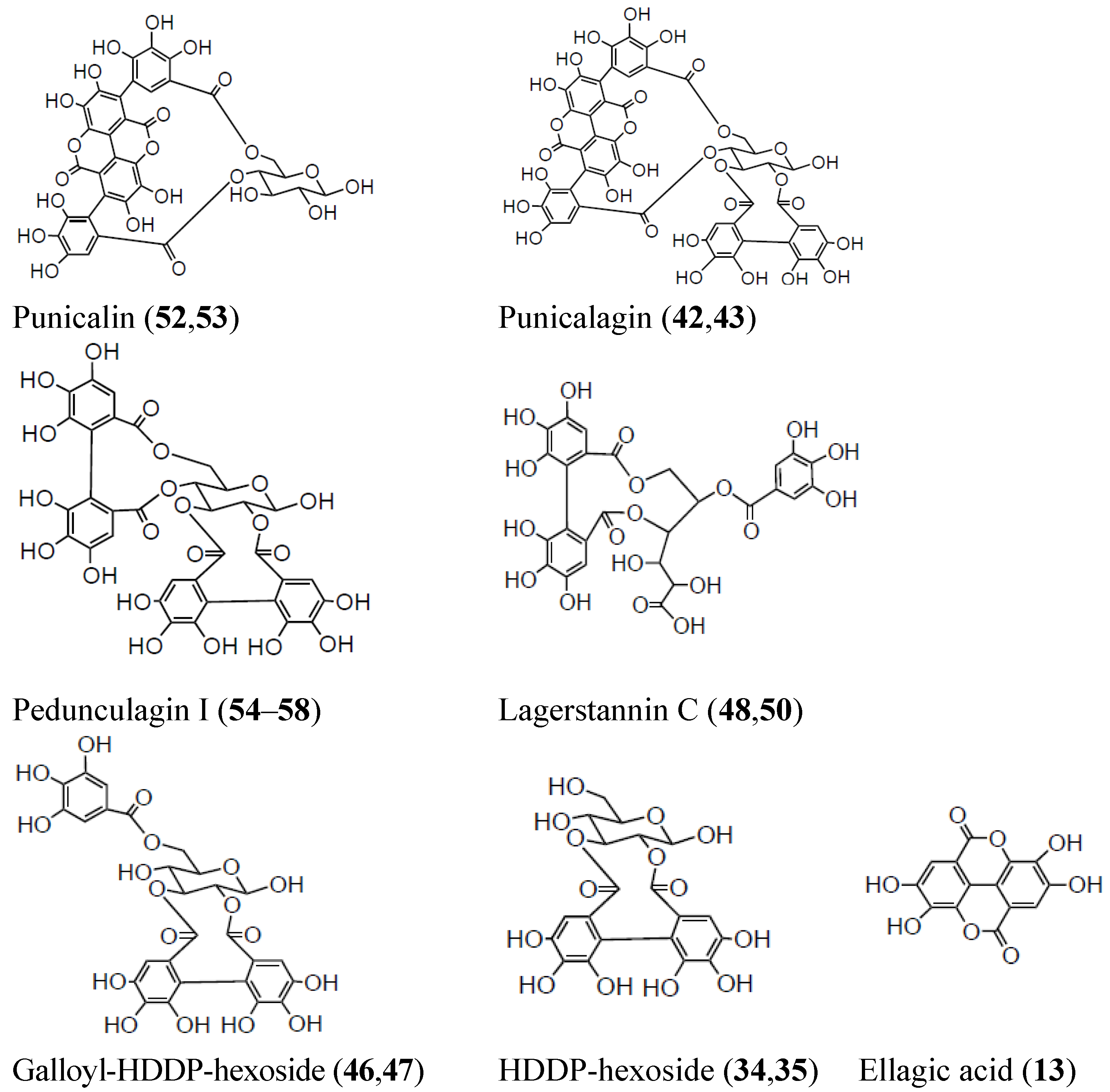
| 13 | Ellagic acid a | 301 | 301, 257, 229, 185 | 257, 229, 301, 284, 185 | 8.18 | x | x | 8.04 | x | x | 8.18 | x | |
| 14 | (+)-Gallocatechin a | 305 | 179, 221, 219, 261 | 164, 151, 137 | 6.08 | x | |||||||
| 15 | Vanillic acid-hexoside | 329 | 167 | 123, 152, 108 | 4.93 | x | x | ||||||
| 16 | Galloyl-hexoside | 331 | 169, 241, 125, 223 | 125, 169 | 1.60 | x | 1.60 | x | |||||
| 17 | Galloyl-hexoside | 331 | 169, 271, 211, 193, 241 | 125, 169 | 1.79 | x | x | 1.79 | x | x | 1.79 | x | x |
| 18 | Galloyl-hexoside | 331 | 169, 193, 211, 271, 313 | 125, 169 | 1.96 | x | 1.96 | x | x | ||||
| 19 | Galloyl-hexoside | 331 | 271, 169 | 211, 169 | 2.25 | x | x | 2.25 | x | x | 2.25 | x | |
| 20 | Galloyl-hexoside | 331 | 241, 271, 169, 313 | 169 | 3.02 | x | |||||||
| 21 | Galloyl-hexoside | 331 | 271, 169, 241 | 211, 169 | 3.25 | x | 3.25 | x | x | 3.25 | x | ||
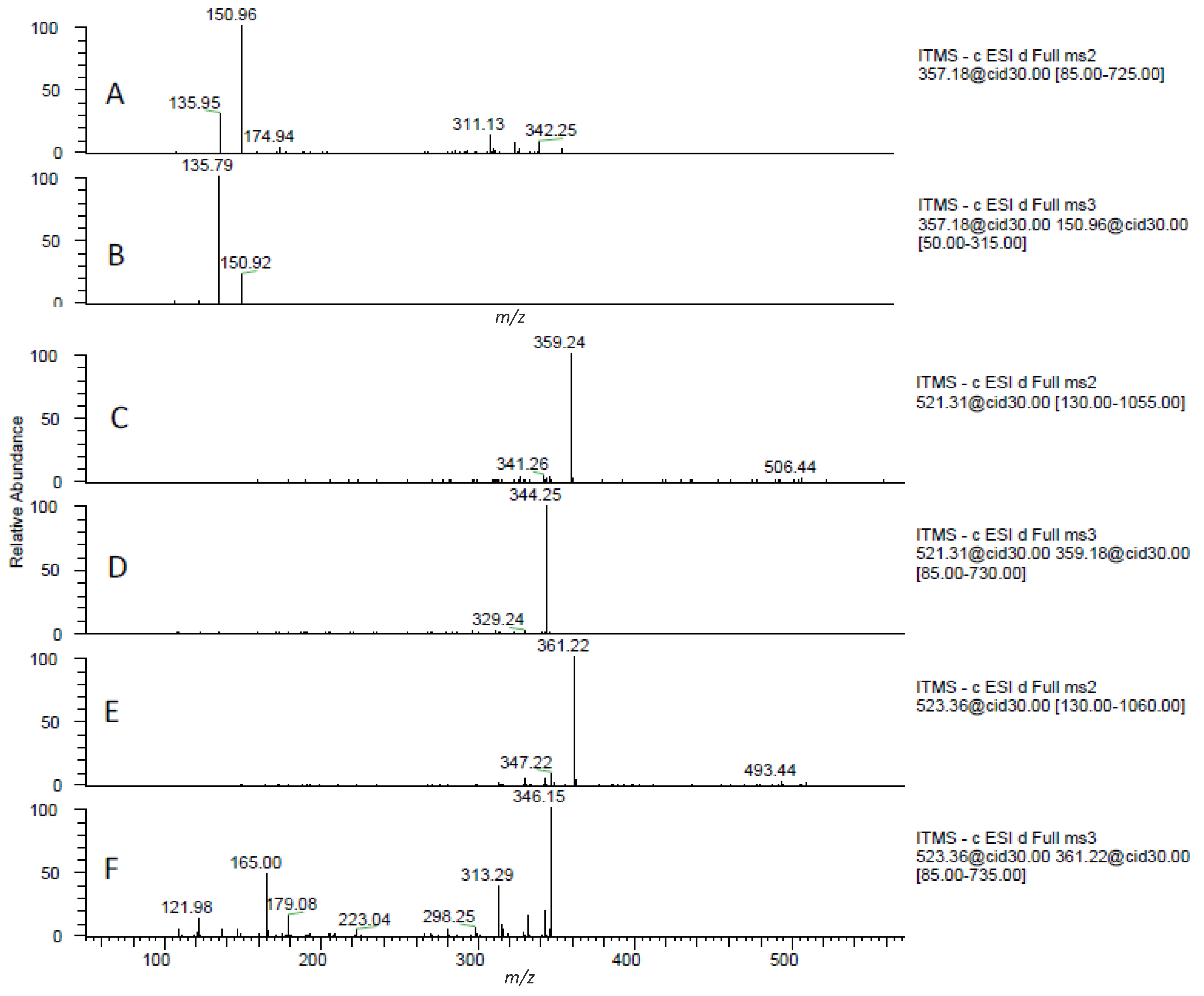
| 22 | Pinoresinol | 357 | 151, 136, 311, 327 | 136, 151 | 8.78 | x | |||||||
| 23 | Secoisolariciresinol | 361 | 346, 165, 179, 313 | 165, 179, 223, 122, 315 | 7.80 | x | |||||||
| 24 | Citric acid derivative | 391 | 217, 191, 373 | 111, 155 | 1.00 | x | 1.40 | x | 1.58 | x | |||
| 25 | Coumaric acid derivative b | 429 | 163, 265, 235, 325, 307 | 145, 119, 103, 89, 127 | 7.80 | x | 8.16 | x | 7.31 | x | |||
| 26 | Ellagic acid-pentoside | 433 | 301 | 300, 257, 229 | 7.93 | x | 7.72 | x | x | ||||
| 27 | Phloretin-hexoside (Phlorizin) a,b | 435 | 273, 297 | 167 | 10.38 | x | |||||||
| 28 | Ellagic acid-deoxyhexoside | 447 | 300 | 300, 257, 229 | 7.99 | x | x | 7.81 | x | x | 7.77 | x | x |
| 29 | Kaempferol-hexoside | 447 | 285, 284, 327, 255 | 257, 267, 229, 241 | 9.10 | x | |||||||
| 30 | Datiscetin-hexoside b | 447 | 285 | 241, 257, 125, 217, 243 | 7.01 | x | 6.85 | x | x | 6.86 | x | ||
| 31 | Dihydrokaempferol-hexoside | 449 | 287, 269, 259, 431 | 259, 243, 269 | 7.32 | x | x | 7.49 | x | x | 7.18 | x | x |
| 32 | Ellagic acid-hexoside | 463 | 301 | 301, 257, 229 | 7.22 | x | x | 7.14 | x | x | 7.10 | x | x |
| 33 | Quercetin-hexoside | 463 | 301 | 179, 151, 257, 301, 273 | 8.46 | x | |||||||
| 34 | HHDP-hexoside | 481 | 301, 275 | 301, 257, 229 | 1.06 | x | x | 1.21 | x | x | 1.06 | x | x |
| 35 | HHDP-hexoside | 481 | 301, 275 | 301, 257, 229 | 1.37 | x | x | 1.51 | x | x | 1.37 | x | x |
| 36 | Digalloyl-hexoside | 483 | 331, 313, 169 | 193, 169, 271, 211, 313 | 2.85 | x | |||||||
| 37 | Syringetin hexoside | 507 | 327, 345, 315 | 312, 283 | 8.65 | x | 8.20 | x | 8.00 | x | |||
| 38 | Feruloyl coniferin b | 517 | 337 | 193, 175, 217, 277 | 8.05 | x | |||||||
| 39 | Cyclolariciresinol hexoside b | 521 | 359 | 344 | 10.17 | x | x | 8.16 | x | x | |||
| 40 | Secoisolariciresinol hexoside b | 523 | 361, 347 | 346, 165, 179, 313 | 8.62 | x | 8.73 | x | |||||
| 41 | Guaiacyl(8-5)ferulic acid hexoside b | 533 | 353, 473, 443, 425, 371 | 338, 353, 413, 395, 371 | 9.24 | x | |||||||
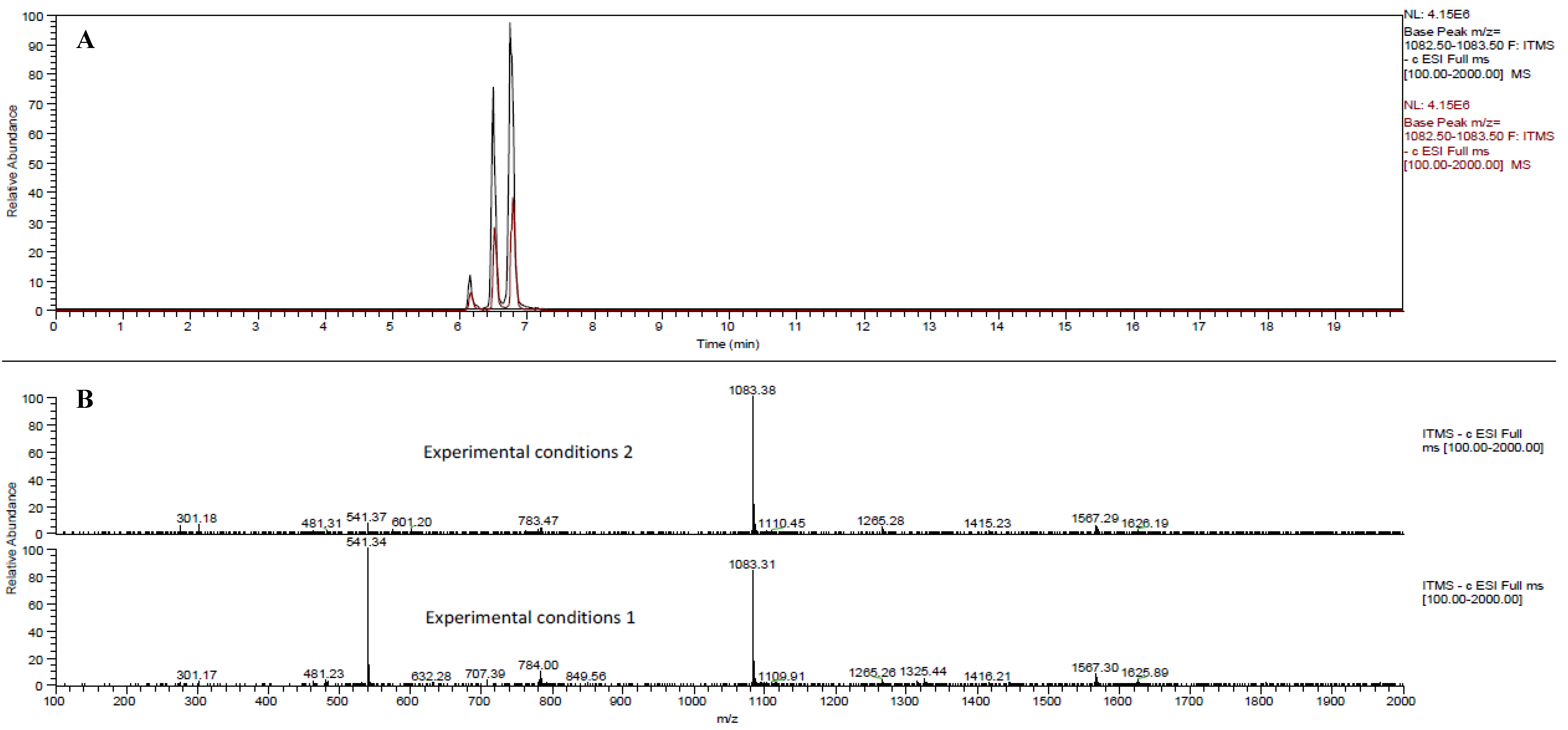
| 42 | Punicalagin isomers a | 541 | 301, 781, 601, 532, 275 | 301, 257, 229 | 6.42 | x | 6.42 | x | 6.42 | x | |||
| 43 | Punicalagin isomers a | 541 | 301, 781, 601, 532, 275 | 301, 257, 229 | 6.75 | x | x | 6.75 | x | 6.75 | x | ||
| 44 | Kaempferol rutinoside | 593 | 285, 547 | 257, 267, 241 | 8.90 | x | |||||||
| 45 | Dehydro-galloyl-HHDP-hexoside | 615 | 463, 301, 257, 229 | 301 | 7.76 | x | |||||||
| 46 | Galloyl-HHDP-hexoside | 633 | 301, 463, 275, 481 | 301, 257, 229 | 4.59 | x | x | 4.11 | x | ||||
| 47 | Galloyl-HHDP-hexoside | 633 | 301, 463, 275, 481 | 301, 257, 229 | 7.08 | x | x | 7.22 | x | x | |||
| 48 | Galloyl-HHDP-gluconate (lagerstannin C) isomer | 649 | 301, 497 | 301, 257, 229 | 1.70 | x | 1.96 | x | 2.30 | x | |||
| 49 | Trisgalloyl glucose b | 649 | 605, 479, 301 | 481, 299, 301, 425 | 4.29 | x | |||||||
| 50 | Galloyl-HHDP-gluconate (lagerstannin C) isomer | 649 | 497, 301 | 301 | 5.78 | x | x | 5.90 | x | 6.06 | x | ||
| 51 | di(HHDP-galloylglucose)-pentose b | 707 | 783, 613, 633, 1113, 933 | 481, 301, 765, 721, 275 | 6.22 | x | 6.68 | x | 6.25 | x | |||
| 52 | Punicalin α/A | 781 | 601, 721 | 299, 271 | 3.66 | x | x | 2.59 | x | x | 3.10 | x | x |
| 53 | Punicalin β/B | 781 | 601, 721 | 299, 271 | 3.90 | x | x | 2.80 | x | x | 3.30 | x | x |
| 54 | Pedunculagin I isomer | 783 | 481, 721, 765, 301 | 437, 419, 299, 275 | 3.48 | x | x | 3.53 | x | x | 5.64 | x | x |
| 55 | Pedunculagin I isomer | 783 | 301, 481, 765 | 301, 257, 229 | 5.57 | x | x | 5.16 | x | x | 5.57 | x | |
| 56 | Pedunculagin I isomer | 783 | 301, 481, 765 | 301, 257, 229 | 5.94 | x | x | 5.69 | x | x | 6.00 | x | |
| 57 | Pedunculagin I isomer | 783 | 301, 481, 765 | 301, 257, 229 | 6.53 | x | x | 6.53 | x | ||||
| 58 | Pedunculagin I isomer | 783 | 301, 481, 275, 765 | 301, 257, 229 | 6.65 | x | x | 6.65 | x | 6.55 | x | x | |
| 70 | Punicalagin isomer | 1083 | 1065, 807, 601, 1021, 721 | 721, 575, 1047, 1021, 601 | 6.08 | x | x | 5.40 | x | x | 6.11 | x | |
| 42 | Punicalagin α a | 1083 | 781, 601 | 601, 721 | 6.45 | x | x | 6.49 | x | x | |||
| 43 | Punicalagin β a | 1083 | 781, 601 | 601, 721 | 6.75 | x | x | 6.71 | x | x | |||
| 71 | Digalloyl-gallagyl-hexoside | 1085 | 765, 783, 633, 451 | 597, 613, 301 | 9.56 | x | x | ||||||
| 72 | Punicalagin-like | 1101 | 1083, 781, 601 | 781, 601, 575, 721 | 2.56 | x | 2.56 | x | x | ||||
| 51 | Di(HHDP-galloylglucose)-pentose b | 1415 | 1397, 783, 933, 1113, 633 | 765, 907, 631, 1121, 1077 | 6.28 | x | 6.66 | x | 6.30 | x | |||
| 73 | Digalloyl triHHDP-diglucose (sanguiin H10) isomer b | 1567 | 765, 935, 783, 915, 1209 | 597, 401, 301, 613, 533 | 6.75 | x | |||||||

2.1.2. Gallotannins
2.1.3. Non-Coloured Flavonoids
2.1.4. Phenolic Acid Derivatives

2.1.5. Lignans
2.1.6. Organic Acids
2.1.7. Anthocyanin Identification by Using Positive Mode
| Id. | Compounds | [M]+ (m/z) | MS2 ion fragments (m/z) a | MS3 ion fragments (m/z) a | BlueOrchid C18 | Hypersil Gold C18 | Kinetex PFP | |||
|---|---|---|---|---|---|---|---|---|---|---|
| RT (min) | RT (min) | RT (min) | ||||||||
| POS1 | Pelargonidin-3-glucoside | 433 | 271 b | 6,40 | x | 6,12 | x | 6,22 | x | |
| POS2 | Cyanidin-3-glucoside | 449 | 287 | 6,08 | x | 5,80 | x | 5,82 | x | |
| POS3 | Delphinidin-3-glucoside | 465 | 303 | 5,80 | x | 5,52 | x | 5,50 | x | |
| POS4 | Pelargonidin-3,5-diglucoside | 595 | 433, 271 | 271 | 5,70 | x | 5,47 | x | 5,38 | x |
| POS5 | Cyanidin-3,5-diglucoside | 611 | 449, 287 | 287 | 5,52 | x | 5,16 | x | 5,17 | x |
| POS6 | Delphinidin-3,5-diglucoside | 627 | 465, 303 | 303 | 5,28 | x | 5,00 | x | 4,88 | x |
| POS7 | (epi)gallocatechin-cyanidin-3-hexoside | 753 | 591 | 423, 573, 329, 465, 287 | 5,13 | x | ||||
2.1.8. General Discussion about the Use of This New Approach for Phytochemical Identification
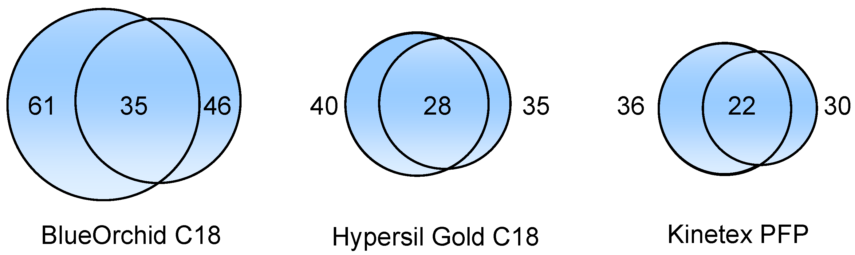
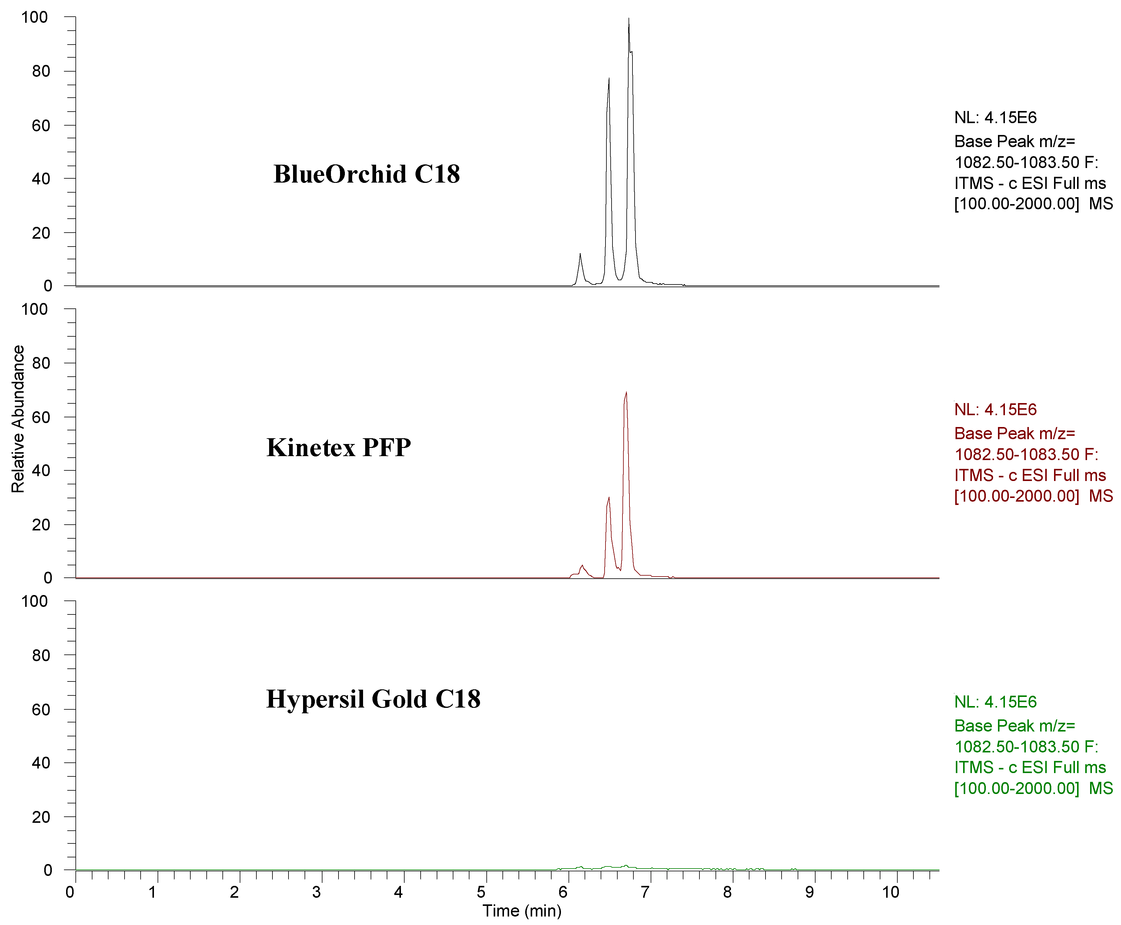
2.2. Comparison of Pomegranate Juice LC/MS Based Fingerprinting among Different Chromatographic Columns
3. Experimental
3.1. Pomegranate Juice and Sample Preparation
3.2. UHPLC-MSn Analyses
4. Conclusions
Acknowledgments
- Sample Availability: Samples of analysed pomegranate juice are available from the authors.
References
- Gasperotti, M.; Masuero, D.; Vrhovsek, U.; Guella, G.; Mattivi, F. Profiling and accurate quantification of Rubus ellagitannins and ellagic acid conjugates using direct UPLC-Q-TOF hdms and HPLC-DAD analysis. J. Agric. Food Chem. 2010, 58, 4602–4616. [Google Scholar]
- Mulabagal, V.; Calderón, A.I. Liquid chromatography/mass spectrometry based fingerprinting analysis and mass profiling of Euterpe oleracea (açaí) dietary supplement raw materials. Food Chem. 2012, 134, 1156–1164. [Google Scholar] [CrossRef]
- Mena, P.; Gironés-Vilaplana, A.; Moreno, D.A.; García-Viguera, C. “Pomegranate fruit for health promotion: Myths and realities” In Antioxidant Properties of Crops III. Funct. Plant Sci. Biotechnol. 2011, 5, 33–42. [Google Scholar]
- De Pascual-Teresa, S.; Santos-Buelga, C.; Rivas-Gonzalo, J.G. Quantitative analysis of flavan-3-ols in Spanish foodstuffs and beverages. J. Agric. Food Chem. 2000, 48, 5331–5337. [Google Scholar] [CrossRef]
- Fischer, U.A.; Carle, R.; Kammerer, D.R. Identification and quantification of phenolic compounds from pomegranate (Punica granatum L.) peel, mesocarp, aril and differently produced juices by HPLC-DAD-ESI/MSn. Food Chem. 2011, 127, 807–821. [Google Scholar] [CrossRef]
- Gil, M.I.; García-Viguera, C.; Artés, F.; Tomás-Barberán, F.A. Changes in pomegranate juice pigmentation during ripening. J. Sci. Food Agric. 1995, 68, 77–81. [Google Scholar] [CrossRef]
- Gil, M.I.; Tomás-Barberán, F.A.; Hess-Pierce, B.; Holcroft, D.M.; Kader, A.A. Antioxidant activity of pomegranate juice and its relationship with phenolic composition and processing. J. Agric. Food Chem. 2000, 48, 4581–4589. [Google Scholar]
- Ben Nasr, C.; Ayed, N.; Metche, M. Quantitative determination of the polyphenolic content of pomegranate peel. Z. Lebensm. Unters. Forsch. 1996, 203, 374–378. [Google Scholar] [CrossRef]
- Zhang, Y.; Krueger, D.; Durst, R.; Lee, R.; Wang, D.; Seeram, N.; Heber, D. International multidimensional authenticity specification (IMAS) algorithm for detection of commercial pomegranate juice adulteration. J. Agric. Food Chem. 2009, 57, 2550–2557. [Google Scholar] [CrossRef]
- Plumb, G.W.; de Pascual-Teresa, S.; Santos-Buelga, C.; Rivas-Gonzalo, J.C.; Williamson, G. Antioxidant properties of gallocatechin and prodelphinidins from pomegranate peel. Redox Rep. 2002, 7, 41–46. [Google Scholar] [CrossRef]
- Borges, G.; Mullen, W.; Crozier, A. Comparison of the polyphenolic composition and antioxidant activity of European commercial fruit juices. Food Funct. 2010, 1, 73–83. [Google Scholar] [CrossRef]
- Cristofori, V.; Caruso, D.; Latini, G.; Dell’Agli, M.; Cammilli, C.; Rugini, E.; Bignami, C.; Muleo, R. Fruit quality of Italian pomegranate (Punica granatum L.) autochthonous varieties. Eur. Food Res. Technol. 2010, 232, 397–403. [Google Scholar]
- Romani, A.; Campo, M.; Pinelli, P. HPLC/DAD/ESI-MS analyses and anti-radical activity of hydrolyzable tannins from different vegetal species. Food Chem. 2012, 130, 214–221. [Google Scholar] [CrossRef]
- Borges, G.; Crozier, A. HPLC-PDA-MS fingerprinting to assess the authenticity of pomegranate beverages. Food Chem. 2012, 135, 1863–1867. [Google Scholar] [CrossRef]
- Törrönen, A.R. Sources and health effects of dietary ellagitannins. In Chemistry and Biology of Ellagitannins; Quideau, S., Ed.; World Scientific Publishing: Singapore, 2009; pp. 298–319. [Google Scholar]
- Filigenzi, M.S.; Ehrke, N.; Aston, L.S.; Poppenga, R.H. Evaluation of a rapid screening method for chemical contaminants of concern in four food-related matrices using QuEChERS extraction, UHPLC and high resolution mass spectrometry. Food Addit. Contam. A 2011, 28, 1324–1339. [Google Scholar] [CrossRef]
- Di Stefano, V.; Avellone, G.; Bongiorno, D.; Cunsolo, V.; Muccilli, V.; Sforza, S.; Dossena, A.; Drahos, L.; Vékey, K. Applications of liquid chromatography-mass spectrometry for food analysis. J. Chromatogr. A 1259, 74–85. [Google Scholar]
- Rak, G.; Fodor, P.; Abrankó, L. Three-step HPLC-ESI-MS/MS procedure for screening and identifying non-target flavonoid derivatives. Int. J. Mass Spectrom. 2010, 290, 32–38. [Google Scholar] [CrossRef]
- Del Rio, D.; Rodríguez-Mateos, A.; Spencer, J.P.; Tognolini, M.; Borges, G.; Crozier, A. Dietary (Poly)phenolics in Human Health: Structures, Bioavailability, and Evidence of Protective Effects Against Chronic Diseases. Antioxid. Redox Signal. 2012. [Google Scholar] [CrossRef]
- Hukkanen, A.T.; Kokko, H.I.; Buchala, A.J.; McDougall, G.J.; Stewart, D.; Kärenlampi, S.O.; Karjalainen, R.O. Benzothiadiazole induces the accumulation of phenolics and improves resistance to powdery mildew in strawberries. J. Agric. Food Chem. 2007, 55, 1862–1870. [Google Scholar]
- Fischer, U.A.; Dettmann, J.S.; Carle, R.; Kammerer, D.R. Impact of processing and storage on the phenolic profiles and contents of pomegranate (Punica granatum L.) juices. Eur. Food Res. Technol. 2011, 233, 797–816. [Google Scholar] [CrossRef]
- Kähkönen, M.; Kylli, P.; Ollilainen, V.; Salminen, J.P.; Heinonen, M. Antioxidant activity of isolated ellagitannins from red raspberries and cloudberries. J. Agric. Food Chem. 2012, 60, 1167–1174. [Google Scholar]
- Barry, K.M.; Pearce, R.B.; Mohammed, C.M. Properties of reaction zones associated with decay from pruning wounds in plantation-grown Eucalyptus nitens. For. Pathol. 2000, 30, 233–245. [Google Scholar] [CrossRef]
- De Rijke, E.; Zappey, H.; Ariese, F.; Gooijer, C.; Brinkman, U.A.T. Liquid chromatography with atmospheric pressure chemical ionization and electrospray ionization mass spectrometry of flavonoids with triple-quadrupole and ion-trap instruments. J. Chromatogr. A 2003, 984, 45–58. [Google Scholar] [CrossRef]
- Arnao, M.B.; Cano, A.; Alcolea, J.F.; Acosta, M. Identification of hydrolysable tannins in the reaction zone of Eucalyptus nitens wood by high performance liquid chromatography-electrospray ionisation mass spectrometry. Phytochem. Anal. 2001, 12, 120–127. [Google Scholar] [CrossRef]
- Boulekbache-Makhlouf, L.; Meudec, E.; Chibane, M.; Mazauric, J.P.; Slimani, S.; Henry, M.; Cheynier, V.; Madani, K. Analysis by high-performance liquid chromatography diode array detection mass spectrometry of phenolic compounds in fruit of Eucalyptus globulus cultivated in Algeria. J. Agric. Food Chem. 2010, 58, 12615–12624. [Google Scholar] [CrossRef]
- Venzie, J.L.; Castro, J.; Balarama Krishna, M.V.; Nelson, D.M.; Marcus, R.K. Electron-impact and glow-discharge ionization LC-MS analysis of green tea tincture. Anal. Bioanal. Chem. 2007, 387, 321–333. [Google Scholar]
- Zywicki, B.; Reemtsma, T.; Jekel, M. Analysis of commercial vegetable tanning agents by reversed-phase liquid chromatography-electrospray ionization-tandem mass spectrometry and its application to wastewater. J. Chromatogr. A 2002, 970, 191–200. [Google Scholar] [CrossRef]
- Vrhovsek, U.; Guella, G.; Gasperotti, M.; Pojer, E.; Zancato, M.; Mattivi, F. Clarifying the identity of the main ellagitannin in the fruit of the strawberry, Fragaria vesca and Fragaria ananassa Duch. J. Agric. Food Chem. 2012, 60, 2507–2516. [Google Scholar] [CrossRef]
- Mena, P.; Gironés-Vilaplana, A.; Martí, N.; García-Viguera, C. Pomegranate varietal wines: Phytochemical composition and quality parameters. Food Chem. 2012, 133, 108–115. [Google Scholar] [CrossRef]
- He, L.; Xu, H.; Liu, X.; He, W.; Yuan, F.; Hou, Z.; Gao, Y. Identification of phenolic compounds from pomegranate (Punica granatum L.) seed residues and investigation into their antioxidant capacities by HPLC-ABTS+ assay. Food Res. Int. 2011, 44, 1161–1167. [Google Scholar] [CrossRef]
- McNab, H.; Ferreira, E.S.B.; Hulme, A.N.; Quye, A. Negative ion ESI-MS analysis of natural yellow dye flavonoids-An isotopic labelling study. Int. J. Mass Spectrom. 2009, 284, 57–65. [Google Scholar] [CrossRef]
- Petroviciu, I.; Vanden Berghe, I.; Cretu, I.; Albu, F.; Medvedovici, A. Identification of natural dyes in historical textiles from Romanian collections by LC-DAD and LC-MS (single stage and tandem MS). J. Cult. Herit. 2012, 13, 89–97. [Google Scholar] [CrossRef]
- Liu, R.; Ye, M.; Guo, H.; Bi, K.; Guo, D.A. Liquid chromatography/electrospray ionization mass spectrometry for the characterization of twenty-three flavonoids in the extract of Dalbergia odorifera. Rapid Commun. Mass Spectrum. 2005, 19, 1557–1565. [Google Scholar] [CrossRef]
- Li, H.; Qiu, J.; Chen, F.; Lv, X.; Fu, C.; Zhao, D.; Hua, X.; Zhao, Q. Molecular characterization and expression analysis of dihydroflavonol 4-reductase (DFR) gene in Saussurea medusa. Mol. Biol. Rep. 2012, 39, 2991–2999. [Google Scholar] [CrossRef]
- Wang, Y.; Yang, L.; He, Y.Q.; Wang, C.H.; Welbeck, E.W.; Bligh, S.W.A.; Wang, Z.T. Characterization of fifty-one flavonoids in a Chinese herbal prescription Longdan Xiegan Decoction by high-performance liquid chromatography coupled to electrospray ionization tandem mass spectrometry and photodiode array detection. Rapid Commun. Mass Spectrum. 2008, 22, 1767–1778. [Google Scholar]
- Perestrelo, R.; Lu, Y.; Santos, S.A.O.; Silvestre, A.J.D.; Neto, C.P.; Câmara, J.S.; Rocha, S.M. Phenolic profile of Sercial and Tinta Negra Vitis vinifera L. grape skins by HPLC-DAD-ESI-MSn: Novel phenolic compounds in Vitis vinifera L. grape. Food Chem. 2012, 135, 94–104. [Google Scholar]
- Sanz, M.; Fernández de Simón, B.; Esteruelas, E.; Muñoz, A.M.; Cadahía, E.; Hernández, T.; Estrella, I.; Pinto, E. Effect of Toasting Intensity at Cooperage on Phenolic Compounds in Acacia (Robinia pseudoacacia) Heartwood. J. Agric. Food Chem. 2011, 59, 3135–3145. [Google Scholar] [CrossRef]
- Eklund, P.C.; Backman, M.J.; Kronberg, L.Å.; Smeds, A.I.; Sjöholm, R.E. Identification of lignans by liquid chromatography-electrospray ionization ion-trap mass spectrometry. J. Mass Spectrom. 2008, 43, 97–107. [Google Scholar]
- Huis, R.; Morreel, K.; Fliniaux, O.; Lucau-Danila, A.; Fénart, S.; Grec, S.; Neutelings, G.; Chabbert, B.; Mesnard, F.; Boerjan, W.; et al. Natural hypolignification is associated with extensive oligolignol accumulation in flax stems. Plant Physiol. 2012, 158, 1893–1915. [Google Scholar] [CrossRef]
- Bonzanini, F.; Bruni, R.; Palla, G.; Serlataite, N.; Caligiani, A. Identification and distribution of lignans in Punica granatum L. fruit endocarp, pulp, seeds, wood knots and commercial juices by GC-MS. Food Chem. 2009, 117, 745–749. [Google Scholar] [CrossRef]
- Fischer, U.A.; Jaksch, A.V.; Carle, R.; Kammerer, D.R. Determination of lignans in edible and nonedible parts of pomegranate (Punica granatum L.) and products derived therefrom, particularly focusing on the quantitation of isolariciresinol using HPLC-DAD-ESI/MSn. J. Agric. Food Chem. 2012, 60, 283–292. [Google Scholar] [CrossRef]
- Sawada, Y.; Akiyama, K.; Sakata, A.; Kuwahara, A.; Otsuki, H.; Sakurai, T.; Saito, K.; Hirai, M.Y. Widely targeted metabolomics based on large-scale MS/MS data for elucidating metabolite accumulation patterns in plants. Plant Cell Physiol. 2009, 50, 37–47. [Google Scholar] [CrossRef]
- Mena, P.; García-Viguera, C.; Navarro-Rico, J.; Moreno, D.A.; Bartual, J.; Saura, D.; Martí, N. Phytochemical characterisation for industrial use of pomegranate (Punica granatum L.) cultivars grown in Spain. J. Sci. Food Agric. 2011, 91, 1893–1906. [Google Scholar]
- Sun, J.; Lin, L.Z.; Chen, P. Study of the mass spectrometric behaviors of anthocyanins in negative ionization mode and its applications for characterization of anthocyanins and non-anthocyanin polyphenols. Rapid Commun. Mass Spectrum. 2012, 26, 1123–1133. [Google Scholar] [CrossRef]
- Sentandreu, E.; Navarro, J.L.; Sendra, J.M. LC-DAD-ESI/MSn determination of direct condensation flavanol-anthocyanin adducts in pressure extracted Pomegranate (Punica granatum L.) juice. J. Agric. Food Chem. 2010, 58, 10560–10567. [Google Scholar] [CrossRef]
- Regos, I.; Treutter, D. Optimization of a high-performance liquid chromatography method for the analysis of complex polyphenol mixtures and application for sainfoin extracts (Onobrychis viciifolia). J. Chromatogr. A 2010, 1217, 6169–6177. [Google Scholar] [CrossRef]
- Haghedooren, E.; Janssens, T.; Nijs, R.; Park, S.K.; Farkas, E.; Dragovic, S.; Noszál, B.; Hoogmartens, J.; Adams, E. Selecting a suitable LC column for pharmaceutical separations using a column characterisation system. J. Liq. Chromatogr. Relat. Technol. 2009, 32, 747–771. [Google Scholar] [CrossRef]
- Mena, P.; Martí, N.; Saura, D.; Valero, M.; García-Viguera, C. Combinatory Effect of Thermal Treatment and Blending on the Quality of Pomegranate Juices. Food Bioprocess Technol. 2012. [Google Scholar] [CrossRef]
- Pérez-Vicente, A.; Serrano, P.; Abellán, P.; García-Viguera, C. Influence of packaging material on pomegranate juice colour and bioactive compounds, during storage. J. Sci. Food Agric. 2004, 84, 639–644. [Google Scholar]
© 2012 by the authors; licensee MDPI, Basel, Switzerland. This article is an open-access article distributed under the terms and conditions of the Creative Commons Attribution license (http://creativecommons.org/licenses/by/3.0/).
Share and Cite
Mena, P.; Calani, L.; Dall'Asta, C.; Galaverna, G.; García-Viguera, C.; Bruni, R.; Crozier, A.; Del Rio, D. Rapid and Comprehensive Evaluation of (Poly)phenolic Compounds in Pomegranate (Punica granatum L.) Juice by UHPLC-MSn. Molecules 2012, 17, 14821-14840. https://doi.org/10.3390/molecules171214821
Mena P, Calani L, Dall'Asta C, Galaverna G, García-Viguera C, Bruni R, Crozier A, Del Rio D. Rapid and Comprehensive Evaluation of (Poly)phenolic Compounds in Pomegranate (Punica granatum L.) Juice by UHPLC-MSn. Molecules. 2012; 17(12):14821-14840. https://doi.org/10.3390/molecules171214821
Chicago/Turabian StyleMena, Pedro, Luca Calani, Chiara Dall'Asta, Gianni Galaverna, Cristina García-Viguera, Renato Bruni, Alan Crozier, and Daniele Del Rio. 2012. "Rapid and Comprehensive Evaluation of (Poly)phenolic Compounds in Pomegranate (Punica granatum L.) Juice by UHPLC-MSn" Molecules 17, no. 12: 14821-14840. https://doi.org/10.3390/molecules171214821