Antibiotics 2021, 10(6), 719; https://doi.org/10.3390/antibiotics10060719 - 15 Jun 2021
Cited by 46 | Viewed by 22857
Abstract
Bacterial vaginosis (BV) has been reported in one-third of women worldwide at different life stages, due to the complex balance in the ecology of the vaginal microbiota. It is a common cause of abnormal vaginal discharge and is associated with other health issues.
[...] Read more.
Bacterial vaginosis (BV) has been reported in one-third of women worldwide at different life stages, due to the complex balance in the ecology of the vaginal microbiota. It is a common cause of abnormal vaginal discharge and is associated with other health issues. Since the first description of anaerobic microbes associated with BV like Gardnerella vaginalis in the 1950s, researchers have stepped up the game by incorporating advanced molecular tools to monitor and evaluate the extent of dysbiosis within the vaginal microbiome, particularly on how specific microbial population changes compared to a healthy state. Moreover, treatment failure and BV recurrence rate remain high despite the standard antibiotic treatment. Consequently, researchers have been probing into alternative or adjunct treatments, including probiotics or even vaginal microbiota transplants, to ensure successful treatment outcomes and reduce the colonization by pathogenic microbes of the female reproductive tract. The current review summarizes the latest findings in probiotics use for BV and explores the potential of vaginal microbiota transplants in restoring vaginal health.
Full article
(This article belongs to the Special Issue Alternative Approaches to Treating Antimicrobial Resistant Infections)
►
Show Figures
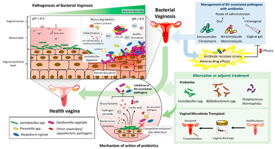
Figure 1


