by
Daniele Marra 1 , Irene Perna 1, Giulio Pota 1, Giuseppe Vitiello 1,2, Alessandro Pezzella 1, Giuseppe Toscano 1, Giuseppina Luciani 1 and Sergio Caserta 1,3,*
, Irene Perna 1, Giulio Pota 1, Giuseppe Vitiello 1,2, Alessandro Pezzella 1, Giuseppe Toscano 1, Giuseppina Luciani 1 and Sergio Caserta 1,3,*
1
DICMaPI, Università di Napoli Federico II, Piazzale V. Tecchio 80, 80125 Napoli, Italy
2
CSGI, Center for Colloid and Surface Science, Via della Lastruccia 3, 50019 Florence, Italy
3
CEINGE, Advanced Biotechnologies, 80145 Naples, Italy
Microorganisms 2023, 11(3), 621; https://doi.org/10.3390/microorganisms11030621 - 28 Feb 2023
Cited by 9 | Viewed by 3629
Abstract
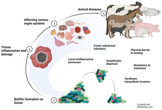
Microbial colonization of surfaces is a sanitary and industrial issue for many applications, leading to product contamination and human infections. When microorganisms closely interact with a surface, they start to produce an exo-polysaccaridic matrix to adhere to and protect themselves from adverse environmental
[...] Read more.
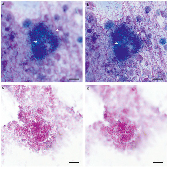
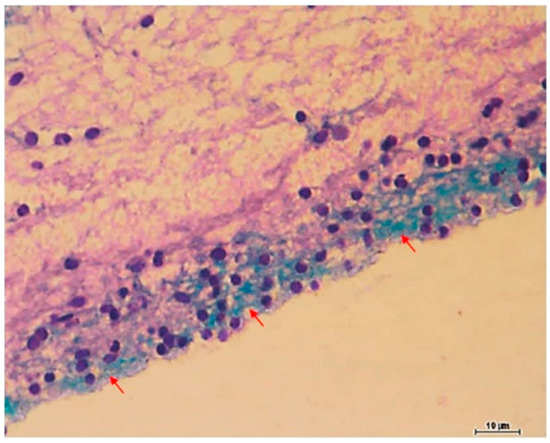
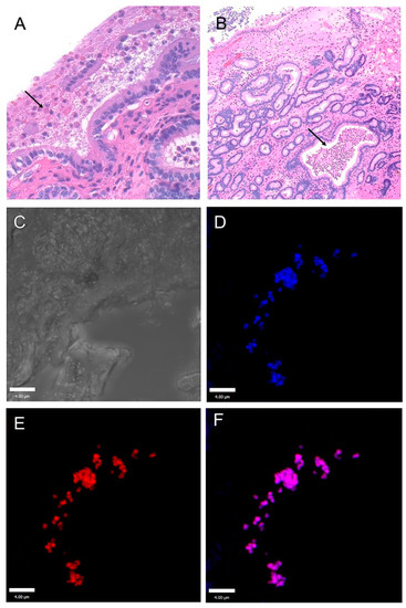
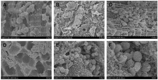
Microbial colonization of surfaces is a sanitary and industrial issue for many applications, leading to product contamination and human infections. When microorganisms closely interact with a surface, they start to produce an exo-polysaccaridic matrix to adhere to and protect themselves from adverse environmental conditions. This type of structure is called a biofilm. The aim of our work is to investigate novel technologies able to prevent biofilm formation by surface coatings. We coated glass surfaces with melanin-ZnO2, melanin-TiO2, and TiO2 hybrid nanoparticles. The functionalization was performed using cold plasma to activate glass-substrate-coated surfaces, that were characterized by performing water and soybean oil wetting tests. A quantitative characterization of the antibiofilm properties was done using Pseudomonas fluorescens AR 11 as a model organism. Biofilm morphologies were observed using confocal laser scanning microscopy and image analysis techniques were used to obtain quantitative morphological parameters. The results highlight the efficacy of the proposed surface coating to prevent biofilm formation. Melanin-TiO2 proved to be the most efficient among the particles investigated. Our results can be a valuable support for future implementation of the technique proposed here in an extended range of applications that may include further testing on other strains and other support materials.
Full article
(This article belongs to the Special Issue Feature Papers in Microbial Biofilm Formation)
▼
Show Figures

Figure 1


