Diagnostics 2023, 13(1), 123; https://doi.org/10.3390/diagnostics13010123 - 30 Dec 2022
Cited by 9 | Viewed by 3106
| Correction
Abstract
Colorectal Cancer is one of the most common cancers found in human beings, and polyps are the predecessor of this cancer. Accurate Computer-Aided polyp detection and segmentation system can help endoscopists to detect abnormal tissues and polyps during colonoscopy examination, thereby reducing the
[...] Read more.
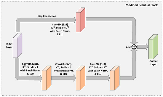
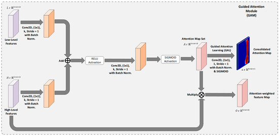
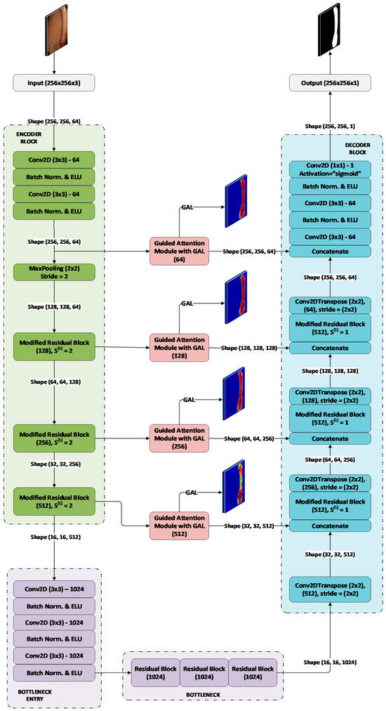
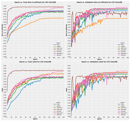
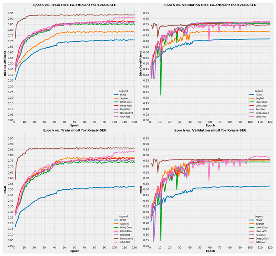
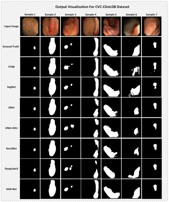
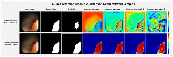
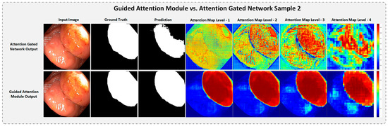
Colorectal Cancer is one of the most common cancers found in human beings, and polyps are the predecessor of this cancer. Accurate Computer-Aided polyp detection and segmentation system can help endoscopists to detect abnormal tissues and polyps during colonoscopy examination, thereby reducing the chance of polyps growing into cancer. Many of the existing techniques fail to delineate the polyps accurately and produce a noisy/broken output map if the shape and size of the polyp are irregular or small. We propose an end-to-end pixel-wise polyp segmentation model named Guided Attention Residual Network (GAR-Net) by combining the power of both residual blocks and attention mechanisms to obtain a refined continuous segmentation map. An enhanced Residual Block is proposed that suppresses the noise and captures low-level feature maps, thereby facilitating information flow for a more accurate semantic segmentation. We propose a special learning technique with a novel attention mechanism called Guided Attention Learning that can capture the refined attention maps both in earlier and deeper layers regardless of the size and shape of the polyp. To study the effectiveness of the proposed GAR-Net, various experiments were carried out on two benchmark collections viz., CVC-ClinicDB (CVC-612) and Kvasir-SEG dataset. From the experimental evaluations, it is shown that GAR-Net outperforms other previously proposed models such as FCN8, SegNet, U-Net, U-Net with Gated Attention, ResUNet, and DeepLabv3. Our proposed model achieves 91% Dice co-efficient and 83.12% mean Intersection over Union (mIoU) on the benchmark CVC-ClinicDB (CVC-612) dataset and 89.15% dice co-efficient and 81.58% mean Intersection over Union (mIoU) on the Kvasir-SEG dataset. The proposed GAR-Net model provides a robust solution for polyp segmentation from colonoscopy video frames.
Full article
(This article belongs to the Special Issue Artificial Intelligence in Gastrointestinal Disease: Diagnosis and Management)
►
Show Figures
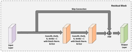
Figure 1


