by
Yanqing Chen †, Zhen Liu † , Ping Gong, Haibo Zhang, Yijun Chen, Songquan Yao, Wei Li, Yan Zhang and Yang Yu *
, Ping Gong, Haibo Zhang, Yijun Chen, Songquan Yao, Wei Li, Yan Zhang and Yang Yu *
1
School of Pharmacy, Shanghai Jiao Tong University, Shanghai 200240, China
†
These authors contributed equally to this work.
Int. J. Mol. Sci. 2022, 23(16), 9041; https://doi.org/10.3390/ijms23169041 - 12 Aug 2022
Cited by 26 | Viewed by 4343
| Correction
Abstract
The accumulation of microglia around senile plaques is one of the pathological features of Alzheimer’s disease (AD). Chemerin is an adipokine with immune-modulating properties. Our previous study showed that chemokine-like receptor 1 (CMKLR1), the receptor for chemerin, is also a functional receptor of
[...] Read more.
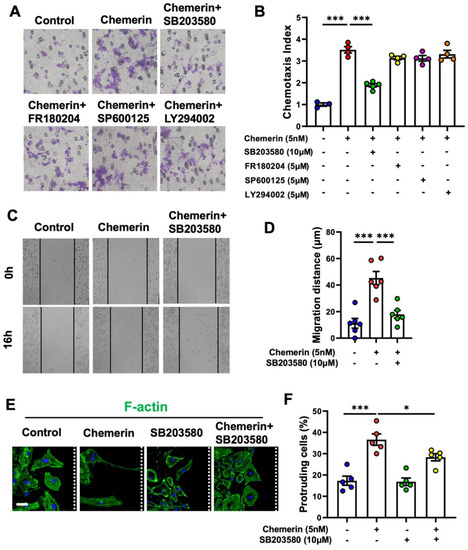
The accumulation of microglia around senile plaques is one of the pathological features of Alzheimer’s disease (AD). Chemerin is an adipokine with immune-modulating properties. Our previous study showed that chemokine-like receptor 1 (CMKLR1), the receptor for chemerin, is also a functional receptor of Aβ. However, it remains unclear whether and how the chemerin/CMKLR1 axis affects the migration of microglia. The impact of CMKLR1 on microglial activation and recruitment toward Aβ deposits was examined in APP/PS1 mice mated with CMKLR1 knockout (CMKLR1−/−) mice. CMKLR1 deficiency reduced the number of microglia around Aβ deposits in aged APP/PS1-CMKLR1−/− mice compared with APP/PS1 mice. Chemerin expression was significantly decreased in the hippocampus and cortex of aged APP/PS1 mice compared with WT mice. In vitro assays demonstrated that activation of the chemerin/CMKLR1 axis promoted the migration of primary cultures of microglia and murine microglial N9 cells. Mechanistic studies found that chemerin/CMKLR1 induced polarization and protrusion formation of microglia by promoting the remodeling of actin filaments and microtubules, and Golgi apparatus reorientation. The inhibition of p38 MAPK attenuated the promotion of the chemerin/CMKLR1 axis on microglial migration and polarization. In addition, chemerin inhibited Aβ-induced microglial clustering. The inhibition of p38 MAPK alleviated the suppressive effect of chemerin on Aβ-induced microglial aggregation. Our data indicate that the chemerin/CMKLR1 axis is involved in the migration and recruitment of microglia to senile plaques via the p38 MAPK pathway. Modulation of the chemerin/CMKLR1 axis is a potential new strategy for AD therapy.
Full article
(This article belongs to the Collection G Protein-Coupled Receptors and Their Kinases in Cell Biology and Disease)
▼
Show Figures
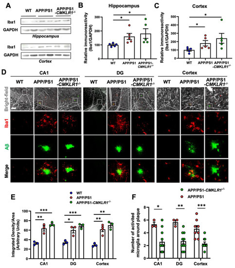
Figure 1


