9 pages, 276 KiB  
Review
Importance of Coagulation Factors as Critical Components of Premature Cardiovascular Disease in Familial Hypercholesterolemia
by Uffe Ravnskov 1,*, Michel de Lorgeril 2, Malcolm Kendrick 3 and David M. Diamond 4
1 Independent Researcher, Magle Stora Kyrkogata 9, 22350 Lund, Sweden
2 Laboratoire Coeur et Nutrition, TIMC-CNRS, Faculté de Médecine et Pharmacie-Université Grenoble Alpes, 38000 Grenoble, France
3 East Cheshire Trust, Macclesfield District General Hospital, Macclesfield SK10 3BL, UK
4 Departments of Psychology, University of South Florida, Tampa, FL 33620, USA
Int. J. Mol. Sci. 2022, 23(16), 9146; https://doi.org/10.3390/ijms23169146 - 15 Aug 2022
Cited by 9 | Viewed by 13957
Abstract
For almost a century, familial hypercholesterolemia (FH) has been considered a serious disease, causing atherosclerosis, cardiovascular disease, and ischemic stroke. Closely related to this is the widespread acceptance that its cause is greatly increased low-density-lipoprotein cholesterol (LDL-C). However, numerous observations and experiments in [...] Read more.
For almost a century, familial hypercholesterolemia (FH) has been considered a serious disease, causing atherosclerosis, cardiovascular disease, and ischemic stroke. Closely related to this is the widespread acceptance that its cause is greatly increased low-density-lipoprotein cholesterol (LDL-C). However, numerous observations and experiments in this field are in conflict with Bradford Hill’s criteria for causality. For instance, those with FH demonstrate no association between LDL-C and the degree of atherosclerosis; coronary artery calcium (CAC) shows no or an inverse association with LDL-C, and on average, the life span of those with FH is about the same as the surrounding population. Furthermore, no controlled, randomized cholesterol-lowering trial restricted to those with FH has demonstrated a positive outcome. On the other hand, a number of studies suggest that increased thrombogenic factors—either procoagulant or those that lead to high platelet reactivity—may be the primary risk factors in FH. Those individuals who die prematurely have either higher lipoprotein (a) (Lp(a)), higher factor VIII and/or higher fibrinogen compared with those with a normal lifespan, whereas their LDL-C does not differ. Conclusions: Many observational and experimental studies have demonstrated that high LDL-C cannot be the cause of premature cardiovascular mortality among people with FH. The number who die early is also much smaller than expected. Apparently, some individuals with FH may have inherited other, more important risk factors than a high LDL-C. In accordance with this, our review has shown that increased coagulation factors are the commonest cause, but there may be other ones as well. Full article
(This article belongs to the Special Issue Lipids Metabolism and Cardiometabolic Diseases)
15 pages, 9221 KiB  
Article
Phytic Acid-Iron/Laponite Coatings for Enhanced Flame Retardancy, Antidripping and Mechanical Properties of Flexible Polyurethane Foam
by Qi Jiang, Ping Li, Yun Liu * and Ping Zhu
College of Textiles & Clothing, Institute of Functional Textiles and Advanced Materials, National Engineering Research Center for Advanced Fire-Safety Materials D & A (Shandong), State Key Laboratory of Bio-Fibers and Eco-Textiles, Qingdao University, Qingdao 266071, China
Int. J. Mol. Sci. 2022, 23(16), 9145; https://doi.org/10.3390/ijms23169145 - 15 Aug 2022
Cited by 19 | Viewed by 2389
Abstract
The use of flexible polyurethane foam (FPUF) is severely limited due to its flammability and dripping, which can easily cause major fire hazards. Therefore, choosing an appropriate flame retardant to solve this problem is an urgent need. A coating was prepared on the [...] Read more.
The use of flexible polyurethane foam (FPUF) is severely limited due to its flammability and dripping, which can easily cause major fire hazards. Therefore, choosing an appropriate flame retardant to solve this problem is an urgent need. A coating was prepared on the FPUF surface by dipping with phytic acid (PA), Fe2(SO4)3·xH2O, and laponite (LAP). The influence of PA-Fe/LAP coating on FPUF flame-retardant performance was explored by thermal stability, flame retardancy, combustion behavior, and smoke density analysis. FPUF/PA-Fe/LAP has a good performance in the small fire test, which can pass the UL-94 V-0 rating and the limiting oxygen index reaches 24.5%. Meanwhile, the peak heat release rate values and maximum smoke density of FPUF/PA-Fe/LAP are reduced by 38.7% and 38.5% compared with those of neat FPUF. After applying PA-Fe/LAP coating, the value of fire growth rate index decreases from 10.5 kW/(m2·s) to 5.1 kW/(m2·s), dramatically reducing the fire risk. Encouragingly, the effect of PA-Fe/LAP coating on cyclic compression and permanent deformation is small, which is close to that of neat FPUF. This work provides an effective strategy for making a flame-retardant FPUF with antidripping and keeping mechanical properties. Full article
(This article belongs to the Special Issue Advanced Flame Retardant Polymeric Materials)
Show Figures

Figure 1

25 pages, 5536 KiB  
Article
Essential Acidovorax citrulli Virulence Gene hrpE Activates Host Immune Response against Pathogen
by Weiqin Ji 1,†, Mei Zhao 2,3,†, Nuoya Fei 1, Linlin Yang 1, Pei Qiao 1, Ron Walcott 3, Yuwen Yang 1,* and Tingchang Zhao 1,*
1 State Key Laboratory for Biology of Plant Diseases and Insect Pests, Institute of Plant Protection, Chinese Academy of Agricultural Sciences, Beijing 100193, China
2 Department of Plant Pathology, College of Plant Protection, China Agricultural University, Beijing 100193, China
3 Department of Plant Pathology, University of Georgia, Athens, GA 30602, USA
These authors contributed equally to this work.
Int. J. Mol. Sci. 2022, 23(16), 9144; https://doi.org/10.3390/ijms23169144 - 15 Aug 2022
Cited by 15 | Viewed by 2792
Abstract
Bacterial fruit blotch (BFB) caused by Acidovorax citrulli (Ac) is a devastating watermelon disease that severely impacts the global watermelon industry. Like other Gram-negative bacteria, the type three secretion system (T3SS) is the main pathogenicity factor of A. citrulli. The T3SS apparatus [...] Read more.
Bacterial fruit blotch (BFB) caused by Acidovorax citrulli (Ac) is a devastating watermelon disease that severely impacts the global watermelon industry. Like other Gram-negative bacteria, the type three secretion system (T3SS) is the main pathogenicity factor of A. citrulli. The T3SS apparatus gene hrpE codes for the Hrp pilus and serves as a conduit to secret effector proteins into host cells. In this study, we found that the deletion of hrpE in A. citrulli results in the loss of pathogenicity on hosts and the hypersensitive response on non-hosts. In addition, the A. citrulli hrpE mutant showed a reduction in in vitro growth, in planta colonization, swimming and twitching motility, and displayed increases in biofilm formation ability compared to the wild type. However, when HrpE was transiently expressed in hosts, the defense responses, including reactive oxygen species bursts, callose deposition, and expression of defense-related genes, were activated. Thus, the A. Citrulli growth in HrpE-pretreated hosts was suppressed. These results indicated that HrpE is essential for A. citrulli virulence but can also be used by hosts to help resist A. citrulli. Our findings provide a better understanding of the T3SS pathogenesis in A. citrulli, thus providing a molecular basis for biopesticide development, and facilitating the effective control of BFB. Full article
(This article belongs to the Section Molecular Plant Sciences)
Show Figures

Figure 1

23 pages, 3103 KiB  
Article
Age-Dependent Variations in Functional Quality and Proteomic Characteristics of Canine (Canis lupus familiaris) Epididymal Spermatozoa
by Anna Zmudzinska 1, Jerzy Wisniewski 2, Piotr Mlynarz 2, Beata Olejnik 3 and Marzena Mogielnicka-Brzozowska 1,*
1 Department of Animal Biochemistry and Biotechnology, University of Warmia and Mazury in Olsztyn, Oczapowskiego 5, 10-719 Olsztyn, Poland
2 Department of Biochemistry, Molecular Biology and Biotechnology, Wroclaw University of Science and Technology, Wyspianskiego 27, 50-370 Wroclaw, Poland
3 Department of Biochemistry and Immunochemistry, Wroclaw Medical University, Chalubinskiego 10, 50-368 Wroclaw, Poland
Int. J. Mol. Sci. 2022, 23(16), 9143; https://doi.org/10.3390/ijms23169143 - 15 Aug 2022
Cited by 9 | Viewed by 2823
Abstract
Increased male age is associated with a significant reduction in semen quality. Little is known about the sperm proteome changes resulting from the aging process. This study aimed to investigate the relationship between the functional quality and proteome of epididymal spermatozoa of dogs [...] Read more.
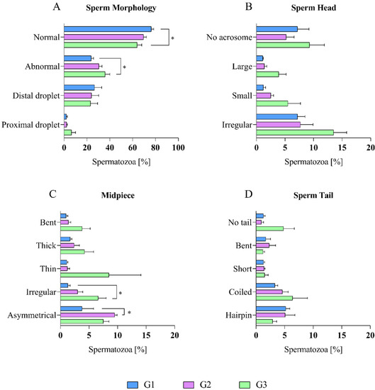
Increased male age is associated with a significant reduction in semen quality. Little is known about the sperm proteome changes resulting from the aging process. This study aimed to investigate the relationship between the functional quality and proteome of epididymal spermatozoa of dogs that were differing in age. The study was conducted on 30 male dogs that were divided into three age groups. G1—12 to 41 months old, G2—42 to 77 months old, and G3—78 to 132 months old. The sperm samples were assessed using a computer-assisted semen analysis (CASA). The epididymal sperm proteins were analyzed using gel electrophoresis (SDS-PAGE), nano-liquid chromatography coupled to quadrupole time of flight mass spectrometry (NanoUPLC-Q-TOF/MS) and bioinformatic tools. The sperm quality parameters were significantly lower in older dogs. NanoUPLC-Q-TOF/MS identification resulted in 865 proteins that were found in the G1, 472 in G2, and 435 in G3. There were seven proteins that were present in all three age groups, and four of them (ACTB, CE10, NPC2, CRISP2) showed significant changes among the studied groups. Age-dependent variations were detected in the sperm proteome composition and were related to important metabolite pathways, which might suggest that several proteins are implicated in sperm maturation and could be potential aging biomarkers. Full article
(This article belongs to the Special Issue Omics Strategies in Male Reproduction)
Show Figures

Figure 1

18 pages, 15816 KiB  
Article
Stability of DMPC Liposomes Externally Conjugated with Branched Polyglycerol
by Alexander A. Beketov 1, Ivan V. Mikhailov 2 and Anatoly A. Darinskii 2,*
1 Center for Chemical Engineering, St. Petersburg National Research University of Information Technologies, Mechanics and Optics, 197101 St. Petersburg, Russia
2 Institute of Macromolecular Compounds, Russian Academy of Sciences, 199004 St. Petersburg, Russia
Int. J. Mol. Sci. 2022, 23(16), 9142; https://doi.org/10.3390/ijms23169142 - 15 Aug 2022
Cited by 2 | Viewed by 2376
Abstract
Vesicles formed by DMPC liposomes externally conjugated with branched polyglycerol-dendrons as well as linear PEG in water solution were simulated using the DPD method. Such a structure of vesicles corresponds to the structure of polymer-grafted liposomes obtained experimentally by the post-insertion method, in [...] Read more.
Vesicles formed by DMPC liposomes externally conjugated with branched polyglycerol-dendrons as well as linear PEG in water solution were simulated using the DPD method. Such a structure of vesicles corresponds to the structure of polymer-grafted liposomes obtained experimentally by the post-insertion method, in which polymer chains are fixed on the outer surface of the liposome. The grafting density, generation number and spacer length of grafted dendrons were varied. It was shown that modification of the outer surface of liposomes due to grafting of hydrophilic dendrons has practically no effect on the size and shape of the vesicle, as well as on the morphology of the lipid membrane up to certain critical thresholds of grafting density, degree of polymerization, and generation number of grafted molecules. Exceeding the threshold values of these structural parameters leads to irreversible deformation of the lipid membrane. Diffusion through the membrane and the transition of grafted molecules from the outer surface of the liposome to the inner surface is not observed for dendrons with a generation number higher than one, even at high grafting densities. The critical values of the generation number and the characteristics of the molecular coating at these values were determined for various grafting densities and spacer lengths of the grafted chains. It was shown that the chemical potential of the grafted dendron can serve as a stability metric for the conjugated liposome. The chemical potential of grafted molecules was calculated using the mean field model of the spherical brush on the liposome surface. An analysis of the simulation data shows that, within the framework of the applicability of the mean field approach, the value of the chemical potential is a sufficient criterion for separating vesicles into stable and unstable forms. These results can be used as a guide for the experimental design of nanocontainers based on lipid vesicles with an external protective coating of branched macromolecules. Full article
(This article belongs to the Special Issue Modern Trends in Polymer Brushes: Experiment, Theory and Simulation)
Show Figures

Figure 1

20 pages, 2971 KiB  
Article
Plasma Microbiome in COVID-19 Subjects: An Indicator of Gut Barrier Defects and Dysbiosis
by Ram Prasad 1,*, Michael John Patton 2, Jason Levi. Floyd 1, Seth Fortmann 1, Mariana DuPont 1, Angela Harbour 1, Justin Wright 3, Regina Lamendella 3, Bruce R. Stevens 4, Gavin Y. Oudit 5 and Maria B. Grant 1,*
1 Department of Ophthalmology and Visual Sciences, University of Alabama at Birmingham, 1670 University BLVD, VH490, Birmingham, AL 35294, USA
2 Hugh Kaul Precision Medicine Institute, Department of Medicine, University of Alabama at Birmingham, Birmingham, AL 35294, USA
3 Wright Labs, LLC, Huntingdon, PA 16652, USA
4 Department of Physiology and Functional Genomics, University of Florida, Gainesville, FL 32611, USA
5 Division of Cardiology, Department of Medicine, University of Alberta, Mazankowski Alberta Heart Institute, Edmonton, AB T6G 2B7, Canada
Int. J. Mol. Sci. 2022, 23(16), 9141; https://doi.org/10.3390/ijms23169141 - 15 Aug 2022
Cited by 46 | Viewed by 4297
Abstract
The gut is a well-established route of infection and target for viral damage by SARS-CoV-2. This is supported by the clinical observation that about half of COVID-19 patients exhibit gastrointestinal (GI) complications. We aimed to investigate whether the analysis of plasma could provide [...] Read more.
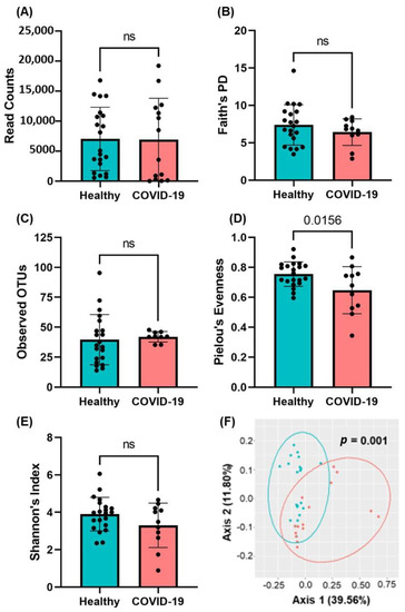
The gut is a well-established route of infection and target for viral damage by SARS-CoV-2. This is supported by the clinical observation that about half of COVID-19 patients exhibit gastrointestinal (GI) complications. We aimed to investigate whether the analysis of plasma could provide insight into gut barrier dysfunction in patients with COVID-19 infection. Plasma samples of COVID-19 patients (n = 146) and healthy individuals (n = 47) were collected during hospitalization and routine visits. Plasma microbiome was analyzed using 16S rRNA sequencing and gut permeability markers including fatty acid binding protein 2 (FABP2), peptidoglycan (PGN), and lipopolysaccharide (LPS) in both patient cohorts. Plasma samples of both cohorts contained predominately Proteobacteria, Firmicutes, Bacteroides, and Actinobacteria. COVID-19 subjects exhibit significant dysbiosis (p = 0.001) of the plasma microbiome with increased abundance of Actinobacteria spp. (p = 0.0332), decreased abundance of Bacteroides spp. (p = 0.0003), and an increased Firmicutes:Bacteroidetes ratio (p = 0.0003) compared to healthy subjects. The concentration of the plasma gut permeability marker FABP2 (p = 0.0013) and the gut microbial antigens PGN (p < 0.0001) and LPS (p = 0.0049) were significantly elevated in COVID-19 patients compared to healthy subjects. These findings support the notion that the intestine may represent a source for bacteremia and contribute to worsening COVID-19 outcomes. Therapies targeting the gut and prevention of gut barrier defects may represent a strategy to improve outcomes in COVID-19 patients. Full article
(This article belongs to the Section Molecular Microbiology)
Show Figures

Figure 1

13 pages, 3276 KiB  
Article
Blood Test for Breast Cancer Screening through the Detection of Tumor-Associated Circulating Transcripts
by Sunyoung Park 1,†, Sungwoo Ahn 1,†, Jee Ye Kim 2, Jungho Kim 3, Hyun Ju Han 4, Dasom Hwang 1, Jungmin Park 2, Hyung Seok Park 2, Seho Park 2, Gun Min Kim 5, Joohyuk Sohn 5, Joon Jeong 6, Yong Uk Song 7, Hyeyoung Lee 1,* and Seung Il Kim 2,*
1 Department of Biomedical Laboratory Science, College of Health Sciences, Yonsei University, Wonju 26493, Korea
2 Department of Surgery, Yonsei University College of Medicine, Seoul 03722, Korea
3 Department of Clinical Laboratory Science, College of Health Sciences, Catholic University of Pusan, Busan 46252, Korea
4 Avison Biomedical Research Center, Yonsei University College of Medicine, Seoul 03722, Korea
5 Department of Medical Oncology, Yonsei University College of Medicine, Seoul 03722, Korea
6 Department of Surgery, Gangnam Severance Hospital, Yonsei University College of Medicine, Seoul 06273, Korea
7 Division of Business Administration, College of Government and Business, Yonsei University, Wonju 26493, Korea
These authors contributed equally to this work.
Int. J. Mol. Sci. 2022, 23(16), 9140; https://doi.org/10.3390/ijms23169140 - 15 Aug 2022
Cited by 6 | Viewed by 3672
Abstract
Liquid biopsy has been emerging for early screening and treatment monitoring at each cancer stage. However, the current blood-based diagnostic tools in breast cancer have not been sufficient to understand patient-derived molecular features of aggressive tumors individually. Herein, we aimed to develop a [...] Read more.
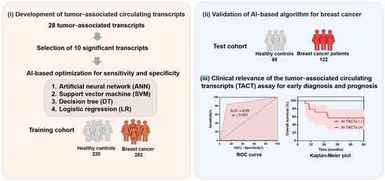
Liquid biopsy has been emerging for early screening and treatment monitoring at each cancer stage. However, the current blood-based diagnostic tools in breast cancer have not been sufficient to understand patient-derived molecular features of aggressive tumors individually. Herein, we aimed to develop a blood test for the early detection of breast cancer with cost-effective and high-throughput considerations in order to combat the challenges associated with precision oncology using mRNA-based tests. We prospectively evaluated 719 blood samples from 404 breast cancer patients and 315 healthy controls, and identified 10 mRNA transcripts whose expression is increased in the blood of breast cancer patients relative to healthy controls. Modeling of the tumor-associated circulating transcripts (TACTs) is performed by means of four different machine learning techniques (artificial neural network (ANN), decision tree (DT), logistic regression (LR), and support vector machine (SVM)). The ANN model had superior sensitivity (90.2%), specificity (80.0%), and accuracy (85.7%) compared with the other three models. Relative to the value of 90.2% achieved using the TACT assay on our test set, the sensitivity values of other conventional assays (mammogram, CEA, and CA 15-3) were comparable or much lower, at 89%, 7%, and 5%, respectively. The sensitivity, specificity, and accuracy of TACTs were appreciably consistent across the different breast cancer stages, suggesting the potential of the TACTs assay as an early diagnosis and prediction of poor outcomes. Our study potentially paves the way for a simple and accurate diagnostic and prognostic tool for liquid biopsy. Full article
(This article belongs to the Special Issue Molecular Biomarkers in Cancer and Their Applications)
Show Figures

Figure 1

18 pages, 2307 KiB  
Article
An Association Study of DNA Methylation and Gene Expression in Angelman Syndrome: A Bioinformatics Approach
by Julia Panov and Hanoch Kaphzan *
Laboratory for Neurobiology of Psychiatric Disorders, Sagol Department of Neurobiology, University of Haifa, Haifa 3498838, Israel
Int. J. Mol. Sci. 2022, 23(16), 9139; https://doi.org/10.3390/ijms23169139 - 15 Aug 2022
Cited by 2 | Viewed by 3060
Abstract
Angelman syndrome (AS) is a neurodevelopmental disorder caused by the loss of function of the E3-ligase UBE3A. Despite multiple studies, AS pathophysiology is still obscure and has mostly been explored in rodent models of the disease. In recent years, a growing body of [...] Read more.
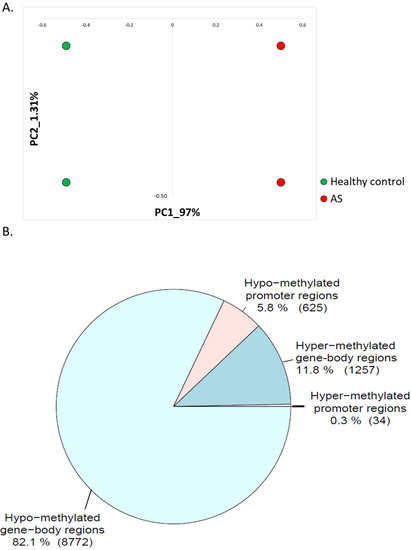
Angelman syndrome (AS) is a neurodevelopmental disorder caused by the loss of function of the E3-ligase UBE3A. Despite multiple studies, AS pathophysiology is still obscure and has mostly been explored in rodent models of the disease. In recent years, a growing body of studies has utilized omics datasets in the attempt to focus research regarding the pathophysiology of AS. Here, for the first time, we utilized a multi-omics approach at the epigenomic level and the transcriptome level, for human-derived neurons. Using publicly available datasets for DNA methylation and gene expression, we found genome regions in proximity to gene promoters and intersecting with gene-body regions that were differentially methylated and differentially expressed in AS. We found that overall, the genome in AS postmortem brain tissue was hypo-methylated compared to healthy controls. We also found more upregulated genes than downregulated genes in AS. Many of these dysregulated genes in neurons obtained from AS patients are known to be critical for neuronal development and synaptic functioning. Taken together, our results suggest a list of dysregulated genes that may be involved in AS development and its pathological features. Moreover, these genes might also have a role in neurodevelopmental disorders similar to AS. Full article
Show Figures

Figure 1

14 pages, 671 KiB  
Review
Glymphatic System a Window on TBI Pathophysiology: A Systematic Review
by Michela Ferrara 1,†, Giuseppe Bertozzi 2,†, Gianpietro Volonnino 1, Nicola Di Fazio 1, Paola Frati 1, Luigi Cipolloni 2, Raffaele La Russa 2 and Vittorio Fineschi 1,*
1 Department of Anatomical, Histological, Forensic and Orthopaedic Sciences, Sapienza University of Rome, 00161 Rome, Italy
2 Department of Clinical and Experimental Medicine, Section of Legal Medicine, University of Foggia, 71122 Foggia, Italy
These authors equally contributed to this work.
Int. J. Mol. Sci. 2022, 23(16), 9138; https://doi.org/10.3390/ijms23169138 - 15 Aug 2022
Cited by 34 | Viewed by 3804
Abstract
Background: In recent years, the attention of the scientific world has focused on a clearance system of brain waste metabolites, called the glymphatic system, based on its similarity to the lymphatic system in peripheral tissue and the relevant role of the AQP4 glial [...] Read more.
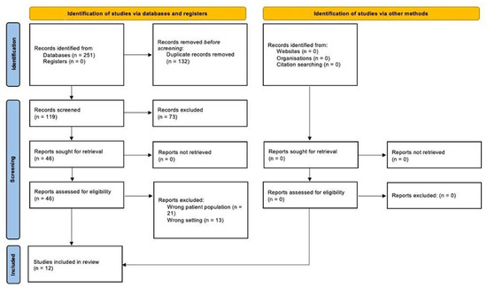
Background: In recent years, the attention of the scientific world has focused on a clearance system of brain waste metabolites, called the glymphatic system, based on its similarity to the lymphatic system in peripheral tissue and the relevant role of the AQP4 glial channels and described for the first time in 2012. Consequently, numerous studies focused on its role in organ damage in cases of neuropathologies, including TBI. Methods: To evaluate the role that the glymphatic system has in the pathogenesis of TBI, on 23 March 2022, a systematic review of the literature according to PRISMA guidelines was carried out using the SCOPUS and Medline (via PubMed) databases, resulting in 12 articles after the selection process. Discussion and conclusion: The present review demonstrated that an alteration of AQP4 is associated with the accumulation of substances S100b, GFAP, and NSE, known markers of TBI in the forensic field. In addition, the alteration of the functionality of AQP4 favors edema, which, as already described, constitutes alterations of secondary brain injuries. Moreover, specific areas of the brain were demonstrated to be prone to alterations of the glymphatic pathway, suggesting their involvement in post-TBI damage. Therefore, further studies are mandatory. In this regard, a study protocol on cadavers is also proposed, based on the analyzed evidence. Full article
(This article belongs to the Special Issue Oxidative Stress and Brain Injury 3.0)
Show Figures

Figure 1

14 pages, 6512 KiB  
Article
Stabilization of the c-Myc Protein via the Modulation of Threonine 58 and Serine 62 Phosphorylation by the Disulfiram/Copper Complex in Oral Cancer Cells
by Gunng-Shinng Chen 1, Ssu-Yu Chen 2, Shu-Ting Liu 2, Cheng-Chih Hsieh 3,4, Shiao-Pieng Lee 1,* and Shih-Ming Huang 2,*
1 School of Dentistry, Department of Dentistry of Tri-Service General Hospital, National Defense Medical Center, Taipei City 114, Taiwan
2 Department of Biochemistry, National Defense Medical Center, Taipei City 114, Taiwan
3 School of Pharmacy and Institute of Pharmacy, National Defense Medical Center, Taipei City 114, Taiwan
4 Department of Pharmacy, Kaohsiung Veterans General Hospital, Kaohsiung 813, Taiwan
Int. J. Mol. Sci. 2022, 23(16), 9137; https://doi.org/10.3390/ijms23169137 - 15 Aug 2022
Cited by 5 | Viewed by 2771
Abstract
MYC has a short half-life that is tightly regulated through phosphorylation and proteasomal degradation. Many studies have claimed that treatment with disulfiram (DSF) with or without copper ions can cause cancer cell death in a reactive oxygen species (ROS)-dependent manner in cancer cells. [...] Read more.
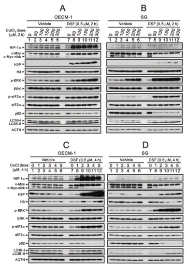
MYC has a short half-life that is tightly regulated through phosphorylation and proteasomal degradation. Many studies have claimed that treatment with disulfiram (DSF) with or without copper ions can cause cancer cell death in a reactive oxygen species (ROS)-dependent manner in cancer cells. Our previous study showed that the levels of c-Myc protein and the phosphorylation of threonine 58 (T58) and serine 62 (S62) increased in DSF-Cu-complex-treated oral epidermoid carcinoma Meng-1 (OECM-1) cells. These abovementioned patterns were suppressed by pretreatment with an ROS scavenger, N-acetyl cysteine. The overexpression of c-Myc failed to induce hypoxia-inducible factor 1α protein expression, which was stabilized by the DSF-Cu complex. In this study, we further examined the regulatory mechanism behind the induction of the c-Myc of the DSF-Cu complex in an OECM-1 cell compared with a Smulow–Glickman (SG) human normal gingival epithelial cell. Our data showed that the downregulation of c-Myc truncated nick and p62 and the induction of the ratio of H3P/H3 and p-ERK/ERK might not be involved in the increase in the amount of c-Myc via the DSF/copper complexes in OECM-1 cells. Combined with the inhibitors for various signaling pathways and cycloheximde treatment, the increase in the amount of c-Myc with the DSF/copper complexes might be mediated through the increase in the stabilities of c-Myc (T58) and c-Myc (S62) proteins in OECM-1 cells. In SG cells, only the c-Myc (T58) protein was stabilized by the DSF-Cu (I and II) complexes. Hence, our findings could provide novel regulatory insights into the phosphorylation-dependent stability of c-Myc in DSF/copper-complex-treated oral squamous cell carcinoma. Full article
Show Figures

Figure 1

19 pages, 1785 KiB  
Review
Nutrition as Personalized Medicine against SARS-CoV-2 Infections: Clinical and Oncological Options with a Specific Female Groups Overview
by Miriam Dellino 1,2,*,†, Eliano Cascardi 3,4,*,†, Marina Vinciguerra 1, Bruno Lamanna 1,5, Antonio Malvasi 1, Salvatore Scacco 6, Silvia Acquaviva 6, Vincenzo Pinto 1, Giovanni Di Vagno 2, Gennaro Cormio 7, Raffaele De Luca 8, Miria Lafranceschina 8, Gerardo Cazzato 9, Giuseppe Ingravallo 9, Eugenio Maiorano 9, Leonardo Resta 9, Antonella Daniele 8,‡ and Daniele La Forgia 8,‡
1 Department of Biomedical Sciences and Human Oncology, University of Bari, 70100 Bari, Italy
2 Clinic of Obstetrics and Gynecology, “San Paolo” Hospital, 70123 Bari, Italy
3 Department of Medical Sciences, University of Turin, 10124 Turin, Italy
4 Pathology Unit, FPO-IRCCS Candiolo Cancer Institute, Str. Provinciale 142, Km 3.95, 10060 Candiolo, Italy
5 Fetal Medicine Research Institute, King’s College Hospital, London SE5 9RS, UK
6 Department of Basic Medical Sciences and Neurosciences, University of Bari “Aldo Moro”, 70121 Bari, Italy
7 Gynecologic Oncology Unit, IRCCS Istituto Tumori Giovanni Paolo II, Department of Interdisciplinary Medicine (DIM), University of Bari “Aldo Moro”, 70121 Bari, Italy
8 IRCCS Istituto Tumori Giovanni Paolo II, 70124 Bari, Italy
9 Department of Emergency and Organ Transplantation, University of Bari “Aldo Moro”, 70121 Bari, Italy
These authors contributed equally to this work.
add Show full affiliation list remove Hide full affiliation list
Int. J. Mol. Sci. 2022, 23(16), 9136; https://doi.org/10.3390/ijms23169136 - 15 Aug 2022
Cited by 22 | Viewed by 4462
Abstract
Coronavirus disease 2019 (COVID-19) is a respiratory disease caused by severe acute respiratory syndrome coronavirus-2 (SARS-CoV-2). It is acknowledged that vulnerable people can suffer from mortal complications of COVID-19. Therefore, strengthening the immune system particularly in the most fragile people could help to [...] Read more.
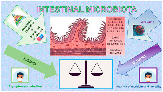
Coronavirus disease 2019 (COVID-19) is a respiratory disease caused by severe acute respiratory syndrome coronavirus-2 (SARS-CoV-2). It is acknowledged that vulnerable people can suffer from mortal complications of COVID-19. Therefore, strengthening the immune system particularly in the most fragile people could help to protect them from infection. First, general nutritional status and food consumption patterns of everyone affect the effectiveness of each immune system. The effects of nutrition could impact the level of intestinal and genital microbiota, the adaptive immune system, and the innate immune system. Indeed, immune system cells and mediators, which are crucial to inflammatory reaction, are in the structures of fats, carbohydrates, and proteins and are activated through vitamins (vit) and minerals. Therefore, the association of malnutrition and infection could damage the immune response, reducing the immune cells and amplifying inflammatory mediators. Both amount and type of dietary fat impact on cytokine biology, that consequently assumes a crucial role in inflammatory disease. This review explores the power of nutrition in the immune response against COVID-19 infection, since a specific diet could modify the cytokine storm during the infection phase. This can be of vital importance in the most vulnerable subjects such as pregnant women or cancer patients to whom we have deemed it necessary to dedicate personalized indications. Full article
(This article belongs to the Special Issue Cancer Prevention with Molecular Target Therapies 3.0)
Show Figures

Figure 1

13 pages, 1575 KiB  
Review
Vitamin D and the Kidney: Two Players, One Console
by Fulvia Zappulo, Maria Cappuccilli, Alessandra Cingolani, Anna Scrivo, Anna Laura Croci Chiocchini, Miriam Di Nunzio, Chiara Donadei, Marianna Napoli, Francesco Tondolo, Giuseppe Cianciolo and Gaetano La Manna *
Nephrology, Dialysis and Renal Transplant Unit, IRCCS-Azienda Ospedaliero-Universitaria di Bologna, Alma Mater Studiorum University of Bologna, 40138 Bologna, Italy
Int. J. Mol. Sci. 2022, 23(16), 9135; https://doi.org/10.3390/ijms23169135 - 15 Aug 2022
Cited by 19 | Viewed by 13599
Abstract
Vitamin D belongs to the group of liposoluble steroids mainly involved in bone metabolism by modulating calcium and phosphorus absorption or reabsorption at various levels, as well as parathyroid hormone production. Recent evidence has shown the extra-bone effects of vitamin D, including glucose [...] Read more.
Vitamin D belongs to the group of liposoluble steroids mainly involved in bone metabolism by modulating calcium and phosphorus absorption or reabsorption at various levels, as well as parathyroid hormone production. Recent evidence has shown the extra-bone effects of vitamin D, including glucose homeostasis, cardiovascular protection, and anti-inflammatory and antiproliferative effects. This narrative review provides an overall view of vitamin D’s role in different settings, with a special focus on chronic kidney disease and kidney transplant. Full article
(This article belongs to the Special Issue The Role of Vitamin D in Human Health and Diseases 2.0)
Show Figures

Figure 1

17 pages, 4069 KiB  
Article
Metformin Mitigated Obesity-Driven Cancer Aggressiveness in Tumor-Bearing Mice
by Chun-Jung Chen 1,2, Chih-Cheng Wu 3,4,5, Cheng-Yi Chang 6,7, Jian-Ri Li 8, Yen-Chuan Ou 9, Wen-Ying Chen 7, Su-Lan Liao 1 and Jiaan-Der Wang 10,11,*
1 Department of Medical Research, Taichung Veterans General Hospital, Taichung City 407, Taiwan
2 Department of Medical Laboratory Science and Biotechnology, China Medical University, Taichung City 404, Taiwan
3 Department of Anesthesiology, Taichung Veterans General Hospital, Taichung City 407, Taiwan
4 Department of Financial Engineering, Providence University, Taichung City 433, Taiwan
5 Department of Data Science and Big Data Analytics, Providence University, Taichung City 433, Taiwan
6 Department of Surgery, Feng Yuan Hospital, Taichung City 420, Taiwan
7 Department of Veterinary Medicine, National Chung Hsing University, Taichung City 402, Taiwan
8 Division of Urology, Taichung Veterans General Hospital, Taichung City 407, Taiwan
9 Division of Urology, Tungs’ Taichung Metro Harbor Hospital, Taichung City 435, Taiwan
10 Children’s Medical Center, Taichung Veterans General Hospital, Taichung City 407, Taiwan
11 Department of Industrial Engineering and Enterprise Information, Tunghai University, Taichung City 407, Taiwan
add Show full affiliation list remove Hide full affiliation list
Int. J. Mol. Sci. 2022, 23(16), 9134; https://doi.org/10.3390/ijms23169134 - 15 Aug 2022
Cited by 14 | Viewed by 3285
Abstract
Metformin may offer benefits to certain cancer populations experiencing metabolic abnormalities. To extend the anticancer studies of metformin, a tumor model was established through the implantation of murine Lewis Lung Carcinoma (LLC) cells to Normal Diet (ND)-fed and High-Fat Diet (HFD)-fed C57BL/6 mice. [...] Read more.
Metformin may offer benefits to certain cancer populations experiencing metabolic abnormalities. To extend the anticancer studies of metformin, a tumor model was established through the implantation of murine Lewis Lung Carcinoma (LLC) cells to Normal Diet (ND)-fed and High-Fat Diet (HFD)-fed C57BL/6 mice. The HFD-fed mice displayed metabolic and pro-inflammatory alterations together with accompanying aggressive tumor growth. Metformin mitigated tumor growth in HFD-fed mice, paralleled by reductions in circulating glucose, insulin, soluble P-selectin, TGF-β1 and High Mobility Group Box-1 (HMGB1), as well as tumor expression of cell proliferation, aerobic glycolysis, glutaminolysis, platelets and neutrophils molecules. The suppressive effects of metformin on cell proliferation, migration and oncogenic signaling molecules were confirmed in cell study. Moreover, tumor-bearing HFD-fed mice had higher contents of circulating and tumor immunopositivity of Neutrophil Extracellular Traps (NETs)-associated molecules, with a suppressive effect from metformin. Data taken from neutrophil studies confirmed the inhibitory effect that metformin has on NET formation induced by HMGB1. Furthermore, HMGB1 was identified as a promoting molecule to boost the transition process towards NETs. The current study shows that metabolic, pro-inflammatory and NET alterations appear to play roles in the obesity-driven aggressiveness of cancer, while also representing candidate targets for anticancer potential of metformin. Full article
(This article belongs to the Special Issue How Does Obesity Cause Cancer?)
Show Figures

Figure 1

12 pages, 2886 KiB  
Article
Renin as a Marker of Tissue Perfusion, Septic Shock and Mortality in Septic Patients: A Prospective Observational Study
by Patrycja Leśnik 1,*, Lidia Łysenko 2, Małgorzata Krzystek-Korpacka 3, Ewa Woźnica-Niesobska 2, Magdalena Mierzchała-Pasierb 3 and Jarosław Janc 1
1 Department of Anaesthesiology and Intensive Therapy, 4th Military Clinical Hospital, 50-981 Wroclaw, Poland
2 Department of Anaesthesiology and Intensive Therapy, Wroclaw Medical University, 50-556 Wroclaw, Poland
3 Department of Biochemistry and Immunochemistry, Wroclaw Medical University, 50-369 Wroclaw, Poland
Int. J. Mol. Sci. 2022, 23(16), 9133; https://doi.org/10.3390/ijms23169133 - 15 Aug 2022
Cited by 15 | Viewed by 2704
Abstract
Sepsis is a life-threatening organ dysfunction caused by the dysregulation of the host’s response to an infection, where the dominant mechanism is tissue hypoperfusion. Currently, the marker used to define tissue disorders is lactate levels, which may be elevated in other disease states [...] Read more.
Sepsis is a life-threatening organ dysfunction caused by the dysregulation of the host’s response to an infection, where the dominant mechanism is tissue hypoperfusion. Currently, the marker used to define tissue disorders is lactate levels, which may be elevated in other disease states as well. Renin is an essential hormone for the proper functioning of the renin-angiotensin-aldosterone (RASS) system. It is secreted in the glomerular apparatus in response to hypoperfusion. This study aimed to assess the usefulness of renin as a marker of tissue hypoperfusion in patients with sepsis and septic shock. A final group of 48 patients treated for sepsis and septic shock in the intensive care unit was included. Blood samples for renin quantification were collected in the morning as a part of routine blood analysis on the first, third, and fifth days. Sepsis was diagnosed in 19 patients (39.6%), and septic shock was diagnosed in 29 patients (60.4%). There was no significant difference in renin concentration between patients who received and did not receive continuous renal replacement therapy (CRRT) on any study day. Therefore, all samples were analyzed together in subsequent analyses. There was a significant difference in renin concentration between sepsis survivors and non-survivors on the third (31.5 and 119.9 pg/mL, respectively) and fifth (18.2 and 106.7 pg/mL, respectively) days. As a survival marker, renin was characterized by 69% and 71% overall accuracy if determined on the third and fifth days, respectively. There was a significant difference in renin concentration between sepsis and septic shock patients on the first (45.8 and 103.4 pg/mL, respectively) and third (24.7 and 102.1 pg/mL, respectively) days. At an optimal cut-off of 87 pg/mL, renin had very good specificity and a positive likelihood ratio. Renin was a strong predictor of mortality in patients with sepsis and septic shock. Further, the level of renin in patients with septic shock was significantly higher than in patients with sepsis. In combination with the assessment of lactate concentration, renin seems to be the optimal parameter for monitoring tissue hypoperfusion and could be helpful for septic shock diagnosis, as well as for identifying candidate patients for CRRT. Full article
Show Figures

Figure 1

20 pages, 3918 KiB  
Article
Sensitive Detection of Hydroxytyrosol in Extra Virgin Olive Oils with a Novel Biosensor Based on Single-Walled Carbon Nanotubes and Tyrosinase
by Alexandra Virginia Bounegru and Constantin Apetrei *
Department of Chemistry, Physics and Environment, Faculty of Sciences and Environment, “Dunărea de Jos” University of Galaţi, 47 Domnească Street, 800008 Galaţi, Romania
Int. J. Mol. Sci. 2022, 23(16), 9132; https://doi.org/10.3390/ijms23169132 - 15 Aug 2022
Cited by 13 | Viewed by 2921
Abstract
Hydroxytyrosol (HT) is an important marker for the authenticity and quality assessment of extra virgin olive oils (EVOO). The aim of the study was the qualitative and quantitative determination of hydroxytyrosol in commercial extra virgin olive oils of different origins and varieties using [...] Read more.
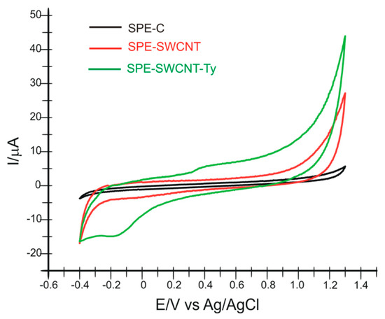
Hydroxytyrosol (HT) is an important marker for the authenticity and quality assessment of extra virgin olive oils (EVOO). The aim of the study was the qualitative and quantitative determination of hydroxytyrosol in commercial extra virgin olive oils of different origins and varieties using a newly developed biosensor based on a screen-printed electrode modified with single-layer carbon nanotubes and tyrosinase (SPE-SWCNT-Ty). The enzyme was immobilized on a carbon-based screen-printed electrode previously modified with single-layer carbon nanotubes (SPE-SWCNT-Ty) by the drop-and-dry method, followed by cross-linking with glutaraldehyde. The modified electrode surface was characterized by different methods, including electrochemical (cyclic voltammetry (CV), differential pulse voltammetry (DPV), electrochemical impedance spectroscopy (EIS)) and spectrometric (Fourier transform infrared (FTIR) spectroscopy) methods. Cyclic voltammetry was used for the quantitative determination of HT, obtaining a detection limit of 3.49 × 10⁻⁸ M and a quantification limit of 1.0 × 10⁻⁷ M, with a wide linearity range (0.49–15.602 µM). The electrochemical performance of the SPE-SWCNT-Ty biosensor was compared with that of the modified SPE-SWCNT sensor, and the results showed increased selectivity and sensitivity of the biosensor due to the electrocatalytic activity of tyrosinase. The results obtained from the quantitative determination of HT showed that commercial EVOOs contain significant amounts of HT, proving the high quality of the finished products. The determination of the antiradical activity of HT was carried out spectrophotometrically using the free reagent galvinoxyl. The results showed that there is a very good correlation between the antiradical capacity of EVOOs, the voltammetric response and implicitly the increased concentration of HT. SPE-SWCNT-Ty has multiple advantages such as sensitivity, selectivity, feasibility and low cost and could be used in routine analysis for quality control of food products such as vegetable oils. Full article
Show Figures

Figure 1