


Cells 2024, 13(6), 543; https://doi.org/10.3390/cells13060543 - 19 Mar 2024
Viewed by 679
Abstract
►
Show Figures
White matter injury (WMI) is a common neurological issue in premature-born neonates, often causing long-term disabilities. We recently demonstrated a key beneficial role of Wharton’s jelly mesenchymal stromal cell-derived small extracellular vesicles (WJ-MSC-sEVs) microRNAs (miRNAs) in WMI-related processes in vitro. Here, we studied
[...] Read more.
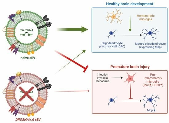
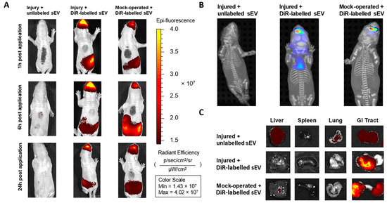
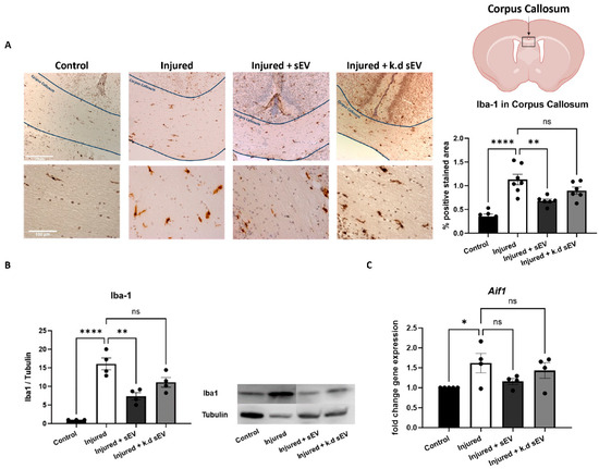
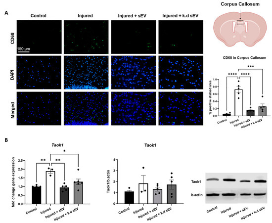
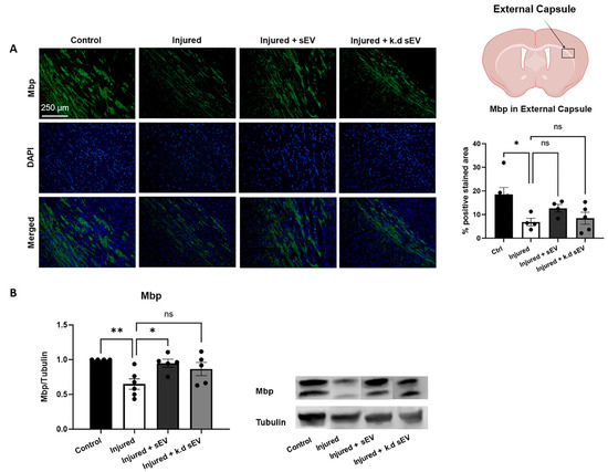
White matter injury (WMI) is a common neurological issue in premature-born neonates, often causing long-term disabilities. We recently demonstrated a key beneficial role of Wharton’s jelly mesenchymal stromal cell-derived small extracellular vesicles (WJ-MSC-sEVs) microRNAs (miRNAs) in WMI-related processes in vitro. Here, we studied the functions of WJ-MSC-sEV miRNAs in vivo using a preclinical rat model of premature WMI. Premature WMI was induced in rat pups through inflammation and hypoxia-ischemia. Small EVs were purified from the culture supernatant of human WJ-MSCs. The capacity of WJ-MSC-sEV-derived miRNAs to decrease microglia activation and promote oligodendrocyte maturation was evaluated by knocking down (k.d) DROSHA in WJ-MSCs, releasing sEVs containing significantly less mature miRNAs. Wharton’s jelly MSC-sEVs intranasally administrated 24 h upon injury reached the brain within 1 h, remained detectable for at least 24 h, significantly reduced microglial activation, and promoted oligodendrocyte maturation. The DROSHA k.d in WJ-MSCs lowered the therapeutic capabilities of sEVs in experimental premature WMI. Our results strongly indicate the relevance of miRNAs in the therapeutic abilities of WJ-MSC-sEVs in premature WMI in vivo, opening the path to clinical application.
Full article
Graphical abstract


