Review

Review of Animal Models of Prostate Cancer Bone Metastasis

1
Department of Veterinary Biosciences, The Ohio State University, 1925 Coffey Road, Columbus, OH 43210, USA
2
Department of Pathology and Unit for Laboratory Animal Medicine, School of Medicine, University of Michigan, Ann Arbor, MI 48109, USA
3
Department of Periodontics & Oral Medicine, School of Dentistry, University of Michigan, 1011 N University Avenue, Ann Arbor, MI 48109, USA
*
Author to whom correspondence should be addressed.
Vet. Sci. 2014, 1(1), 16-39; https://doi.org/10.3390/vetsci1010016
Submission received: 29 March 2014 / Revised: 27 May 2014 / Accepted: 30 May 2014 / Published: 5 June 2014
(This article belongs to the Special Issue Animal Models of Disease)

Abstract

:
Prostate cancer bone metastases are associated with a poor prognosis and are considered incurable. Insight into the formation and growth of prostate cancer bone metastasis is required for development of new imaging and therapeutic strategies to combat this devastating disease. Animal models are indispensable in investigating cancer pathogenesis and evaluating therapeutics. Multiple animal models of prostate cancer bone metastasis have been developed, but few effectively model prostatic neoplasms and osteoblastic bone metastases as they occur in men. This review discusses the animal models that have been developed to investigate prostate cancer bone metastasis, with a focus on canine models and also includes human xenograft and rodent models. Adult dogs spontaneously develop benign prostatic hyperplasia and prostate cancer with osteoblastic bone metastases. Large animal models, such as dogs, are needed to develop new molecular imaging tools and effective focal intraprostatic therapy. None of the available models fully reflect the metastatic disease seen in men, although the various models have provided important insight into the metastatic process. As additional models are developed and knowledge from the different models is combined, the molecular mechanisms of prostate cancer bone metastasis can be deciphered and targeted for development of novel therapies and molecular diagnostic imaging.

1. Introduction

Prostate cancer is the most common newly diagnosed cancer in men in the United States and it is estimated to have resulted in 10% of all cancer deaths in men in 2013 [1]. Between 70% and 100% of patients that die due to prostate cancer have bone metastases [2,3]. Patients with skeletal metastases have a five-year survival rate of 25% and median survival of 40 months [4]. The most common sites of bone metastasis in prostate cancer are pelvic bones, vertebral column, ribs, and long bones (primarily femur and humerus) [5,6]. Bone metastases in prostate cancer are primarily osteoblastic, or bone-forming lesions. These metastases result in pain, fractures and nerve compressions that reduce quality of life and represent incurable disease to clinicians [7].
In vivo animal models are the key to understanding the pathogenesis of prostate cancer bone metastasis to develop better therapeutics. The perfect model would include a species with a prostate structurally and functionally similar to humans, have slowly developing disease that progresses through prostatic intra-epithelial neoplasia (PIN), locally invasive carcinoma and late-stage osteoblastic metastasis to the pelvis, vertebrae, ribs, and long bones [8]. No model exists at this time that perfectly recapitulates human disease; however, each available model has its applicability towards particular aspects of bone metastasis. As new models are developed that more fully encompass the spectrum of human prostate cancer bone metastasis, novel strategies for palliative and curative treatments can be developed.
In an effort to accurately replicate different stages of tumor progression and metastasis, multiple injection models have been developed. These include subcutaneous, orthotopic, intracardiac, tail vein, and bone (Figure 1). The injection models can be controversial because they fail to fully recapitulate human disease by eliminating the early stages of carcinogenesis; however, they remain one of the most widely used investigative techniques of cancer until more complete models are developed.

2. Prostate Cancer in Dogs

The dog is the only species other than man to spontaneously develop benign prostatic hyperplasia (BPH) and prostate cancer at a significant incidence [9]. However, even though intact adult male dogs have a high incidence of BPH, they have a low incidence of spontaneous prostate cancer compared to men. One study reported the incidence of prostate cancer to be 0.2% in dogs and 12% in men [10]. Unlike the single-lobed architecture of the human prostate, the canine prostate is bilobed and lacks the differential regions or zones that are characteristic of the human prostate gland (peripheral, transitional, and central zones) [11,12]. All aged, intact male dogs will develop benign prostatic hyperplasia to a variable degree [11]. BPH can be severe enough to cause clinical disease, which includes dorsal compression of the colon and dyschezia. Dysuria is not a typical clinical problem in dogs with BPH compared to men. BPH does not appear to be a preneoplastic condition of the prostate in dogs, similar to men.
Figure 1. Different injection models of prostate cancer to investigate cancer progression and metastasis. The metastatic cascade involves the local invasion of tumor cells from the prostate, intravasation into a blood or lymph vessel, extravasation out of the circulation, and survival and growth at a secondary site (distant metastasis). At this time, metastasis models are not able to recapitulate this process from cancer initiation to metastatic disease. Therefore, different injection models are used to investigate this complex process. Prostatic (orthotopic) injections model invasion, extravasation, and distant metastasis. Intravascular injections (intracardiac, tail vein) model extravasation and metastasis. Tail vein injections tend to favor lung metastases, whereas intracardiac injection of cancer cells into the left ventricle permits cancer cells to localize to any tissue of the body depending on its inherent metastatic phenotype. Finally, intratibial injections model the ability to grow in and modify the bone microenvironment. Together, the different injection models reveal important information about the pathogenesis of the metastatic cascade.
Vetsci 01 00016 g001
Prostatic intra-epithelial neoplasia (PIN) has been reported in dogs with or without invasive carcinoma [13]. The incidence of PIN in the clinically normal canine prostate gland has been reported to be approximately 3%; however, the incidence increased to 69% in glands that also had invasive carcinoma [14]. However, the role of PIN in dogs has not been thoroughly investigated. PIN may be a preneoplastic condition in dogs, but it has not been commonly reported. It may be that the incidence of both PIN and prostate carcinoma are lower in dogs compared to men. It is common practice in some countries to neuter pet dogs. Interestingly, the incidence of prostate cancer is similar or greater in neutered dogs compared to intact dogs [15]. This suggests that prostate cancer in dogs may develop from androgen-independent stem cells that are present in the atrophied ducts of the prostate glands of neutered dogs [16]. An alternative theory is that non-testicular sources of androgen in the body may be sufficient to provide androgen activity on the prostate cells.
A striking difference between men and dogs is the role of androgens in prostate cancer pathogenesis. Androgens play a prominent role in the development and function of male accessory sex organs, and the androgen signaling pathway is considered essential to the development of prostate cancer in men [17]. Androgen deprivation therapy is an established early therapy for androgen-dependent prostate cancer in men, and even after progression to androgen-resistance the androgen signaling pathway is still the primary target of many therapeutics [18]. In dogs, the androgen receptor is present in normal prostatic tissue, is important for normal accessory sex organ maintenance and function, and necessary for the development of BPH in adult intact male dogs. In contrast to humans, most dogs with prostate cancer have absent expression of the androgen receptor (AR) [19]. Since castration of dogs does not decrease the incidence of prostate cancer and androgen receptor expression is not present in canine prostate cancer, it is possible that the AR does not play a central role in the pathogenesis of canine prostate cancer. Interestingly, secretory cells of the prostatic acini are androgen dependent; however, the prostatic ducts and urothelium of the prostatic urethra are maintained independent of androgen [20]. This suggests that canine prostate cancer may originate from prostatic ductular epithelium rather than the acini as occurs in men.
In men, immunohistochemical markers are often used to distinguish between prostatic and urothelial carcinoma. A prominent example is prostate specific antigen (PSA), which is found in prostatic epithelial cells, but not transitional cells of the urothelium [21]. PSA is not produced in dogs, but instead a related kallikrein enzyme, arginine esterase, is considered to be the main secretory product of the canine prostate [22]. Unfortunately, arginine esterase does not differentiate between prostatic or urothelial origin of canine prostate cancer [23]. Another marker used often in men is prostate specific membrane antigen (PSMA). In the past it has been reported that PSMA was not expressed in the canine prostate; however, recent work has shown it to be present in at least two canine prostate cancer cell lines (Ace-1 and Leo) [24,25]. Prostatic acid phosphatase has also been used in men as a prostate lineage specific marker; yet investigation of its use in canine prostate cancer has not been extensively pursued [26,27]. In men, keratin 7 expression is also used for differentiating urothelial carcinoma from prostatic adenocarcinoma. Studies have shown that keratin 7 does not differentiate between these two tumors types in dogs, making it an unsuitable marker [23,28]. At this time, there is no definitive marker in dogs that can confidently differentiate between prostate tumors originating from the acinar secretory cells and the ductular epithelium or urothelium.
Prostate cancer affects older dogs (mean age of 10 years old) and the tumors can be heterogeneous. The most common diagnosis is prostatic adenocarcinoma, but as many as 53% of canine prostate carcinomas show mixed morphologic features [12,29]. The most common pattern of growth is intra-alveolar, where the cells form papillary or cribiform projections of epithelium filling dilated ducts or alveoli [30]. The second pattern is acinar, in which one to two layers of cuboidal to columnar cells line acini (though in some cases, acini can be completely filled with a solid mass of cells) [30]. Unlike in men, grading is not routine. All canine prostate cancers are considered malignant with a high metastatic potential, and no survival difference has been found for the different histologic subtypes [31]. At the time of necropsy, it has been reported that up to 80% of dogs with a primary prostate carcinoma will have gross metastases with skeletal metastases (predominantly in the axial skeleton) present in approximately 20% of cases [29]. Greater than 75% of canine prostate cancer skeletal metastases are osteoblastic or mixed osteoblastic/osteolytic in nature, similar to men [31,32].

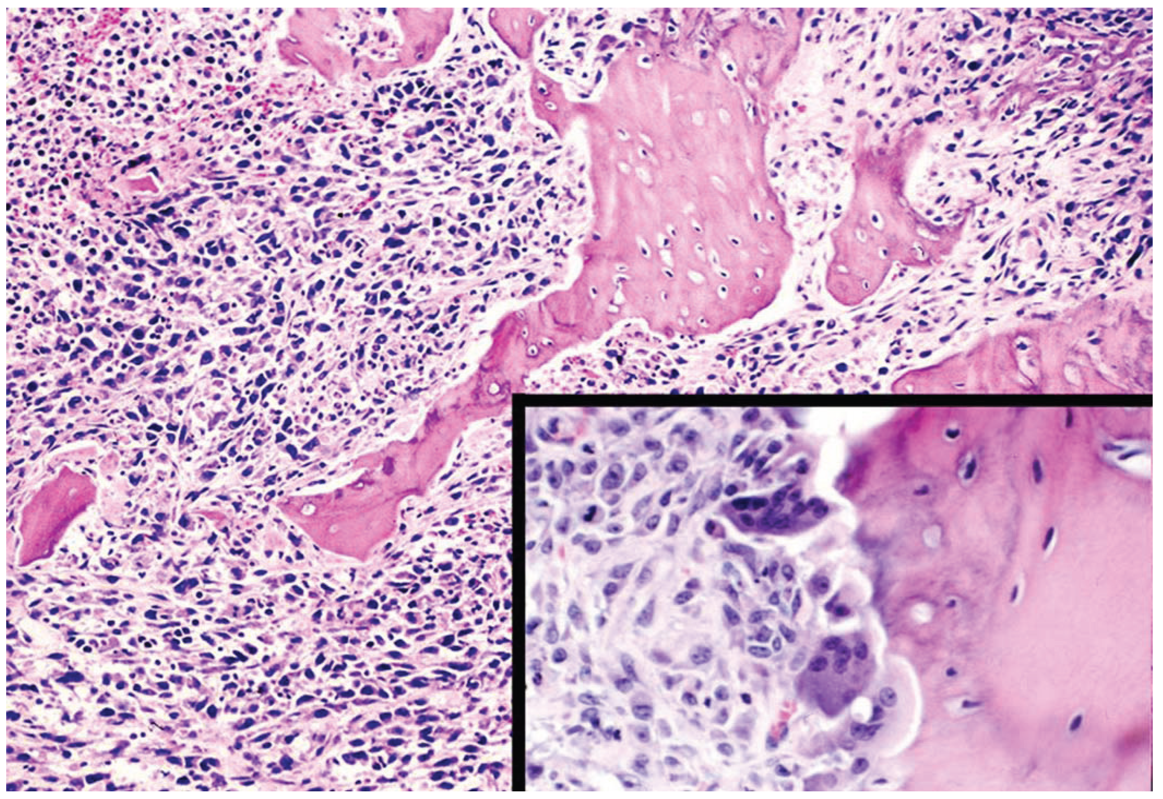
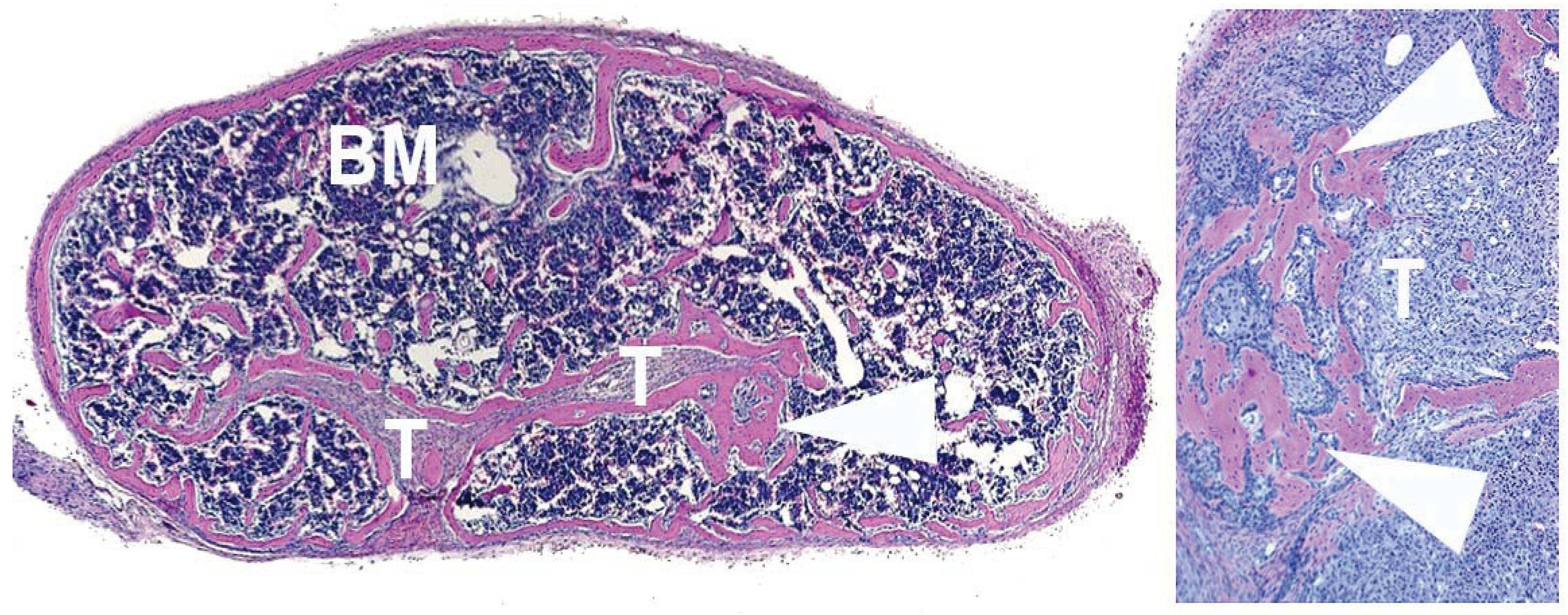
3. Dogs in Prostate Cancer Research

Prostate Tissue–Bone Interaction in vivo: Since a unique characteristic of prostate cancer bone metastasis is the ability to cause dramatic new bone formation, methods of investigating the tissue-specific mechanisms responsible for the osteoblastic phenotype are vital to understanding its pathogenesis. Dogs have proven to be an excellent model for these investigations. A novel approach was utilized to investigate the prostate-specific effects on bone [33]. Canine prostate, kidney, urinary bladder, spleen, and skeletal muscle were implanted adjacent to the calvaria of immunodeficient mice in vivo. Only the prostatic tissue resulted in significant rapid new bone formation (Figure 2). A possible mechanism for the new bone formation was discovered through in vitro studies co-culturing rat calvaria with homogenates of normal canine prostate. These experiments revealed that osteoblasts are activated by the canine prostatic tissue through an endothelin-dependent mechanism [34]. The importance of this work is highlighted by the fact that endothelin has become an important target in human prostate cancer metastases in the last five years [35,36,37,38,39]. This particular model has the advantage of providing an easily manipulated system for investigating prostate-specific mechanisms of new bone formation. Another benefit is the use of prostatic tissue rather than a cell line, which may more closely represent the complex microenvironment of metastases. Canine prostate, in particular, is uniquely useful since it is known to produce bioactive factors (such as endothelin and parathyroid hormone-related peptide) that are also produced in the human prostate and are thought to be important in the pathogenesis of osteoblastic metastases [40,41].
Canine Prostate Cancer Cell Lines: In order to model human prostate carcinoma bone metastasis, multiple canine cell lines have been created and utilized in vivo. There are six canine prostate cancer cell lines that have been reported: DPC-1 [42], CPA-1 [43], Ace-1 [44], Leo [45], CT-1258 [46] and Probasco. To date, only DPC-1, Ace-1, Leo, and Probasco cells have been used to study bone metastasis (Table 1).
DPC-1: DPC-1 originated from an 11-year-old Doberman Pinscher with a poorly differentiated prostatic adenocarcinoma [42]. The DPC-1 cells are tumorigenic in both immunosuppressed mice (subcutaneous injection) and dogs (orthotopic injection). In studies where the cells were injected into the prostate glands of immunosuppressed dogs, it was found that they metastasized to the pelvic bones in two out of 12 dogs, forming mixed osteoblastic/osteolytic metastases [42,47].
Figure 2. Effects of canine prostatic tissue implanted adjacent to murine calvaria in vivo. (A) Decalcified calvarium with prostate tissue implanted between the skin and calvarium (above and out of the plane of the image). Note pre-existing calvaria (C) and marked periosteal thickening caused by dramatic new bone formation (arrowheads) (H&E). (B) Undecalcified calcein-labeled prostate-implanted calvaria. Calcein is a green fluorescent calcium-binding dye that is taken up at sites of active bone mineralization. Note calcein fluorescence in mineralized periosteal new bone (arrowheads). Pre-existing calvaria (C) had minimal fluorescence (Unstained).
Vetsci 01 00016 g002
Table 1. Canine Cell Lines that Metastasize to Bone.
Cell LineModelsNotes
DCP-1Subcutaneous xenograft in miceMixed osteoblastic/osteolytic metastases to pelvic bones in allograft model
Orthotopic allograft in dogs
Ace-1Subcutaneous, intratibial, intracardiac, and intra-vossicle xenografts in miceMixed osteoblastic/osteolytic tumors in intratibial, intracardiac and intra-vossicle models. Metastasis to long bones, ribs and vertebrae in intracardiac model
Orthotopic allograft in dogsMetastasis to lymph nodes and bone in allograft model
LeoSubcutaneous, intratibial and intracardiac xenografts in miceOsteolytic tumors in intratibial and intracardiac models. Metastasis primarily to brain and spinal cord, but also long bones in intracardiac model
ProbascoSubcutaneous, intratibial and intracardiac xenografts in miceOsteoblastic tumors in intratibial and intracardiac models. Metastasis primarily to long bones
Figure 3. Intraprostatic Ace-1 tumor in an immunosuppressed dog. Ace-1 cells formed poorly demarcated masses (right side of image) that invaded the prostatic glands and ducts. The cells were polygonal to mildly spindle-shaped (epithelial-mesenchymal transition) and formed nests and papillary projections, frequently filling acini. Larger papillary projections were associated with coagulation necrosis. Lymphoplasmacytic inflammation and desmoplasia surrounded the affected acini and between the border of the invasive neoplastic cells into the normal prostatic tissue to the left. The left side of the image is composed of numerous variably sized, hypercellular glands lined by tall columnar to cuboidal epithelium and occasional prominent intra-luminal papillary projections (benign prostatic hyperplasia) (H&E).
Vetsci 01 00016 g003
Ace-1: The Ace-1 cell line was developed from a primary prostatic carcinoma (intra-alveolar type) of an eight-year-old male castrated Labrador Retriever. The cells are tumorigenic in immunocompromised mice and rats and form mixed osteoblastic/osteolytic metastases after intratibial, intracardiac, and intra-vossicle injection in nude mice [44,48,49,50]. In bone, the cells initially induce the formation of new woven bone on metaphyseal trabecular bone and then on periosteal surfaces in areas with osteoclastic bone resorption of cortical bone. After intracardiac injection in mice, affected bones included humeri, femurs, tibias, ribs, and lumbar vertebrae. The Ace-1 cells have also been injected orthotopically in immunosuppressed dogs to develop an experimental model of prostate cancer (Figure 3) [51]. The Ace-1 cells are versatile and have been used both for metastasis studies and also as a model of tumor-induced bone pain [52,53]. The Ace-1 cells are very permissive to transfection and transduction. They have been stably transduced with yellow fluorescent protein (YFP) and luciferase (luc) to enable bioluminescent imaging for easier in vivo monitoring of metastasis and growth. Ace-1 cells have also been stably transfected with human Dickkopf-1 (Dkk-1) to determine the role of Wnt signaling in prostate cancer bone metastasis [54]. It was found that Dkk-1 upregulated non-canonical Wnt/JNK signaling and increased tumor growth and the number of metastases in vivo. Ace-1 cells have also been transfected with parathyroid hormone-related peptide (PTHrP) and it was found that PTHrP expression resulted in increased subcutaneous tumor growth and increased osteoclastogenesis, osteoblastogenesis, and angiogenesis in vossicles (neonatal murine vertebrae; Figure 4) transplanted in nude mice [49]. Ace-1 cells have proven to be a valuable model of mixed osteoblastic/osteolytic prostate cancer metastasis.
Figure 4. Nude mouse subcutaneously transplanted vertebral vossicle with canine Ace-1 tumor cells (T). The vossicle has cortical and trabecular bone with normal bone marrow (BM) hematopoietic cells (darkly stained cells). Note the new trabecular bone formation (arrowhead) induced by the Ace-1 tumor cells that surrounds the tumor cells. The image on the right is a magnification of the hyperplastic trabecular bone (arrowheads) induced by the Ace-1 tumor cells (T) (H&E).
Vetsci 01 00016 g004
Leo: The Leo cell line originated from a primary prostate carcinoma from a five-year-old mixed breed dog. Similar to Ace-1, these cells have been transduced with YFP/Luc to allow for in vivo bioluminescent imaging. After intra-cardiac injection into nude mice, the most common site of metastasis was the brain and spinal cord followed by metastases to long bones [45]. Bone metastases were characterized by loss of cortical and trabecular bone. Brain metastasis in prostate cancer is rare, and there are few models available to study its pathogenesis. This unique cell line has the potential to contribute to this field.
Probasco: The Probasco cells came from the primary prostate carcinoma of a 10.5-year-old castrated mixed-breed dog that had been previously treated with palliative radiation therapy and metronomic chemotherapy (piroxicam, cyclophosphamide, toceranib phosphate, and chlorambucil) [55]. Similar to the Ace-1 and Leo cell lines, this cell line was also developed in the laboratory of Thomas Rosol. Like the Ace-1 cells, these cells have been transduced with YFP/Luc and are very permissive to transduction or transfection. Following intracardiac injection, Probasco cells metastasized primarily to the appendicular skeleton, and both intratibial and intracardiac injections produced markedly osteoblastic tumors in bone (Figure 5). Metastases had extensive new bone formation on both the endosteal and periosteal surfaces. In the early intra-tibial lesions (three weeks post-injection), the new woven bone was confined to the endosteal surfaces and marrow spaces. At six weeks post-injection, the Probasco cells penetrated the cortical bone and induced marked periosteal new bone formation. The overall effect on the bone was markedly osteoblastic, with mild osteolysis. These cells have been transfected with PTHrP and it was found to increase osteolysis and tumor growth in vivo. The osteoblastic nature and ease of transfection/transduction make these cells useful for studying bone metastasis pathogenesis and osteoblastic metastases.
Figure 5. Intratibial Probasco tumor in a nude mouse. (A) Micro-computed tomography (µCT) and of a nude mouse tibia injected with Probasco cells (right) and control tibia (left) at three weeks. Tibias in the Probasco-injected group had dramatic new bone formation signified by the increased radio-opacity in the diaphyseal region compared to the control bone; (B) Photomicrograph of intratibial tumor. Densely packed large polygonal neoplastic cells formed sheets and irregular glands with surrounding large irregular trabeculae of induced woven bone. Numerous polygonal to cuboidal osteoblasts line the trabecular bone and in some areas there is apposition of the prostate cancer cells to the new woven bone surfaces. Mitotic figures are frequent (H&E).
Vetsci 01 00016 g005
Canine Cell Lines in Immunocompromised Dogs: Dogs provide a useful large animal model for prostate cancer because they share several anatomic and physiologic similarities with men. The larger size of dogs also makes them helpful to investigate new molecular diagnostics for imaging and surgical procedures that cannot be performed readily in mice. However, the genetic heterogeneity of dogs as well as their cost can make them a challenging research model [56]. Canine cell lines have been injected orthotopically in immunosuppressed dogs to investigate their growth and metastasis in vivo and response to histotripsy. The Ace-1 cells formed prostate neoplasms in all injected dogs and metastases to the lungs and regional lymph nodes occurred in four out of 10 dogs. No bone metastases were present at the time of euthanasia; however, the experimental time course was relatively short (2–6 weeks). The tumors could be imaged by transrectal ultrasound and PET [57]. It was shown that histotripsy was effective in causing necrosis of the tumor tissue [51]. The DPC-1 cells have also been successfully implanted in dogs using a CT-guided transperitoneal approach with subsequent metastasis to lymph nodes (9/12), lungs (5/12) and bones (mixed osteoblastic/osteolytic metastasis) in two out of 12 dogs after 3–4 months [47].

4. Human Prostate Cancer Xenografts in Immunocompromised Mice

Much research on prostate cancer bone metastasis is performed with human prostate cancer xenografts. The most commonly used cell lines are PC3 (derived from a bone metastasis), DU145 (derived from a brain metastasis), and LNCaP (derived from a lymph node metastasis). The PC3 and LNCaP cell lines and their sublines have been widely used to study bone metastasis, as well as several less commonly used cell lines. The DU145 cell lines do not metastasize or grow in bone in vivo. Of the human prostate cancer cell lines available, PC3, PC3M, LAPC-4, LAPC-9, LNCaP, LNCaPC4-2, MDA PCa 2b, Wish-pc2, CL-1, CWR22R and ARCaP have been shown to produce bone metastases in vivo (Table 2).
PC3 and PC3M: PC3 cells are androgen insensitive and form osteolytic metastases after orthotopic, intra-cardiac and intratibial injection [58,59,60]. After orthotopic or intra-cardiac injection, sites of bone metastasis include the skull, ribs, pelvis, femur and tibia, mandible, and occasionally the brain and spinal cord. PC3 cells will also form osteolytic metastases in human fetal and adult bones implanted subcutaneously after tail vein injection of neoplastic cells [61,62] and when cells are injected adjacent to adult human bone implanted subcutaneously in immunocompromised mice [58]. This cell line has been transduced with green fluorescent protein (GFP) and luciferase (luc), allowing for more efficient imaging of metastases. A variant of PC3 which has a higher incidence of metastasis, PC3M, also forms osteolytic metastases (frequently within the mandible) after orthotopic and intracardiac injection [63,64]. Mandibular metastases in prostate cancer (and cancer in general) is an uncommon, but devastating event [65].
LAPC-4 and LAPC-9: Both LAPC-4 and LAPC-9 were derived from a femoral metastasis and are androgen dependent [66]. LAPC-4 produced mixed osteoblastic/osteolytic bone tumors when injected either orthotopically or adjacent to subcutaneously implanted fragments of adult human bone [67,68]. LAPC-9, a more recently developed cell line, has been shown to produce predominantly osteoblastic tumors after intratibial injection [58]. Since osteoblastic tumors are unusual in human cell lines, the LAPC-9 model should prove to be a valuable tool to investigate the pathogenesis of osteoblastic metastasis. For both cell lines, severely immunocompromised mice (such as NOD/SCID) must be used to allow for tumor growth in vivo.
Table 2. Human Cell and Tumor Lines used to Model Bone Metastasis in Mice.
Cell lineModelsNotes
PC3Orthotopic, intratibial, and intracardiac injectionOsteolytic tumors
Metastasizes to skull, ribs, pelvis, femur and tibia after orthotopic or intracardiac injection
Human fetal and adult bone with tail vein injection
PC3MOrthotopic and intracardiac injectionOsteolytic tumors with metastasis primarily to mandible
LAPC-4Orthotopic injectionMixed osteoblastic/osteolytic tumors
Human adult bone with adjacent injection
LAPC-9Intratibial injectionOsteoblastic tumors
LNCAPHuman adult bone with intra-bone or tail vain injectionMixed osteoblastic/osteolytic tumors
LNCaP C4-2Subcutaneous, orthotopic, and intracardiac injectionVertebral osteolytic metastases
LNCaP C4-2B4IntrafemoralMixed osteoblastic/osteolytic tumors
LNCaP CL-1Orthotopic injectionOsteolytic tumors
Direct invasion and metastasis to skull, rib, pelvis, femur and tibia
MDA PCa 2bIntrafemoral injectionOsteoblastic tumors
Injected human adult bone
CWR22RSubcutaneous injectionLacZ-positive bone micro-metastases
Metastases not histologically confirmed
ARCaPOrthotopic injectionMixed osteoblastic/osteolytic tumors
LuCap 35Intratibial injection
Orthotopic injection
Osteolytic tumors in intratibial injections
Bone marrow PSA positive sites after orthotopic injection (not histologically confirmed)
LuCaP 23.1Intratibial injectionOsteoblastic tumors
Wish-pc2Intrafemoral or intratibial injectionOsteolytic tumors
LNCaP and Sublines: The LNCap cell line and two of its sublines, LNCaP C4-2 and CL-1, have been used to study prostate cancer bone metastasis in vivo. LNCaP is androgen dependent and has been shown to form mixed osteoblastic/osteolytic tumors in subcutaneously implanted human adult bone after either direct injection into human bone or tail vein injection in immunocompromised mice [62,69]. Interestingly, LNCaP does not metastasize to murine bone after orthotopic, intracardiac, or tail vein injection [63]. The LNCaP cell line has been transfected with luciferase for bioluminescent imaging [70,71]. The subline LNCaP C4-2 is castration resistant and has been reported to metastasize to vertebrae and form mixed bone metastases after subcutaneous, orthotopic, and intracardiac injection [59,72]. The LNCaP C4-2B4 cell line was derived from a LNCaP C4-2 bone metastasis [72]. The LNCaP C4-2B4 cell line is also castration-resistant and forms mixed osteoblastic/osteolytic metastases after intra-femoral injection [59]. The CL-1 subline is also castration resistant and has been reported to directly invade and induce lysis of the lumbar vertebral column after orthotopic injection. Metastases to bone after orthotopic injection have been reported using green fluorescent protein (GFP) expression, although these were not confirmed histologically [60,73]. A potential disadvantage of LNCaP cells is that they can be challenging to work with in vivo since they are slow growing and require the use of severely immunocompromised mice (such as NOD/SCID) and/or the use of the basement membrane product, Matrigel™ (Corning, Tewksbury, MA), with the cell injections.
MDA PCa 2b: The MDA PCa 2b cells were derived from a prostate cancer vertebral bone metastasis and demonstrate androgen-sensitive growth [56,74]. The MDA PCA 2b line forms predominantly osteoblastic tumors when injected into mouse long bones (femurs and tibias) or when directly injected into subcutaneously implanted human adult bone [75,76]. This cell line has been used extensively to investigate androgen signaling and progression to androgen independence [77,78,79,80].
CWR22R: The CWR22R cells were derived from a primary prostatic carcinoma and are castration resistant [81]. Transfection of the CWR22R cell line with lacZ allowed for visualization of micro-metastases after subcutaneous injection; however, histology was not used to confirm the metastases and no bone pathology was reported [82].
ARCaP: ARCaP cells were derived from the ascites fluid of a man diagnosed with metastatic prostate carcinoma. Mixed osteoblastic/osteolytic metastases were reported when the cells were injected orthotopically [83]. The cell line is unique in that they are characterized by an androgen-repressed phenotype (growth is suppressed when exposed to androgens).
LuCaP 23.1 and LuCaP35: Both LuCaP 23.1 and 35 are serially transplantable, prostate specific antigen-producing tumor lines that do not grow in vitro and were developed from prostate cancer lymph node metastases [84,85]. Both are androgen sensitive; however, LuCaP 35 can transition to androgen independence [84]. When injected intra-tibially, LuCaP 35 forms osteolytic metastases, while LuCaP 23.1 produces predominantly osteoblastic lesions [86]. After orthotopic implantation there was PSA mRNA detected in the bone marrow by real-time polymerase chain reaction (RT-PCR), suggesting micrometastases were present [87]. A challenge with the tumor lines is the lack of in vitro capacity for propagation and hence an inability to perform in vitro assays to investigate molecular mechanisms of pathogenesis.
Wish-pc2: The Wish-pc2 tumor line was developed from a poorly differentiated prostate carcinoma. These cells are castration resistant and considered to be prostatic neuroendocrine cells and represent a model for small cell carcinoma of the prostate. Intraosseous (femur or tibia) injection of the cells into mice results in primarily osteolytic tumors [88].

5. Prostate Cancer in Mice

The prostate gland of the mouse is significantly different to men and dogs. The anatomy of the murine prostate consists of four grossly and histologically distinct lobes (ventral, dorsal, lateral, and anterior) unlike the single lobe of the human prostate. The ventral lobe of the mouse prostate has no human counterpart. In the past, the dorsal and lateral lobes were considered analogous to the peripheral zone of the human prostate, and the anterior lobe is similar to the central zone in men; however, a 2004 consensus report determined that there was no evidence for a direct relationship between the mouse prostate lobes and human prostate zones [89,90]. Wild-type mice rarely develop spontaneous prostate cancer; therefore, transgenic mice have been developed to study particular mutations or transgenes in prostate cancer pathogenesis and treatment. A potential limitation in the use of transgenic mice is that few mutations can be studied concurrently, which does not reflect spontaneous invasive and metastatic prostate cancer. However, transgenic models do permit investigations on the interactions between neoplastic cells and the surrounding stroma and the role of stroma in cancer progression. Another challenge to developing a useful transgenic mouse model for prostate cancer research is that very few develop bone metastases. Most mouse models of prostate cancer do not progress beyond the stage of invasive carcinoma [91]. This is unfortunate, since most morbidity and complications in prostate cancer are due to advanced or metastatic disease, which cannot be treated surgically. However, transgenic models can be useful to study chemoprevention of prostate cancer [92,93,94]. Only the TRAMP, LPB-Tag/PB-hepsin, CR2-T-Ag, and 12T-7f transgenic models have been reported to develop metastasis to bone, albeit infrequently.
TRAMP: Transgenic adenocarcinoma of the mouse prostate (TRAMP) mice are considered a model of neuroendocrine prostate carcinoma [95]. Bone metastasis has been infrequently reported in transgenic adenocarcinoma of the mouse prostate (TRAMP) mice and only in the FVB background strain. Uncommon skeletal metastasis (one study reported 5/20 mice) has been reported to occur at approximately 23 weeks of age in the [TRAMP X FVB] F1strain, although histology revealed only minimal osteolytic/osteoblastic response to the presence of the neoplastic cells [96,97,98,99].
LPB-Tag/PB-Hepsin: Femoral metastases were found in a small percentage of LPB-Tag/PB-hepsin mice by 21 weeks; however, there was no associated osteolysis or new bone formation [100]. It is important to note that only femoral bones were examined.
CR2-T-Ag: Similar to TRAMP mice, Cryptdin-2/sv40 T (CR2-T-Ag) transgenic mice are considered a model of neuroendocrine prostate carcinoma and are reported to occasionally develop bone metastases by 24 weeks, though associated bone pathology was not described [101]. Similarly to the LPB-Tag/PB-hepsin study, only femurs were examined for metastases.
12T-7f: 12T-7f/MT-DNIIR and 12T-7f mice are two additional models of neuroendocrine prostate carcinoma and both rarely develop bone metastases, although no associated bone pathology has been described [102]. Only lumbar spine was evaluated.
G3/4 LSL-mTert PB-Pten/p53: These mice were generated by crossing p53/Pten conditional knockout mice with LSL-mTert mice that have Cre-mediated telomerase reactivation [103]. By 24 weeks of age, 25% of mice develop lumbar spine metastases. The osteoblastic or osteolytic nature of these metastases was not characterized.
Pten/p53/Smad4: These mice were generated by crossing p53/Pten double null mice with a Smad4 conditional knockout model [103]. By 17 weeks of age, approximately 12% of Pten/P53/Smad4 mice developed lumbar spine metastases. Similar to the G3/4 LSL-mTert PB-Pten/p53 model, the osteoblastic or osteolytic nature of these metastases was not characterized.
There are other transgenic mice that are reported to have bone metastases; however, this was based on positive PCR from bone marrow or from areas of bone remodeling without histologic confirmation of neoplastic cells. These include the prostate-specific PTEN knockout mouse [104] and the Gγ/T-15 stains [105]. These mice were not included in this review as their usefulness in studying the pathogenesis of bone metastases is limited by the lack of histologically confirmed disease.

6. Mouse Prostate Reconstitution Model

The basic concept of the mouse prostate reconstitution model is to combine prostate cells (neoplastic or non-neoplastic) with urogenital sinus mesenchyme cells (rat or mouse) and implantation of the cells beneath the renal capsule of an immunodeficient mouse [32,106]. This model has been used to study the differentiation of putative prostate cancer stem cells, the significance of carcinogenic genes that cannot be investigated with whole-animal knockouts, as well as epithelial and mesenchymal interactions in prostate cancer [107,108,109]. One model that developed frequent micrometastases to bone is mouse prostate tissue over-expressing both ras and myc co-implanted with p53 knockout mouse urogenital sinus cells [110]. While the mouse prostate reconstitution model provides a fascinating avenue to further explore the genetic pathogenesis of prostate cancer progression and metastasis, its use is limited by the complexity and high technical skill required to isolate and implant the tissue.

7. End-Stage Mouse Models of Tumor/Bone Interactions

The ability to address specific questions regarding how tumors interact with the bone microenvironment is critical to understanding the cellular and molecular mechanisms of prostate cancer skeletal morbidity. Despite the fact that bone homing and the prostate cancer metastatic cascade in mice is quite different from dogs and humans, tissue reconstitution models can address specific mechanistic questions. The intratibial injection model has the advantage of direct placement of tumor cells into bone [111]. A disadvantage is that the trauma associated with injecting the tumor cells represents an atypical metastatic dissemination with wound healing consequences that are dramatic in bone. In addition, intra-tibial injection may force some of the tumor cells directly into bone marrow sinusoids and mimic an intravenous injection. Another novel model termed the “vossicle model” involves co-implantation of tumor cells and neonatal vertebral bodies [111]. This relatively simple model allows combinations of tumor cells or explants with murine bone. Isolated vertebral bones can be isolated from different types of genetically modified mice and/or luciferase-expressing mice in order to determine the impact of altering a gene in bone alone without systemic impact and to monitor tumor and/or bone growth over time.

8. Prostate Cancer in Rats

Similar to mice, the incidence of spontaneous prostate cancer in rats is rare in most strains. Some strains of rats, however, have a notable incidence of prostate cancer. As many as 30% and 16% of aged Lobund-Wistar and ACI/Seg rats, respectively, develop prostate carcinoma, although bone metastases do not occur [112]. To increase prostate tumorigenicity in Lobund-Wistar rats, intravenous injection of N-methyl-N-nitrosourea (MNU) with or without subcutaneous implantation of testosterone proprionate pellets has been shown to produce more rapid and consistent development of prostate cancer with metastasis to the lung and lymph nodes; however, bone metastases were not reported [113]. Most prostate cancer bone metastasis research performed is limited to two rat models: the MATLyLu subline and the PA-III cell line.
MATLyLu: This subline was developed from an original prostate carcinoma from a 22-month-old inbred Copenhagen male rat and the designation MATLyLu is an abbreviation for Metastatic Anaplastic Tumor Metastasizing to Lymph node and Lungs. This subline is considered a poorly differentiated carcinoma, is androgen insensitive and has been used to produce osteoclastic or, with variant R3327, osteoblastic bone metastases in Copenhagen rats after tail vein, intracardiac, and intraosseous injection [114,115,116,117,118]. The “osteoblastic” bone metastases represented osteolytic tumors with secondary periosteal bone proliferation due cortical bone erosion. Blomme et al. have reported that the bone metastases that occur with MATLyLu cells are predominantly osteolytic (Figure 6) [117].
PA-III: The PA-III cell line was derived from a spontaneous prostate carcinoma from a Lobund-Wistar rat. Similar to MayLyLu, this line is considered androgen insensitive and has been used to produce mixed osteolytic/osteoblastic lesions when implanted over the calvaria or scapula after periosteal disruption [112,119].
Figure 6. MatLyLu vertebral metastasis in a Copenhagen rat. There is marked replacement of hematopoietic cells with anaplastic carcinoma cells with dramatic loss of the medullary bone. The surfaces of the remaining trabecular bone are scalloped and eroded and they have severely eroded endosteal surfaces due to osteoclastic bone resorption (H&E). Inset: Numerous multinucleated osteoclasts line the scalloped surface of an eroded trabeculae (H&E).
Vetsci 01 00016 g006

9. Conclusions

There are a considerable number of prostate cancer models available for scientific studies although relatively few of them can be used to consistently model osteoblastic bone metastases as occurs in men. Since bone metastasis represents end-stage disease, it is important that useful animal models are available to investigate novel prevention and treatment strategies. Specific animal models should be carefully chosen for individual experimental designs so that they have the pathogenic characteristics to enable them to test hypotheses that are relevant to human prostate cancer. The canine and human osteoblastic prostate cancer lines have the greatest utility for in vivo experiments; however, no model fully recapitulates the disease as it occurs in men.
While there is no perfect animal model of prostate cancer, a few better represent prostate cancer skeletal metastasis. In the author’s opinion, the most representative model available is the LAPC-9 human cell line. It is one of the few available cell lines that was derived from a bone metastasis, and one of the very few that form osteoblastic metastases when injected in bone. It can be used for in vitro experiments, unlike the LuCaP 23.1 and 35 tumor lines. However, as most prostate cancer bone metastases are castration-resistant in men, the androgen-dependent nature of the LAPC-9 cell line does makes it less representative of the condition in men. The canine cell lines Ace-1 and Probasco are also valuable model systems since they grow well in vivo and in vitro, are easily transfectable and transducible, form bone metastases after intracardiac injection, and form osteoblastic (Probasco) or mixed osteoblastic/osteolytic (Ace-1) metastases. These two canine cell lines are exceptional model systems for the pathogenesis of bone metastasis and tumor-induced new bone formation; however, the lack of androgen receptor expression does limit their value in the investigation of drugs that inhibit the AR.
Recent breakthroughs in therapeutics have come from targeting metastatic prostate cancer with radiopharmaceuticals such as radium-223 [120]. The success of these therapies in slowing metastatic disease and improving survival indicate a need for increased research in this area, and may result in a future where bone metastases are considered curable. Animal models that best recapitulate human metastatic disease are essential for the development of these novel therapies. The characterization of novel large animal models, such as experimental dogs with orthotopic implantation of canine prostate cancer cells lines, will be helpful to develop and assess new molecular imaging tools and effective intraprostatic therapies. It is hoped that additional animal models are continually developed and critically evaluated as compared to human prostate cancer so the tools are available to effectively target prostate cancer to prevent, slow, or even reverse metastatic disease.

Acknowledgments

This work was funded in part by NCI F32 CA165689, NCI P01 CA093900 and The Ohio State University College of Veterinary Medicine Canine Research Funds. The authors thank Alan Flechtner and Anne Saulsbery for tissue processing and preparation of slides; Shelly Haramia for slide scanning; and Tim Vojt for assistance with figures.

Conflicts of Interest

The authors declare no conflict of interest.

References

  1. Siegel, R.; Naishadham, D.; Jemal, A. Cancer statistics, 2013. CA: Cancer J. Clin. 2013, 63, 11–30. [Google Scholar] [CrossRef]
  2. Coleman, R.E. Clinical features of metastatic bone disease and risk of skeletal morbidity. Clin. Cancer Res. 2006, 12, 6243s–6249s. [Google Scholar] [CrossRef]
  3. Mehra, R.; Kumar-Sinha, C.; Shankar, S.; Lonigro, R.J.; Jing, X.; Philips, N.E.; Siddiqui, J.; Han, B.; Cao, X.; Smith, D.C.; et al. Characterization of bone metastases from rapid autopsies of prostate cancer patients. Clin. Cancer Res. 2011, 17, 3924–3932. [Google Scholar] [CrossRef]
  4. Sturge, J.; Caley, M.P.; Waxman, J. Bone metastasis in prostate cancer: Emerging therapeutic strategies. Nat. Rev. Clin. Oncol. 2011, 8, 357–368. [Google Scholar]
  5. Heidenreich, A. Bisphosphonates in the management of metastatic prostate cancer. Oncology 2003, 65, 5–11. [Google Scholar] [CrossRef]
  6. Weilbaecher, K.N.; Guise, T.A.; McCauley, L.K. Cancer to bone: A fatal attraction. Nat. Rev. Cancer 2011, 11, 411–425. [Google Scholar] [CrossRef]
  7. Morgans, A.K.; Smith, M.R. Bone-targeted agents: Preventing skeletal complications in prostate cancer. Urol. Clin. North Am. 2012, 39, 533–546. [Google Scholar] [CrossRef]
  8. Langdon, S.P. Animal modeling of cancer pathology and studying tumor response to therapy. Curr. Drug Targets 2012, 13, 1535–1547. [Google Scholar] [CrossRef]
  9. Waters, D.J.; Hayden, D.W.; Bell, F.W.; Klausner, J.S.; Qian, J.; Bostwick, D.G. Prostatic intraepithelial neoplasia in dogs with spontaneous prostate cancer. Prostate 1997, 30, 92–97. [Google Scholar] [CrossRef]
  10. Gamlem, H.; Nordstoga, K.; Glattre, E. Canine neoplasia—Introductory paper. APMIS. Suppl. 2008, 125, 5–18. [Google Scholar] [CrossRef]
  11. Rosol, T.J.; Tannehill-Gregg, S.H.; LeRoy, B.E.; Mandl, S.; Contag, C.H. Animal models of bone metastasis. Cancer 2003, 97, 748–757. [Google Scholar] [CrossRef]
  12. Smith, J. Canine prostatic disease: A review of anatomy, pathology, diagnosis, and treatment. Theriogenology 2008, 70, 375–383. [Google Scholar] [CrossRef]
  13. Waters, D.J.; Bostwick, D.G. Prostatic intraepithelial neoplasia occurs spontaneously in the canine prostate. J. Urol. 1997, 157, 713–716. [Google Scholar] [CrossRef]
  14. Madewell, B.R.; Gandour-Edwards, R.; DeVere White, R.W. Canine prostatic intraepithelial neoplasia: Is the comparative model relevant? Prostate 2004, 58, 314–317. [Google Scholar] [CrossRef]
  15. Teske, E.; Naan, E.C.; van Dijk, E.M.; van Garderen, E.; Schalken, J.A. Canine prostate carcinoma: Epidemiological evidence of an increased risk in castrated dogs. Mol. Cell. Endocrinol. 2002, 197, 251–255. [Google Scholar] [CrossRef]
  16. Moulay, M.; Liu, W.; Willenbrock, S.; Sterenczak, K.A.; Carlson, R.; Ngezahayo, A.; Murua Escobar, H.; Nolte, I. Evaluation of stem cell marker gene expression in canine prostate carcinoma- and prostate cyst-derived cell lines. Anticancer Res. 2013, 33, 5421–5431. [Google Scholar]
  17. Sharifi, N. Mechanisms of androgen receptor activation in castration-resistant prostate cancer. Endocrinology 2013, 154, 4010–4017. [Google Scholar] [CrossRef]
  18. Derleth, C.L.; Yu, E.Y. Targeted therapy in the treatment of castration-resistant prostate cancer. Oncology (Williston Park) 2013, 27, 620–628. [Google Scholar]
  19. Gallardo, F.; Mogas, T.; Baro, T.; Rabanal, R.; Morote, J.; Abal, M.; Reventos, J.; Lloreta, J. Expression of androgen, oestrogen alpha and beta, and progesterone receptors in the canine prostate: Differences between normal, inflamed, hyperplastic and neoplastic glands. J. Comp. Pathol. 2007, 136, 1–8. [Google Scholar] [CrossRef]
  20. Leav, I.; Schelling, K.H.; Adams, J.Y.; Merk, F.B.; Alroy, J. Role of canine basal cells in postnatal prostatic development, induction of hyperplasia, and sex hormone-stimulated growth; and the ductal origin of carcinoma. Prostate 2001, 48, 210–224. [Google Scholar] [CrossRef]
  21. Genega, E.M.; Hutchinson, B.; Reuter, V.E.; Gaudin, P.B. Immunophenotype of high-grade prostatic adenocarcinoma and urothelial carcinoma. Mod. Pathol. 2000, 13, 1186–1191. [Google Scholar] [CrossRef]
  22. Gobello, C.; Castex, G.; Corrada, Y. Serum and seminal markers in the diagnosis of disorders of the genital tract of the dog: A mini-review. Theriogenology 2002, 57, 1285–1291. [Google Scholar] [CrossRef]
  23. LeRoy, B.E.; Nadella, M.V.; Toribio, R.E.; Leav, I.; Rosol, T.J. Canine prostate carcinomas express markers of urothelial and prostatic differentiation. Vet. Pathol. 2004, 41, 131–140. [Google Scholar] [CrossRef]
  24. Aggarwal, S.; Ricklis, R.M.; Williams, S.A.; Denmeade, S.R. Comparative study of psma expression in the prostate of mouse, dog, monkey, and human. Prostate 2006, 66, 903–910. [Google Scholar] [CrossRef]
  25. Wu, L.Y.; Johnson, J.M.; Simmons, J.K.; Mendes, D.E.; Geruntho, J.J.; Liu, T.; Dirksen, W.P.; Rosol, T.J.; Davis, W.C.; Berkman, C.E. Biochemical characterization of prostate-specific membrane antigen from canine prostate carcinoma cells. Prostate 2014, 74, 451–457. [Google Scholar] [CrossRef]
  26. Paner, G.P.; Luthringer, D.J.; Amin, M.B. Best practice in diagnostic immunohistochemistry: Prostate carcinoma and its mimics in needle core biopsies. Arch. Pathol. Lab. Med. 2008, 132, 1388–1396. [Google Scholar]
  27. McEntee, M.; Isaacs, W.; Smith, C. Adenocarcinoma of the canine prostate: Immunohistochemical examination for secretory antigens. Prostate 1987, 11, 163–170. [Google Scholar] [CrossRef]
  28. LeRoy, B.; Painter, A.; Sheppard, H.; Popiolek, L.; Samuel-Foo, M.; Andacht, T.M. Protein expression profiling of normal and neoplastic canine prostate and bladder tissue. Vet. Comp. Oncol. 2007, 5, 119–130. [Google Scholar] [CrossRef]
  29. Chen, Y.; Scher, H.I. Prostate cancer in 2011: Hitting old targets better and identifying new targets. Nat. Rev. Clin. Oncol. 2012, 9, 70–72. [Google Scholar] [CrossRef]
  30. Meuten, D.J. Tumors in Domestic Animals, 4th ed.; Wiley-Blackwell: Ames, IA, USA, 2002. [Google Scholar]
  31. Cornell, K.K.; Bostwick, D.G.; Cooley, D.M.; Hall, G.; Harvey, H.J.; Hendrick, M.J.; Pauli, B.U.; Render, J.A.; Stoica, G.; Sweet, D.C.; et al. Clinical and pathologic aspects of spontaneous canine prostate carcinoma: A retrospective analysis of 76 cases. Prostate 2000, 45, 173–183. [Google Scholar] [CrossRef]
  32. Valkenburg, K.C.; Williams, B.O. Mouse models of prostate cancer. Prostate Cancer 2011, 2011. [Google Scholar] [CrossRef]
  33. LeRoy, B.E.; Bahnson, R.R.; Rosol, T.J. New bone formation in nude mouse calvaria induced by canine prostate tissue. Mol. Cell. Endocrinol. 2002, 197, 257–263. [Google Scholar] [CrossRef]
  34. LeRoy, B.E.; Sellers, R.S.; Rosol, T.J. Canine prostate stimulates osteoblast function using the endothelin receptors. Prostate 2004, 59, 148–156. [Google Scholar] [CrossRef]
  35. Heidegger, I.; Massoner, P.; Eder, I.E.; Pircher, A.; Pichler, R.; Aigner, F.; Bektic, J.; Horninger, W.; Klocker, H. Novel therapeutic approaches for the treatment of castration-resistant prostate cancer. J. Steroid Biochem. Mol. Biol. 2013, 138, 248–256. [Google Scholar] [CrossRef]
  36. Rove, K.O.; Crawford, E.D. Evolution of treatment options for patients with CRPC and bone metastases: Bone-targeted agents that go beyond palliation of symptoms to improve overall survival. Oncology (Williston Park) 2011, 25, 1362–1370, 1375–1381, 1387. [Google Scholar]
  37. Pinto, A.; Merino, M.; Zamora, P.; Redondo, A.; Castelo, B.; Espinosa, E. Targeting the endothelin axis in prostate carcinoma. Tumour Biol. 2012, 33, 421–426. [Google Scholar] [CrossRef]
  38. Roh, M.; Abdulkadir, S.A. Targeting the endothelin receptor in prostate cancer bone metastasis: Back to the mouse? Cancer Biol. Ther. 2010, 9, 615–617. [Google Scholar]
  39. Shao, N.; Wang, Y.; Jiang, W.Y.; Qiao, D.; Zhang, S.G.; Wu, Y.; Zhang, X.X.; Wang, J.L.; Ding, Y.; Feng, N.H. Immunotherapy and endothelin receptor antagonists for treatment of castration-resistant prostate cancer. Int. J. Cancer. 2013, 133, 1743–1750. [Google Scholar] [CrossRef]
  40. Ishizaka, K.; Azuma, H.; Matsubara, O.; Kitahara, S.; Oshima, H. Production of endothelin by canine prostatic epithelial cells and its stimulatory effects on their growth. J. Androl. 1999, 20, 529–536. [Google Scholar]
  41. Blomme, E.A.; Sugimoto, Y.; McCauley, L.K.; Lin, Y.C.; Capen, C.C.; Rosol, T.J. Stromal and epithelial cells of the canine prostate express parathyroid hormone-related protein, but not the pth/pthrp receptor. Prostate 1998, 36, 110–120. [Google Scholar] [CrossRef]
  42. Anidjar, M.; Villette, J.M.; Devauchelle, P.; Delisle, F.; Cotard, J.P.; Billotey, C.; Cochand-Priollet, B.; Copin, H.; Barnoux, M.; Triballeau, S.; et al. In vivo model mimicking natural history of dog prostate cancer using DPC-1, a new canine prostate carcinoma cell line. Prostate 2001, 46, 2–10. [Google Scholar] [CrossRef]
  43. Eaton, C.L.; Pierrepoint, C.G. Growth of a spontaneous canine prostatic adenocarcinoma in vivo and in vitro: Isolation and characterization of a neoplastic prostatic epithelial cell line, CPA 1. Prostate 1988, 12, 129–143. [Google Scholar] [CrossRef]
  44. LeRoy, B.E.; Thudi, N.K.; Nadella, M.V.; Toribio, R.E.; Tannehill-Gregg, S.H.; van Bokhoven, A.; Davis, D.; Corn, S.; Rosol, T.J. New bone formation and osteolysis by a metastatic, highly invasive canine prostate carcinoma xenograft. Prostate 2006, 66, 1213–1222. [Google Scholar] [CrossRef]
  45. Thudi, N.K.; Shu, S.T.; Martin, C.K.; Lanigan, L.G.; Nadella, M.V.; Van Bokhoven, A.; Werbeck, J.L.; Simmons, J.K.; Murahari, S.; Kisseberth, W.C.; et al. Development of a brain metastatic canine prostate cancer cell line. Prostate 2011, 71, 1251–1263. [Google Scholar]
  46. Fork, M.A.; Murua Escobar, H.; Soller, J.T.; Sterenczak, K.A.; Willenbrock, S.; Winkler, S.; Dorsch, M.; Reimann-Berg, N.; Hedrich, H.J.; Bullerdiek, J.; et al. Establishing an in vivo model of canine prostate carcinoma using the new cell line ct1258. BMC Cancer 2008, 8. [Google Scholar] [CrossRef]
  47. Anidjar, M.; Scarlata, E.; Cury, F.L.; Rocha, J.; Hamel, L.; Luz, M.; Chevalier, S. Refining the orthotopic dog prostate cancer (DPC)-1 model to better bridge the gap between rodents and men. Prostate 2012, 72, 752–761. [Google Scholar] [CrossRef]
  48. Li, X.; Liao, J.; Park, S.I.; Koh, A.J.; Sadler, W.D.; Pienta, K.J.; Rosol, T.J.; McCauley, L.K. Drugs which inhibit osteoclast function suppress tumor growth through calcium reduction in bone. Bone 2011, 48, 1354–1361. [Google Scholar] [CrossRef]
  49. Liao, J.; Li, X.; Koh, A.J.; Berry, J.E.; Thudi, N.; Rosol, T.J.; Pienta, K.J.; McCauley, L.K. Tumor expressed pthrp facilitates prostate cancer-induced osteoblastic lesions. Int. J. Cancer. 2008, 123, 2267–2278. [Google Scholar] [CrossRef]
  50. Wise-Milestone, L.; Akens, M.K.; Rosol, T.J.; Hojjat, S.P.; Grynpas, M.D.; Whyne, C.M. Evaluating the effects of mixed osteolytic/osteoblastic metastasis on vertebral bone quality in a new rat model. J. Orthop. Res. 2012, 30, 817–823. [Google Scholar] [CrossRef]
  51. Schade, G.R.; Keller, J.; Ives, K.; Cheng, X.; Rosol, T.J.; Keller, E.; Roberts, W.W. Histotripsy focal ablation of implanted prostate tumor in an ACE-1 canine cancer model. J. Urol. 2012, 188, 1957–1964. [Google Scholar] [CrossRef]
  52. Halvorson, K.G.; Kubota, K.; Sevcik, M.A.; Lindsay, T.H.; Sotillo, J.E.; Ghilardi, J.R.; Rosol, T.J.; Boustany, L.; Shelton, D.L.; Mantyh, P.W. A blocking antibody to nerve growth factor attenuates skeletal pain induced by prostate tumor cells growing in bone. Cancer Res. 2005, 65, 9426–9435. [Google Scholar] [CrossRef]
  53. Halvorson, K.G.; Sevcik, M.A.; Ghilardi, J.R.; Rosol, T.J.; Mantyh, P.W. Similarities and differences in tumor growth, skeletal remodeling and pain in an osteolytic and osteoblastic model of bone cancer. Clin. J. Pain 2006, 22, 587–600. [Google Scholar] [CrossRef]
  54. Thudi, N.K.; Martin, C.K.; Murahari, S.; Shu, S.T.; Lanigan, L.G.; Werbeck, J.L.; Keller, E.T.; McCauley, L.K.; Pinzone, J.J.; Rosol, T.J. Dickkopf-1 (DKK-1) stimulated prostate cancer growth and metastasis and inhibited bone formation in osteoblastic bone metastases. Prostate 2011, 71, 615–625. [Google Scholar] [CrossRef]
  55. Simmons, J.K.; Dirksen, W.P.; Hildreth, B.E., 3rd; Dorr, C.; Williams, C.; Thomas, R.; Breen, M.; Toribio, R.E.; Rosol, T.J. Canine prostate cancer cell line (probasco) produces osteoblastic metastases in vivo. Prostate 2014, PROS-14-178, in press. [Google Scholar]
  56. Navone, N.M.; Logothetis, C.J.; von Eschenbach, A.C.; Troncoso, P. Model systems of prostate cancer: Uses and limitations. Cancer Metastasis Rev. 1998, 17, 361–371. [Google Scholar] [CrossRef]
  57. Keller, J.M.; Schade, G.R.; Ives, K.; Cheng, X.; Rosol, T.J.; Piert, M.; Siddiqui, J.; Roberts, W.W.; Keller, E.T. A novel canine model for prostate cancer. Prostate 2013, 73, 952–959. [Google Scholar] [CrossRef]
  58. Lee, Y.P.; Schwarz, E.M.; Davies, M.; Jo, M.; Gates, J.; Zhang, X.; Wu, J.; Lieberman, J.R. Use of zoledronate to treat osteoblastic versus osteolytic lesions in a severe-combined-immunodeficient mouse model. Cancer Res. 2002, 62, 5564–5570. [Google Scholar]
  59. Wu, T.T.; Sikes, R.A.; Cui, Q.; Thalmann, G.N.; Kao, C.; Murphy, C.F.; Yang, H.; Zhau, H.E.; Balian, G.; Chung, L.W. Establishing human prostate cancer cell xenografts in bone: Induction of osteoblastic reaction by prostate-specific antigen-producing tumors in athymic and SCID/bg mice using LNCaP and lineage-derived metastatic sublines. Int. J. Cancer 1998, 77, 887–894. [Google Scholar] [CrossRef]
  60. Yang, M.; Jiang, P.; Sun, F.X.; Hasegawa, S.; Baranov, E.; Chishima, T.; Shimada, H.; Moossa, A.R.; Hoffman, R.M. A fluorescent orthotopic bone metastasis model of human prostate cancer. Cancer Res. 1999, 59, 781–786. [Google Scholar]
  61. Nemeth, J.A.; Harb, J.F.; Barroso, U., Jr.; He, Z.; Grignon, D.J.; Cher, M.L. Severe combined immunodeficient-hu model of human prostate cancer metastasis to human bone. Cancer Res. 1999, 59, 1987–1993. [Google Scholar]
  62. Yonou, H.; Yokose, T.; Kamijo, T.; Kanomata, N.; Hasebe, T.; Nagai, K.; Hatano, T.; Ogawa, Y.; Ochiai, A. Establishment of a novel species- and tissue-specific metastasis model of human prostate cancer in humanized non-obese diabetic/severe combined immunodeficient mice engrafted with human adult lung and bone. Cancer Res. 2001, 61, 2177–2182. [Google Scholar]
  63. Pettaway, C.A.; Pathak, S.; Greene, G.; Ramirez, E.; Wilson, M.R.; Killion, J.J.; Fidler, I.J. Selection of highly metastatic variants of different human prostatic carcinomas using orthotopic implantation in nude mice. Clin. Cancer Res. 1996, 2, 1627–1636. [Google Scholar]
  64. Kozlowski, J.M.; Fidler, I.J.; Campbell, D.; Xu, Z.L.; Kaighn, M.E.; Hart, I.R. Metastatic behavior of human tumor cell lines grown in the nude mouse. Cancer Res. 1984, 44, 3522–3529. [Google Scholar]
  65. Parkins, G.E.; Klufio, G.O. Prostate cancer metastasis to the mandible: Case report. East Afr. Med. J. 2009, 86, 251–252. [Google Scholar]
  66. Craft, N.; Chhor, C.; Tran, C.; Belldegrun, A.; DeKernion, J.; Witte, O.N.; Said, J.; Reiter, R.E.; Sawyers, C.L. Evidence for clonal outgrowth of androgen-independent prostate cancer cells from androgen-dependent tumors through a two-step process. Cancer Res. 1999, 59, 5030–5036. [Google Scholar]
  67. Davies, M.R.; Lee, Y.P.; Lee, C.; Zhang, X.; Afar, D.E.; Lieberman, J.R. Use of a scid mouse model to select for a more aggressive strain of prostate cancer. Anticancer Res. 2003, 23, 2245–2252. [Google Scholar]
  68. Tsingotjidou, A.S.; Zotalis, G.; Jackson, K.R.; Sawyers, C.; Puzas, J.E.; Hicks, D.G.; Reiter, R.; Lieberman, J.R. Development of an animal model for prostate cancer cell metastasis to adult human bone. Anticancer Res. 2001, 21, 971–978. [Google Scholar]
  69. Yonou, H.; Kanomata, N.; Goya, M.; Kamijo, T.; Yokose, T.; Hasebe, T.; Nagai, K.; Hatano, T.; Ogawa, Y.; Ochiai, A. Osteoprotegerin/osteoclastogenesis inhibitory factor decreases human prostate cancer burden in human adult bone implanted into nonobese diabetic/severe combined immunodeficient mice. Cancer Res. 2003, 63, 2096–2102. [Google Scholar]
  70. Dias, S.J.; Li, K.; Rimando, A.M.; Dhar, S.; Mizuno, C.S.; Penman, A.D.; Levenson, A.S. Trimethoxy-resveratrol and piceatannol administered orally suppress and inhibit tumor formation and growth in prostate cancer xenografts. Prostate 2013, 73, 1135–1146. [Google Scholar] [CrossRef]
  71. Scatena, C.D.; Hepner, M.A.; Oei, Y.A.; Dusich, J.M.; Yu, S.F.; Purchio, T.; Contag, P.R.; Jenkins, D.E. Imaging of bioluminescent LNCaP-luc-M6 tumors: A new animal model for the study of metastatic human prostate cancer. Prostate 2004, 59, 292–303. [Google Scholar] [CrossRef]
  72. Thalmann, G.N.; Anezinis, P.E.; Chang, S.M.; Zhau, H.E.; Kim, E.E.; Hopwood, V.L.; Pathak, S.; von Eschenbach, A.C.; Chung, L.W. Androgen-independent cancer progression and bone metastasis in the LNCaP model of human prostate cancer. Cancer Res. 1994, 54, 2577–2581. [Google Scholar]
  73. Tso, C.L.; McBride, W.H.; Sun, J.; Patel, B.; Tsui, K.H.; Paik, S.H.; Gitlitz, B.; Caliliw, R.; van Ophoven, A.; Wu, L.; et al. Androgen deprivation induces selective outgrowth of aggressive hormone-refractory prostate cancer clones expressing distinct cellular and molecular properties not present in parental androgen-dependent cancer cells. Cancer J. 2000, 6, 220–233. [Google Scholar]
  74. Navone, N.M.; Olive, M.; Ozen, M.; Davis, R.; Troncoso, P.; Tu, S.M.; Johnston, D.; Pollack, A.; Pathak, S.; von Eschenbach, A.C.; et al. Establishment of two human prostate cancer cell lines derived from a single bone metastasis. Clin. Cancer Res. 1997, 3, 2493–2500. [Google Scholar]
  75. Yang, J.; Fizazi, K.; Peleg, S.; Sikes, C.R.; Raymond, A.K.; Jamal, N.; Hu, M.; Olive, M.; Martinez, L.A.; Wood, C.G.; et al. Prostate cancer cells induce osteoblast differentiation through a cbfa1-dependent pathway. Cancer Res. 2001, 61, 5652–5659. [Google Scholar]
  76. Goya, M.; Miyamoto, S.; Nagai, K.; Ohki, Y.; Nakamura, K.; Shitara, K.; Maeda, H.; Sangai, T.; Kodama, K.; Endoh, Y.; et al. Growth inhibition of human prostate cancer cells in human adult bone implanted into nonobese diabetic/severe combined immunodeficient mice by a ligand-specific antibody to human insulin-like growth factors. Cancer Res. 2004, 64, 6252–6258. [Google Scholar] [CrossRef]
  77. Zhao, X.Y.; Malloy, P.J.; Krishnan, A.V.; Swami, S.; Navone, N.M.; Peehl, D.M.; Feldman, D. Glucocorticoids can promote androgen-independent growth of prostate cancer cells through a mutated androgen receptor. Nat. Med. 2000, 6, 703–706. [Google Scholar] [CrossRef]
  78. Hara, T.; Nakamura, K.; Araki, H.; Kusaka, M.; Yamaoka, M. Enhanced androgen receptor signaling correlates with the androgen-refractory growth in a newly established MDA PCa 2b-hr human prostate cancer cell subline. Cancer Res. 2003, 63, 5622–5628. [Google Scholar]
  79. Zhao, X.Y.; Boyle, B.; Krishnan, A.V.; Navone, N.M.; Peehl, D.M.; Feldman, D. Two mutations identified in the androgen receptor of the new human prostate cancer cell line MDA PCa 2a. J. Urol. 1999, 162, 2192–2199. [Google Scholar] [CrossRef]
  80. Liu, X.H.; Kirschenbaum, A.; Yao, S.; Liu, G.; Aaronson, S.A.; Levine, A.C. Androgen-induced wnt signaling in preosteoblasts promotes the growth of MDA-PCa-2b human prostate cancer cells. Cancer Res. 2007, 67, 5747–5753. [Google Scholar] [CrossRef]
  81. Pretlow, T.G.; Wolman, S.R.; Micale, M.A.; Pelley, R.J.; Kursh, E.D.; Resnick, M.I.; Bodner, D.R.; Jacobberger, J.W.; Delmoro, C.M.; Giaconia, J.M.; et al. Xenografts of primary human prostatic carcinoma. J. Natl. Cancer Inst. 1993, 85, 394–398. [Google Scholar] [CrossRef]
  82. Holleran, J.L.; Miller, C.J.; Culp, L.A. Tracking micrometastasis to multiple organs with lacZ-tagged CWR22R prostate carcinoma cells. J. Histochem. Cytochem. 2000, 48, 643–651. [Google Scholar] [CrossRef]
  83. Zhau, H.Y.; Chang, S.M.; Chen, B.Q.; Wang, Y.; Zhang, H.; Kao, C.; Sang, Q.A.; Pathak, S.J.; Chung, L.W. Androgen-repressed phenotype in human prostate cancer. Proc. Natl. Acad. Sci. U.S.A 1996, 93, 15152–15157. [Google Scholar] [CrossRef]
  84. Corey, E.; Quinn, J.E.; Buhler, K.R.; Nelson, P.S.; Macoska, J.A.; True, L.D.; Vessella, R.L. LuCaP 35: A new model of prostate cancer progression to androgen independence. Prostate 2003, 55, 239–246. [Google Scholar] [CrossRef]
  85. Ellis, W.J.; Vessella, R.L.; Buhler, K.R.; Bladou, F.; True, L.D.; Bigler, S.A.; Curtis, D.; Lange, P.H. Characterization of a novel androgen-sensitive, prostate-specific antigen-producing prostatic carcinoma xenograft: LuCaP 23. Clin. Cancer Res. 1996, 2, 1039–1048. [Google Scholar]
  86. Corey, E.; Quinn, J.E.; Bladou, F.; Brown, L.G.; Roudier, M.P.; Brown, J.M.; Buhler, K.R.; Vessella, R.L. Establishment and characterization of osseous prostate cancer models: Intra-tibial injection of human prostate cancer cells. Prostate 2002, 52, 20–33. [Google Scholar] [CrossRef]
  87. Corey, E.; Quinn, J.E.; Vessella, R.L. A novel method of generating prostate cancer metastases from orthotopic implants. Prostate 2003, 56, 110–114. [Google Scholar] [CrossRef]
  88. Pinthus, J.H.; Waks, T.; Schindler, D.G.; Harmelin, A.; Said, J.W.; Belldegrun, A.; Ramon, J.; Eshhar, Z. WISH-PC2: A unique xenograft model of human prostatic small cell carcinoma. Cancer Res. 2000, 60, 6563–6567. [Google Scholar]
  89. Lee, C.H.; Akin-Olugbade, O.; Kirschenbaum, A. Overview of prostate anatomy, histology, and pathology. Endocrinol. Metab. Clin. North Am. 2011, 40, 565–575. [Google Scholar] [CrossRef]
  90. Shappell, S.B.; Thomas, G.V.; Roberts, R.L.; Herbert, R.; Ittmann, M.M.; Rubin, M.A.; Humphrey, P.A.; Sundberg, J.P.; Rozengurt, N.; Barrios, R.; et al. Prostate pathology of genetically engineered mice: Definitions and classification. The consensus report from the bar harbor meeting of the mouse models of human cancer consortium prostate pathology committee. Cancer Res. 2004, 64, 2270–2305. [Google Scholar] [CrossRef]
  91. Kasper, S. Survey of genetically engineered mouse models for prostate cancer: Analyzing the molecular basis of prostate cancer development, progression, and metastasis. J. Cell. Biochem. 2005, 94, 279–297. [Google Scholar] [CrossRef]
  92. Berman-Booty, L.D.; Sargeant, A.M.; Rosol, T.J.; Rengel, R.C.; Clinton, S.K.; Chen, C.S.; Kulp, S.K. A review of the existing grading schemes and a proposal for a modified grading scheme for prostatic lesions in tramp mice. Toxicol. Pathol. 2012, 40, 5–17. [Google Scholar] [CrossRef]
  93. Berman-Booty, L.D.; Chu, P.C.; Thomas-Ahner, J.M.; Bolon, B.; Wang, D.; Yang, T.; Clinton, S.K.; Kulp, S.K.; Chen, C.S. Suppression of prostate epithelial proliferation and intraprostatic progrowth signaling in transgenic mice by a new energy restriction-mimetic agent. Cancer Prev. Res. 2013, 6, 232–241. [Google Scholar] [CrossRef]
  94. Sargeant, A.M.; Klein, R.D.; Rengel, R.C.; Clinton, S.K.; Kulp, S.K.; Kashida, Y.; Yamaguchi, M.; Wang, X.; Chen, C.S. Chemopreventive and bioenergetic signaling effects of PDK1/Akt pathway inhibition in a transgenic mouse model of prostate cancer. Toxicol. Pathol. 2007, 35, 549–561. [Google Scholar] [CrossRef]
  95. Patel, S.J.; Molinolo, A.A.; Gutkind, S.; Crawford, N.P. Germline genetic variation modulates tumor progression and metastasis in a mouse model of neuroendocrine prostate carcinoma. PLoS One 2013, 8. [Google Scholar] [CrossRef]
  96. Gingrich, J.R.; Barrios, R.J.; Morton, R.A.; Boyce, B.F.; DeMayo, F.J.; Finegold, M.J.; Angelopoulou, R.; Rosen, J.M.; Greenberg, N.M. Metastatic prostate cancer in a transgenic mouse. Cancer Res. 1996, 56, 4096–4102. [Google Scholar]
  97. Hensley, P.J.; Kyprianou, N. Modeling prostate cancer in mice: Limitations and opportunities. J. Androl. 2012, 33, 133–144. [Google Scholar] [CrossRef]
  98. Roy-Burman, P.; Wu, H.; Powell, W.C.; Hagenkord, J.; Cohen, M.B. Genetically defined mouse models that mimic natural aspects of human prostate cancer development. Endocr.-Relat. Cancer 2004, 11, 225–254. [Google Scholar] [CrossRef]
  99. Gupta, S.; Hastak, K.; Ahmad, N.; Lewin, J.S.; Mukhtar, H. Inhibition of prostate carcinogenesis in tramp mice by oral infusion of green tea polyphenols. Proc. Natl. Acad. Sci. USA 2001, 98, 10350–10355. [Google Scholar] [CrossRef]
  100. Klezovitch, O.; Chevillet, J.; Mirosevich, J.; Roberts, R.L.; Matusik, R.J.; Vasioukhin, V. Hepsin promotes prostate cancer progression and metastasis. Cancer Cell 2004, 6, 185–195. [Google Scholar]
  101. Garabedian, E.M.; Humphrey, P.A.; Gordon, J.I. A transgenic mouse model of metastatic prostate cancer originating from neuroendocrine cells. Proc. Natl. Acad. Sci. USA 1998, 95, 15382–15387. [Google Scholar] [CrossRef]
  102. Tu, W.H.; Thomas, T.Z.; Masumori, N.; Bhowmick, N.A.; Gorska, A.E.; Shyr, Y.; Kasper, S.; Case, T.; Roberts, R.L.; Shappell, S.B.; et al. The loss of tgf-beta signaling promotes prostate cancer metastasis. Neoplasia 2003, 5, 267–277. [Google Scholar] [CrossRef]
  103. Ding, Z.; Wu, C.J.; Jaskelioff, M.; Ivanova, E.; Kost-Alimova, M.; Protopopov, A.; Chu, G.C.; Wang, G.; Lu, X.; Labrot, E.S.; et al. Telomerase reactivation following telomere dysfunction yields murine prostate tumors with bone metastases. Cell 2012, 148, 896–907. [Google Scholar] [CrossRef]
  104. Wang, S.; Gao, J.; Lei, Q.; Rozengurt, N.; Pritchard, C.; Jiao, J.; Thomas, G.V.; Li, G.; Roy-Burman, P.; Nelson, P.S.; et al. Prostate-specific deletion of the murine pten tumor suppressor gene leads to metastatic prostate cancer. Cancer Cell 2003, 4, 209–221. [Google Scholar] [CrossRef]
  105. Perez-Stable, C.; Altman, N.H.; Mehta, P.P.; Deftos, L.J.; Roos, B.A. Prostate cancer progression, metastasis, and gene expression in transgenic mice. Cancer Res. 1997, 57, 900–906. [Google Scholar]
  106. Cai, Y.; Kregel, S.; Vander Griend, D.J. Formation of human prostate epithelium using tissue recombination of rodent urogenital sinus mesenchyme and human stem cells. J. Vis. Exp. 2013, 22. [Google Scholar] [CrossRef]
  107. Buttyan, R. The mouse prostate reconstitution model of prostate diseases. Prostate 1997, 33, 164–165. [Google Scholar] [CrossRef]
  108. Thompson, T.C.; Timme, T.L.; Park, S.H.; Yang, G.; Ren, C. Mouse prostate reconstitution model system: A series of in vivo and in vitro models for benign and malignant prostatic disease. Prostate 2000, 43, 248–254. [Google Scholar] [CrossRef]
  109. Cunha, G.R.; Hayward, S.W.; Wang, Y.Z. Role of stroma in carcinogenesis of the prostate. Differentiation 2002, 70, 473–485. [Google Scholar] [CrossRef]
  110. Thompson, T.C.; Park, S.H.; Timme, T.L.; Ren, C.; Eastham, J.A.; Donehower, L.A.; Bradley, A.; Kadmon, D.; Yang, G. Loss of p53 function leads to metastasis in ras + myc-initiated mouse prostate cancer. Oncogene 1995, 10, 869–879. [Google Scholar]
  111. Park, S.I.; Kim, S.J.; McCauley, L.K.; Gallick, G.E. Pre-clinical mouse models of human prostate cancer and their utility in drug discovery. Curr. Protoc. Pharmacol. 2010. [Google Scholar] [CrossRef]
  112. Blouin, S.; Basle, M.F.; Chappard, D. Rat models of bone metastases. Clin. Exp. Metastasis 2005, 22, 605–614. [Google Scholar] [CrossRef]
  113. Pollard, M.; Suckow, M.A. Hormone-refractory prostate cancer in the lobund-wistar rat. Exp. Biol. Med. 2005, 230, 520–526. [Google Scholar]
  114. Liepe, K.; Geidel, H.; Haase, M.; Hakenberg, O.W.; Runge, R.; Kotzerke, J. New model for the induction of osteoblastic bone metastases in rat. Anticancer Res. 2005, 25, 1067–1073. [Google Scholar]
  115. Geldof, A.A.; Rao, B.R. Prostatic tumor (r3327) skeletal metastasis. Prostate 1990, 16, 279–290. [Google Scholar] [CrossRef]
  116. Haq, M.; Goltzman, D.; Tremblay, G.; Brodt, P. Rat prostate adenocarcinoma cells disseminate to bone and adhere preferentially to bone marrow-derived endothelial cells. Cancer Res. 1992, 52, 4613–4619. [Google Scholar]
  117. Blomme, E.A.; Dougherty, K.M.; Pienta, K.J.; Capen, C.C.; Rosol, T.J.; McCauley, L.K. Skeletal metastasis of prostate adenocarcinoma in rats: Morphometric analysis and role of parathyroid hormone-related protein. Prostate 1999, 39, 187–197. [Google Scholar] [CrossRef]
  118. Tennant, T.R.; Kim, H.; Sokoloff, M.; Rinker-Schaeffer, C.W. The dunning model. Prostate 2000, 43, 295–302. [Google Scholar] [CrossRef]
  119. Koutsilieris, M. PA-III rat prostate adenocarcinoma cells (review). In Vivo 1992, 6, 199–203. [Google Scholar]
  120. Lorente, D.; de Bono, J.S. Molecular alterations and emerging targets in castration resistant prostate cancer. Eur. J. Cancer 2014, 50, 753–764. [Google Scholar] [CrossRef]

