Article

Filarial Nematode Infection in Ixodes scapularis Ticks Collected from Southern Connecticut

Department of Biology and Environmental Science, University of New Haven, West Haven, CT 06516, USA
*
Author to whom correspondence should be addressed.
Vet. Sci. 2014, 1(1), 5-15; https://doi.org/10.3390/vetsci1010005
Submission received: 8 February 2014 / Revised: 24 March 2014 / Accepted: 7 May 2014 / Published: 12 May 2014

Abstract

:
It was recently demonstrated that the lone star tick Amblyomma americanum could harbor filarial nematodes within the genus Acanthocheilonema. In this study, Ixodes scapularis (deer) ticks collected from Southern Connecticut were evaluated for their potential to harbor filarial nematodes. Non-engorged nymphal and adult stage Ixodes scapularis ticks were collected in Southern Connecticut using the standard drag method. In situ hybridization with filarial nematode specific sequences demonstrated the presence of filarial nematodes in Ixodes ticks. Filarial nematode specific DNA sequences were amplified and confirmed by direct sequencing in Ixodes nymphal and adult ticks using either general filarial nematode or Onchocercidae family specific PCR primers. Phylogenetic analysis of the 12S rDNA gene sequence indicated that the filarial nematode infecting Ixodes scapularis ticks is most closely related to the species found in Amblyoma americanum ticks and belongs to the genus of Acanthocheilonema. Our data also demonstrated that infection rate of these filarial nematode in Ixodes ticks is relatively high (about 22% and 30% in nymphal and adult Ixodes ticks, respectively). In summary, the results from our studies demonstrated that filarial nematode infection was found in Ixodes ticks similar to what has been found in Amblyomma americanum ticks.

Graphical Abstract

1. Introduction

The tick Ixodes scapularis is the host of variety pathogens with medical and veterinary importance such as bacterial pathogens belonging to genera Borrelia, Anaplasma, Bartonella, Rickettsia, Francisella and Coxiella [1,2,3,4,5,6,7]. Ixodes ticks can also harbor protozoan parasites; the best known being Babesia spp [8]. In 1984, W. Burgdorfer reported the presence of parasitic worms in Ixodes ticks collected from Shelter Island, NY. He discovered 30 microfilarial worms (species was not identified) in one adult Ixodes dammini tick [9]. The presence of microfilarial nematodes has been reported in ticks such as the soft tick Ornithodoros, the hard tick Rhipicephalus, and recently in Amblyoma americanum (lone star) ticks [10,11,12]. The species they identified in those studies were closely related to Acanthocheilonema (formerly Dipetalonema) filarial nematodes which are known animal parasites. Development and transmission of Acanthocheilonema species by both soft and hard ticks has also been demonstrated [10,11].
In this study, we further investigated the potential presence of filarial nematodes in the Ixodes scapularis (deer tick) collected from Southern Connecticut. The goal was to visualize filarial nematode infections in Ixodes scapularis using filarial nematode DNA specific in situ hybridization techniques. Polymerase chain reaction (PCR) combined with direct sequencing methods were used to characterize the potential genus and species as well as the rate of microfilarial infection in nymphal and adult ticks collected from Southern Connecticut.

2. Experimental Section

2.1. Tick Collection and DNA Preparation

Non-engorged nymphal- and adult-stage ticks were collected by the standard drag method from Southern Connecticut (Fairfield County, five different sites) in Spring/Summer 2009–2010 and transferred to 15 mL polypropylene Falcon tubes with or without 70% alcohol. Ticks were washed extensively with 70% alcohol to remove any external contaminants and crushed in liquid nitrogen. Genomic DNA was prepared using the DNeasy Blood and Tissue kit (Qiagen) following the manufacturer’s instructions. DNA concentration was measured using the Nanodrop 3300 Fluorophore following the manufacturer’s instructions.

2.2. PCR Protocols

The isolated genomic DNA was used as a template for the various PCR experiments. To confirm the tick species, each sample was first assessed using Ixodes scapularis-specific primers targeting the mitochondrial 16S rDNA gene (Ixo-F 5'-TAAACAATTAAAAG-3' and Ixo-R 5'-AATCGCTAAAAACG-3') as described previously [4]. The PCR amplifications were performed using standard PCR buffer (Promega buffer B), plus 2.5 µL of 25 mM MgCl2, 1 µL dNTPs mix (10 mM each), 1 µL of each PCR primer (50 pmol/µL) and 0.25 µL of Taq DNA Polymerase (Promega) in a 25 µL reaction volume. PCR reaction included an initial denaturation at 94 °C for 5 min, 35 cycles of 94 °C for 45 sec, 50 °C for 45 sec, 72 °C for 1 min, and a final extension step at 72 °C for 2 min.
Initial filarial nematode-specific PCR reactions were carried out using previously described filarial nematode genus-specific primers FL1 and ITSR (18S and 5.8S rDNA containing an internal transcribed sequence region) [13], FL1: 5'-TTCCGTAGGTGAACCTGC-3' and ITSR: 5'-ACCCTCAACCAGACGTAC-3'). Cycling conditions consisted of an initial denaturation at 94 °C for 5 min and 35 subsequent cycles of 45 sec at 94 °C, 45 sec at 54 °C and 1 min at 72 °C.
For further PCR characterization, Onchocerciadea family-specific primers were designed for conserved mitochondrial 12S rDNA regions (5'-TGACTGACTTTAGATTTTTCTTTGG-3' and 5'-AATTACTTTCTTTTCCAATTTCACA-3'). Cycling conditions consisted of an initial denaturation at 94 °C for 5 min and 40 subsequent cycles of 45 sec at 94 °C, 45 sec at 56 °C and 1 min at 72 °C, followed by a final extension step at 72 °C for 2 min.
All primers were custom made using Eurofins MWG Operon oligonucleotide service. All PCR reactions included a negative control using water in the place of template DNA. PCR products were evaluated by standard 2% agarose gel electrophoresis.

2.3. Sequencing and Sequence Analyses

PCR products were extracted from the agarose gel using QIAquick Gel Extraction kit (Qiagen) according to the manufacturer’s instructions. The eluates from each sample were sequenced in both directions using the primers that generated the products. Sequencing reactions were performed by Keck Sequencing Laboratory at Yale University (New Haven, CT, USA). Obtained sequences were compared by searching the GenBank database (National Center for Biotechnology Information) using the Basic Local Alignment Tool (BLAST). Sequence alignment (Clustel W) and neighbor-joining phylogenetic analyses were conducted using MEGA version 5 [14]. Tree support was evaluated by bootstrapping with 500 replications.

2.4. Nucleotide Sequence Accession Numbers

Nucleotide sequences for the 18S/5.8S internal transcribed spacer sequence (ITS) and for the 12S rDNA loci have been deposited in GenBank (http://www.ncbi.nlm.nih.gov) with the following accession numbers: JX117890 and JF701608 through JF701611 respectively.

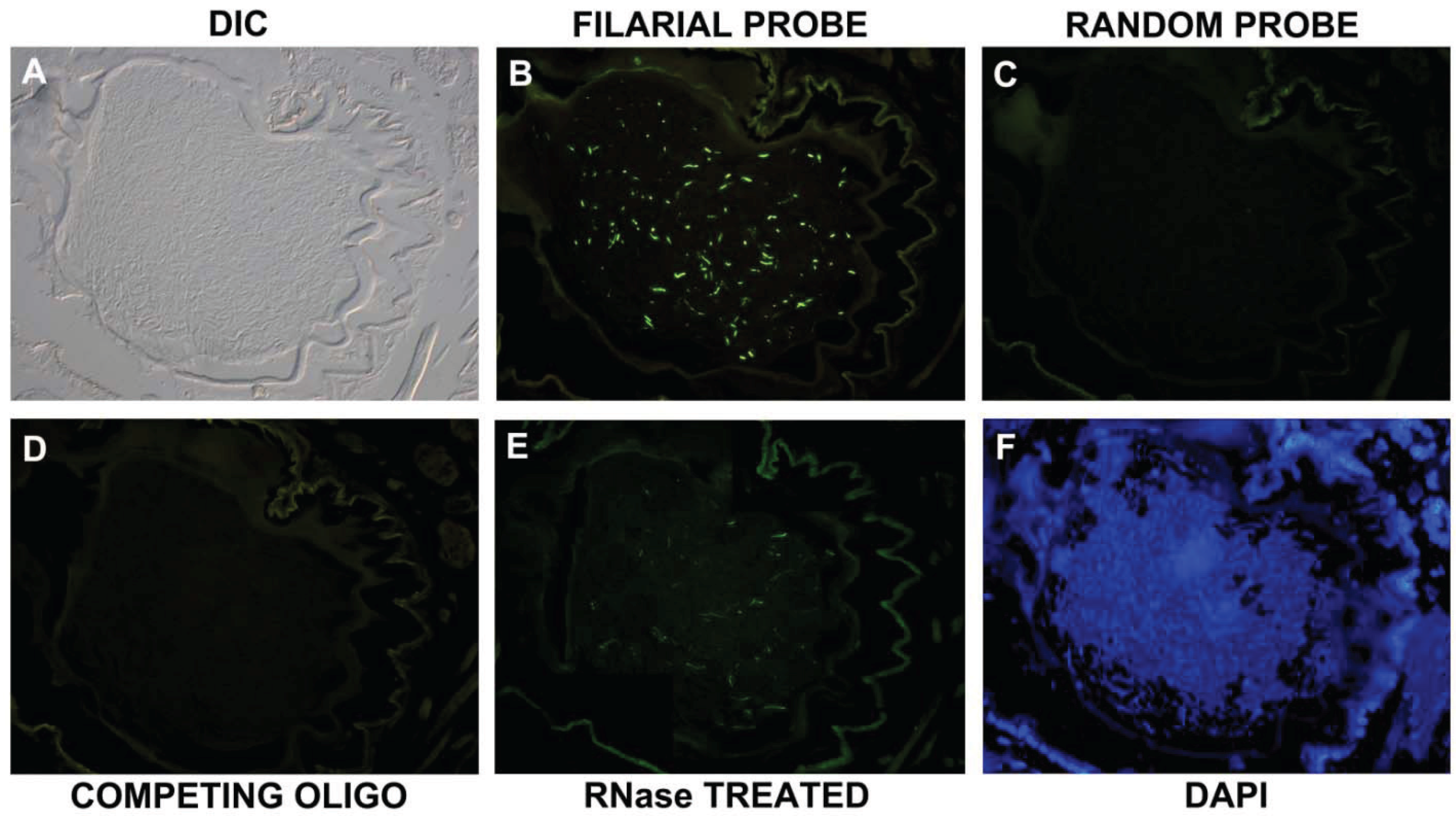
2.5. In Situ Hybridization

In situ hybridization was used to confirm the presence of filarial nematodes in ticks. Adult ticks were dissected under a dissecting microscope. The live ticks were first fixed in wax on the slides and dissected starting from the thoracic region to downward to the anus region and then the slides were reversed to continue dissecting from anus region to thoracic region on the other side. The mouthpart region was dissected carefully to remove the dorsal side of the hard body. A drop of double distilled sterile water was put on the dissected tick and whole gut region along with the salivary glands were separated. The whole gut was transferred to another clean slide. They were separated and fixed with freshly made paraformaldehyde (4%, w/v) and in alcohol:formaline:aldehyde solution (1:2:1, v/v/v) for 24 hours at 4 °C.
A 27-base long oligonucleotide probe was designed to the 18S/5.8S rDNA internal transcribed sequence (5'-GAAACATTCAATTACCTCAAACTTGGG-3') based on the sequence obtained for the filarial-specific PCR reaction (see above). As a negative control, a 27-base long random oligonucleotide probe (5'-GCATAGACATGAGATATACTGTACTAG-3') was also designed. Both probes were synthesized and labeled with FITC at 5’ end by Eurofins MWG Operon oligonuclotide services. For the in situ experiments, 100 ng of labeled probe was mixed first with 2.5 µg of salmon sperm DNA (Sigma) and precipitated at −20 °C for 30 min with 0.1 volume of 3 M sodium acetate and two volumes of ice-cold ethanol. The mixture was then centrifuged at 22,000 × g for 20 min. The resultant pellet was resuspended in 1XTE buffer pH 8.0. The probe was incubated at 95 °C for 10 min, immediately placed on ice for 5 min and then incubated at 37 °C for 15 min. The prefixed whole gut tissue sections first were treated with 1 mg/mL Proteinase K in Proteinase K Buffer at 56 °C for 5 min and then denatured in denaturating solution (70% v/v formamide, 2× SSC, 0.1 mM EDTA pH 7) at 95 °C before hybridization. The sections were hybridized with the probe mixture 56 °C overnight. They were then washed thrice in 2× SSC for 3 min each at room temperature followed by two 20 min washes in 0.1× SSC at 42 °C and a final wash in 0.1× SSC at room temperature. Sections were then washed with freshly made blocking solution (3% w/v BSA in 4× SSC, Triton X-100) for 3 min and wash solution (4× SSC, 0.05% v/v Triton X-100) for 3 min. To visualize the structures of the tick tissue, all tissue samples were counterstained with 300 ng/mL DAPI for 90 seconds in dark and washed with 1× PBS pH 7.4 (phosphate buffer saline) once and mounted with Permafluor mounting medium (Thermo Scientific). All steps were repeated with several controls such as: (1) 100 ng negative control, random oligonucleotide, (2) 200 ng of a competing oligonucleotide present during the hybridization step (5'-CCCAAGTTTGAGGTAATTGAATGTTTC-3'), (3) RNase treatment of the sections before the hybridization step (20 µg/mL for 30 min at 37 °C) and (4) no DNA probe control. Images were acquired using a Leica DM2500 biomedical microscope equipped with dark field, differential interference contrast (DIC) and epifluorescent illumination.

3. Results and Discussion
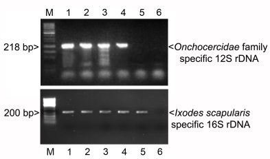
3.1. PCR

To demonstrate the presence of filarial nematodes in Ixodes scapularis ticks, genomic DNA from 20 nymphal ticks collected from Southern Connecticut was then tested for the presence of filarial nematode DNA using a universal filarial nematode-specific PCR technique. Previously published [13] filarial nematode primers FIL and ITSR were then used to amplify the 18S and 5.8S rDNA regions for all species in the phylum Filaria. The expected bands of 400 bp were successfully amplified in 3 tick samples (data not shown) and positive DNA fragments were isolated for direct sequencing. A representative sequence was deposited into NCBI Genbank (JX117890). The sequences were further analyzed by the BLAST sequence analysis program (NCBI). The resulting sequences revealed that the amplified products had the highest similarity with various genera of the Onchocercidae family in Filarial phylum showing greatest similarities to Onchocerca, Dirofilarie, Brugia, Mansonella and Acanthocheilonema genera. Total scores were used to identify the greatest similarity. Onchocerca/Dirofilarie genera had the maximum query coverage ranging from 60% to 86% and 60% to 63% respectively. Sequences had maximum identity of 71%–81% for Onchocerca species and 78%–79% for Dirofilarie species. However, based on these similarity scores, assignment of the sequence found in Ixodes ticks to a specific genus was not possible.

3.2. In Situ Confirmation

To further provide evidence for the presence of filarial nematodes in Ixodes ticks, in situ hybridization was performed with an Onchocercidae family specific oligonucleotide derived from the sequences identified above. The probe was labeled with an FITC fluorescent dye and hybridized on tick gut sections of Ixodes scapularis (Figure 1). DIC microscopy and DAPI counterstain were used to visualize the structure and the nuclei of the tissue (Figure 1, Panel A and F respectively). The in situ staining revealed that filarial nematode-like structures could be observed in tick guts using Onchocercidae family DNA specific probes (Figure 1B). As controls; an FITC labeled random probe sequence, a competing oligonucleotide and RNAase treated samples were utilized in consecutive sections as described in the experimental section (Panel C–E respectively). No positive staining was seen on sections with an FITC labeled random sequence probe, or with the experiment with competing oligonucleotide (Figure 1, Panel C and D). Significant signals were observed even when the sections were RNAse treated demonstrating DNA staining on the section (Panel E). Also, in the “no DNA control” experiments, there were no detectable fluorescent signals on the slides (data not shown).
Figure 1. Representative image of an in situ hybridization experiment using Onchocercidae family filarial nematode specific probe (18S/5.8S internal transcribed spacer sequence) on whole mount tick consecutive sections. Panel A and Panel F show the structure and the nuclei of the tissue using differential interphase microscopy (DIC) and DAPI staining (blue staining) respectively. Panel B show the filarial specific probe staining (bright green staining) while Panels C–E show different control experiments: Panel C = random probe, Panel D = competing oligonucleotide and Panel D = RNase treated sections). 400× magnification.
Vetsci 01 00005 g001

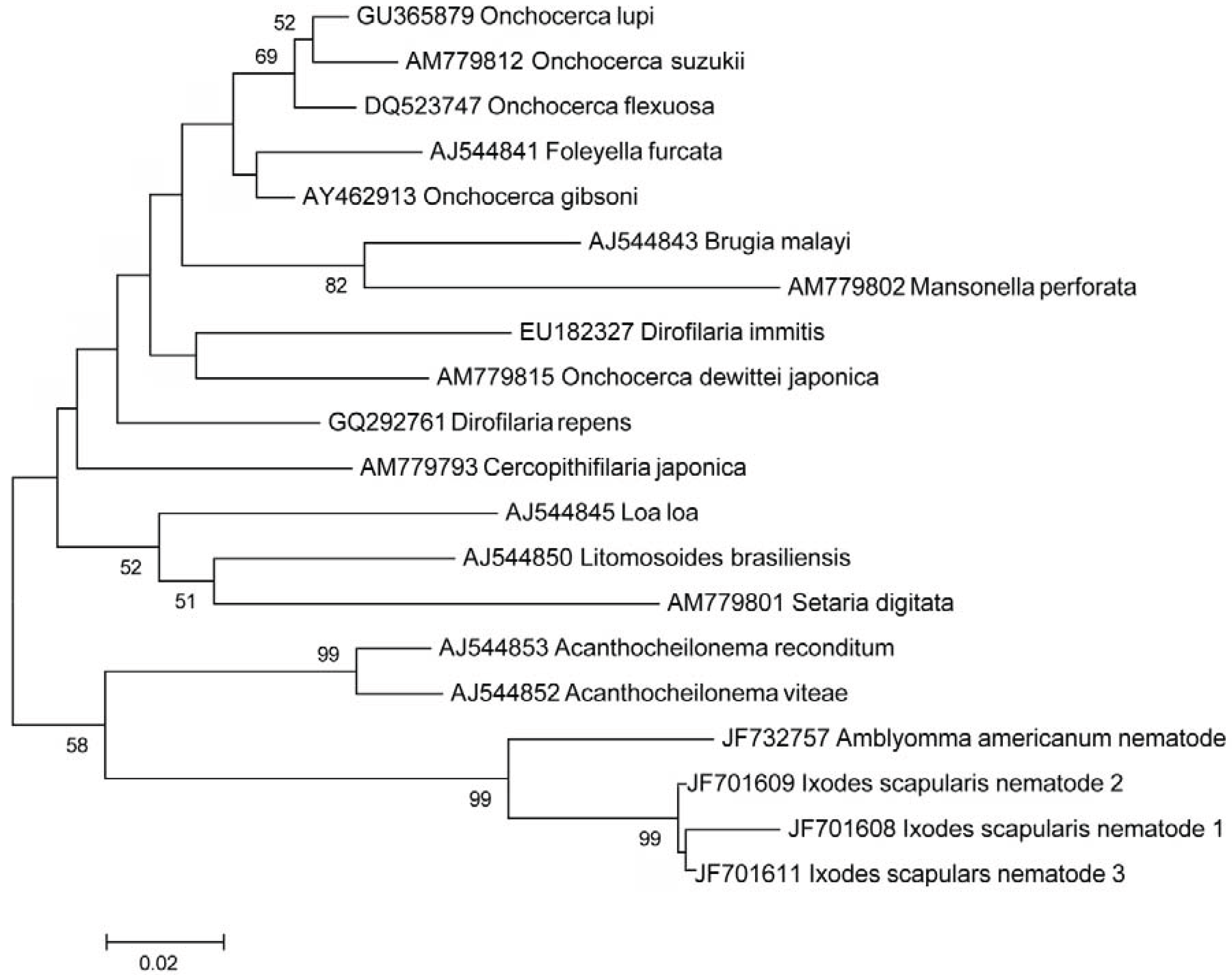
3.3. Additional PCR and Sequencing Analyses

In order to further narrow down the potential species for this microfilaria discovered in Ixodes tick samples, the Onchocercidae family was targeted using Onchocercidae family-specific mitochondrial 12S rDNA PCR primers in the 20 individual nymphal tick samples. PCR experiments with previously published 12S rDNA primer sets did not produce the predicted PCR target [13,15,16], therefore a novel 12S rDNA primer set was designed and used in Ixodes nymphal tick samples. Figure 2 shows a representative PCR experiment with five individual nymphal tick samples showing the successful amplification of a 218 bp fragment of an Onchocercidae family specific 12 rDNA (Figure 2, lanes 2–5). As positive controls, the same tick DNA samples were also analyzed for Ixodes scapularis specific 16S rDNA sequence and found to be positive (Figure 2, lanes 2–5). As a negative control no tick DNA added to lane 6 samples, which showed no amplifications for either the Onchocercidae family specific filarial or Ixodes scapularis specific PCR experiments. BLAST analyses identified three types of isolates with 99% identity (1 or 2 nucleotide differences in the 218 bp sequence). Representative sequences were submitted to the NCBI National Genome Database (Accession numbers: JF701608-11). Sequence alignment (ClustelW) and neighbor-joining phylogenetic analysis were conducted on a 200 bp alignment of the three types of 12S rDNA sequences obtained and compared to corresponding genomic segments of the 12S rDNA gene sequences from different genera of Onchocercidae family using MEGA 5 Evolutionary Genetics Analysis program [14]. Tree support was evaluated by bootstrapping with 500 replications. Figure 3 shows that the result of the phylogenetic analysis indicated that the filarial nematode infecting Ixodes scapularis ticks were most closely related to filarial nematodes in the genus Acanthocheilonema.
Figure 2. Polymerase chain reaction (PCR) performed using Onchocercidae family specific primers for mitochondrial 12S rDNA gene (upper panel) and for Ixodes scapularis specific 16S rDNA (lower panel). DNA isolated from five individual nymphal tick DNA samples (lanes 1–5). Lane 6 shows the negative control (no DNA) for both PCR reactions. Lane M is a 50 bp molecular weight ladder from Fisher Scientific.
Vetsci 01 00005 g002
Figure 3. A neighbor-joining phylogenetic analysis of a 200 bp alignment of the mitochondrial 12S rDNA gene of the three microfilarial nematode isolates obtained from our study (JF70108-11) compared to other species in Onchocercidae family using MEGA 5 Evolutionary Genetics Analysis program. Alphanumeric codes represent GenBank (NCBI) ID numbers. Numbers at nodes indicate bootstrap support values (500 replicates).
Vetsci 01 00005 g003
The closest potential filarial species to what was found in Ixodes ticks were the filarial species found in Amblyoma ticks (92% identity). Comparing filarial species to known Acanthocheilonema species such as Acanthocheilonema vita, and Acanthocheilonema reconditum we have found 85 and 84% identity in the 200 bp region of 12S rDNA gene respectively. We attempted to further identify the species found in Ixodes ticks using additional published primer sets targeting Onchocerciadea genus specific genes such as loci for mitochondrial nicotinamide adenine dehydrogenase (NADH) and cytochrome oxidase I (COI) major sperm protein 1 and 2 (OVMSP1 and OVMSP2) [15,16,17,18], with no successful amplification (data not shown).

3.4. Rate of Infection

To determine the rate of infection, a total of 100 nymphal and 100 adults tick samples were tested individually to evaluate the percentage of deer ticks (Ixodes scapularis) infected with filarial nematodes using the same Onchocercidae family specific primers for mitochondrial 12S rDNA gene. Our results found that 22% of the Ixodes scapularis nymphal ticks and 30% of the adults tick samples were positive for this filarial gene.
In this paper, we identified an Acanthocheilonema spp filarial infection in Ixodes scapularis ticks collected in Southern Connecticut. Our PCR results showed that filarial nematode DNA sequence could be amplified from Ixodes nymphal and adult ticks using either filarial nematode or Onchocercidae family specific PCR primers. Direct sequencing of the resulting sequences further confirmed Onchocercidae family specific sequences. In situ hybridization with an Onchocercidae specific DNA probe also demonstrated the presence of filarial nematodes in tick tissue sections. Phylogenetic analysis of the 12S mitochondrial rDNA gene sequence indicated that the filarial nematodes infecting Ixodes scapularis ticks were most closely related to the species found in Amblyoma americanum ticks and in the genus Acanthocheilonema. Comparing the 12S rDNA filarial sequences found in Ixodes ticks to well characterized species in the genus Acanthocheilonema, we have the most similarity to Acanthocheilonema reconditum (formerly Dipetalonema reconditum) and Acanthocheilonema vitae (formerly Dipetalonema viteae).
Acanthocheilonema reconditum is a known parasite found in dogs while Acanthocheilonema viteae is a rodent parasite [19,20]. The latter is used as a model system for human filarial infections because it shares antigenic homology with the human filarial worm Onchocerca volvulus [20]. Based on this information, one can speculate that the species found in Ixodes tick is most likely an animal parasite. Interestingly, an Acanthocheilonema reconditum infection was also found in human subjects [21,22], therefore further investigation is necessary to identify the potential host(s) of the filarial species found in Ixodes ticks.
In this study, we attempted to further identify the filarial nematode species found in Ixodes ticks using published PCR primer sets targeting Onchocerciadea specific genes other than those targeting ribosomal DNA genetic segments, with no success, suggesting that filarial nematodes found in Ixodes ticks have unique sequences for those targets. In future projects, we will use a second-generation whole genome sequencing approach to more specifically identify this potential species.
We have visualized the filarial nematodes with in situ hybridization. In the literature, the filarial nematodes are usually found to be in the hindgut portion of the arthropod’s body after a blood meal [21,22]. They either remain in the hindgut and accessory glands or they migrate to the salivary glands for manifestation of the disease to the human host. Our in situ staining results with whole mount tick sections of nymphal Ixodes scapularis ticks showed the hindgut regions of the ticks were the primary location of these filarial nematodes. Stage specific development of Acanthocheilonema filarial nematodes was already found in nymphal brown dog ticks (Rhipicephalus sanguineus) [11]. Our sequencing data shows that filarial nematodes found in Amblyomma and Ixodes ticks are closely related. This data combined with our in situ data further suggest it is likely that hard ticks can support the stage specific development of these filarial nematodes.
Our 12S rDNA Onchocercidae specific PCR results further revealed that 22% nymphal and 30% adult ticks, collected from the Southern Connecticut area were infected with filarial nematodes. Comparing to the filarial infection rate found in Amblyomma ticks collected in Maryland (~3.5%), this rate is significantly higher, however it is comparable to a recent report where ~29% of another haematophagous arthropod, Culicidae mosquities, were found to be positive for Setaria and unidentified filarial nematodes in Germany [23].

4. Conclusions

Our study concludes that Ixodes scapularis tick can indeed be a potential vector for filarial nematodes. Based on our in situ and additional data from the literature, it is probable that Ixodes ticks can support the different developmental stages of this filarial nematode as well as its transmission to animals or even humans in the endemic area where ticks are prevalent. Our study warrants further investigation regarding the potential transmission of this type of infection to different hosts.

Acknowledgments

This work was supported by grants from University of New Haven and from Tick-Borne Disease Alliance (TBDA, formally Turn the Corner) to ES. The Schwartz Foundation, the Lyme Disease Association and the Lyme Research Alliance donated microscope, microscope attachments and computers for image analyses. The funders had no role in study design, data collection and analysis, decision to publish, or preparation of the manuscript. The authors thank to Roman Zajac (University of New Haven, West Haven, CT, USA) for his helpful discussion in the preparation of the phylogenetic tree.

Author Contributions

NP: As part of her Master’s thesis she conducted a significant part of the tick collection field-work, executed PCR and in situ staining works, analyzed the data and contributed to the manuscript drafting. JMM contributed in the designs and execution of PCR experiments, sequence analysis and phylogenetic tree output. SM and PRR extracted DNAs and performed PCR experiments. CB was involved in sample collection, additional in situ staining works and in manuscript drafting and MJR gave significant input to the study design and involved in manuscript drafting.
ES contributed to the overall study design and analyses of the data, supervised the laboratory work and wrote the manuscript. All authors read and approved the final version of the manuscript.

Conflicts of Interest

The authors declare no conflict of interest.

References

  1. Piesman, J.; Hicks, T.C.; Sinsky, R.J.; Obiri, G. Simultaneous transmission of Borrelia burgdorferi and Babesia microti by individual nymphal Ixodes dammini ticks. J. Clin. Microbiol. 1987, 25, 2012–2013. [Google Scholar]
  2. Levin, M.L.; Fish, D. Acquisition of coinfection and simultaneous transmission of Borrelia burgdorferi and Ehrlichia phagocytophila by Ixodes scapularis ticks. Infect. Immun. 2000, 68, 2183–2186. [Google Scholar] [CrossRef]
  3. Courtney, J.W.; Dryden, R.L.; Montgomery, J.; Schneider, BS.; Smith, G.; Massung, R.F. Molecular characterization of Anaplasma phagocytophilum and Borrelia burgdorferi in Ixodes scapularis ticks from Pennsylvania. J. Clin. Microbiol. 2003, 41, 1569–1573. [Google Scholar] [CrossRef]
  4. Adelson, M.E.; Rao, R.V.; Tilton, R.C.; Cabets, K.; Eskow, E.; Fein, L.; Occi, J.L.; Mordechai, E. Prevalence of Borrelia burgdorferi, Bartonella spp., Babesia microti, and Anaplasma phagocytophila in Ixodes scapularis Ticks collected in northern New Jersey. J. Clin. Microbiol. 2004, 42, 2799–2801. [Google Scholar] [CrossRef]
  5. Holman, M.S.; Caporale, D.A.; Goldberg, J.; Lacombe, E.; Lubelczyk, C.; Rand, P.W.; Smith, R.P. Anaplasma phagocytophilum, Babesia microti, and Borrelia burgdorferi in Ixodes scapularis, southern coastal Maine. Emerg. Infect. Dis. 2004, 10, 744–746. [Google Scholar] [CrossRef]
  6. Swanson, S.J.; Neitzel, D.; Reed, K.D.; Belongia, E.A. Coinfections acquired from Ixodes ticks. Clin. Microbiol. 2006, 19, 708–727. [Google Scholar] [CrossRef]
  7. Tokarz, R.; Jain, K.; Bennett, A.; Briese, T.; Lipkin, W.I. Assessment of polymicrobial infections in ticks in New York State. Vector Borne Zoonotic Dis. 2010, 10, 217–221. [Google Scholar] [CrossRef]
  8. Kjemtrup, AM.; Conrad, P.A. Human babesiosis: An emerging tick-borne disease. Int. J. Parasitol. 2000, 30, 1323–1337. [Google Scholar] [CrossRef]
  9. Beaver, P.C.; Burgdorfer, W. A microfilaria of exceptional size from the Ixodid tick, Ixodes dammini from Shelter Island New York. J. Parasitol. 1984, 70, 963–966. [Google Scholar] [CrossRef]
  10. Londono, M.I. Behaviour and characteristics of the filarial Dipetalonema viteae in the tick Ornithodoros tartakowskyi. Antioquia Medica 1973, 23, 515–516. [Google Scholar]
  11. Olmeda-García, A.S.; Rodríguez-Rodríguez, J.A. Stage specific development of filarial nematodes in vector ticks. J. Helminthol. 1994, 68, 231–235. [Google Scholar] [CrossRef]
  12. Zhang, X.; Norris, D.E.; Rasgon, J.L. Distribution and molecular characterization of Wolbachia endosymbionts and filarial nematodes in Maryland populations of the lone star tick (Amblyomma americanum). FEMS Microbiol. Ecol. 2011, 77, 50–56. [Google Scholar] [CrossRef]
  13. Nuchprayoon, S.; Junpee, A.; Poovorawn, Y.; Scott, A. Detection and differentiation of filarial parasites by universal primer and polymerase chain reaction–restriction fragment length polymorphism analysis. Am. J. Trop. Med. Hy. 2005, 73, 895–900. [Google Scholar]
  14. Tamura, K.; Peterson, D.; Nei, M. MEGA5: Molecular evolutionary genetics analysis using maximum likelihood, evolutionary distance and maximum parsimony methods. Mol. Biol Evol. 2011, 28, 2731–2739. [Google Scholar] [CrossRef]
  15. Casiraghi, M.; Anderson, T.J.C.; Bandi, C.; Bazzocchi, C.; Genchi, C. A Phylogenetic analysis of filarial nematode: Comparison with the phylogeny of Wolbachia endosymbionts. Parasitology 2001, 122, 93–103. [Google Scholar] [CrossRef]
  16. Otranto, D.; Brianti, E.; Dantas-Torres, F.; Weigl, S.; Latrofa, M.S.; Gaglio, G.; Cauquil, L.; Giannetto, S.; Bain, O. Morphological and molecular data on the dermal microfilariae of a species of Cercopithi filaria from a dog in Sicily. Vet. Parasitol. 2011, 182, 221–229. [Google Scholar] [CrossRef]
  17. Li, B.W.; Rush, A.C.; Tan, J.; Weil, G.J. Quantitative analysis of gender-regulated transcripts in the filarial Nematode Brugia malayi by real-time RT-PCR. Mol. Biochem. Parasitol. 2004, 137, 329–337. [Google Scholar] [CrossRef]
  18. Hojas, R.M.; Post, R.J. Regional genetic variation in the major sperm protein genes of Onchocerca volvulus and Mansonella ozzardi (Nematoda: Filarioidea). Int. J. Parasitol. 2000, 30, 1459–1465. [Google Scholar] [CrossRef]
  19. Brianti, B.; Gagglio, G.; Napoli, E.; Giannetto, S.; Dantas-Torres, P.; Bain, O.; Otranto, D. New insights into the ecology and biology of Acanthocheilonema reconditum causing canine subcutaneous filariosis. Parasitology 2012, 139, 530–536. [Google Scholar] [CrossRef]
  20. Eisenbeiss, W.F.; Apfel, H.; Meyer, T.F. Recovery, distribution, and development of Acanthocheilonema viteae third- and early fourth-stage larvae in adult jirds. J. Parasitol. 1991, 77, 580–586. [Google Scholar] [CrossRef]
  21. Beaver, P.C.; Meyer, E.A.; Jarroll, E.L. Dipetalonema from the eye of a man in Oregon, USA. A case report. Am. J. Trop. Med. Hyg. 1980, 29, 367–372. [Google Scholar]
  22. Huynh, T.; Thean, J.; Maini, R. Dipetalonema reconditum in the human eye. Br. J. Ophtalmol. 2001, 85. [Google Scholar] [CrossRef]
  23. Czajka, C.; Becker, N.; Poppert, S.; Jöst, H.; Schmidt-Chanasit, J.; Krüge, A. Molecular detection of Setaria tundra (Nematoda: Filarioidea) and an unidentified filarial species in mosquitoes in Germany. Parasit. Vectors 2012, 5. [Google Scholar] [CrossRef]

