Next Issue
Volume 12, April
Previous Issue
Volume 12, February


Biomedicines, Volume 12, Issue 3 (March 2024) – 235 articles

Cover Story (view full-size image): 
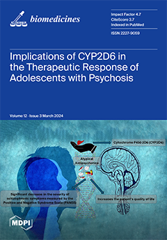
The plasma levels of antipsychotics and their metabolites depend on the activity of the cytochrome P450 (CYP) system in the liver. This research aimed to test the variability of individual responses to atypical antipsychotic drugs, depending on CYP2D6 enzyme activity in adolescents diagnosed with schizophrenia. It was hypothesized that interindividual variation in the metabolic activity of the CYP2D6 enzyme may influence responses to antipsychotic treatment in terms of therapeutic efficacy and the risk of adverse effects, depending on the type of metabolism of the patient. Evaluating CYP2D6 polymorphism may be useful in pediatric psychiatry for improving clinical symptoms and increasing patients' quality of life. View this paper
  • Issues are regarded as officially published after their release is announced to the table of contents alert mailing list.
  • You may sign up for e-mail alerts to receive table of contents of newly released issues.
  • PDF is the official format for papers published in both, html and pdf forms. To view the papers in pdf format, click on the "PDF Full-text" link, and use the free Adobe Reader to open them.
Order results
Result details
Section
Select all
Export citation of selected articles as:
18 pages, 2379 KiB  
Article
Comparing the Efficacy of Two Generations of EGFR-TKIs: An Integrated Drug–Disease Mechanistic Model Approach in EGFR-Mutated Lung Adenocarcinoma
by Hippolyte Darré, Perrine Masson, Arnaud Nativel, Laura Villain, Diane Lefaudeux, Claire Couty, Bastien Martin, Evgueni Jacob, Michaël Duruisseaux, Jean-Louis Palgen, Claudio Monteiro and Adèle L’Hostis
Biomedicines 2024, 12(3), 704; https://doi.org/10.3390/biomedicines12030704 - 21 Mar 2024
Viewed by 1063
Abstract
Mutationsin epidermal growth factor receptor (EGFR) are found in approximately 48% of Asian and 19% of Western patients with lung adenocarcinoma (LUAD), leading to aggressive tumor growth. While tyrosine kinase inhibitors (TKIs) like gefitinib and osimertinib target this mutation, treatments often face challenges [...] Read more.
Mutationsin epidermal growth factor receptor (EGFR) are found in approximately 48% of Asian and 19% of Western patients with lung adenocarcinoma (LUAD), leading to aggressive tumor growth. While tyrosine kinase inhibitors (TKIs) like gefitinib and osimertinib target this mutation, treatments often face challenges such as metastasis and resistance. To address this, we developed physiologically based pharmacokinetic (PBPK) models for both drugs, simulating their distribution within the primary tumor and metastases following oral administration. These models, combined with a mechanistic knowledge-based disease model of EGFR-mutated LUAD, allow us to predict the tumor’s behavior under treatment considering the diversity within the tumor cells due to different mutations. The combined model reproduces the drugs’ distribution within the body, as well as the effects of both gefitinib and osimertinib on EGFR-activation-induced signaling pathways. In addition, the disease model encapsulates the heterogeneity within the tumor through the representation of various subclones. Each subclone is characterized by unique mutation profiles, allowing the model to accurately reproduce clinical outcomes, including patients’ progression, aligning with RECIST criteria guidelines (version 1.1). Datasets used for calibration came from NEJ002 and FLAURA clinical trials. The quality of the fit was ensured with rigorous visual predictive checks and statistical tests (comparison metrics computed from bootstrapped, weighted log-rank tests: 98.4% (NEJ002) and 99.9% (FLAURA) similarity). In addition, the model was able to predict outcomes from an independent retrospective study comparing gefitinib and osimertinib which had not been used within the model development phase. This output validation underscores mechanistic models’ potential in guiding future clinical trials by comparing treatment efficacies and identifying patients who would benefit most from specific TKIs. Our work is a step towards the design of a powerful tool enhancing personalized treatment in LUAD. It could support treatment strategy evaluations and potentially reduce trial sizes, promising more efficient and targeted therapeutic approaches. Following its consecutive prospective validations with the FLAURA2 and MARIPOSA trials (validation metrics computed from bootstrapped, weighted log-rank tests: 94.0% and 98.1%, respectively), the model could be used to generate a synthetic control arm. Full article
(This article belongs to the Special Issue Recent Advances of Receptor Tyrosine Kinases in Solid Tumors)
Show Figures

Figure 1

19 pages, 3274 KiB  
Article
Gut Microbiota Signatures with Potential Clinical Usefulness in Colorectal and Non-Small Cell Lung Cancers
by Sofía Tesolato, Juan Vicente-Valor, Mateo Paz-Cabezas, Dulcenombre Gómez-Garre, Silvia Sánchez-González, Adriana Ortega-Hernández, Sofía de la Serna, Inmaculada Domínguez-Serrano, Jana Dziakova, Daniel Rivera, Jose-Ramón Jarabo, Ana-María Gómez-Martínez, Florentino Hernando, Antonio Torres and Pilar Iniesta
Biomedicines 2024, 12(3), 703; https://doi.org/10.3390/biomedicines12030703 - 21 Mar 2024
Viewed by 759
Abstract
The application of bacterial metagenomic analysis as a biomarker for cancer detection is emerging. Our aim was to discover gut microbiota signatures with potential utility in the diagnosis of colorectal cancer (CRC) and non-small cell lung cancer (NSCLC). A prospective study was performed [...] Read more.
The application of bacterial metagenomic analysis as a biomarker for cancer detection is emerging. Our aim was to discover gut microbiota signatures with potential utility in the diagnosis of colorectal cancer (CRC) and non-small cell lung cancer (NSCLC). A prospective study was performed on a total of 77 fecal samples from CRC and NSCLC patients and controls. DNA from stool was analyzed for bacterial genomic sequencing using the Ion Torrent™ technology. Bioinformatic analysis was performed using the QIIME2 pipeline. We applied logistic regression to adjust for differences attributable to sex, age, and body mass index, and the diagnostic accuracy of our gut signatures was compared with other previously published results. The feces of patients affected by different tumor types, such as CRC and NSCLC, showed a differential intestinal microbiota profile. After adjusting for confounders, Parvimonas (OR = 53.3), Gemella (OR = 6.01), Eisenbergiella (OR = 5.35), Peptostreptococcus (OR = 9.42), Lactobacillus (OR = 6.72), Salmonella (OR = 5.44), and Fusobacterium (OR = 78.9) remained significantly associated with the risk of CRC. Two genera from the Ruminococcaceae family, DTU089 (OR = 20.1) and an uncharacterized genus (OR = 160.1), were associated with the risk of NSCLC. Our two panels had better diagnostic capacity for CRC (AUC = 0.840) and NSLC (AUC = 0.747) compared to the application of two other published panels to our population. Thus, we propose a gut bacteria panel for each cancer type and show its potential application in cancer diagnosis. Full article
(This article belongs to the Special Issue Advanced Research of Gut Microbiota in Health and Diseases)
Show Figures

Figure 1

24 pages, 1007 KiB  
Review
Putting Functional Gastrointestinal Disorders within the Spectrum of Inflammatory Disorders Can Improve Classification and Diagnostics of These Disorders
by Dunja Šojat, Mile Volarić, Tanja Keškić, Nikola Volarić, Venija Cerovečki and Ljiljana Trtica Majnarić
Biomedicines 2024, 12(3), 702; https://doi.org/10.3390/biomedicines12030702 - 21 Mar 2024
Viewed by 797
Abstract
The spectrum, intensity, and overlap of symptoms between functional gastrointestinal disorders (FGIDs) and other gastrointestinal disorders characterize patients with FGIDs, who are incredibly different in their backgrounds. An additional challenge with regard to the diagnosis of FGID and the applicability of a given [...] Read more.
The spectrum, intensity, and overlap of symptoms between functional gastrointestinal disorders (FGIDs) and other gastrointestinal disorders characterize patients with FGIDs, who are incredibly different in their backgrounds. An additional challenge with regard to the diagnosis of FGID and the applicability of a given treatment is the ongoing expansion of the risk factors believed to be connected to these disorders. Many cytokines and inflammatory cells have been found to cause the continuous existence of a low level of inflammation, which is thought to be a basic pathophysiological process. The idea of the gut–brain axis has been created to offer a basic framework for the complex interactions that occur between the nervous system and the intestinal functions, including the involvement of gut bacteria. In this review paper, we intend to promote the hypothesis that FGIDs should be seen through the perspective of the network of the neuroendocrine, immunological, metabolic, and microbiome pathways. This hypothesis arises from an increased understanding of chronic inflammation as a systemic disorder, that is omnipresent in chronic health conditions. A better understanding of inflammation’s role in the pathogenesis of FGIDs can be achieved by clustering markers of inflammation with data indicating symptoms, comorbidities, and psycho-social factors. Finding subclasses among related entities of FGIDs may reduce patient heterogeneity and help clarify the pathophysiology of this disease to allow for better treatment. Full article
Show Figures

Graphical abstract

18 pages, 318 KiB  
Review
Exploring the Landscape of Anti-Inflammatory Trials: A Comprehensive Review of Strategies for Targeting Inflammation in Acute Myocardial Infraction
by Andreas Mitsis, Michaela Kyriakou, Stefanos Sokratous, Georgia Karmioti, Michail Drakomathioulakis, Michael Myrianthefs, Antonios Ziakas, Stergios Tzikas and George Kassimis
Biomedicines 2024, 12(3), 701; https://doi.org/10.3390/biomedicines12030701 - 21 Mar 2024
Viewed by 752
Abstract
The role of inflammation in the pathophysiology of acute myocardial infarction (AMI) is well established. In recognizing inflammation’s pivotal role in AMI, this manuscript systematically traces the historical studies spanning from early attempts to the present landscape. Several anti-inflammatory trials targeting inflammation in [...] Read more.
The role of inflammation in the pathophysiology of acute myocardial infarction (AMI) is well established. In recognizing inflammation’s pivotal role in AMI, this manuscript systematically traces the historical studies spanning from early attempts to the present landscape. Several anti-inflammatory trials targeting inflammation in post-AMI have been performed, and this review includes the key trials, as well as examines their designs, patient demographics, and primary outcomes. Efficacies and challenges are analyzed, thereby shedding light on the translational implications of trial outcomes. This article also discusses emerging trends, ongoing research, and potential future directions in the field. Practical applications and implications for clinical practice are considered by providing a holistic view of the evolving landscape of anti-inflammatory interventions in the context of AMI. Full article
(This article belongs to the Special Issue Molecular Insights into Myocardial Infarction)
13 pages, 1435 KiB  
Article
Development of a Novel Endometrial Signature Based on Endometrial microRNA for Determining the Optimal Timing for Embryo Transfer
by Ching-Hung Chen, Farn Lu, Wen-Jui Yang, Wei-Ming Chen, Pok Eric Yang, Shih-Ting Kang, Tiffany Wang, Po-Chang Chang, Chi-Ting Feng, Jung-Hsuan Yang, Chen-Yu Liu, Chi-An Hsieh, Lily Hui-Ching Wang and Jack Yu-Jen Huang
Biomedicines 2024, 12(3), 700; https://doi.org/10.3390/biomedicines12030700 - 21 Mar 2024
Viewed by 641
Abstract
Though tremendous advances have been made in the field of in vitro fertilization (IVF), a portion of patients are still affected by embryo implantation failure issues. One of the most significant factors contributing to implantation failure is a uterine condition called displaced window [...] Read more.
Though tremendous advances have been made in the field of in vitro fertilization (IVF), a portion of patients are still affected by embryo implantation failure issues. One of the most significant factors contributing to implantation failure is a uterine condition called displaced window of implantation (WOI), which refers to an unsynchronized endometrium and embryo transfer time for IVF patients. Previous studies have shown that microRNAs (miRNAs) can be important biomarkers in the reproductive process. In this study, we aim to develop a miRNA-based classifier to identify the WOI for optimal time for embryo transfer. A reproductive-related PanelChip® was used to obtain the miRNA expression profiles from the 200 patients who underwent IVF treatment. In total, 143 out of the 167 miRNAs with amplification signals across 90% of the expression profiles were utilized to build a miRNA-based classifier. The microRNA-based classifier identified the optimal timing for embryo transfer with an accuracy of 93.9%, a sensitivity of 85.3%, and a specificity of 92.4% in the training set, and an accuracy of 88.5% in the testing set, showing high promise in accurately identifying the WOI for the optimal timing for embryo transfer. Full article
(This article belongs to the Special Issue Non-coding RNAs in Health and Disease 2.0)
Show Figures

Figure 1

20 pages, 1872 KiB  
Review
Development of Cell Technologies Based on Dendritic Cells for Immunotherapy of Oncological Diseases
by Vasily Kurilin, Alina Alshevskaya and Sergey Sennikov
Biomedicines 2024, 12(3), 699; https://doi.org/10.3390/biomedicines12030699 - 21 Mar 2024
Viewed by 888
Abstract
Immunotherapy using dendritic cell-based vaccination is a natural approach using the capabilities and functions inherent in the patient’s immune system to eliminate tumor cells. The development of dendritic cell-based cell technologies evolved as the disorders of dendritic cell differentiation and function in cancer [...] Read more.
Immunotherapy using dendritic cell-based vaccination is a natural approach using the capabilities and functions inherent in the patient’s immune system to eliminate tumor cells. The development of dendritic cell-based cell technologies evolved as the disorders of dendritic cell differentiation and function in cancer were studied; some of these functions are antigen presentation, priming of cytotoxic T-lymphocytes and induction of antigen-specific immune responses. At the initial stage of technology development, it was necessary to develop protocols for the in vitro generation of functionally mature dendritic cells that were capable of capturing tumor antigens and processing and presenting them in complex with MHC to T-lymphocytes. To achieve this, various forms of tumor-associated antigen delivery systems were tested, including lysates, tumor cell proteins (peptides), and DNA and RNA constructs, and it was shown that the use of DNA and RNA constructs was the most effective method, as it made it possible not only to deliver the most immunogenic epitopes of tumor-associated antigens to dendritic cells, but also to enhance their ability to induce antigen-specific cytotoxic T-lymphocytes. Currently, cell therapy based on dendritic cells is a modern basis for antigen-specific immunotherapy of cancer due to the simplicity of creating DNA and RNA constructs encoding information about both target tumor antigens and regulatory molecules. The potential development of cell technologies based on dendritic cells aims to obtain antigen-specific cytotoxic T-lymphocytes induced by dendritic cells, study their functional activity and develop cell-based therapy. Full article
(This article belongs to the Section Immunology and Immunotherapy)
Show Figures

Figure 1

15 pages, 3121 KiB  
Article
Repurposing of the Cardiovascular Drug Statin for the Treatment of Cancers: Efficacy of Statin–Dipyridamole Combination Treatment in Melanoma Cell Lines
by Nanami Irie, Kana Mizoguchi, Tomoko Warita, Mirai Nakano, Kasuga Sasaki, Jiro Tashiro, Tomohiro Osaki, Takuro Ishikawa, Zoltán N. Oltvai and Katsuhiko Warita
Biomedicines 2024, 12(3), 698; https://doi.org/10.3390/biomedicines12030698 - 21 Mar 2024
Viewed by 989
Abstract
Metastatic melanoma has a very poor prognosis. Statins, 3-hydroxy-3-methyl-glutaryl-coenzyme A reductase (HMGCR) inhibitors, are cholesterol-lowering agents with a potential for cancer treatment. The inhibition of HMGCR by statins, however, induces feedback, which paradoxically upregulates HMGCR expression via sterol regulatory element-binding protein-2 (SREBP2). Dipyridamole, [...] Read more.
Metastatic melanoma has a very poor prognosis. Statins, 3-hydroxy-3-methyl-glutaryl-coenzyme A reductase (HMGCR) inhibitors, are cholesterol-lowering agents with a potential for cancer treatment. The inhibition of HMGCR by statins, however, induces feedback, which paradoxically upregulates HMGCR expression via sterol regulatory element-binding protein-2 (SREBP2). Dipyridamole, an antiplatelet agent, is known to inhibit SREBP2 upregulation. We aimed to demonstrate the efficacy of statin–dipyridamole combination treatment in both human and spontaneously occurring canine melanoma cell lines. The half maximal inhibitory concentration (IC50) of atorvastatin showed a 68–92% reduction when combined with dipyridamole, compared with that of atorvastatin alone. In some melanoma cell lines, cell proliferation was suppressed to almost zero by the combination treatment (≥3 μM atorvastatin). Finally, the BRAF inhibitor, vemurafenib, further potentiated the effects of the combined statin–dipyridamole treatment in BRAF V600E mutation-bearing human melanoma cell lines. In conclusion, the inexpensive and frequently prescribed statin–dipyridamole combination therapy may lead to new developments in the treatment of melanoma and may potentiate the effects of vemurafenib for the targeted therapy of BRAF V600E-mutation bearing melanoma patients. The concordance between the data from canine and human melanoma cell lines reinforces this possibility. Full article
Show Figures

Figure 1

12 pages, 3015 KiB  
Article
Cimetidine Attenuates Therapeutic Effect of Anti-PD-1 and Anti-PD-L1 and Modulates Tumor Microenvironment in Colon Cancer
by Feng-Chi Kuo, Jerry Cheng-Yen Lai, Hui-Ru Shieh, Wan-Zu Liou, Ming-Jong Bair and Yu-Jen Chen
Biomedicines 2024, 12(3), 697; https://doi.org/10.3390/biomedicines12030697 - 21 Mar 2024
Viewed by 655
Abstract
Histamine modulates immunity by binding to histamine receptor 2 (H2R). Cimetidine, an H2R antagonist that inhibits gastric acid secretion and treats gastrointestinal ulcers, interferes with histamine-mediated immunomodulation and may have anticancer activity. This study examined cimetidine’s effect on the anticancer effect of anti-PD-L1 [...] Read more.
Histamine modulates immunity by binding to histamine receptor 2 (H2R). Cimetidine, an H2R antagonist that inhibits gastric acid secretion and treats gastrointestinal ulcers, interferes with histamine-mediated immunomodulation and may have anticancer activity. This study examined cimetidine’s effect on the anticancer effect of anti-PD-L1 in colon cancer. The MTT assay, colony formation assay, and DNA histograms assessed cell viability, clonogenicity, and cell cycle distribution, respectively. Flow cytometry measured H2R and PD-L1 expression and estimated specific immune cell lineages. For the in vivo study, tumor cells were subcutaneously implanted into the right flank of BALB/c mice. Cimetidine had no significant effect on CT26 cell viability, clonogenicity, or cell cycle distribution. It also did not affect H2R and PD-L1 expression levels in CT26 cells. In vivo, anti-PD-1 and anti-PD-L1 suppressed CT26 tumor growth, whereas cimetidine showed mild antitumor activity. In the combined experiment, cimetidine significantly attenuated anti-PD-1 and anti-PD-L1′ antitumor effects without major toxicity. In the tumor microenvironment, anti-PD-L1 increased CD3+ T, CD4+ T, and CD8+ T cells and M1 macrophages. Combined treatment with cimetidine reversed this. Cimetidine also reversed anti-PD-1 and anti-PD-L1′s decrease in circulating and tumor-associated neutrophils. Cimetidine attenuated anti-PD-L1′s antitumor effect and modulated the tumor microenvironment in colon cancer. Full article
(This article belongs to the Special Issue Cancer Immunotherapy: The Multiple Impacts of Tumor Microenvironment)
Show Figures

Figure 1

17 pages, 5258 KiB  
Article
Gene-Expression Patterns of Tumor and Peritumor Tissues of Smoking and Non-Smoking HPV-Negative Patients with Head and Neck Squamous Cell Carcinoma
by Anna Soboleva, Irina Arutyunyan, Enar Jumaniyazova, Polina Vishnyakova, Daria Zarubina, Eldar Nimatov, Andrey Elchaninov and Timur Fatkhudinov
Biomedicines 2024, 12(3), 696; https://doi.org/10.3390/biomedicines12030696 - 21 Mar 2024
Viewed by 690
Abstract
We studied the gene-expression patterns in specimens of tumor and peritumor tissue biopsies of 26 patients with head and neck carcinomas depending on smoking status. Histological and immunohistochemical examinations verified that all tumors belonged to the “classical” subgroup of head and neck carcinomas, [...] Read more.
We studied the gene-expression patterns in specimens of tumor and peritumor tissue biopsies of 26 patients with head and neck carcinomas depending on smoking status. Histological and immunohistochemical examinations verified that all tumors belonged to the “classical” subgroup of head and neck carcinomas, and the HPV-negative tumor status was confirmed. The expression of 28 tumor-associated genes determined by RT-PCR was independent of patients’ sex or age, TNM status, degree of differentiation, or tissue localization. Moreover, in peritumor tissue, none of the 28 genes were differentially expressed between the groups of smoking and nonsmoking patients. During oncotransformation in both studied groups, there were similar processes typical for HNSCC progression: the expression levels of paired keratins 4 and 13 were reduced, while the expression levels of keratin 17 and CD44 were significantly increased. However, further investigation revealed some distinctive features: the expression of the genes EGFR and TP63 increased significantly only in the nonsmoking group, and the expression of IL6, CDKN2A, EGF, and PITX1 genes changed only in the smoking group. In addition, correlation analysis identified several clusters within which genes displayed correlations in their expression levels. The largest group included 10 genes: TIMP1, TIMP2, WEE1, YAP, HIF1A, PI3KCA, UTP14A, APIP, PTEN, and SLC26A6. The genetic signatures associated with smoking habits that we have found may serve as a prerequisite for the development of diagnostic panels/tests predicting responses to different therapeutic strategies for HNSCC. Full article
(This article belongs to the Special Issue Head and Neck Tumors, 3rd Edition)
Show Figures

Figure 1

11 pages, 753 KiB  
Article
APOE Gene Variation’s Impact on Cardiovascular Health: A Case-Control Study
by Aya Badeea Ismail, Özlem Balcıoğlu, Barçın Özcem and Mahmut Çerkez Ergoren
Biomedicines 2024, 12(3), 695; https://doi.org/10.3390/biomedicines12030695 - 21 Mar 2024
Viewed by 628
Abstract
Chronic venous insufficiency (CVI) is a common medical condition characterized by impaired functioning of the venous system in the lower extremities. It leads to various symptoms, including varicose veins, leg edema, and skin pigmentation. It is believed that a combination of genetic and [...] Read more.
Chronic venous insufficiency (CVI) is a common medical condition characterized by impaired functioning of the venous system in the lower extremities. It leads to various symptoms, including varicose veins, leg edema, and skin pigmentation. It is believed that a combination of genetic and environmental factors affect the development of CVI. The APOE gene is of particular interest in this context, as it plays a role in lipid metabolism and inflammation. The ε4 allele (rs429358) has been associated with an increased risk of Alzheimer’s disease, while the ε2 allele (rs7412) has shown a protective effect against Alzheimer’s disease but a strong association with cardiovascular inflammation. This research aimed to investigate the presence of APOE gene variants in individuals with chronic venous insufficiency disease and validate the relationship between this gene and cardiovascular diseases. The study analyzed the expression of APOE gene variants in varicose vein tissue samples from patients and a normal vein in the control group. The results indicated no significant expression of the ε4 allele in either group. However, there was a significant decrease in the expression of the ε2 allele in the patient group. Additionally, a negative correlation was observed between the two single nucleotide polymorphisms (SNPs) in vein tissue. The lower expression of the ε2 allele in patients suggests a potentially reduced risk of cardiovascular disease in these individuals. Consequently, there appears to be a weaker association between the expression of the APOE gene ε2 allele and cardiovascular diseases. Full article
(This article belongs to the Special Issue Phenotypic Consequences of Human Genetic Diversity)
Show Figures

Figure 1

15 pages, 1998 KiB  
Article
Developing Bioactive Hydrogels with Peptides for Dental Application
by Alexandrina Muntean, Codruta Sarosi, Ioan Petean, Stanca Cuc, Rahela Carpa, Ioana Andreea Chis, Aranka Ilea, Ada Gabriela Delean and Marioara Moldovan
Biomedicines 2024, 12(3), 694; https://doi.org/10.3390/biomedicines12030694 - 21 Mar 2024
Viewed by 680
Abstract
Dental caries is an avoidable and complex condition impacting billions of individuals worldwide, posing a specific concern among younger generations, despite the progress of oral hygiene products. This deterioration occurs due to the acid demineralization of tooth enamel, leading to the loss of [...] Read more.
Dental caries is an avoidable and complex condition impacting billions of individuals worldwide, posing a specific concern among younger generations, despite the progress of oral hygiene products. This deterioration occurs due to the acid demineralization of tooth enamel, leading to the loss of minerals from the enamel subsurface. The remineralisation of early enamel carious lesions could prevent the cavitation of teeth. The enamel protein amelogenin constitutes 90% of the total enamel matrix protein and plays a key role in the bio mineralisation process. The aim of this study is to investigate the self-assembly microstructure and reticulation behaviour of a newly developed bioactive hydrogel with leucine-rich amelogenin peptide (LRAP) intended for enamel remineralisation. SEM, AFM, UV-VIS, and FTIR analyses emphasize the ability of peptides to promote cell adhesion and the treatment of early carious lesions. In conclusion, short-chain peptides can be used in hydrogels for individual or professional use. Full article
(This article belongs to the Topic Advanced Functional Materials for Regenerative Medicine)
Show Figures

Figure 1

27 pages, 1257 KiB  
Review
Zebrafish as a Model for Cardiovascular and Metabolic Disease: The Future of Precision Medicine
by Ramcharan Singh Angom and Naga Malleswara Rao Nakka
Biomedicines 2024, 12(3), 693; https://doi.org/10.3390/biomedicines12030693 - 20 Mar 2024
Viewed by 905
Abstract
The zebrafish (Danio rerio) has emerged as an appreciated and versatile model organism for studying cardiovascular and metabolic diseases, offering unique advantages for both basic research and drug discovery. The genetic conservation between zebrafish and humans and their high fecundity and [...] Read more.
The zebrafish (Danio rerio) has emerged as an appreciated and versatile model organism for studying cardiovascular and metabolic diseases, offering unique advantages for both basic research and drug discovery. The genetic conservation between zebrafish and humans and their high fecundity and transparent embryos allow for efficient large-scale genetic and drug-oriented screening studies. Zebrafish possess a simplified cardiovascular system that shares similarities with mammals, making them particularly suitable for modeling various aspects of heart development, function, and disease. The transparency of zebrafish embryos enables the real-time visualization of cardiovascular dynamics, offering insights into early embryonic events and facilitating the study of heart-related anomalies. In metabolic research, zebrafish provide a cost-effective platform for modeling obesity, type 2 diabetes, hyperlipidemia, and other metabolic disorders. Their high reproductive rate allows for the generation of large cohorts for robust statistical analyses, while advanced genetic tools, such as CRISPR/Cas9, enable precise gene editing with which to model specific genetic mutations associated with human diseases. Zebrafish metabolic models have been instrumental in elucidating the molecular mechanisms underlying metabolic diseases, studying the effects of environmental factors, and identifying potential therapeutic targets. Additionally, the permeability of zebrafish embryos to small molecules facilitates drug discovery and screening, offering a rapid and economical approach to identifying compounds with therapeutic potential. In conclusion, zebrafish cardiovascular and metabolic disease models continue to contribute significantly to our perception of disease pathogenesis, providing a platform for translational research and developing novel therapeutic interventions. The versatility, scalability, and genetic manipulability of zebrafish position them as an invaluable asset in unraveling the complexities of cardiovascular and metabolic diseases. This review presents an overview of the zebrafish model’s key features and contributions to investigating cardiovascular and metabolic disorders. We discuss the benefits and drawbacks of using zebrafish models to study human disease and the critical findings revealed by the progress in this endeavor to date. Full article
Show Figures

Figure 1

15 pages, 1398 KiB  
Review
Bridging Metabolic-Associated Steatotic Liver Disease and Cardiovascular Risk: A Potential Role for Ketogenesis
by Rafael Suárez del Villar-Carrero, Agustín Blanco, Lidia Daimiel Ruiz, Maria J. García-Blanco, Ramón Costa Segovia, Rocío García de la Garza and Diego Martínez-Urbistondo
Biomedicines 2024, 12(3), 692; https://doi.org/10.3390/biomedicines12030692 - 20 Mar 2024
Viewed by 1181
Abstract
The prevalence of cardiovascular diseases (CVDs) is a growing global health concern. Recent advances have demonstrated significant reductions in acute cardiovascular events through the management of modifiable cardiovascular risk factors. However, these factors are responsible for about 50% of the global cardiovascular disease [...] Read more.
The prevalence of cardiovascular diseases (CVDs) is a growing global health concern. Recent advances have demonstrated significant reductions in acute cardiovascular events through the management of modifiable cardiovascular risk factors. However, these factors are responsible for about 50% of the global cardiovascular disease burden. Considering that CVDs are one of the top mortality causes worldwide, the concept of residual cardiovascular risk is an important emerging area of study. Different factors have been proposed as sources of residual risk markers, including non-HDL particles characterization, as well as inflammation measured by serum and imaging technics. Among these, metabolic-associated steatotic liver disease (MASLD) remains controversial. Two opposing viewpoints contend: one positing that fatty liver disease merely reflects classical risk factors and thus adds no additional risk and another asserting that fatty liver disease independently impacts cardiovascular disease incidence. To address this dilemma, one hypothetical approach is to identify specific hepatic energy-yielding mechanisms and assess their impact on the cardiovascular system. Ketogenesis, a metabolic intermediate process particularly linked to energy homeostasis during fasting, might help to link these concepts. Ketogenic metabolism has been shown to vary through MASLD progression. Additionally, newer evidence supports the significance of circulating ketone bodies in cardiovascular risk prediction. Furthermore, ketogenic metabolism modification seems to have a therapeutic impact on cardiovascular and endothelial damage. Describing the relationship, if any, between steatotic liver disease and cardiovascular disease development through ketogenesis impairment might help to clarify MASLD’s role in cardiovascular risk. Furthermore, this evidence might help to solve the controversy surrounding liver steatosis impact in CVD and might lead to a more accurate risk assessment and therapeutic targets in the pursuit of precision medicine. Full article
(This article belongs to the Special Issue Metabolic- and Genetic-Associated Fatty Liver Diseases Volume II)
Show Figures

Graphical abstract

23 pages, 1651 KiB  
Review
Functions of Differentially Regulated miRNAs in Breast Cancer Progression: Potential Markers for Early Detection and Candidates for Therapy
by Kumar Subramanian and Raghu Sinha
Biomedicines 2024, 12(3), 691; https://doi.org/10.3390/biomedicines12030691 - 20 Mar 2024
Viewed by 766
Abstract
Breast cancer remains a major global health concern, emphasizing the need for reliable biomarkers to enhance early detection and therapeutic interventions. MicroRNAs (miRNAs) are evolutionarily conserved small non-coding RNA (~22 nt in length) molecules, which are aberrantly expressed in cancer and seem to [...] Read more.
Breast cancer remains a major global health concern, emphasizing the need for reliable biomarkers to enhance early detection and therapeutic interventions. MicroRNAs (miRNAs) are evolutionarily conserved small non-coding RNA (~22 nt in length) molecules, which are aberrantly expressed in cancer and seem to influence tumor behavior and progression. Specific miRNA dysregulation has been associated with breast cancer initiation, proliferation, invasion, and metastasis. Understanding the functional roles of these miRNAs provides valuable insights into the intricate molecular mechanisms underlying breast cancer progression. The diagnostic potential of miRNAs as non-invasive biomarkers for early breast cancer detection is a burgeoning area of research. This review aims to elucidate the functions of differentially regulated miRNAs in breast cancer progression and assess their potential as markers for early detection, stage-specific biomarkers, and therapeutic targets. Furthermore, the ability of specific miRNAs to serve as prognostic indicators and predictors of treatment response highlights their potential clinical utility in guiding personalized therapeutic interventions. Full article
(This article belongs to the Special Issue Feature Reviews in Cancer Biomarkers)
Show Figures

Graphical abstract

12 pages, 2990 KiB  
Article
Short-Term Periodic Fasting Reduces Ischemia-Induced Necrosis in Musculocutaneous Flap Tissue
by Andrea Weinzierl, Maximilian Coerper, Yves Harder, Michael D. Menger and Matthias W. Laschke
Biomedicines 2024, 12(3), 690; https://doi.org/10.3390/biomedicines12030690 - 20 Mar 2024
Viewed by 595
Abstract
Periodic fasting (PF) as a form of dietary restriction has been shown to induce tissue-protective effects against ischemic injury in several different tissues. Accordingly, in this study we analyzed whether a short-term 24 h fast is suitable to prevent necrosis of musculocutaneous flap [...] Read more.
Periodic fasting (PF) as a form of dietary restriction has been shown to induce tissue-protective effects against ischemic injury in several different tissues. Accordingly, in this study we analyzed whether a short-term 24 h fast is suitable to prevent necrosis of musculocutaneous flap tissue undergoing acute persistent ischemia. C57BL/6N mice were randomly divided into a PF group (n = 8) and a control group that was given unrestricted access to standard chow (n = 8). The PF animals underwent a 24 h fast immediately before flap elevation and had unrestricted access to food for the rest of the 10 day observation period. Musculocutaneous flaps with a random pattern design were dissected on the animals’ backs and mounted into dorsal skinfold chambers. On days 1, 3, 5, 7 and 10 after surgery, nutritive tissue perfusion, angiogenesis and flap necrosis were evaluated using intravital fluorescence microscopy. Thereafter, the flap tissue was excised and fixed for histological and immunohistochemical analyses. The flaps of PF-treated animals exhibited a higher functional capillary density and more newly formed microvessels, resulting in a significantly increased flap survival rate. Moreover, they contained a lower number of myeloperoxidase (MPO)-positive neutrophilic granulocytes and cleaved caspase-3-positive apoptotic cells in the transition zone between vital and necrotic flap tissue. These findings indicate that short-term PF improves tissue survival in ischemically challenged musculocutaneous flaps by maintaining nutritive blood perfusion and dampening ischemia-induced inflammation. Full article
(This article belongs to the Section Molecular and Translational Medicine)
Show Figures

Figure 1

6 pages, 197 KiB  
Editorial
Crohn’s Disease and Ulcerative Colitis: From Pathophysiology to Novel Therapeutic Approaches
by Dingpei Long
Biomedicines 2024, 12(3), 689; https://doi.org/10.3390/biomedicines12030689 - 19 Mar 2024
Viewed by 831
Abstract
Inflammatory bowel disease (IBD) is a non-specific autoimmune condition impacting the gastrointestinal tract, encompassing Crohn’s disease (CD) and ulcerative colitis (UC) [...] Full article
14 pages, 1824 KiB  
Article
Evaluation of the Prognostic Impact of SP263-Evaluated PD-L1 Expression in Patients with Stage III Non-Small Cell Lung Cancer (NSLC) Treated with Radio-Chemotherapy
by Jan Nicolai Wagner, Julia Roeper, Lukas Heukamp, Markus Falk, Kay Willborn and Frank Griesinger
Biomedicines 2024, 12(3), 688; https://doi.org/10.3390/biomedicines12030688 - 19 Mar 2024
Viewed by 601
Abstract
Background: The PACIFIC study showed that after radio-chemotherapy, patients with NSCLC derived a benefit in PFS and OS when treated with durvalumab. This effect was limited to patients with a PD-L1 expression of >1%, partly because the outcome in the observational control arm [...] Read more.
Background: The PACIFIC study showed that after radio-chemotherapy, patients with NSCLC derived a benefit in PFS and OS when treated with durvalumab. This effect was limited to patients with a PD-L1 expression of >1%, partly because the outcome in the observational control arm was surprisingly favorable. Thus, it could be speculated that a lack of PD-L1 expression confers a favorable outcome for patients with stage III NSCLC. Methods: Clinical data, PD-L1 expression, predictive blood markers, and the outcomes of 99 homogeneously treated patients with stage III NSCLC were retrospectively captured. Statistical analyses using the log rank test were performed. Results: The median OS of patients with an expression of PD-L1 < 1% was 20 months (CI 10.5–29.5) and the median OS of patients with an expression of PD-L1 ≥ 1% was 28 months (CI 16.5–39.2) (p = 0.734). The median PFS of patients with an expression of PD-L1 < 1% was 9 months (CI 6.3–11.6) and the median PFS of patients with an expression of PD-L1 ≥ 1% was 12 months (CI 9.8–14.2) (p = 0.112). Conclusions: The assumption that the lack of PD-L1 expression represents a favorable prognostic factor after radio-chemotherapy vs. PD-L1 expression > 1% was not confirmed. Full article
Show Figures

Figure 1

15 pages, 1350 KiB  
Article
Association of Circulating Endothelial Nitric Oxide Synthase Levels with Phosphataemia in Patients on Haemodialysis
by Leszek Niepolski and Kamila Malinowska-Loba
Biomedicines 2024, 12(3), 687; https://doi.org/10.3390/biomedicines12030687 - 19 Mar 2024
Viewed by 530
Abstract
The amount of evidence indicates that hyperphosphataemia (HP) can induce endothelial damage and significantly impair endothelial nitric oxide synthase (eNOS) expression. There are no clinical studies that have assessed HP and its correlation with circulating eNOS concentration in patients with end-stage renal disease [...] Read more.
The amount of evidence indicates that hyperphosphataemia (HP) can induce endothelial damage and significantly impair endothelial nitric oxide synthase (eNOS) expression. There are no clinical studies that have assessed HP and its correlation with circulating eNOS concentration in patients with end-stage renal disease (ESRD). Our preliminary study aimed to evaluate the relationship between plasma inorganic phosphorus (P) levels and circulating plasma eNOS concentration in patients on haemodialysis (HD). A total of 50 patients on HD were enrolled to the study. They were divided into groups according to the tertiles of P. The examined HD group was also analysed and compared with controls as a whole group; then, the group was divided into patients with and without dyslipidaemia (D) as well as into those with and without type 2 diabetes mellitus (type 2 DM). A total of 26 age-matched healthy volunteers were included in the study as the control group. The plasma levels of eNOS in HD patients are reduced in comparison to those in healthy subjects. There was no difference in plasma eNOS concentrations between HD patients with type 2 DM and those without DM as well as between those with D and without D. In the entire group of HD patients, there were positive correlations between circulating levels of eNOS and plasma P concentrations. In HD patients with D, higher systolic and diastolic blood pressure were accompanied by decreased plasma eNOS concentrations. In conclusion, HP and high blood pressure appear to decrease the circulating eNOS levels. These findings demonstrate an additional negative impact of HP on eNOS activity. Full article
Show Figures

Figure 1

11 pages, 1318 KiB  
Article
Mesenchymal Stem Cells Reduce the Extracellular Mitochondrial DNA-Mediated TLR9 Activation in Neonatal Hyperoxia-Induced Lung Injury
by Young Eun Kim, So Yoon Ahn, Se In Sung, Misun Yang, Dong Kyung Sung, Won Soon Park and Yun Sil Chang
Biomedicines 2024, 12(3), 686; https://doi.org/10.3390/biomedicines12030686 - 19 Mar 2024
Viewed by 683
Abstract
Mitochondrial DNA (mtDNA) released from dead or injured cells can activate inflammation, and mesenchymal stem cell (MSC) transplantation can reduce inflammation and injury. However, it has not been tested whether the release of mtDNA can be reduced by MSC transplantation. We hypothesized that [...] Read more.
Mitochondrial DNA (mtDNA) released from dead or injured cells can activate inflammation, and mesenchymal stem cell (MSC) transplantation can reduce inflammation and injury. However, it has not been tested whether the release of mtDNA can be reduced by MSC transplantation. We hypothesized that the level of extracellular mtDNA would be increased after hyperoxia-induced lung injury but reduced after lung injury attenuation by MSC therapy in our newborn rat model. In an in vitro study using a rat lung epithelial L2 cell line, we found that the level of extracellular mtDNA was significantly increased with H2O2-induced cell death but reduced after MSC co-incubation. In an in vivo study, we confirmed that the levels of cell death, extracellular mtDNA, and inflammatory cytokines were significantly increased in hyperoxic newborn rat lungs but reduced after MSC transplantation. The levels of extracellular mtDNA were significantly and positively correlated with the levels of the inflammatory cytokines. The TLR9/MyD88/NF-κB pathway, which is activated by binding to mtDNA, was also significantly upregulated but downregulated after MSC transplantation. We found a significant positive correlation between inflammatory cytokines and extracellular mtDNA in intubated neonates. The levels of inflammatory cytokines and extracellular mtDNA changed over time in a similar pattern in transtracheal aspirate samples from intubated neonates. In conclusion, increased levels of extracellular mtDNA are associated with increased inflammation in hyperoxia-induced lung injury, and attenuation of lung inflammation by MSC therapy is associated with reduced levels of extracellular mtDNA. Full article
Show Figures

Figure 1

15 pages, 781 KiB  
Review
Linking Periodontitis with Inflammatory Bowel Disease through the Oral–Gut Axis: The Potential Role of Porphyromonas gingivalis
by Xinyi Huang, Yilin Li, Jun Zhang and Qiang Feng
Biomedicines 2024, 12(3), 685; https://doi.org/10.3390/biomedicines12030685 - 19 Mar 2024
Viewed by 1079
Abstract
Periodontitis and inflammatory bowel disease (IBD) are both chronic inflammatory diseases that are characterized by abnormal host immune responses and microbiota dysbiosis. Emerging evidence implies potential associations between periodontitis and IBD. Porphyromonas gingivalis (P. gingivalis), a primary cause of periodontitis, is [...] Read more.
Periodontitis and inflammatory bowel disease (IBD) are both chronic inflammatory diseases that are characterized by abnormal host immune responses and microbiota dysbiosis. Emerging evidence implies potential associations between periodontitis and IBD. Porphyromonas gingivalis (P. gingivalis), a primary cause of periodontitis, is thought to play a role in the development of IBD through the oral–gut disease axis. However, the precise mechanisms of its involvement remain enigmatic. In this narrative review, we begin with a discussion of the bidirectional relationship between periodontitis and IBD and the involvement of P. gingivalis in each of the two diseases. Further, we summarize the possible routes by which P. gingivalis links periodontitis and IBD through the oral–gut axis, as well as the underlying mechanisms of its involvement in the pathogenesis of IBD. Collectively, P. gingivalis participates in the progression of IBD through gut dysbiosis, impairment of the intestinal barrier, release of inflammatory mediators, and disturbance of the immune response. The above findings may provide new insights for exploring novel biomarkers and potential therapeutic approaches for IBD. Full article
(This article belongs to the Section Microbiology in Human Health and Disease)
Show Figures

Figure 1

13 pages, 2987 KiB  
Article
Melatonin Decreases Alveolar Bone Loss in Rats with Experimental Periodontitis and Osteoporosis: A Morphometric and Histopathologic Study
by Suat Serhan Altıntepe Doğan, Hülya Toker and Ömer Fahrettin Göze
Biomedicines 2024, 12(3), 684; https://doi.org/10.3390/biomedicines12030684 - 19 Mar 2024
Viewed by 564
Abstract
Background: Periodontitis and post-menopausal osteoporosis include common chronic bone disorders worldwide, with similar etiopathogenetic events. This study evaluated the effect of systemic melatonin administration on the alveolar bone destruction of periodontitis progression in an experimental periodontitis model in osteoporotic rats. Methods: Forty-four Wistar [...] Read more.
Background: Periodontitis and post-menopausal osteoporosis include common chronic bone disorders worldwide, with similar etiopathogenetic events. This study evaluated the effect of systemic melatonin administration on the alveolar bone destruction of periodontitis progression in an experimental periodontitis model in osteoporotic rats. Methods: Forty-four Wistar rats were randomly divided into six experimental groups: control (C; n = 6); osteoporosis (O; n = 6); ligated periodontitis (LP; n = 8); osteoporosis- and periodontitis-induced (O+LP; n = 8); osteoporosis- and periodontitis-induced through 30 mg/kg/day melatonin administration (ML30; n = 8); and osteoporosis- and periodontitis-induced through 50 mg/kg/day melatonin administration (ML50; n = 8). The rats underwent bilateraloophorectomy and were maintained for 4 months to induce osteoporosis. After 4 months, 4-0 silk ligatures were placed submarginally around the mandibular first molar of each rat to induce experimental periodontitis, and melatonin was administered in the ML30 and ML50 groups for 30 days. Changes in alveolar bone levels were clinically measured, and tissues were histopathologically examined. Results: Osteoclastic activity in the LP and O+LP groups was significantly higher than in the other groups (p < 0.05), but was similar in the C, O, and ML30 groups (p > 0.05). RANKL activity was the highest in the O+LP group, while melatonin decreased RANKL activity in the melatonin-administered groups (p < 0.05). Systemically administered melatonin significantly decreased alveolar bone loss in the ML30 and ML50 groups compared with that in the periodontitis groups (p < 0.05). Conclusions: Melatonin inhibited alveolar bone destruction by decreasing the RANKL expression and inflammatory cell infiltration and increased osteoblastic activity in a rat model with osteoporosis and periodontitis. Full article
(This article belongs to the Topic Animal Model in Biomedical Research, 2nd Volume)
Show Figures

Figure 1

17 pages, 1974 KiB  
Review
Advancements in Spinal Cord Injury Repair: Insights from Dental-Derived Stem Cells
by Xueying Wen, Wenkai Jiang, Xiaolin Li, Qian Liu, Yuanyuan Kang and Bing Song
Biomedicines 2024, 12(3), 683; https://doi.org/10.3390/biomedicines12030683 - 19 Mar 2024
Viewed by 906
Abstract
Spinal cord injury (SCI), a prevalent and disabling neurological condition, prompts a growing interest in stem cell therapy as a promising avenue for treatment. Dental-derived stem cells, including dental pulp stem cells (DPSCs), stem cells from human exfoliated deciduous teeth (SHED), stem cells [...] Read more.
Spinal cord injury (SCI), a prevalent and disabling neurological condition, prompts a growing interest in stem cell therapy as a promising avenue for treatment. Dental-derived stem cells, including dental pulp stem cells (DPSCs), stem cells from human exfoliated deciduous teeth (SHED), stem cells from the apical papilla (SCAP), dental follicle stem cells (DFSCs), are of interest due to their accessibility, minimally invasive extraction, and robust differentiating capabilities. Research indicates their potential to differentiate into neural cells and promote SCI repair in animal models at both tissue and functional levels. This review explores the potential applications of dental-derived stem cells in SCI neural repair, covering stem cell transplantation, conditioned culture medium injection, bioengineered delivery systems, exosomes, extracellular vesicle treatments, and combined therapies. Assessing the clinical effectiveness of dental-derived stem cells in the treatment of SCI, further research is necessary. This includes investigating potential biological mechanisms and conducting Large-animal studies and clinical trials. It is also important to undertake more comprehensive comparisons, optimize the selection of dental-derived stem cell types, and implement a functionalized delivery system. These efforts will enhance the therapeutic potential of dental-derived stem cells for repairing SCI. Full article
(This article belongs to the Special Issue Combined Treatments and Therapies to Cure Spinal Cord Injury)
Show Figures

Graphical abstract

19 pages, 3194 KiB  
Review
Role of Genetics in Diagnosis and Management of Hypertrophic Cardiomyopathy: A Glimpse into the Future
by Mohammed Tiseer Abbas, Nima Baba Ali, Juan M. Farina, Ahmed K. Mahmoud, Milagros Pereyra, Isabel G. Scalia, Moaz A. Kamel, Timothy Barry, Steven J. Lester, Charles R. Cannan, Rohit Mital, Susan Wilansky, William K. Freeman, Chieh-Ju Chao, Said Alsidawi, Chadi Ayoub and Reza Arsanjani
Biomedicines 2024, 12(3), 682; https://doi.org/10.3390/biomedicines12030682 - 19 Mar 2024
Viewed by 959
Abstract
Hypertrophic cardiomyopathy (HCM) is the most common inherited cardiomyopathy. It follows an autosomal dominant inheritance pattern in most cases, with incomplete penetrance and heterogeneity. It is familial in 60% of cases and most of these are caused by pathogenic variants in the core [...] Read more.
Hypertrophic cardiomyopathy (HCM) is the most common inherited cardiomyopathy. It follows an autosomal dominant inheritance pattern in most cases, with incomplete penetrance and heterogeneity. It is familial in 60% of cases and most of these are caused by pathogenic variants in the core sarcomeric genes (MYH7, MYBPC3, TNNT2, TNNI3, MYL2, MYL3, TPM1, ACTC1). Genetic testing using targeted disease-specific panels that utilize next-generation sequencing (NGS) and include sarcomeric genes with the strongest evidence of association and syndrome-associated genes is highly recommended for every HCM patient to confirm the diagnosis, identify the molecular etiology, and guide screening and management. The yield of genetic testing for a disease-causing variant is 30% in sporadic cases and up to 60% in familial cases and in younger patients with typical asymmetrical septal hypertrophy. Genetic testing remains challenging in the interpretation of results and classification of variants. Therefore, in 2015 the American College of Medical Genetics and Genomics (ACMG) established guidelines to classify and interpret the variants with an emphasis on the necessity of periodic reassessment of variant classification as genetic knowledge rapidly expands. The current guidelines recommend focused cascade genetic testing regardless of age in phenotype-negative first-degree relatives if a variant with decisive evidence of pathogenicity has been identified in the proband. Genetic test results in family members guide longitudinal clinical surveillance. At present, there is emerging evidence for genetic test application in risk stratification and management but its implementation into clinical practice needs further study. Promising fields such as gene therapy and implementation of artificial intelligence in the diagnosis of HCM are emerging and paving the way for more effective screening and management, but many challenges and obstacles need to be overcome before establishing the practical implications of these new methods. Full article
(This article belongs to the Section Molecular Genetics and Genetic Diseases)
Show Figures

Graphical abstract

17 pages, 2315 KiB  
Review
Macroscopic and Microscopic Cerebral Findings in Drug and Alcohol Abusers: The Point of View of the Forensic Pathologist
by Angelo Montana, Letizia Alfieri, Margherita Neri, Denise Piano, Eva Renier, Matteo Marti, Marco Palpacelli, Giuseppe Basile, Giovanni Tossetta and Francesco Paolo Busardò
Biomedicines 2024, 12(3), 681; https://doi.org/10.3390/biomedicines12030681 - 19 Mar 2024
Viewed by 696
Abstract
Drug abuse still represents a significant challenge for forensic pathologists; it must always be considered during the autopsy examination when the brain morphological alterations observed are not characteristic of any known disease of the central nervous system (CNS). Nonetheless, no specific brain lesions [...] Read more.
Drug abuse still represents a significant challenge for forensic pathologists; it must always be considered during the autopsy examination when the brain morphological alterations observed are not characteristic of any known disease of the central nervous system (CNS). Nonetheless, no specific brain lesions had been found to characterize the precise drug that caused the poisoning. In fact, a broad spectrum of changes affecting the CNS are seen in drug abusers. Thus, forensic pathology plays a key role in identifying the encephalic morphological alterations underlying the death. The aim of this review is to present an updated overview of the literature regarding the correlation between the main substances of abuse and the morphological alterations of the CNS to help the forensic pathologist to discriminate drug-induced alterations of the brain. The authors used the PRISMA criteriology to perform the bibliographic search for the present review. Among the articles identified according to the selected search criteria, 116 articles were chosen which allow us to define a picture of the main macroscopic and microscopic alterations of the brain in drug abuse. Full article
Show Figures

Figure 1

16 pages, 3400 KiB  
Article
Comparison of Intact Fish Skin Graft and Allograft as Temporary Coverage for Full-Thickness Burns: A Non-Inferiority Study
by Randolph Stone II, Emily C. Saathoff, David A. Larson, John T. Wall, Nathan A. Wienandt, Skuli Magnusson, Hilmar Kjartansson, Robert J. Christy and Shanmugasundaram Natesan
Biomedicines 2024, 12(3), 680; https://doi.org/10.3390/biomedicines12030680 - 18 Mar 2024
Viewed by 798
Abstract
The extent and depth of burn injury may mandate temporary use of cadaver skin (allograft) to protect the wound and allow the formation of granulation tissue while split-thickness skin grafts (STSGs) are serially harvested from the same donor areas. However, allografts are not [...] Read more.
The extent and depth of burn injury may mandate temporary use of cadaver skin (allograft) to protect the wound and allow the formation of granulation tissue while split-thickness skin grafts (STSGs) are serially harvested from the same donor areas. However, allografts are not always available and have a high cost, hence the interest in identifying more economical, readily available products that serve the same function. This study evaluated intact fish skin graft (IFSG) as a temporary cover to prepare the wound bed for STSG application. Thirty-six full-thickness (FT) 5 × 5 cm burn wounds were created on the dorsum of six anesthetized Yorkshire pigs on day −1. To mimic the two-stage clinical situation, on day 0, wounds were excised down to a bleeding wound bed and a temporary cover (either IFSG or cadaver porcine skin) was applied; then, on day 7, wounds were debrided to a viable wound bed prior to the application of autologous 1.5:1 meshed STSG (mSTSG). Rechecks were performed on days 14, 21, 28, 45, and 60 with digital images, non-invasive measurements, and punch biopsies. The IFSG created a granulated wound bed receptive to the application of an mSTSG. FT burn wounds treated with an IFSG had similar outcome measures, including contraction rates, trans-epidermal water loss (TEWL) measurements, hydration, and blood perfusion levels, compared to cadaver skin-treated burn wounds. Pathology scoring indicated significant differences between the allograft- and IFSG-treated wounds on day 7, with the IFSG having increased angiogenesis, granulation tissue formation, and immune cells. Pathology scoring indicated no significant differences once mSTSGs were applied to wounds. The IFSG performed as well as cadaver skin as a temporary cover and was not inferior to the standard of care, suggesting the potential to transition IFSGs into clinical use for burns. Full article
Show Figures

Figure 1

13 pages, 3948 KiB  
Article
THZ2 Ameliorates Mouse Colitis and Colitis-Associated Colorectal Cancer
by Sheng-Te Wang, Ying-Ying Wang, Jia-Rong Huang, Yu-Bin Shu, Ke He and Zhi Shi
Biomedicines 2024, 12(3), 679; https://doi.org/10.3390/biomedicines12030679 - 18 Mar 2024
Viewed by 770
Abstract
Colorectal cancer is a global malignancy with a high incidence and mortality rate. THZ2, a small inhibitor targeted CDK7, could inhibit multiple human tumor growths including small cell lung cancer, triple-negative breast cancer, ovarian cancer. However, the effect of THZ2 on inflammation, especially [...] Read more.
Colorectal cancer is a global malignancy with a high incidence and mortality rate. THZ2, a small inhibitor targeted CDK7, could inhibit multiple human tumor growths including small cell lung cancer, triple-negative breast cancer, ovarian cancer. However, the effect of THZ2 on inflammation, especially on colitis-associated colorectal cancer, is still unknown. In this study, we assessed the anti-inflammatory and anti-tumor effect of THZ2 in the mouse models of dextran sulfate sodium (DSS)-induced acute colitis and azoxymethane (AOM)/DSS-induced colitis-associated colorectal cancer. We found that THZ2 ameliorated inflammatory symptoms, including bleeding and diarrhea, in mouse models of DSS-induced acute colitis and AOM/DSS-induced colorectal cancer. The results of Western blot and immunohistochemistry showed that THZ2 rescued the up-regulated expression of COX2, IL-6, β-catenin, and snail in the mouse models. Moreover, THZ2 inhibits the development of colorectal cancer in the mouse model of AOM/DSS-induced colitis-associated colorectal cancer. Generally, THZ2 not only can inhibit DSS-induced colitis, but also can hinder AOM/DSS-induced colorectal cancer. Full article
(This article belongs to the Section Drug Discovery, Development and Delivery)
Show Figures

Figure 1

23 pages, 6981 KiB  
Article
Circular RNA CircFOXO3 Functions as a Competitive Endogenous RNA for Acid-Sensing Ion Channel Subunit 1 Mediating Oxeiptosis in Nucleus Pulposus
by Xi Chen, Ying Song, Guanghui Chen, Baoliang Zhang, Yang Bai, Chuiguo Sun, Dongwei Fan and Zhongqiang Chen
Biomedicines 2024, 12(3), 678; https://doi.org/10.3390/biomedicines12030678 - 18 Mar 2024
Viewed by 622
Abstract
Oxeiptosis is a reactive oxygen species (ROS)-induced pathway of cell death. The involvement of circular RNAs (circRNAs) has been confirmed in the incidence and progression of intervertebral disc degeneration (IVDD). However, whether oxeiptosis occurs in IVDD and how circRNAs regulate oxeiptosis is still [...] Read more.
Oxeiptosis is a reactive oxygen species (ROS)-induced pathway of cell death. The involvement of circular RNAs (circRNAs) has been confirmed in the incidence and progression of intervertebral disc degeneration (IVDD). However, whether oxeiptosis occurs in IVDD and how circRNAs regulate oxeiptosis is still unclear. In this study, we discovered that oxeiptosis could be induced in nucleus pulposus cells (NPCs), and circFOXO3 was significantly upregulated after oxeiptosis induction. Transfection using circFOXO3 small interfering RNA (siRNA) significantly inhibited oxeiptosis in NPCs. Mechanistically, circFOXO3 upregulated acid-sensing ion channel subunit 1 (ASIC1) expression by functioning as a molecular sponge for miR-185-3p and miR-939-5p. Subsequent rescue experiments validated that circFOXO3 could regulate oxeiptosis in NPCs via the miR-185-3p/miR-939-5p-ASIC1 axis. Further research on ASIC1 functions indicated that this regulation was achieved by affecting the Calcium ion (Ca2+) influx mediated by ASIC1. A mouse IVDD model was established, and silencing circFOXO3 in vivo was found to inhibit IVDD development and the activation of the oxeiptosis-related pathway. Overall, circFOXO3 is one of the factors contributing to the progression of IVDD by mediating oxeiptosis. Full article
(This article belongs to the Special Issue Advanced Research on Muscle and Bone Diseases)
Show Figures

Figure 1

13 pages, 285 KiB  
Article
Beyond Pain Relief: Unveiling the Multifaceted Impact of Anti-CGRP/R mAbs on Comorbid Symptoms in Resistant Migraine Patients
by Alessandra Della Vecchia, Ciro De Luca, Lucrezia Becattini, Letizia Curto, Elena Ferrari, Gabriele Siciliano, Sara Gori and Filippo Baldacci
Biomedicines 2024, 12(3), 677; https://doi.org/10.3390/biomedicines12030677 - 18 Mar 2024
Viewed by 847
Abstract
The study aimed to evaluate the effects of monoclonal antibodies (mAbs) acting on the calcitonin gene-related peptide (CGRP) or its receptor (anti-CGRP/R mAbs) on migraine comorbidities of depression, anxiety, and fatigue in patients resistant to traditional therapies. The issue addressed in this study [...] Read more.
The study aimed to evaluate the effects of monoclonal antibodies (mAbs) acting on the calcitonin gene-related peptide (CGRP) or its receptor (anti-CGRP/R mAbs) on migraine comorbidities of depression, anxiety, and fatigue in patients resistant to traditional therapies. The issue addressed in this study is pivotal to unveiling the role of this neurotransmitter beyond pain processing. We conducted an open-label prospective study assessing comorbidities in patients with high frequency (HFEM) and chronic migraine (CM), medication overuse headache (MOH), and resistance to traditional prophylaxis. All patients were treated with anti-CGRP/R mAbs for 3 months. Seventy-seven patients were enrolled with either HFEM (21%) or CM (79%) with or without MOH (56% and 44%, respectively). We identified 21 non-responders (27%) and 56 responders (73%), defined on the reduction ≥50% of headache frequency. The two groups were highly homogeneous for the investigated comorbidities. Disease severity in terms of headache frequency, migraine-related disability, and affective comorbid symptoms was reduced in both groups with different thresholds; allodynia and fatigue were ameliorated only in responders. We found that anti-CGRP/R antibodies improved pain together with affection, fatigue, and sensory sensitization in a cohort of migraine patients resistant to traditional prophylaxis. Our results offer novel perspectives on the early efficacy of anti-CGRP/R mAbs in difficult-to-treat patients focusing on clinical features other than pain relief. Full article
15 pages, 635 KiB  
Review
The Use and Potential Benefits of N-Acetylcysteine in Non-Acetaminophen Acute Liver Failure: An Etiology-Based Review
by Mihai Popescu, Angelica Bratu, Mihaela Agapie, Tudor Borjog, Mugurel Jafal, Romina-Marina Sima and Carmen Orban
Biomedicines 2024, 12(3), 676; https://doi.org/10.3390/biomedicines12030676 - 18 Mar 2024
Viewed by 1677
Abstract
Acute liver failure represents a life-threatening organ dysfunction with high mortality rates and an urgent need for liver transplantation. The etiology of the disease varies widely depending on various socio-economic factors and is represented mainly by paracetamol overdose and other drug-induced forms of [...] Read more.
Acute liver failure represents a life-threatening organ dysfunction with high mortality rates and an urgent need for liver transplantation. The etiology of the disease varies widely depending on various socio-economic factors and is represented mainly by paracetamol overdose and other drug-induced forms of liver dysfunction in the developed world and by viral hepatitis and mushroom poisoning in less developed countries. Current medical care constitutes either specific antidotes or supportive measures to ensure spontaneous recovery. Although it has been proven to have beneficial effects in paracetamol-induced liver failure, N-acetylcysteine is widely used for all forms of acute liver failure. Despite this, few well-designed studies have been conducted on the assessment of the potential benefits, dose regimens, or route of administration of N-acetylcysteine in non-acetaminophen liver failure. This review aims to summarize the current evidence behind the use of this drug in different forms of liver failure. Full article
(This article belongs to the Special Issue Physiopathology and Pharmacology of the Gastrointestinal Tract)
Show Figures

Figure 1

16 pages, 2726 KiB  
Review
Current Knowledge on the Use of Neuromonitoring in Thyroid Surgery
by Beata Wojtczak, Karolina Sutkowska-Stępień, Mateusz Głód, Krzysztof Kaliszewski, Krzysztof Sutkowski and Marcin Barczyński
Biomedicines 2024, 12(3), 675; https://doi.org/10.3390/biomedicines12030675 - 18 Mar 2024
Viewed by 673
Abstract
Thyroid surgery rates have tripled over the past three decades, making it one of the most frequently performed procedures within general surgery. Thyroid surgery is associated with the possibility of serious postoperative complications which have a significant impact on the patient’s quality of [...] Read more.
Thyroid surgery rates have tripled over the past three decades, making it one of the most frequently performed procedures within general surgery. Thyroid surgery is associated with the possibility of serious postoperative complications which have a significant impact on the patient’s quality of life. Recurrent laryngeal nerve (RLN) palsy and external branch of the superior laryngeal nerve (EBSLN) palsy are, next to hypoparathyroidism and postoperative bleeding, some of the most common complications. The introduction of neuromonitoring into thyroid surgery, which enabled both the confirmation of anatomical integrity and the assessment of laryngeal nerve function, was a milestone that began a new era in thyroid surgery. The International Neural Monitoring Study Group has produced a standardization of the technique of RLN and EBSLN monitoring during thyroid and parathyroid surgery, which in turn increased the prevalence of neural monitoring during thyroidectomy. The current status of IONM and the benefits of its use have been presented in this publication. Full article
(This article belongs to the Section Molecular and Translational Medicine)
Show Figures

Figure 1

Previous Issue
Next Issue
Back to TopTop