Article

Humanized-Single Domain Antibodies (VH/VHH) that Bound Specifically to Naja kaouthia Phospholipase A2 and Neutralized the Enzymatic Activity

1
Graduate Program in Immunology, Faculty of Medicine Siriraj Hospital, Mahidol University, Bangkok 10700, Thailand
2
Laboratory for Research and Technology Development, Department of Parasitology, Faculty of Medicine Siriraj Hospital, Mahidol University, Bangkok 10700, Thailand
3
Department of Biochemistry, Kasetsart University, Bangkok 10900, Thailand
4
Department of Research and Development, Faculty of Medicine Siriraj Hospital, Mahidol University, Bangkok 10700, Thailand
*
Author to whom correspondence should be addressed.
Toxins 2012, 4(7), 554-567; https://doi.org/10.3390/toxins4070554
Submission received: 19 June 2012 / Revised: 26 June 2012 / Accepted: 6 July 2012 / Published: 19 July 2012
(This article belongs to the Special Issue Toxin-Antibody Interactions)

Abstract

:
Naja kaouthia (monocled cobra) venom contains many isoforms of secreted phospholipase A2 (sPLA2). The PLA2 exerts several pharmacologic and toxic effects in the snake bitten subject, dependent or independent on the enzymatic activity. N. kaouthia venom appeared in two protein profiles, P3 and P5, after fractionating the venom by ion exchange column chromatography. In this study, phage clones displaying humanized-camel single domain antibodies (VH/VHH) that bound specifically to the P3 and P5 were selected from a humanized-camel VH/VHH phage display library. Two phagemid transfected E. coli clones (P3-1 and P3-3) produced humanized-VHH, while another clone (P3-7) produced humanized-VH. At the optimal venom:antibody ratio, the VH/VHH purified from the E. coli homogenates neutralized PLA2 enzyme activity comparable to the horse immune serum against the N. kaouthia holo-venom. Homology modeling and molecular docking revealed that the VH/VHH covered the areas around the PLA2 catalytic groove and inserted their Complementarity Determining Regions (CDRs) into the enzymatic cleft. It is envisaged that the VH/VHH would ameliorate/abrogate the principal toxicity of the venom PLA2 (membrane phospholipid catabolism leading to cellular and subcellular membrane damage which consequently causes hemolysis, hemorrhage, and dermo-/myo-necrosis), if they were used for passive immunotherapy of the cobra bitten victim. The speculation needs further investigations.

1. Introduction

Venoms of poisonous snakes contain several isoforms of secreted phospholipase A2 (sPLA2) [1,2,3]. The principal role of the snake venom PLA2 is for digesting the prey. Nevertheless, PLA2 is regarded as one of the major and the most toxic components of the venoms causing several pharmacologic effects and toxicities, either dependent or independent of the catalytic activity, to the snake bitten subjects [4,5,6]. This enzyme has phosphatidylcholine 2-acid hydrolase activity which specifically hydrolyses the ester bonds at position 2 of 1,2 diacyl-sn-3-phosphoglycerides to produce free fatty acid and lysophospholipids [6]. PLA2 digests cell membrane phospholipids yielding arachidonic acid which is metabolized further to form pro-inflammatory eicosanoids, including prostaglandins (cyclooxygenase metabolic pathway) and leukotrienes and platelet activating factors (PAF) (lypooxygenase metabolic pathway) [7]. Besides the inflammation, particularly tissue edema and pain caused by the eicosanoids, the PLA2 causes, as a consequence of the cellular and subcellular (mitochondrial) membrane damage, lysis of erythrocytes, hemorrhage and dermo-/myo-necrosis [4,8,9]. The poisonous snake victims suffer also blood coagulopathy due to the PLA2 mediated inhibition of platelet aggregation and fibrinogenolysis [10,11,12,13], cardiotoxicity [4,14] as well as pre- and post-synaptic neurotoxicity [15].
Passive administration of antivenom derived from animal immunized with appropriate holo-snake venom is the only specific treatment for snake bite [4]. Nevertheless, the effectiveness of the antivenom therapy in neutralizing the local dermonecrosis, especially from the cobra bites, is unappreciated [4,15]. Moreover, the animal foreign proteins induce frequently adverse side effects including early phase allergic reactions which may be as severe as anaphylaxis and later the detrimental serum sickness. Several natural inhibitors of snake PLA2 activities have been searched for adjunctive use in attenuation of the enzyme mediated toxicities [8,15]. These include protein inhibitors from endogenous blood of several snake species and mammalian blood, as well as inhibitors from medicinal plants [8]. The mechanism of the natural inhibitors in abolishing/ameliolating the sPLA2 activities was proposed [8]; including inhibition of p38 MAPK phosphorylation which slow-down the transcription factors specific for transcription of various matrix metalloproteinases and inflammatory cytokines.
Camelids including old world camel (one hump) and llama contained in their sera, not only the conventional 2H2L IgG immunoglobulins, but also atypical antibodies which the molecules devoid of L chains and consist of only H chains in homodimeric form. These antibodies are called heavy chain antibodies (HCAb). H chain of the HCAb lacks CH1 domain and the hinge region is exceptionally long. Antigen binding domain of the HCAb is called VHH domain in order to distinguish from the heavy chain variable domain (VH) of the conventional four chain antibodies. VH/VHH (alternatively called single domain antibodies, SdAb) are small (15–20 kDa); thus, they have high tissue penetrating ability and rapid bio-distribution. VHH has been shown to be potent enzyme inhibitor [16,17,18,19,20]. Recently, a humanized-camel VH/VHH phage display library was constructed [19]. Humanized-camel VH/VHH sequences from the phage library showed high sequence identity to the human VH sequences indicating their negligible immunogenicity to the human immune system if they were used for passive immunotherapy. VHH specific to botulinum neurotoxin derived from this library neutralized readily the zinc metalloproteinase activity of the neurotoxin light chain by inserting the CDR3 domain directly into the toxin catalytic groove [19]. This enzyme inhibitory mechanism cannot be achieved from the large sized antibody molecules such as intact IgG (150 kDa). Therefore in this communication, humanized-camel SdAb that bound specifically with PLA2 of Naja kaouthia (monocled cobra) which is a predominant snake species causing high hospitalized cases and relatively high mortality among the bitten victims in Thailand, were produced and tested for neutralization of enzymatic activity of the PLA2.

2. Materials and Methods

2.1. N. kaouthia Venom and Horse Immune Serum against N. kaouthia Venom

N. kaouthia venom and horse immune serum to N. kaouthia venom were obtained from the Queen Saovabha Memorial Institute, Thai Red Cross, Bangkok, Thailand. Universal precaution was followed when handling the venom. The venom was dissolved in small volume of sterile distilled water and the protein content was measured using Bradford reagent. The venom solution was fractionated by cation exchange column chromatography [21,22]. After the cellulose matrix was well equilibrated with 0.09 M ammonim acetate, pH 6.5, the venom solution was loaded onto the column and the column flow-through fluids were collected in three ml fractions; the bound proteins were eluted with a gradient of 0.14 to 1.2 M ammonium acetate, pH 6.5 and also collected in three ml fractions [22]. OD280nm of each fraction was monitored. The protein peaks 3 and 5 (P3 and P5, respectively) which had been shown by LC-MS/MS to be PLA2 of the N. kaouthia [22] were dialysed against distilled water, concentrated, and the protein contents were measured.

2.2. Humanized-Camel VH/VHH Phage Display Library

The humanized-camel VH/VHH phage display library used in this study was constructed previously [19]. Briefly, cDNA was prepared from mRNA of lymphocytes of a naïve camel, Camelus dromedarius, and used as template for amplification of VH and VHH by PCR. The oligonucleotide primers used for the PCR, however, were human degenerate primers designed from all families of human immunoglobulin vh and jh sequences [22]. Thus, the human primers directed amplification of only human-like camel vh/vhh (humanized-) sequences. The humanized-vh/vhh sequences were ligated with pCANTAB5E phagemid DNA and the recombinant phagemids were used to transfect appropriate competent E. coli. After growing the recombinant phagemid transformed E. coli in the presence of helper phage (M13KO7), complete phage particles which displayed humanized-VH/VHH as a fusion protein on the phage coat and also carried the respective vh/vhh in the phage genome could be obtained from the E. coli culture supernatant. They were used in the phage bio-panning below.

2.3. Phage Bio-Panning for Selecting Phage Clones that Displayed P3- and P5-Bound VH/VHH from the Phage Library

The P3 and P5 PLA2 purified from the N. kaouthia venom were used separately as antigens in the single round-phage bio-panning which was done as described previously [19,22]. One microgram of P3/P5 protein was immobilized on the surface of separate wells of microtiter plate (Costar, Corning, USA). The humanized VH/VHH phage display library was added into the antigen coated wells and kept at 25 °C for 1 h. The unbound phage particles were removed by extensive washing with a washing buffer. Bound phage particles were immediately supplemented with log-phase grown HB2151 E. coli bacteria. The phagemid transformed HB2151 E. coli preparations were spread on LB-AG (LB-A containing 2% glucose) agar plates and the plates were incubated at 37 °C overnight. Colonies grown on plates were randomly picked and screened for the recombinant vh/vhh-phagemids by PCR using phagemid specific primers, R1 and R2. The E. coli transformants positive for the recombinant vh/vhh-phagemid vectors were further screened for their ability to express soluble VH/VHH by indirect ELISA.

2.4. Screening of the Transformed E. coli Clones that Could Express VH/VHH

The E. coli clones positive for vh/vhh sequences were grown individually in LB-A broth containing 0.5 mM IPTG for 5 h. The bacterial cells were collected and subjected to sonication and centrifugation. Individual bacterial lysates were screened for the presence of VH/VHH by Western blot analysis (WB). Each lysate was electrophoretically separated in 12% polyacrilamide gel and the separated components were blotted onto nitrocellulose membrane (NC). After blocking the unoccupied sites on the NC with unrelated protein, the NC blot was probed with rabbit anti-E Tag antibody (Abcam®, Cambridge, UK). The VH/VHH bound-rabbit anti-E tag was revealed by using goat anti-rabbit immunoglobulin-alkaline phosphatase (AP) conjugate (Southern Biotech) and BCIP/NBT chromogenic substrate. The VH/VHH contained in each lysate was standardized spectrophotometrically based on the band intensities in the WB. The standardized VH/VHH in the bacterial lysates were subjected to indirect ELISA and Western blot analysis for determining their specific binding to the P3 and P5 proteins, or both.

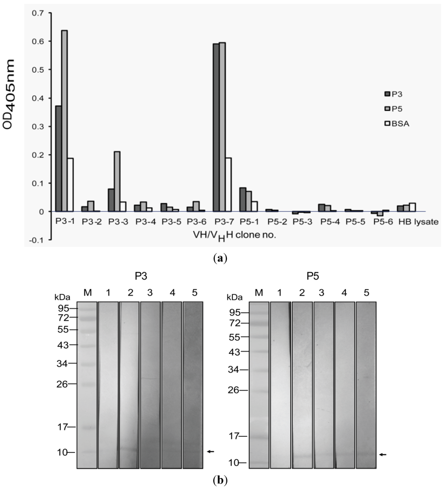
2.5. Determination of Specific Binding of the VH/VHH to the P3 and P5 PLA2

For indirect ELISA, P3 and P5 were used for coating separate ELISA wells. Wells coated with bovine serum albumin (BSA), lysate of original HB2151 E. coli and PBS only were included as antigen control, background, and blank, respectively. After being incubated and washed, wells were added appropriately with individual E. coli lysates containing standardized VH/VHH. Rabbit anti-E tag, goat anti-rabbit immunoglobulin-horseradish peroxidase and substrate were sequentially added with washing between each step. OD405nm of the contents in wells coated with E. coli lysates and BSA were determined against the blank. VH/VHH in E. coli lysates that revealed OD405nm two times higher than the same lysates added to BSA coated wells were regarded as positive binding of the VH/VHH to the P3/P5, or both.
For Western blot analysis, the SDS-PAGE separated P3 and P5 were electrotransblotted onto separate NC. After blocking, each NC blot was cut vertically into strips and immersed appropriately into standardized VH/VHH positive E. coli lysates. The VH/VHH that bound to P3/P5 on NC strip was revealed by using rabbit anti-E tag, goat anti-rabbit immunoglobulin-AP conjugate and enzyme chromogenic substrate, respectively.

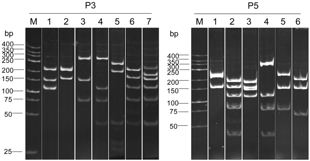
2.6. Determination of the Restriction Fragment Length Polymorphism (RFLP) of the vh/vhh Sequences

RFLP of DNA sequences coding for the VH/VHH in individual transformed HB2151 E. coli clones were determined after digesting with MvaI restriction endonuclease and resolved by 14% polyacrylamide gel electrophoresis followed by ethidium bromide staining [19].

2.7. Amino Acid Sequences, Immunoglobulin Frameworks (FRs) and Complementarity Determining Regions (CDRs) of the VH/VHH

The vh/vhh cDNA insert in the recombinant pCANTAB5E vector from each phagemid-transformed HB2151 E. coli clone was sequenced and deduced into amino acid sequence. The VH/VHH sequences were compared by ClustalW. The immunoglobulin frameworks (FRs) and complementarity determining regions (CRDs) of each VH/VHH were predicted by using the International ImMunoGeneTics (IMGT) information system [23].

2.8. Large Scale Production and Purification of the VH/VHH

The VH/VHH were produced in large scale by subcloning the vh/vhh inserts from pCANTAB5E to the modified pET23b+ vector backbone as described previously [24]. The recombinant vh/vhh-pET23b+vectors were introduced into BL21 (DE3) E. coli. The hexahistidine-tagged VH/VHH fusion proteins were produced from selected transformed bacteria and purified by using Ni-NTA agarose beads. The purified VH/VHH antibodies were tested for their ability to inhibit N. kaouthia PLA2 enzymatic activity in comparison with the horse anti-N. kaouthia venom.

2.9. PLA2 Enzymatic Assay and Neutralization of the Enzymatic Activity by Humanized-VH/VHH and Horse Anti-N. kaouthia Venom

The P3 and P5 enzymatic activity in hydrolyzing phosphatidylcholine substrate was determined by using secretory PLA2 assay kit (Cayman Chemical Company, MI, USA) according to manufacturer’s instruction. To set up the assay, 10 µL of DTNB, 10 µL P3/P5 (containing various amounts: 50, 100 and 150 ng) and 5 µL of assay buffer were mixed in each well of an ELISA plate (Corning). To initiate the reaction, the mixture was added to 200 µL of substrate solution (diheptanoyl thiol-phosphatidylcholine). Mixture added to deionized water and bee venom PLA2 instead of P3/P5 served as blank and positive enzyme control, respectively. A thio-ester bond of phosphatidylcholine was hydrolyzed by PLA2 and the free thiol group was detected by adding 5,5’-dithiol-bis-(2-nitrobenzoic Acid) (DTNB). OD405nm of the reaction mixtures were determined spectrophotometrically at 1-min intervals. Activity of PLA2 (µmol/min/mL) was calculated from duplicate wells by formula:
ΔOD405nm/min = [(OD405nm at time-2) − (OD405nm at time-1)] ÷ [time-2 (min) − time-1 (min)]
PLA2 activity = (ΔOD405nm/min ÷ 10.66 mM−1) × (0.225 mL ÷ 0.01 mL) × sample dilution
For the antibody mediated PLA2 neutralization assay, P3 and P5 was pre-incubated with purified VH/VHH for 1 h at ambient temperature. The P3/P5 pre-incubated with 10 µL of 1:1000 horse anti-N. kaouthia venom served as positive inhibition control. The reaction was initiated by adding DTNB and substrate solution. The OD405nm was measured and PLA2 activity was calculated as above. The experiments were repeated two times with high reproducibility.

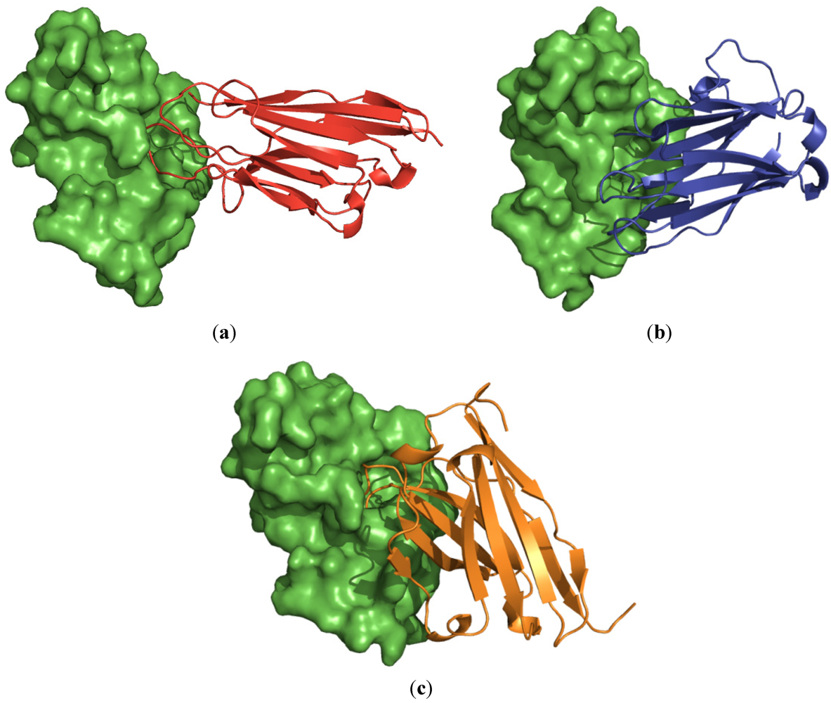
2.10. Homology Modeling and Molecular Docking for Determination of the Interface Binding between the VH/VHH and the P3 and P5 PLA2

The structures of VH/VHH and PLA2 were modeled from their templates derived from BLAST search analysis. Homology modeling technique was used for constructing the antibody and the enzyme models. Steric hindrance of each modeled structure was determined by using RAMACHANDRAN plot. The structures of the VH/VHH and phospholipase A2 were subjected to molecular docking. All experiments were performed by using program Discovery studio 2.5. The enzyme and the antibodies were set as receptor and ligands, respectively. The docked poses from ZDOCK were subjected to structural refinement by using RDOCK program.

3. Results

3.1. PLA2 Fractions from N. kaouthia Venom

After loading the N. kaouthia holo-venom onto the cationic exchange chromatography, 5 protein profiles (P1-P5) were washed through the column by using 0.09 M ammonium acetate, pH 6.5 and 6 profiles (P6-P11) of the column bound proteins were eluted out with a gradient solution of 0.14 to 1.2 M ammonium acetate [22]. Purity of the P3 and P5 profiles of the column flow through were determined by SDS-PAGE (Supplemental Figure 1). The proteins were identified as PLA2 by LC-MS/MS (Supplemental Figure 2). They were dialyzed and concentrated for further use. The P3 and P5 constituted 3.35 and 2.35% of the total venom proteins, respectively.

3.2. Enzymatic Activity of the P3 and P5 from N. kaouthia Venom

By using the sPLA2 assay kit, both P3 and P5 preparations were found to have PLA2 catalytic activity. The optimal amounts of the proteins that gave appropriate enzymatic kinetics were 50 and 100 ng, respectively (data not shown). From duplicate experiments, the calculated PLA2 catalytic activity of the P3 and the P5 were 0.47 and 0.13 (µmol/min/mL), respectively.

3.3. Selection of Phage Clones that Displayed P3/P5-bound VH/VHH and Characterization of the VH/VHH

Both P3 and P5 proteins were used separately as antigens in the single round phage bio-panning. The recombinant pCANTAB5E-transformed HB2151 E. coli colonies appeared on the overnight plate, were screened for the presence of the vh/vhh. It was found that 21 of 30 clones from the P3-panning and 14 of 18 clones from the P5-panning were positive for the antibody gene sequences (~600 bp) by PCR and among them 17 and 12 clones, respectively, could express VH/VHH (Mr ~15–25 kDa) as determined Western blot analysis (representatives are shown in Supplemental Figure 3).
RFLP of the vh/vhh sequences of the 17 and 12 clones were studied using MvaI restriction nuclease. The P3 derived clones revealed 7 different DNA banding patterns while the P5 derived clones showed 6 different RFLP patterns (Figure 1).
Figure 1. MvaI restriction RFLP patterns of the vh/vhh sequences of the clones that bound to the P3 and the P5, respectively. The P3 derived clones revealed 7 different DNA banding patterns while the P5 derived clones showed 6 different patterns.
Toxins 04 00554 g001
VH/VHH of representative clone of each pattern, designated clones P3-1 to P3-7 and P5-1 to P5-6 were tested for specific binding to the P3 and the P5 by indirect ELISA and Western blot analysis. It was found that VH/VHH in the lysates of clones P3-1, P3-3 and P3-7 bound to the P3 as well as the P5 by the indirect ELISA (Figure 2a). They also bound to the SDS-PAGE separated purified P3 and P5 PLA2 in the Western blot analysis (Figure 2b). None of the VH/VHH of clones P5-1 to P5-6 bound to P3 and/or P5 by both assays.
Multiple alignments revealed that the amino acid sequences of VH/VHH of clones P3-1, P3-3 and P3-7 were different especially at the CDR domains (Figure 3). The deduced amino acid sequences of clones P3-1 and P3-3 had the characteristic amino acid tetrad of VHH in the immunoglobulin framework-2 (FR2); they were designated VHH-P3-1 and VHH-P3-3; the clone P3-7 had conventional VH features, designated VH-P3-7 [19].
Figure 2. Binding specificity of VH/VHH in lysates of the selected vh/vhh-phagemid transformed HB2151 E. coli clones. (a) VH/VHH in lysates of 13 HB2151 E. coli clones were tested for binding to the P3 and the P5 by indirect ELISA; lysates of three clones, P3-1, P3-3 and P3-7, gave OD405nm to the immobilized P3 and P5 two times higher than to BSA control. HB, Lysate of normal HB2151 E. coli used as negative VH/VHH control. (b) Western blot analysis for confirmation of the binding of the VH/VHH of the three ELISA positive E. coli clones to SDS-PAGE separated P3 and P5. VH/VHH of all clones bound to both proteins (lanes 3–5 of both panels) (arrows). Lanes M, Pre-stained protein marker; lanes 1, SDS-PAGE separated-P3/P5 probed with normal HB2151 E. coli lysate (negative control); lanes 2, SDS-PAGE separated-P3/P5 probed with horse anti-venom (1:1000) (positive control).
Toxins 04 00554 g002
Figure 3. Multiple alignment of amino acid sequences for determining immunoglobulin frameworks (FRs) and complementarity determining regions (CRDs) of the VH/VHH sequences by using the International Immunogenetics Information System sever. Clones P3-1 and P3-3 showed the amino acid tetrad hallmark of VHH in the FR2 (underlined bold letters); clone P3-7 was conventional VH. Asterisk indicates identical amino acids; colon indicates conserved amino acid substitution; and dot indicates semi-conserved amino acid substitution.
Toxins 04 00554 g003
All of the three humanized-camel VH/VHH showed high homology with human VH sequences (Table 1).
Table 1. Percent amino acid identity of the humanized-camel VH/VHH sequences with the closest human V region frameworks [23].
VH/VHH clone numberClosest human V regionPercent amino acid identity with human FRs
FR1FR2FR3
VHH-P3-1Z27054 IGHV3-66*0292.070.678.9
VHH-P3-3Z27054 IGHV3-66*0284.058.884.2
VH-P3-7HM85593992.088.284.2
* Indicates the allele polymorphism.

3.4. VH/VHH-Mediated Inhibition of PLA2 Enzymatic Activity

The purified VHH-P3-1, VHH-P3-3 and VH-P3-7 when mixed with the P3 and the P5 at the optimal enzyme:antibody ratios 1:3 and 1:5, respectively, could inhibit activity of the enzymes by 32, 52 and 16% and 19, 37 and 26%, respectively (Figure 4). The horse anti-cobra venom (1:1000) inhibited catalytic activity of the P3 and the P5 PLA2 by 35 and 45%, respectively.
Figure 4. Results of VH/VHH- mediated inhibition of the P3 and the P5 enzymatic activity. P3 and P5 were mixed with VHH-P3-1, VHH-P3-3 and VH-P3-7 at molecular ratios 1:3 and 1:5 (antibody:enzyme), respectively. Horse anti-N. kaouthia venom diluted 1:1000 mixed with the P3/P5 was used as positive inhibition control. It was found that the VHH-P3-1, VHH-P3-3 and VH-P3-7 could inhibit the P3 enzyme (homologous system) by 32, 52 and 16%, respectively; the antibodies inhibited P5 enzyme (heterologous system) by 19, 37 and 26%, respectively. The horse immune serum at dilution 1:1000 inhibited the P3 and the P5 PLA2 by 35 and 45%, respectively.
Toxins 04 00554 g004

3.5. Interface Binding of the VH/VHH and the PLA2

BLAST search analysis revealed that the amino acid sequences of VH/VHH of clones no. VHH-P3-1, VHH-P3-3, and VH-P3-7 had the highest sequence identity with PDB entries: 1VHP, 1MVF, and 2GCY, respectively. The PDB entry 1POA has the highest sequence identity with sPLA2. After homology modeling, the qualities of the modeled structures were assessed by RAMACHANDRAN plot. Percentages of amino acids in allowed regions of the VH/VHH clones no. P3-1, P3-3, and P3-7 were 99.1%, 98.2%, and 99.1% respectively (Supplemental Figure 4a). For modeled P3 and P5 PLA2, all amino acids (100%) of both structures were in the allowed regions (Supplemental Figure 4b).
The interface bindings of the antibodies and the PLA2 are shown in Figure 5. It was found that all antibodies occupied the PLA2 catalytic surface areas and protruded their CDRs into the PLA2 enzymatic groove.
Figure 5. Hypothetical models showing binding sites of the antibodies (ribbons) VHH-P3-1 (red; panel a), VHH-P3-3 (blue; panel b) and VH P3-7 (orange; panel c) on the molecular surface of PLA2 (green surface model). All antibodies were found to cover the enzymatic groove surface areas of the modeled PLA2 and inserted the CDRs into the catalytic cleft.
Toxins 04 00554 g005

4. Discussions

Phospholipase A2 (PLA2) of Naja kaouthia venom comprises both basic and acidic proteins [1,5]. The most toxic effect of the PLA2 is believed to be indirectly mediated by the catalytic active acidic PLA2 which consequently enhances cellular or subcellular (mitochondrial) membrane damage through phospholipid hydrolysis and generation of pharmacological mediators including platelet activating factor and eicosanoids causing hemolysis, hemorrhage, edema and loss of organ functions. Besides this, the PLA2 can cause prolonged blood coagulation, dermo-/myo-necrosis and cardiotoxicity [25]. In this study, attempts have been made to produce a humanized-single domain antibody (VH/VHH) that specifically interferes with the N. kaouthia PLA2 catalytic activity.
Two isoforms of N. kaouthia PLA2, i.e., P3 and P5, were isolated from the N. kaouthia venom [1]. These two proteins possessed the phosphatidylcholine hydrolytic activity as assessed in this study by using the commercialized sPLA2 assay kit. The P3 was more enzymatically active than the P5 on the same weight basis (0.47 versus 0.13 µmol/min/mL). When these two proteins were used separately as target antigens in phage bio-panning for selecting specific phage clones that displayed the antigen-bound humanized-camel VH/VHH on the surface and carried also the respective VH/VHH coding sequences in the phage genomes, it was found that only the P3 antigens could be selected for the desired phage clones which not only bind to the homologous P3 but also to the P5. None of the transformed E. coli clones transfected with the P5-derived phage clones produced VH/VHH that specifically bound to the P3/P5 PLA2. It is not known whether the difference in the antigenicity of the two proteins lies in their enzymatic activity, some amino acid differences or due to other factors. Moreover, P5-specific VH/VHH might be relatively rare in the antibody repertoire of the phage library. Similar results were obtained by using another antibody phage display library [26].
It should be noted that the P3-1 and P3-3 bound better to the heterologous P5 than to the homologous P3; the P3-7 bound equally well to both P3 and P5 in the indirect ELISA. Nevertheless, the antibodies of the three clones neutralized enzymatic activity of the homologous P3 enzyme (toxin: antibody 1:3) better than the heterologous P5 enzyme (toxin: antibody 1:5). The ambiguous results should be due to differences of the assay principles and the functions of the epitopes bound by the antibodies. Among the VHH-P3-1, VHH-P3-3 and VH-P3-7, the VHH-P3-3 showed the highest inhibitory activity to the P3 and P5 phospholipase activity, i.e., 52% and 37%, respectively. The percent inhibition of the P3 and P5 PLA2 catalytic activity mediated by the horse immune serum to N. kaouthia venom (positive inhibition control of this study) were 35% and 45%, respectively. A previous study has demonstrated also that at dilution 1:500, the horse anti-N. kaouthia serum inhibited 16% of the snake PLA2 enzymatic activity [5]. Thus, the humanized-VH/VHH produced in this study has, more or less, comparable PLA2 enzyme inhibitory activity to the animal immune sera derived from two different sources. It is not known why the PLA2 specific antibodies in the preparations tested could not completely inhibit the PLA2 enzymatic activity.
Molecular modeling and docking results suggested that the humanized-camel VH/VHH antibodies, bound to the surface areas around the enzymatic pocket and with CDR loops inserted into the PLA2 catalytic groove, would probably interfere with the substrate accessibility of the enzyme and thus accounted for the observed enzymatic activity inhibition. This speculation needs experimental validation. It is known that the surface areas of the PLA2 covered by the humanized-VH/VHH also involved in Ca2+ binding (residues Y27, G29, G31 and D48), active site (H47, D48, Y51 and D93), phospholipid binding (L2, F5, I9, W19, F21, A22, G31 and Y63) and anticoagulant activity (residues 54–66, i.e., AEKISRCWPYFKT) [27,28]. Thus by binding to the PLA2 target, the humanized-VH/VHH might exert not only the neutralization of the PLA2 enzyme, but also other PLA2 bio-toxic functions. Experiments are needed to verify this speculation. Moreover, the ability of the humanized-single domain antibodies (VH/VHH) in blocking the venom PLA2 mediated dermo-/myo-necrosis, hemorrhage and carditoxicity should also be explored.

5. Conclusions

Humanized-camel single domain antibodies (VH/VHH) that cross neutralized enzymatic activity of different isoforms of N. kaouthia PLA2 were produced from an antibody phage display library. The PLA2 neutralizing activities of the VH/VHH were comparable to the horse immune serum against N. kaouthia holo-venom. Homology modeling and molecular docking revealed that the VH/VHH bound to the surface areas around the enzymatic pocket of the PLA2 and inserted the CDRS into the catalytic cleft.

Acknowledgments

The work was supported by the National University Research (NRU) Project, Office of Higher Education Commission, Ministry of Education and The Thailand Research Fund (DPG5380001).

Conflict of Interest

We declare that we have no conflict of interest.

References

  1. Kulkeaw, K.; Chaicumpa, W.; Sakolvaree, Y.; Tongtawe, P.; Tapchaisri, P. Proteome and immunome of the venom of the Thai cobra, Naja kaouthia. Toxicon 2007, 49, 1026–1041. [Google Scholar] [CrossRef]
  2. Mebs, D.; Claus, I. Amino acid sequences and toxicities of snake venom components. In Snake Toxins; Harvey, A.L., Ed.; Pegamon Press: New York, NY, USA, 1991; pp. 425–447. [Google Scholar]
  3. Armugam, A.; Earnest, L.; Chung, M.C.; Copalakrishnakone, P.; Tan, C.H.; Tan, N.H.; Jeyaseelan, K. Cloning and characterization of cDNAs encoding three isoforms of phospholipase A2 in Malayan spitting cobra (Naja naja sputatrix) venom. Toxicon 1997, 35, 27–37. [Google Scholar] [CrossRef]
  4. Warrell, D.A. Snake bite. Lancet 2010, 375, 77–88. [Google Scholar] [CrossRef]
  5. Doley, R.; Mukherjee, A.K. Purification and characterization of an anticoagulant phospholipase A2 from Indian monocled cobra (Naja kaouthia) venom. Toxicon 2003, 41, 81–91. [Google Scholar] [CrossRef]
  6. Dennis, E.A. Diversity of group types, regulation, and function of phospholipase A2. J. Biol. Chem. 1994, 269, 13057–13060. [Google Scholar]
  7. Bingham, C.O., III; Austen, K.F. Phospholipase A2 enzymes in eicosanoid generation. Proc. Assoc. Am. Phys. 1999, 111, 516–524. [Google Scholar] [CrossRef]
  8. Doley, R.; King, G.F.; Mukherjee, A.K. Differential hydrolysis of erythrocyte and mitochondrial membrane phospholipids by two phospholipase A2 isoenzymes (NK-PLA2-I and NK-PLA2-II) from venom of the Indian monocle cobra, Naja kaouthia. Arch. Biochem. Biophys. 2004, 425, 1–13. [Google Scholar] [CrossRef]
  9. Evans, J.; Ownby, C.L. Neutralization of edema, hemorrhage and myonecrosis induced by North American crotalid venoms in simulated first-aid treatments. Toxicon 1999, 37, 633–650. [Google Scholar] [CrossRef]
  10. Samy, R.P.; Gopalakrishnakone, P.; Chow, V.T.K. Therapeutic application of natural inhibitors against snake venom phospholipase A2. Bioinformation 2012, 8, 48–57. [Google Scholar]
  11. Kini, R.M. Anticoagulant proteins from snake venoms: Structure, function and mechanism. Biochem. J. 2006, 397, 377–387. [Google Scholar] [CrossRef]
  12. Khandelwal, G.; Katx, K.D.; Brooks, D.E.; Gonzales, S.M.; Ulishney, C.D. Naja kaouthia: Two cases of Asiatic cobra envenomations. J. Emerg. Med. 2007, 2, 171–174. [Google Scholar]
  13. Sekhar, C.C.; Chakrabarty, D. Fibrinolytic toxin from Indian monocle cobra (Naja kaouthia) venom. J. Biosci. 2011, 36, 355–361. [Google Scholar] [CrossRef]
  14. Zhang, H.L.; Xu, S.J.; Wang, Q.Y.; Song, S.Y.; Shu, Y.Y.; Lin, Z.J. Structure of a cardiotoxic phospholipase A(2) from Ophiophagus hannah with the “pancreatic loop”. J. Struct. Biol. 2002, 138, 207–215. [Google Scholar] [CrossRef]
  15. Lizano, S.; Angulo, Y.; Lomonte, B.; Fox, J.W.; Lambeau, G.; Lazdunski, M.; Guyierrez, M.G. Two phospholipase A2 inhibitors from the plasma of Cerrophidion (Bothrops) godmani which selectively inhibit two different group-II phospholipase A2 myotoxins from its own venom: Isolation, molecular cloning and biological properties. Biochem. J. 2000, 346, 631–639. [Google Scholar] [CrossRef]
  16. Alzogaray, V.; Danquah, W.; Aguirre, A.; Urrutia, M.; Berguer, P.; García Véscovi, E.; Haag, F.; Koch-Nolte, F.; Goldbaum, F.A. Single-domain llama antibodies as specific intracellular inhibitors of SpvB, the actin ADP-ribosylating toxin of Salmonella typhimurium. FASEB J. 2011, 25, 526–534. [Google Scholar]
  17. Harmsen, M.M.; de Haard, H.J. Properties, production and applications of camelid single-domain antibody fragments. Appl. Microbiol. Biotechnol. 2007, 7, 13–22. [Google Scholar]
  18. Lauwereys, M.; Arbabi Ghahroudi, M.; Desmyter, A.; Kinne, J.; Hölzer, W.; de Genst, E.; Wyns, L.; Muyldermans, S. Potent enzyme inhibitors derived from dromedary heavy-chain antibodies. EMBO J. 1998, 17, 3512–3520. [Google Scholar]
  19. Thanongsaksrikul, J.; Srimanote, P.; Maneewatch, S.; Choowongkomon, K.; Tapchaisri, P.; Makino, S.; Kurazono, H.; Chaicumpa, W. A VHH that neutralizes the zinc metalloproteinase activity of botulinum neurotoxin type A. J. Biol. Chem. 2010, 285, 9657–9666. [Google Scholar]
  20. Thanongsaksrikul, J.; Chaicumpa, W. Botulinum neurotoxin and botulinum: A novel therapeutic approach. Toxins 2011, 3, 469–488. [Google Scholar] [CrossRef]
  21. Karlsson, E.; Amberg, H.; Waker, D. Isolation of the principal neurotoxins of two Naja naja subspecies. Eur. J. Biochem. 1971, 21, 1–16. [Google Scholar] [CrossRef]
  22. Kulkeaw, K.; Sakolvaree, Y.; Srimanote, P.; Tongtawe, P.; Maneewatch, S.; Sookrung, N.; Tungtrongchitr, A.; Tapchaisri, P.; Kurazono, H.; Chaicumpa, W. Human monoclonal ScFv neutralize lethal Thai cobra, Naja kaouthia, venom. J. Proteomics 2009, 72, 270–282. [Google Scholar] [CrossRef]
  23. IMGT Home page. IMGT Tools: IMGT/V-QUEST. Available online: http://www.imgt.org (accessed on 19 May 2012).
  24. Poungpair, O.; Pootong, A.; Maneewatch, S.; Srimanote, P.; Tongtawe, P.; Songserm, T.; Tapchaisri, P.; Chaicumpa, W. A human single chain transbody specific to matrix protein (M1) interferes with the replication of influenza A virus. Bioconjug. Chem. 2010, 21, 1134–1141. [Google Scholar] [CrossRef]
  25. Kini, R.M.; Evans, H.J. A model to explain the pharmacological effects of snake venom phospholipases A2. Toxicon 1989, 27, 613–635. [Google Scholar] [CrossRef]
  26. Kulkeaw, K. Production of Human Anti-Naja kaouthia Venom by Antibody Phage Display Library. Thammasat University: Pathumthani, Thailand, 2008. [Google Scholar]
  27. Gu, L.; Wang, Z.; Song, S.; Shu, Y.; Lin, Z. Crystal structures of an acidic phospholipase A2 from the venom of Naja kaouthia. Toxicon 2002, 40, 917–922. [Google Scholar] [CrossRef]
  28. Kang, T.S.; Georgieva, D.; Genov, N.; Murakami, M.T.; Sinha, M.; Kumar, R.P.; Kaur, P.; Kumar, S.; Dey, S.; Sharma, S.; et al. Enzymatic toxins from snake venom: Structural characterization and mechanism of catalysis. FEBS J. 2011, 278, 4544–4576. [Google Scholar]

Supplementary Files

  • Supplementary File 1::

    PDF-Document (PDF, 621 KB)

  • Share and Cite

    MDPI and ACS Style

    Chavanayarn, C.; Thanongsaksrikul, J.; Thueng-in, K.; Bangphoomi, K.; Sookrung, N.; Chaicumpa, W. Humanized-Single Domain Antibodies (VH/VHH) that Bound Specifically to Naja kaouthia Phospholipase A2 and Neutralized the Enzymatic Activity. Toxins 2012, 4, 554-567. https://doi.org/10.3390/toxins4070554

    AMA Style

    Chavanayarn C, Thanongsaksrikul J, Thueng-in K, Bangphoomi K, Sookrung N, Chaicumpa W. Humanized-Single Domain Antibodies (VH/VHH) that Bound Specifically to Naja kaouthia Phospholipase A2 and Neutralized the Enzymatic Activity. Toxins. 2012; 4(7):554-567. https://doi.org/10.3390/toxins4070554

    Chicago/Turabian Style

    Chavanayarn, Charnwit, Jeeraphong Thanongsaksrikul, Kanyarat Thueng-in, Kunan Bangphoomi, Nitat Sookrung, and Wanpen Chaicumpa. 2012. "Humanized-Single Domain Antibodies (VH/VHH) that Bound Specifically to Naja kaouthia Phospholipase A2 and Neutralized the Enzymatic Activity" Toxins 4, no. 7: 554-567. https://doi.org/10.3390/toxins4070554

    Article Metrics

    Back to TopTop