Article

The Versatility of the Helicobacter pylori Vacuolating Cytotoxin VacA in Signal Transduction and Molecular Crosstalk

Ardmore House, School of Biomolecular and Biomedical Sciences, Belfield Campus, University College Dublin, Dublin-4, Ireland
*
Author to whom correspondence should be addressed.
Toxins 2010, 2(1), 69-92; https://doi.org/10.3390/toxins2010069
Submission received: 3 December 2009 / Revised: 31 December 2009 / Accepted: 14 January 2010 / Published: 15 January 2010
(This article belongs to the Special Issue Bacterial Protein Toxins)

Abstract

:
By modulating important properties of eukaryotic cells, many bacterial protein toxins highjack host signalling pathways to create a suitable niche for the pathogen to colonize and persist. Helicobacter pylori VacA is paradigm of pore-forming toxins which contributes to the pathogenesis of peptic ulceration. Several cellular receptors have been described for VacA, which exert different effects on epithelial and immune cells. The crystal structure of VacA p55 subunit might be important for elucidating details of receptor interaction and pore formation. Here we discuss the multiple signalling activities of this important toxin and the molecular crosstalk between VacA and other virulence factors.

1. Introduction

Helicobacter pylori (Hp) is a highly successful pathogen that is estimated to persistently colonize the stomach of about half of the human world population. This bacterium has been recognized as the causative agent of chronic gastric inflammation, peptic ulcer disease, mucosa-associated lymphoid tissue (MALT) lymphoma or gastric cancer [1,2,3,4,5,6]. The clinical outcome of Hp infections is determined by a complex scenario of host-pathogen interactions. The cellular and molecular mechanisms used by Hp to subvert host defences have been investigated intensively over the last few years. Disease development is dependent on several parameters including both the genotype of the bacteria and genetic predisposition of the host, as well as environmental factors [7,8,9,10,11,12]. Hp isolates from different geographical origin are surprisingly diverse both in their genome sequences and their pathogenicity. Dozens of bacterial factors have been shown to influence Hp pathogenesis including the flagella, urease, catalase, neutrophil-activating protein NapA, peptidoglycan, several adhesins (e.g., SabA, BabA, AlpA/B, OipA) and many others [1,13,14,15,16]. They can be classified as colonization factors (also called pathogenicity-associated factors) and virulence factors having direct cell-damaging capabilities. However, the two major disease-associated components are the vacuolating cytotoxin (VacA) and the cag (cytotoxin-associated genes) pathogenicity island (cagPAI). The gene encoding vacA is present in virtually all Hp strains, which suggests that the production of VacA plays an important role in the colonization or persistence of Hp in the human stomach [1,7,8,9,17]. The cagPAI is a 40 kb stretch of DNA, which was acquired by a horizontal DNA transfer event from yet unknown donor, and encodes components of a sophisticated type IV secretion system (T4SS). This T4SS forms a pilus for the injection of virulence factors into host target cells such as the CagA effector protein [7,11,12,18,19,20]. Injected CagA mimics a host cell protein in binding and activation of multiple signalling factors and induces actin-cytoskeletal rearrangements (cell elongation), disruption of cell-to-cell junctions (cell scattering) as well as proliferative, pro-inflammatory and anti-apoptotic nuclear responses [11,20,21,22,23]. In addition, Hp induces several T4SS-dependent but CagA-independent signalling events such as the activation of epidermal growth factor receptor EGFR [24,25] and the small Rho GTPase Rac1 [26,27,28]. The role of CagA and the T4SS have been discussed in numerous recent reviews [7,11,12,20]. Here we review the recent findings on the VacA structure and the various pathways how VacA highjacks host cell signalling cascades. We will also discuss a set of novel findings on the molecular crosstalk between VacA and CagA.

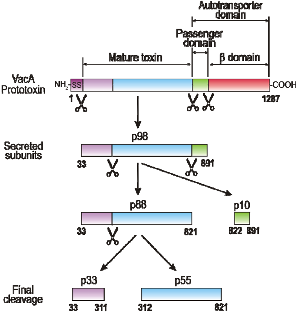
2. VacA Expression, Secretion, Maturation and Allelic Variation

An early work by Leunk and colleagues [29] discovered that broth-culture supernatants of Hp strains contained a proteinaceous cytotoxin that induced cytoplasmic vacuolation when incubated with eukaryotic cells in vitro. Multiple cell types from human and animal species were susceptible to its toxic activity, albeit with great differences. The structural organization of VacA closely resembles that of IgA protease-like factors secreted by pathogenic Neisseriae and Haemophilus species [9], which are today known as the autotransporter or type V secretion family of proteins [18]. Like many other classical autotransporters, VacA contains a 33-amino acid signal sequence and undergoes cleavage during the transport across bacterial membranes (Figure 1). VacA genes are predicted to encode a translated protein of about 135-145 kDa depending on the strain. However, VacA proteins of this molecular weight have never been reported [8]. Instead, three VacA protein spots of the Hp strain 26695 were identified by two-dimensional gel electrophoresis (2-DE). The most predominant VacA protein spots on 2-DE gels were approximately 95.7 kDa and 87.9 kDa in size, and were found both in fractions of the Hp membrane and culture supernatant [30,31]. In addition, a 10.5 kDa VacA spot was found in the secreted protein fraction [30]. Mass spectrometry of the identified peptides are consistent with a model that the 95.7 kDa VacA protein is the secreted toxin which is then cleaved into the 87.9 kDa mature protein (p88) and the 10.5 kDa passenger domain (p10, also known as the alpha-protein) (Figure 1). The exact cleavage site between p88 and p10 was detected between position 991 and 992 [30]. The processed beta-domain of the autotransporter subunit was not detected by 2-DE, but has been observed in total cell lysates [32]. However, the mature translocated p88 domain can undergo further proteolytic cleavage to yield two fragments, known as p33 and p55 (Figure 1, bottom). The latter two processing products are believed to represent the functional domains of VacA (see below). Thus, VacA may exhibit some properties resembling those of classical A/B-toxins. Interestingly, while cleavage between p33 and p55 subunits has been seen in vitro [33], no cleavage in vivo could be detected [34]. A possible explanation is that the rate of VacA cleavage in vivo is too low to be observed. In some studies, VacA and its domains have slightly different sizes, but we will use the p33 and p55 nomenclature throughout this review. However, the basis of vacA size and sequence variation was found in several alleles that have been identified in the signal region (genotypes s1 and s2) and in the mid region (genotypes m1 and m2), occurring in all possible combinations [1,7,8,9]. In addition, two intermediate region variants (called i1 and i2) were identified as other important determinants of VacA toxicity [35]. VacA proteins of the s2 type are inactive in assays for the vacuolating phenotype, while type s1/ml alleles produce extensive vacuolation in a large array of cell categories and type s1/m2 allels induce detectable vacuolation in a more limited range of cell types, which is in concurrence with its location in p55 as the cellular binding domain of VacA [36,37].
Figure 1. VacA domain structure and processing products. The amino-terminal signal sequence (SS) and carboxy-terminal domain are cleaved from the full-length 135-140 kDa VacA protein to yield a 98 kDa secreted toxin (p98) that is transported into the extracellular space via the autotransporter mechanism. Proteins of this autotransporter family are able to mediate their own secretion without the need of additional bacterial proteins. The 33 kDa carboxy-terminal beta-barrel domain of VacA is predicted to insert into the outer membrane and to form a channel, through which the mature VacA toxin is secreted. The secreted toxin can be processed into the mature toxin (p88) and p10. The mature, secreted p88 subunit can undergo further limited proteolytic cleavage to yield two additional fragments, p33 and p55 [7,8,32]. The latter two processing products are considered to represent the functional domains or subunits of VacA.
Toxins 02 00069 g001

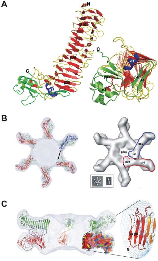
3. Domains of VacA and Crystal Structure of the p55 Subunit

In response to acidic pH, VacA undergoes a structural change, which increases its cellular activity and renders it resistant to proteolysis by pepsin [38]. The relevance of the pH, however, is still not fully understood because VacA has also been shown to be activated at alkaline pH [39]. In fact, it appears that only highly purified VacA is poorly active and needs to be activated by pH change, but this is not necessary during infection or when VacA is simply collected from the bacterial supernatant (broth culture filtrate). Thus, VacA secreted by the bacteria is most likely fully active independently of the luminal gastric pH. This is in agreement with the fact that the mucus layer close to the gastric epithelium, in which Hp resides, is at a mild acidic pH. However, the secreted p88 subunit of VacA can assemble into water-soluble oligomeric structures [33,40], and can insert into planar lipid bilayers to form anion-selective membrane channels [41,42,43]. These structures resemble a flower-like configuration (Figure 2B). Interestingly, low pH treatment of these oligomeric structures break them apart releasing VacA monomers [40]. The first reported high-resolution images were 19-Å cryo electron microscopy maps of VacA dodecamers [44], and atomic-force microscopy and electrophysiological studies implied that VacA membrane channels are single-layered structures [41,43]. When the p33 and p55 domains are expressed separately and then mixed, p33 and p55 can physically interact and restore vacuolating activity in epithelial cells [45,46,47]. The p33 subunit contains a hydrophobic stretch (amino acids 6-27) which is involved in pore formation and/or membrane insertion [48,49], whereas p55 contains one or more cell interaction sites [36,37,50]. Recently, the 2.4-Å crystal structure of p55 has been published [51]. The structure is predominantly a right-handed parallel β-helix, a feature which is characteristic of autotransporter passenger subunits but unique among other known toxins (Figure 2A-C). Notable features of p55 include disruption of β-sheet contacts which result in five β-helix subdomains and a carboxy-terminal domain that contains a disulfide-bond. Gangwer et al. [51] proposed an oligomerization model in which p33 interacts with the amino-terminal portion of p55 from the neighboring subunit. Regions of contact between p33 and p55 are depicted with dashed lines (Figure 2B, right structure). On the left in Figure 2C, the upper circle highlights three loops that mediate p55-p55 contacts between the two layers. The lower circle indicates the two loops that line the conserved pocket that were proposed as a common receptor-binding site [51]. This pocket is located on the side of the molecule and would therefore be accessible in both single-layered and bilayered forms of the toxin. The circle in the middle contains the other conserved surface in p55. This surface protrudes into the central ring of the VacA oligomer and would be accessible to the molecule for contacts that could mediate oligomerization within a hexameric or heptameric plane. The enlarged section to the right in Figure 2C shows the secondary structures that contribute to this surface. Again, p55 contains the aforementioned m1/m2 alleles, and investigation of VacA sequences from unrelated Hp strains allowed the identification of structural surface features which may be important for interactions with host cell receptors [51].
Figure 2. Molecular structure of VacA. (A) The p55 domain of VacA adopts a β-helix structure that is composed of three parallel β-sheets connected by loops of varying length and structure [51]. The carboxy-terminal domain contains a mixture of α/β secondary structure elements and contains a disulfide bond. The carboxy-terminus of the β-helix (right, rotated by 90°) is capped by a β-hairpin from the carboxy-terminal domain and the α1−helix located in one of the long helix loops. (B) Docking the p55 crystal structure into a 19-Å cryo electron microscopy map of the VacA dodecamer results in a model for oligomerization. Twelve p55 subunits are shown docked into a 19-Å cryo-EM map of a VacA dodecamer [44]. An arrow is shown to indicate the space that the upper molecule will occupy if p33 extends the β-helix structure of p55. The inset shows EM images of a VacA hexamer and a VacA monomer [44]. The shape of a VacA hexamer (inset) is similar to the shape of a single layer within the dodecamer [44]. The rod-like shape of the p88 monomer (inset) supports a model in which the β-helix of p55 will extend into p33. (C) The view from the left structure in panel B is rotated by 90°. This figure was kindly provided by Dr. Borden Lacy and originally published by Gangwer et al. [51] with kind permission of the National Academy of Sciences, USA.
Toxins 02 00069 g002

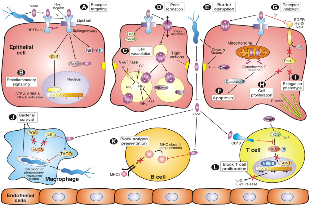
4. VacA Targets Various Surface Factors Both on Epithelial and Immune Cells

The cellular effects of VacA have been investigated intensively both in vivo and in vitro. A simplified model for the complex interactions of VacA with multiple cell types and downstream signalling cascades is presented in Figure 3A-L, while known host cell binding partners are summarized in Table 1. Like many other bacterial toxins, VacA interacts with the plasma membrane of susceptible cells as a first step during intoxication (Figure 3A). Following cell binding, VacA is internalized by a pinocytic-like and clathrin-independent mechanism [52,53]. A remarkable feature of VacA is that it can target multiple cell-surface components in vitro or on epithelial cells including RPTP-alpha [54], RPTP-beta [39,55], various lipids [56,57,41], heparin sulphate [58], sphingomyelin [59], fibronectin [60], the epidermal growth factor receptor (EGFR) [61] as well as integrin β2 (CD18) on T cells [62]. Determining which of these factors is required for VacA uptake and subversion of cellular signalling has been a challenging topic of research and is still not fully studied for each factor. However, the importance of RPTP’s for VacA-dependent activities has been investigated in much detail. For example, VacA causes gastric injury in wild-type mice but not in the RPTP-beta knockout mice [55]. This indicates that interaction of VacA with RPTP-beta on the surface of gastric epithelial cells is mechanistically important in VacA-induced gastric injury. Remarkably, VacA can still induce vacuolation in cultured gastric epithelial cells from RPTP-beta knockout mice or in G401 human kidney cells, which do not express RPTP-beta [54,55], but inhibition of RPTP-beta expression by silencing oligonucleotides in HL-60 cells downregulated VacA-induced vacuolation [63]. These data suggest that RPTP-beta exhibits crucial functions in the process of VacA-induced signalling but not necessarily vacuolation itself. Addition of VacA to target cells has been also shown to induce the tyrosine phosphorylation of G-protein-coupled receptor-kinase-interacting protein-1 (Git-1) and lipid-raft dependent downstream signalling [55,64]. In line with these observations, VacA was reported to bind to the QTTQP-motif in the extracellular domain of RPTP-beta, which was also necessary for inducing Git-1 phosphorylation [54]. Another reported consequence of VacA-binding to RPTP-beta is detachment of primary murine gastric epithelial cells from the extracellular matrix [55]. Furthermore, VacA has been shown to interact with lipid preparations and artificial membranes, but the specificity and importance of VacA-lipid interactions for toxin function was unknown until recently. A new study provided evidence that sphingomyelin modulates the sensitivity of epithelial cells to VacA [59]. Sphingomyelin is a key structural component of the cell membrane (in particular lipid rafts), while at the same time serving a functional role, as it is the parental compound of various lipid mediators. In fact, sphingomyelin was shown to be required for VacA’s association with lipid rafts on the plasma membrane and binding to cells, suggesting that sphingomyelin functions as a receptor molecule of VacA on the epithelial cell surface [59]. However, VacA does not only interact with gastric epithelial cells, but also with immune cells including B cells, T cells and macrophages (Figure 3J-L). Interestingly, VacA efficiently enters activated, migrating primary human T lymphocytes by binding to the integrin β2 (CD18) receptor and exploiting the recycling of LFA-1, the lymphocyte function-associated molecule-1 [62]. LFA-1-deficient Jurkat T-cells were resistant to vacuolation, and genetic complementation of LFA-1 restored the sensitivity of cells to VacA. Remarkably, VacA targeted human CD18 for cell entry, but not its murine counterpart. These findings are consistent with the species-specific adaptation of Hp in its human host [62].
Table 1. Host binding partners of VacA and proposed function a.
VacA binding partnersProposed role in VacA-host interactionsMethods usedExperimental evidencebReference
EGF receptorReceptor on epithelial cellsAntibody blocking, IPInfection in vitro[61]
FibronectinBinding of H. pylori to host cellsBinding and cell adhesion and assaysBinding in vitro[60]
GlycosphingolipidsBinding partner for VacA internalizationBinding assays, Chromatography, MSBinding in vitro[65]
Heparin sulphateReceptor/co-receptor on epithelial cellsSPR-based biosensor studiesBinding in vitro[58]
Integrin β2 (CD18)Receptor on T cellsFlow cytometry, IF, live cell imaging, use of knockout cellsTreatment of cells in vitro, Infection in vitro[62]
Lipid bilayersLow pH-triggered pore formationAFMBinding in vitro[41]
Lipid raftsPutative docking and entering site at cell surfaceCell fractionation, flow cytometry, IF, MCD inhibitor studiesTreatment of cells in vitro[66,67,64]
Lipid vesiclesBinding and host cell entry of the toxinLight scattering, energy transfer studiesBinding in vitro[56]
Phospho-lipidsBinding and host cell entry of the toxinANS-binding studies, PhotolabellingBinding in vitro[57]
RPTP-alphaReceptor on epithelial cellsIP, MS, antisense silencing studiesInfection in vitro, Treatment of cells in vitro[54,68]
RPTP-betaReceptor on epithelial cells, receptor for Git1 phosphorylationFlow cytometry, IF, IP, siRNA, use of knockout mice and cellsBinding in vitro, Infection of mice and cells[39,63,55]
RACK-1Yet unknownPD, Y2HBinding in vitro[69]
Sphingo-myelinReceptor on epithelial cellsBinding studies, ELISA, flow cytometry, treatment with sphingomyelinaseBinding and treatment of cells in vitro[59]

5. VacA Activities on Endosomal Maturation and Cell Vacuolation

As mentioned above, the first identified activity of VacA was its ability to induce vacuolation in numerous eukaryotic cell types [29]. A current model for vacuole formation suggests that VacA binds to the plasma membrane of host cells, forms anion-selective channels in the plasma membrane, becomes internalized into endosomes and the vacuoles finally arise by swelling due to osmotic imbalance of the late endosomal compartments (Figure 3C). In fact, VacA-induced vacuoles constitute hybrid compartments of late endosomal origin that contain some lysosomal markers, and their formation strictly depends on the presence of weak bases such as ammonia, which can be provided by urease activity [34,70]. To achieve this goal, VacA is first pinocytosed in a manner dependent on the small Rho GTPase Cdc42 [53], and is subsequently routed into early endosomal compartments which are enriched with glycosylphosphatidylinositol (GPI)-anchored proteins [52,53]. These early endosomes are also associated with dynamic polymerised (filamentous) actin structures [71]. The CD2-associated protein (CD2AP), a docking protein implicated in intracellular trafficking, bridged the filamentous actin with early endosomes containing VacA. Interestingly, CD2AP regulated these actin structures and was required to transfer VacA from early to late endosomes and vacuolation [71]. Vacuole formation further depends on the presence and activity of a number of other signalling factors including V-ATPase [72], Rab7 [73] and Rac1 [74]. Recent studies showed that at least the role of Rac1 in vacuole formation is due to the fact that Rac1 is required for binding of VacA to the cell membrane [53]. However, protein kinase PIKfyve is also necessary for vacuole production, and microinjection of its substrate phosphatidylinositol-3,5-bisphosphate (PIP2) was demonstrated to be sufficient for vacuole formation [75], whereas involvement of the SNARE protein syntaxin-7 is controversial [76,77]. Also, the role of dynamin in large VacA-dependent vacuole formation is controversial. Dynamin has been shown to play either no role [52] or is fully necessary for cell vacuolation [78]. When expressed intracellularly, the minimum segment of VacA required for vacuolating activity comprises the entire p33 subunit and about 110 amino acids from the amino-terminus of p55 [45,79]. Vacuolating activity also requires di- or oligomerization of VacA [80,81] and depends on the hydrophobic amino-terminus (about 30 amino acids) containing three tandem GXXXG-sequences which are transmembrane dimerization motifs [49,82]. These requirements suggest that channel formation is a prerequisite for vacuolation. This conclusion is also supported by the observation that small anion-channel inhibitors effectively block vacuole production [83].

6. Amino-Terminal VacA Targets Mitochondria to Induce Apoptosis

Infection of epithelial target cells with Hp has been associated with both increased and reduced levels of apoptosis [1,2,7]. In vitro, Hp reproducibly stimulates apoptosis in infected gastric epithelial cells, and this has also been seen in tissues isolated from infected patients or animal models [1,2]. Early studies indicated that purified VacA induced mitochondrial damage as indicated by a significant drop in ATP levels within cells [84], and was then discovered to induce apoptosis by a mitochondrial pathway [85]. In the latter study, HEp-2 cells were transfected with different VacA constructs, and the p33 subunit was found to localize specifically to mitochondria, whereas p55 remained cytosolic. When purified VacA was co-incubated with isolated mitochondria, p33 (but not p55) was translocated into these organelles. In addition, transient expression of p33-GFP or full-length VacA-GFP in HeLa cells induced the dramatic release of cytochrome c from mitochondria and activated apoptotic caspase-3 (Figure 3F), as determined by the cleavage of poly-ADP-ribose polymerase, PARP. Cleavage of PARP was antagonized specifically by co-transfection of DNA vectors expressing Bcl-2, a factor known to inhibit mitochondria-associated apoptotic signals [85]. These observations were further supported by infection studies using wild-type Hp and ΔvacA mutant strains [86,87]. Remarkably, the s1/m1 allel of VacA induced high levels of apoptosis while s2/m1 toxin and truncated VacA mutants lacking the hydrophobic region near the amino-terminus did not. These results suggest that VacA can trigger gastric epithelial cell apoptosis and implicate that differences in levels of gastric mucosal epithelial apoptosis among Hp-infected individuals may arise from strain-dependent vacA sequence variation. Interestingly, studies in MKN-28 cells showed that, unlike cell vacuolation, neither apoptosis nor change of mitochondrial transmembrane potential induced by VacA require ammonia [88]. The latter findings make unlikely the hypothesis that ammonia-dependent swelling and rupture of endosomal vesicles in which VacA is sequestered after cellular uptake may allow the toxin to reach mitochondria and trigger apoptosis, and therefore requires a different pathway.

7. VacA and Other Factors Affecting the Transepithelial Resistance of Polarized Cell Monolayers

An important feature of intact polarized cells is the development of transepithelial electrical resistance (TER), a measurable indicator of epithelial integrity. When purified VacA (acid-activated) was incubated with polarized host cell monolayers, it was shown to decrease TER significantly [89] and may contribute to epithelial barrier disruption induced by Hp (Figure 3E). This effect was confirmed in several types of polarized epithelial cells including EPH4, T84 and MDCK and during infection. The latter effect was independent of VacA’s vacuolating activity as it was also seen with m2-alleles of VacA [90]. It was proposed that selective permeabilization of polarized epithelial monolayers by VacA may lead to the release of certain molecules such as Ni2+, Fe3+, sugars and amino acids, which might support the growth of Hp withinthe gastric mucus [89,90]. As yet, the mechanism by which VacA alters paracellular permeability is little understood. However, it has also been noted that VacA increases the transepithelial flux of certain molecules such as urea the substrate of Hp urease [91]. The release of urea from cells, however, is attributed to the formation of VacA channels in the plasma membrane. Other studies have indicated that disruption of cell-to-cell junctions in polarized MDCK cells required VacA but also a second factor, injected CagA (Figure 3E), which targets both tight and adherens junctions [20,92]. In contrast, it was shown that incubation of polarized T84 epithelia by VacA prepared from broth culture filtrate of Hp did not modify the TER of the epithelia [93]. More complexity arises from another recent study showing that Hp induced a progressive loss of barrier function in INS-GAS mice and MKN-28 gastric epithelial cells, which was attenuated by inactivation of the ΔureB gene, but not ΔvacA or ΔT4SS genes [94]. Remarkably, Hp induced the deregulation of specific tight junction proteins, including phosphorylation of MLC (myosin regulatory light chain) by MLC kinase and internalization of occludin. Thus, epithelial barrier disruption by Hp is a highly complex scenario, requiring various bacterial factors and signalling pathways which still need to be investigated in more detailed future studies [95].
Figure 3. Model of the various activities of VacA to induce altered host cell signalling in epithelial and immune cells which contributes to persitent Hp colonization in the human stomach.
Toxins 02 00069 g003

8. VacA Elicits Suppressive Activities on Immune Cell Proliferation

During the long co-evolution of Hp with humans, the bacteria have accumulated numerous strategies to circumvent host immune cell functions. Some of these functions have been attributed to VacA (Figure 3J-L). A pioneering early study demonstrated that VacA can inhibit processing and presentation of antigenic peptides to human CD4-positive T cells (Figure 3J), and suggested for the first time that VacA may contribute to the persistence of Hp by interfering with protective immune responses [96]. In infected professional phagocytes, such as mouse RAW 264.7 and human THP-1 macrophage-like cell lines, wild-type Hp exhibited enhanced survival as compared to vacA deficient mutants [97]. Therefore, it appears that VacA can arrest phagosome maturation by recruiting and retaining coronin-1 or TACO, the tryptophane aspartate-containing coat protein (Figure 3K). In addition, it was shown that VacA can inhibit the expression of endothelial nitric oxygen synthase (eNOS) which resulted in significantly less production of reactive oxygen species (ROS) in monocyte/macrophage-like U937 cells and enhanced survival of VacA-positive Hp (Figure 3J). The detected pathway involved a suppressive effect of VacA on the expression of ILK (integrin-linked kinase), which provided a direct link to eNOS downsteam of ILK [98]. However, enhanced survival of isogenic ΔvacA mutants as compared to their wild-type strains was not seen in two other reports when freshly isolated human monocytes were infected [99,100]. The latter differences may be attributed to varying infection doses, cell types or Hp strains carrying different vacA alleles.
Co-incubation of purified VacA with or infection of T lymphocytes yields multiple effects. For example, VacA can specifically block the antigen-dependent proliferation of Jurkat T cells by interfering with IL-2-mediated signalling [101,102]. As discussed above, Hp targets CD18 (β2 integrin) as the receptor of VacA on human T lymphocytes [62]. After entry, VacA inhibits Ca2+ mobilization and, subsequently, downregulation of the activity of the Ca2+-dependent phosphatase calcineurin. This in turn inhibits the activation of transcription factor NFAT (nuclear factor of activated T cells). As a consequence, NFAT target genes such as IL-2 and the high-affinity IL-2 receptor (IL-2R-alpha) are not expressed anymore (Figure 3L). VacA, however, exerts a different effect on primary human CD4-positive T cells, whose proliferation is inhibited through the T cell receptor (CD28) and eventually by other VacA activities [103]. In the latter study, VacA was shown to suppress IL-2-induced cell cycle progression and proliferation of primary T cells without affecting NFAT. Therefore, VacA may inhibit the clonal expansion of T cells that have been activated by numerous bacterial antigens. This in turn would allow Hp to evade adaptive immune responses which can permit persistent infection.

9. VacA-Dependent Pro-Inflammatory Signalling Activities

Numerous Hp factors have been implicated in the induction of gastric inflammation. Besides peptidoglycan [14], OipA [13] and CagA [21], VacA has now also been found to activate pro-inflammatory responses during infection with Hp. For example, addition of VacA to the gastric adenocarcinoma cell line AZ-521 activates two classes of MAP kinases (p38 and Erk-1/2) and the activating transcription factor 2 (ATF-2) signalling pathway [104], (Figure 3B). Pharmacological inhibition of p38 kinase activity, however, did not block the vacuolation phenotype or mitochondrial damage, which indicates that VacA-induced activation of the p38/ATF-2 signalling cascade is independent of endosomal and mitochondrial pathways of VacA [104]. VacA has also been reported to induce activation of p38 in several other types of cells [102] and U937 macrophage-like cells [105]. In the latter study, VacA-induced p38 activation in U937 cells revealed a prominent role in stimulation IL-8 production via ATF-2, cAMP response element binding (CREB) protein and nuclear transcription factor kappa B (NF-κB). Interestingly, VacA can also induce NF-κB in T cells [106], thus the toxin obviously has two paradoxical activities on T cells, immunosuppression and pro-inflammatory effects (Figure 3J). In addition, in neutrophils and macrophages, VacA treatment can stimulate the expression of Cox-2 (cyclooxygenase-2), a pro-inflammatory enzyme [102]. The expression of Cox-2 is mediated by VacA-induction of p38 and also enhanced the secretion of prostaglandin E2 [105]. In another cell culture model, binding of VacA to RBL-2H3 mast cells resulted in a rapid change in cytosolic Ca2+ concentrations [107]. This was accompanied by stimulation of pro-inflammatory cytokines (including IL-6 and TNF-α), resulting in chemotaxis and degranulation of these mast cells [107,108].

10. Molecular Crosstalk between VacA and CagA Signalling Pathways

Several recent studies have demonstrated that VacA does not act independently of other bacterial colonization or virulence factors. There are four prime examples of molecular crosstalk between VacA, CagA and the T4SS affecting multiple responses including Hp-induced apoptosis, cell vacuolation, cell elongation, NFAT signalling and EGFR activation, which will be discussed below.

10.1. Role of VacA and CagA in Apoptotic and Anti-Apoptotic Signalling

As described above VacA can target mitochondria and induce apoptotic cell death in vitro. However, for gastric cell apoptosis in vivo, several other parameters are likely to be important. Under normal conditions, rapid self-renewal of the gut epithelium, which occurs by a balance of progenitor proliferation and pit cell apoptosis, serves as a host defence mechanism to limit bacterial colonization. In the Mongolian gerbil (Meriones unguiculatus) model system, it was shown that Hp inhibits the apoptotic loss of pit cells [22]. Suppression of apoptosis contributed to pit hyperplasia and persistent bacterial colonization of the stomach, and this was mediated by injected CagA which stimulated the pro-survival mitogen-activated protein (MAP) kinase member Erk-1/2 and anti-apoptotic protein MCL-1 in the gastric pits. Thus, it appears that CagA counteracts VacA signalling and activates anti-apoptotic and host cell survival signal cascades to overcome self-renewal of the gastric epithelium and contribute to chronic Hp infection [22]. In a very recent study, the transfected carboxy-terminal domain of CagA (38 kDa) inhibited VacA-induced apoptosis by two complementary mechanisms [109]. Tyrosine-phosphorylated CagA prevented pinocytosed VacA to reach its target intracellular compartments. Unphosphorylated CagA triggered an anti-apoptotic activity blocking VacA-induced apoptosis at the mitochondrial level without affecting the intracellular trafficking of the toxin. These results were confirmed during infection with Hp and it was proposed that, once bacteria have colonized the gastric niche, the apoptotic action of VacA might be detrimental for the survival of Hp adherent to the mucosa. Thus, CagA association with VacA maybe a novel, highly ingenious microbial strategy to locally protect its ecological niche against a bacterial virulence factor, with however detrimental consequences for the human host [109].

10.2. Role of VacA and CagA in NFAT Signalling

It has been demonstrated that CagA and VacA have opposing effects in T cells, for example on the activation of transcription factor NFAT [110]. A genome-wide screening of CagA-responsive genes using DNA microarrays identified NFAT transcription factors whose binding sites were overrepresented in the promoter regions of CagA-activated genes. The results of reporter assays confirmed that CagA was capable of activating NFAT in a manner independent of CagA phosphorylation. Expression of CagA in gastric epithelial cells provoked translocation of NFATc3, a member of the NFAT family, from the cytoplasm to the nucleus and activated an NFAT-regulated gene, p21WAF1/Cip1. CagA-mediated NFAT activation was abolished by inhibiting calcineurin or phospholipase C-gamma activity. Furthermore, treatment of gastric epithelial cells with purified VacA, which inhibits NFAT activity in T lymphocytes [101], counteracted the ability of CagA to activate NFAT [110]. These findings indicate that the two major Hp virulence factors inversely control NFAT activity. Considering the pleiotropic roles of NFAT in cell growth and differentiation, deregulation of NFAT, either positively or negatively, depending on the relative exposure of cells to CagA and VacA, may contribute to the various disease outcomes caused by Hp infection.

10.3. Role of VacA and CagA in the Vacuolation and Cell Elongation Phenotypes

It has been demonstrated in vitro that VacA can negatively regulate the CagA-induced elongation phenotype, and CagA can downregulate VacA-induced cell vacuolation [111,112]. In particular, isogenic ΔcagA and T4SS-deficient Hp mutants were found to significantly increase VacA-induced vacuolation of AGS gastric epithelial cells, and the ΔvacA mutants significantly increased the CagA-induced cellular elongation phenotype as compared with their wild-type strains. Although AGS cells incubated with the wild-type Hp strains may display both vacuolation and elongation phenotype formation, it was found that (i) the length of elongated cells was significantly reduced in vacuolated cells compared with those without vacuolation; (ii) the number of vacuoles was significantly reduced in elongated cells; and (iii) cells displaying extensive vacuolation did not subsequently elongate and vice versa. Importantly, VacA did not affect the phosphorylation of CagA [111,112]. These data indicate that VacA and CagA can downregulate each other's phenotypical effects on AGS epithelial cells.

10.4. Role of VacA and T4SS in Epidermal Growth Factor Receptor (EGFR) Signalling

Another recent study indicated that the T4SS and VacA have opposing effects on the activation of members of the EGFR family kinases during infection with Hp [112]. Early studies have demonstrated that Hp can activate EGFR in a T4SS-dependent manner [24,25], and that addition of purified VacA or VacA-positive culture supernatants had suppressive effects on proliferation, migration or wound healing of gastric epithelial cells in vitro [113,114,115,116]. In the latter studies it was shown that VacA can block the activity of externally added EGF. These findings suggest that VacA may interfere with EGFR receptor signalling and cell proliferation (Figure 3G,H). Indeed, preparations of VacA or infection with VacA-expressing Hp exhibited suppressive effects on the phosphorylation and consequently activation of EGFR [112,114] and HER2/Neu, another member of the EGFR family [112]. Immunofluorescence data showed internalized EGFR in infections with VacA-expressing Hp,providing a possible mechanism for how significant portions of EGFR are inactivated by VacA [112]. Interestingly, RPTP-alpha but not vacuolation per se played a role in this process. In addition, it was shown that EGFR-mediated Erk-1/2 activation is necessary for the CagA-induced elongation phenotype (Figure 3I), which can be suppressed in infection with Hp strains expressing highly active VacA [112]. Although further investigation is necessary, these data clearly demonstrate that VacA can downregulate CagA's effects on epithelial cells, a novel molecular mechanism showing how Hp may avoid excessive cellular damage during infection, which may also contribute to ensure persistent infection.

11. Other VacA-Dependent Signalling Activities

In addition to the VacA-triggered activities described above, several other features have been described. For example, Yeast two-hybrid (Y2H) screens and other experiments have demonstrated VacA’s potential for specific binding to the receptor for activated C kinase-1, Rack-1 (Table 1), however, its function for the infection process is widely unknown [69]. The effect of the toxin on endosomal and lysosomal functions was also studied by following procathepsin-D maturation and EGF degradation in HeLa cells exposed to VacA. Interestingly, VacA blocked cleavage of procathepsin-D (53 kDa) into both the intermediate (47 kDa) and the mature (31 kDa) form, and intracellular degradation of EGF was also inhibited [117]. These findings suggest that VacA can also impair the degradative power of late endosomes and lysosomes. Finally, further complexity in the overall scenario arises from the observation that VacA can also activate the beta-catenin signalling pathway [118]. Incubation of AZ-521 cells with VacA resulted in phosphorylation of protein kinase B (Akt) and glycogen synthase kinase-3beta (GSK3beta) through a phosphoinositide-3-kinase (PI3K)-dependent pathway. Following phosphorylation and inhibition of GSK3beta, beta-catenin was released from a GSK3beta/beta-catenin complex, with subsequent nuclear translocation. Inhibitor studies indicated that VacA-induced phosphorylation of Akt does not require VacA internalization and is independent of vacuolation. In contrast, infection with a ΔvacA mutant failed to induce phosphorylation of Akt and GSK3beta, or release of beta-catenin from a GSK3beta/beta-catenin complex [118]. These results support the hypothesis that VacA activates the PI3K→Akt signalling pathway, resulting in phosphorylation and inhibition of GSK3beta. Subsequent translocation of beta-catenin into the host cell nucleus is consistent with effects of VacA on beta-catenin-regulated transcriptional activity and a new possible role in the pathogenesis of Hp, including the development of gastric cancer. Thus, VacA is a highly fascinating bacterial toxin with multiple signalling activities.

12. Conclusions

Hp is the most common chronic bacterial infection worldwide, and, although asymptomatic in the majority of infected persons, it is also the cause of significant rates of morbidity and mortality. The interest in this field is underlined by more than 28,000 scientific publications that have appeared in PubMed since its discovery in 1982. However, Hp infection occurs in approximately half of the world population, with disease being an exception rather than the rule. Understanding how this organism interacts with its host is essential for formulating an effective strategy to deal with its most important clinical consequences. Studies of host-bacterial interactions and bacterial virulence factors in this infection model have provided important insights for clinical practice. Research over the past decade on VacA, CagA and the T4SS has provided us with fundamental insights into Hp biology and has generated a complex and often puzzling scenario. VacA is secreted into the extracellular space and also is, at least in part, retained on the bacterial cell surface. The toxin exists in monomeric and oligomeric forms, and binds to multiple host cell surface receptors. We have discussed here the cellular effects induced by VacA which include pore formation in the plasma membrane, alteration of endo-lysosomal function, vacuolation, apoptosis and immune cell inhibition. VacA has been demonstrated to target several different cell components, including endocytic vesicles, the actin-cytoskeleton and mitochondria. However, it still remains unclear whether VacA has an enzymatic activity, and if it should be categorised as an A/B-type toxin, a pore-forming toxin or both. In addition, it appears that there is substantial molecular crosstalk between VacA and CagA signalling cascades during infection both in epithelial and immune cells. These novel findings further highlight the enormous complexity in Hp-host cell interactions. Thus, it appears that Hp‘s virulence factors will continue to be a fascinating and rewarding research subject in future studies.

Acknowledgements

The authors are very thankful to Patrice Boquet for critical reading and suggestions on the manuscript, and Borden Lacy for providing the pictures in Figure 2. This work was supported through grants by the Deutsche Forschungsgemeinschaft (Ba1671/3-3, Ba1671/8-1) and a research grant from University College Dublin (R11408) to S.B.

References

  1. Montecucco, C.; Rappuoli, R. Living dangerously: How Helicobacter pylori survives in the human stomach. Nat. Rev. Mol. Cell Biol. 2001, 2, 457–466. [Google Scholar]
  2. Peek, R.M.; Blaser, M.J. Helicobacter pylori and gastrointestinal tract adenocarcinomas. Nat. Rev. Cancer 2002, 2, 28–37. [Google Scholar]
  3. Sepulveda, A.R.; Graham, D.Y. Role of Helicobacter pylori in gastric carcinogenesis. Gastroenterol. Clin. North Amer. 2002, 31, 517–535. [Google Scholar]
  4. Monack, D.M.; Mueller, A.; Falkow, S. Persistent bacterial infections: The interface of the pathogen and the host immune system. Nat. Rev. Microbiol. 2004, 2, 747–765. [Google Scholar]
  5. Kusters, J.G.; van Vliet, A.H.; Kuipers, E.J. Pathogenesis of Helicobacter pylori infection. Clin. Microbiol. Rev. 2006, 19, 449–490. [Google Scholar]
  6. Correa, P.; Houghton, J. Carcinogenesis of Helicobacter pylori. Gastroenterology 2007, 133, 659–672. [Google Scholar]
  7. Blaser, M.J.; Atherton, J.C. Helicobacter pylori persistence: Biology and disease. J. Clin. Invest. 2004, 113, 321–333. [Google Scholar]
  8. Cover, T.L.; Blanke, S.R. Helicobacter pylori VacA, a paradigm for toxin multifunctionality. Nat. Rev. Microbiol. 2005, 3, 320–332. [Google Scholar] [CrossRef] [PubMed]
  9. Rieder, G.; Fischer, W.; Haas, R. Interaction of Helicobacter pylori with host cells: Function of secreted and translocated molecules. Curr. Opin. Microbiol. 2005, 8, 67–73. [Google Scholar]
  10. Wilson, K.T.; Crabtree, J.E. Immunology of Helicobacter pylori: Insights into the failure of the immune response and perspectives on vaccine studies. Gastroenterology 2007, 133, 288–308. [Google Scholar]
  11. Backert, S.; Selbach, M. Role of type IV secretion in Helicobacter pylori pathogenesis. Cell Microbiol. 2008, 10, 1573–1581. [Google Scholar]
  12. Amieva, M.R.; El-Omar, E.M. Host-bacterial interactions in Helicobacter pylori infection. Gastroenterology 2008, 134, 306–323. [Google Scholar]
  13. Graham, D.Y.; Yamaoka, Y. Disease-specific Helicobacter pylori virulence factors: The unfulfilled promise. Helicobacter 2000, 5, 3–9. [Google Scholar]
  14. Viala, J.; Chaput, C.; Boneca, I.G.; Cardona, A.; Girardin, S.E.; Moran, A.P.; Athman, R.; Mémet, S.; Huerre, M.R.; Coyle, A.J.; DiStefano, P.S.; Sansonetti, P.J.; Labigne, A.; Bertin, J.; Philpott, D.J.; Ferrero, R.L. Nod1 responds to peptidoglycan delivered by the Helicobacter pylori cag pathogenicity island. Nat. Immunol. 2004, 5, 1166–1174. [Google Scholar]
  15. Dubois, A.; Borén, T. Helicobacter pylori is invasive and it may be a facultative intracellular organism. Cell. Microbiol. 2007, 9, 1108–1116. [Google Scholar]
  16. Suerbaum, S.; Josenhans, C. Helicobacter pylori evolution and phenotypic diversification in a changing host. Nat. Rev. Microbiol. 2007, 5, 441–452. [Google Scholar]
  17. Boquet, P.; Ricci, V.; Galmiche, A.; Gauthier, N.C. Gastric cell apoptosis and H. pylori: Has the main function of VacA finally been identified? Trends Microbiol. 2003, 11, 410–413. [Google Scholar] [CrossRef] [PubMed]
  18. Backert, S.; Meyer, T.F. Type IV secretion systems and their effectors in bacterial pathogenesis. Curr. Opin. Microbiol. 2006, 9, 207–217. [Google Scholar]
  19. Kwok, T.; Zabler, D.; Urman, S.; Rohde, M.; Hartig, R.; Wessler, S.; Misselwitz, R.; Berger, J.; Sewald, N.; König, W.; Backert, S. Helicobacter exploits integrin for type IV secretion and kinase activation. Nature 2007, 449, 862–866. [Google Scholar]
  20. Hatakeyama, M. SagA of CagA in Helicobacter pylori pathogenesis. Curr. Opin. Microbiol. 2008, 11, 30–37. [Google Scholar]
  21. Brandt, S.; Kwok, T.; Hartig, R.; König, W.; Backert, S. NF-κB activation and potentiation of proinflammatory responses by the Helicobacter pylori CagA protein. Proc. Natl. Acad. Sci. USA 2005, 102, 9300–9305. [Google Scholar]
  22. Mimuro, H.; Suzuki, T.; Nagai, S.; Rieder, G.; Suzuki, M.; Nagai, T.; Fujita, Y.; Nagamatsu, K.; Ishijima, N.; Koyasu, S.; Haas, R.; Sasakawa, C. Helicobacter pylori dampens gut epithelial self-renewal by inhibiting apoptosis, a bacterial strategy to enhance colonization of the stomach. Cell Host Microbe 2007, 2, 250–263. [Google Scholar] [CrossRef] [PubMed]
  23. Selbach, M.; Paul, F.E.; Brandt, S.; Guye, P.; Daumke, O.; Backert, S.; Dehio, C.; Mann, M. Host cell interactome of tyrosine-phosphorylated bacterial proteins. Cell Host Microbe 2009, 5, 397–403. [Google Scholar]
  24. Keates, S.; Sougioultzis, S.; Keates, A.C.; Zhao, D.; Peek, R.M.; Shaw, L.M.; Kelly, C.P. cag+ Helicobacterpylori induce transactivation of the epidermal growth factor receptor in AGS gastric epithelial cells. J. Biol. Chem. 2001, 276, 48127–48134. [Google Scholar] [PubMed]
  25. Wallasch, C.; Crabtree, J.E.; Bevec, D.; Robinson, P.A.; Wagner, H.; Ullrich, A. Helicobacter pylori-stimulated EGF receptor transactivation requires metalloprotease cleavage of HB-EGF. Biochem. Biophys. Res. Commun. 2002, 29, 695–701. [Google Scholar]
  26. Churin, Y.; Kardalinou, E.; Meyer, T.F.; Naumann, M. Pathogenicity island-dependent activation of Rho GTPases Rac1 and Cdc42 in Helicobacter pylori infection. Mol. Microbiol. 2001, 40, 815–823. [Google Scholar]
  27. Suzuki, M.; Mimuro, H.; Suzuki, T.; Park, M.; Yamamoto, T.; Sasakawa, C. Interaction of CagA-Crk plays an important role in Helicobacter pylori-induced loss of gastric epithelial cell adhesion. J. Exp. Med. 2005, 202, 1235–1247. [Google Scholar]
  28. Brandt, S.; Shafikhani, S.; Balachandran, P.; Jin, S.; Hartig, R.; König, W.; Engel, J.; Backert, S. Use of a novel coinfection system reveals a role for Rac1, H-Ras, and CrkII phosphorylation in Helicobacter pylori-induced host cell actin cytoskeletal rearrangements. FEMS Immunol. Med. Microbiol. 2007, 50, 190–205. [Google Scholar] [CrossRef] [PubMed]
  29. Leunk, R.D.; Johnson, P.T.; David, B.C.; Kraft, W.G.; Morgan, D.R. Cytotoxic activity in broth-culture filtrates of Campylobacter pylori. J. Med. Microbiol. 1988, 26, 93–99. [Google Scholar]
  30. Bumann, D.; Aksu, S.; Wendland, M.; Janek, K.; Zimny-Arndt, U.; Sabarth, N.; Meyer, T.F.; Jungblut, P.R. Proteome analysis of secreted proteins of the gastric pathogen Helicobacter pylori. Infect. Immun. 2002, 70, 3396–3403. [Google Scholar]
  31. Backert, S.; Kwok, T.; Schmid, M.; Selbach, M.; Moese, S.; Peek, R.M.; König, W.; Meyer, T.F.; Jungblut, P.R. Subproteomes of soluble and structure-bound Helicobacter pylori proteins analyzed by two-dimensional gel electrophoresis and mass spectrometry. Proteomics 2005, 5, 1331–1345. [Google Scholar]
  32. Telford, J.L.; Ghiara, P.; Dell'Orco, M.; Comanducci, M.; Burroni, D.; Bugnoli, M.; Tecce, M.F.; Censini, S.; Covacci, A.; Xiang, Z. Gene structure of the Helicobacter pylori cytotoxin and evidence of its key role in gastric disease. J. Exp. Med. 1994, 179, 1653–1658. [Google Scholar]
  33. Lupetti, P.; Heuser, J.E.; Manetti, R.; Massari, P.; Lanzavecchia, S.; Bellon, P.L.; Dallai, R.; Rappuoli, R.; Telford, J.L. Oligomeric and subunit structure of the Helicobacter pylori vacuolating cytotoxin. J. Cell Biol. 1996, 133, 801–807. [Google Scholar]
  34. Ricci, V.; Sommi, P.; Fiocca, R.; Romano, M.; Solcia, E.; Ventura, U. Helicobacter pylori vacuolating toxin accumulates within the endosomal-vacuolar compartment of cultured gastric cells and potentiates the vacuolating activity of ammonia. J. Pathol. 1997, 183, 453–459. [Google Scholar]
  35. Rhead, J.L.; Letley, D.P.; Mohammadi, M.; Hussein, N.; Mohagheghi, M.A.; Eshagh Hosseini, M.; Atherton, J.C. A new Helicobacter pylori vacuolating cytotoxin determinant, the intermediate region, is associated with gastric cancer. Gastroenterology 2007, 133, 926–936. [Google Scholar] [CrossRef] [PubMed]
  36. Pagliaccia, C.; de Bernard, M.; Lupetti, P.; Ji, X.; Burroni, D.; Cover, T.L.; Papini, E.; Rappuoli, R.; Telford, J.L.; Reyrat, J.M. The m2 form of the Helicobacter pylori cytotoxin has cell type-specific vacuolating activity. Proc. Natl. Acad. Sci. USA 1998, 95, 10212–10217. [Google Scholar]
  37. Reyrat, J.M.; Lanzavecchia, S.; Lupetti, P.; de Bernard, M.; Pagliaccia, C.; Pelicic, V.; Charrel, M.; Ulivieri, C.; Norais, N.; Ji, X.; Cabiaux, V.; Papini, E.; Rappuoli, R.; Telford, J.L. 3D imaging of the 58 kDa cell binding subunit of the Helicobacter pylori cytotoxin. J. Mol. Biol. 1999, 290, 459–470. [Google Scholar]
  38. de Bernard, M.; Papini, E.; de Filippis, V.; Gottardi, E.; Telford, J.; Manetti, R.; Fontana, A.; Rappuoli, R.; Montecucco, C. Low pH activates the vacuolating toxin of Helicobacter pylori, which becomes acid and pepsin resistant. J. Biol. Chem. 1995, 270, 23937–23940. [Google Scholar] [PubMed]
  39. Yahiro, K.; Niidome, T.; Kimura, M.; Hatakeyama, T.; Aoyagi, H.; Kurazono, H.; Imagawa, K.; Wada, A.; Moss, J.; Hirayama, T. Activation of Helicobacter pylori VacA toxin by alkaline or acid conditions increases its binding to a 250-kDa receptor protein-tyrosine phosphatase β. J. Biol. Chem. 1999, 274, 36693–36699. [Google Scholar]
  40. Cover, T.L.; Hanson, P.I.; Heuser, J.E. Acid-induced dissociation of VacA, the Helicobacter pylori vacuolating cytotoxin, reveals its pattern of assembly. J. Cell Biol. 1997, 138, 759–769. [Google Scholar] [CrossRef] [PubMed]
  41. Czajkowsky, D.M.; Iwamoto, H.; Cover, T.L.; Shao, Z. The vacuolating toxin from Helicobacter pylori forms hexameric pores in lipid bilayers at low pH. Proc. Natl. Acad. Sci. USA 1999, 96, 2001–2006. [Google Scholar]
  42. Tombola, F.; Carlesso, C.; Szabó, I.; de Bernard, M.; Reyrat, J.M.; Telford, J.L.; Rappuoli, R.; Montecucco, C.; Papini, E.; Zoratti, M. Helicobacter pylori vacuolating toxin forms anion-selective channels in planar lipid bilayers: Possible implications for the mechanism of cellular vacuolation. Biophys. J. 1999, 76, 1401–1409. [Google Scholar] [CrossRef] [PubMed]
  43. Iwamoto, H.; Czajkowsky, D.M.; Cover, T.L.; Szabo, G.; Shao, Z. VacA from Helicobacter pylori: A hexameric chloride channel. FEBS Lett. 1999, 450, 101–104. [Google Scholar]
  44. El-Bez, C.; Adrian, M.; Dubochet, J.; Cover, T.L. High resolution structural analysis of Helicobacter pylori VacA toxin oligomers by cryo-negative staining electron microscopy. J. Struct. Biol. 2005, 151, 215–228. [Google Scholar]
  45. Ye, D.; Willhite, D.C.; Blanke, S.R. Identification of the minimal intracellular vacuolating domain of the Helicobacter pylori vacuolating toxin. J. Biol. Chem. 1999, 274, 9277–9282. [Google Scholar]
  46. Torres, V.J.; McClain, M.S.; Cover, T.L. Interactions between p-33 and p-55 domains of the Helicobacter pylori vacuolating cytotoxin (VacA). J. Biol. Chem. 2004, 279, 2324–2331. [Google Scholar]
  47. Torres, V.J.; Ivie, S.E.; McClain, M.S.; Cover, T.L. Functional properties of the p33 and p55 domains of the Helicobacter pylori vacuolating cytotoxin. J. Biol. Chem. 2005, 280, 21107–21114. [Google Scholar]
  48. Vinion-Dubiel, A.D.; McClain, M.S.; Czajkowsky, D.M.; Iwamoto, H.; Ye, D.; Cao, P.; Schraw, W.; Szabo, G.; Blanke, S.R.; Shao, Z.; Cover, T.L. A dominant negative mutant of Helicobacter pylori vacuolating toxin (VacA) inhibits VacA-induced cell vacuolation. J. Biol. Chem. 1999, 274, 37736–37742. [Google Scholar]
  49. McClain, M.S.; Iwamoto, H.; Cao, P.; Vinion-Dubiel, A.D.; Li, Y.; Szabo, G.; Shao, Z.; Cover, T.L. Essential role of a GXXXG motif for membrane channel formation by Helicobacter pylori vacuolating toxin. J. Biol. Chem. 2003, 278, 12101–12108. [Google Scholar]
  50. Wang, H.J.; Wang, W.C. Expression and binding analysis of GST-VacA fusions reveals that the C-terminal approximately 100-residue segment of exotoxin is crucial for binding in HeLa cells. Biochem. Biophys. Res. Commun. 2000, 278, 449–454. [Google Scholar]
  51. Gangwer, K.A.; Mushrush, D.J.; Stauff, D.L.; Spiller, B.; McClain, M.S.; Cover, T.L.; Lacy, D.B. Crystal structure of the Helicobacter pylori vacuolating toxin p55 domain. Proc. Natl. Acad. Sci. USA 2007, 104, 16293–16298. [Google Scholar]
  52. Ricci, V.; Galmiche, A.; Doye, A.; Necchi, V.; Solcia, E.; Boquet, P. High cell sensitivity to Helicobacter pylori VacA toxin depends on a GPI-anchored protein and is not blocked by inhibition of the clathrin-mediated pathway of endocytosis. Mol. Biol. Cell. 2000, 11, 3897–3909. [Google Scholar]
  53. Gauthier, N.C.; Monzo, P.; Kaddai, V.; Doye, A.; Ricci, V.; Boquet, P. Helicobacter pylori VacA cytotoxin: A probe for a clathrin-independent and Cdc42-dependent pinocytic pathway routed to late endosomes. Mol. Biol. Cell. 2005, 16, 4852–4866. [Google Scholar]
  54. Yahiro, K.; Wada, A.; Nakayama, M.; Kimura, T.; Ogushi, K.; Niidome, T.; Aoyagi, H.; Yoshino, K.; Yonezawa, K.; Moss, J.; Hirayama, T. Protein-tyrosine phosphatase alpha, RPTPα, is a Helicobacter pylori VacA receptor. J. Biol. Chem. 2003, 278, 19183–19189. [Google Scholar] [PubMed]
  55. Fujikawa, A.; Shirasaka, D.; Yamamoto, S.; Ota, H.; Yahiro, K.; Fukada, M.; Shintani, T.; Wada, A.; Aoyama, N.; Hirayama, T.; Fukamachi, H.; Noda, M. Mice deficient in protein tyrosine phosphatase receptor type Z are resistant to gastric ulcer induction by VacA of Helicobacter pylori. Nat. Genet. 2003, 33, 375–381. [Google Scholar]
  56. Moll, G.; Papini, E.; Colonna, R.; Burroni, D.; Telford, J.; Rappuoli, R.; Montecucco, C. Lipid interaction of the 37-kDa and 58-kDa fragments of the Helicobacter pylori cytotoxin. Eur. J. Biochem. 1995, 234, 947–952. [Google Scholar]
  57. Molinari, M.; Galli, C.; de Bernard, M.; Norais, N.; Ruysschaert, J.M.; Rappuoli, R.; Montecucco, C. The acid activation of Helicobacter pylori toxin VacA: Structural and membrane binding studies. Biochem. Biophys. Res. Commun. 1998, 248, 334–340. [Google Scholar]
  58. Utt, M.; Danielsson, B.; Wadström, T. Helicobacter pylori vacuolating cytotoxin binding to a putative cell surface receptor, heparan sulfate, studied by surface plasmon resonance. FEMS Immunol. Med. Microbiol. 2001, 30, 109–113. [Google Scholar] [CrossRef] [PubMed]
  59. Gupta, V.R.; Patel, H.K.; Kostolansky, S.S.; Ballivian, R.A.; Eichberg, J.; Blanke, S.R. Sphingomyelin functions as a novel receptor for Helicobacter pylori VacA. PLoS Pathog. 2008, 4. [Google Scholar]
  60. Hennig, E.E.; Godlewski, M.M.; Butruk, E.; Ostrowski, J. Helicobacter pylori VacA cytotoxin interacts with fibronectin and alters HeLa cell adhesion and cytoskeletal organization in vitro. FEMS Immunol. Med. Microbiol. 2005, 44, 143–150. [Google Scholar]
  61. Seto, K.; Hayashi-Kuwabara, Y.; Yoneta, T.; Suda, H.; Tamaki, H. Vacuolation induced by cytotoxin from Helicobacter pylori is mediated by the EGF receptor in HeLa cells. FEBS Lett. 1998, 431, 347–350. [Google Scholar]
  62. Sewald, X.; Gebert-Vogl, B.; Prassl, S.; Barwig, I.; Weiss, E.; Fabbri, M.; Osicka, R.; Schiemann, M.; Busch, D.H.; Semmrich, M.; Holzmann, B.; Sebo, P.; Haas, R. Integrin subunit CD18 Is the T-lymphocyte receptor for the Helicobacter pylori vacuolating cytotoxin. Cell Host Microbe 2008, 3, 20–29. [Google Scholar]
  63. Padilla, P.I.; Wada, A.; Yahiro, K.; Kimura, M.; Niidome, T.; Aoyagi, H.; Kumatori, A.; Anami, M.; Hayashi, T.; Fujisawa, J.; Saito, H.; Moss, J.; Hirayama, T. Morphologic differentiation of HL-60 cells is associated with appearance of RPTPβ and induction of Helicobacter pylori VacA sensitivity. J. Biol. Chem. 2000, 275, 15200–15206. [Google Scholar] [PubMed]
  64. Nakayama, M.; Hisatsune, J.; Yamasaki, E.; Nishi, Y.; Wada, A.; Kurazono, H.; Sap, J.; Yahiro, K.; Moss, J.; Hirayama, T. Clustering of Helicobacter pylori VacA in lipid rafts, mediated by its receptor, receptor-like protein tyrosine phosphatase β, is required for intoxication in AZ-521 Cells. Infect. Immun. 2006, 74, 6571–6580. [Google Scholar] [CrossRef] [PubMed]
  65. Roche, N.; Ilver, D.; Angstrom, J.; Barone, S.; Telford, J.L.; Teneberg, S. Human gastric glycosphingolipids recognized by Helicobacter pylori vacuolating cytotoxin VacA. Microbes Infect. 2007, 9, 605–614. [Google Scholar]
  66. Schraw, W.; Li, Y.; McClain, M.S.; van der Goot, F.G.; Cover, T.L. Association of Helicobacter pylori vacuolating toxin (VacA) with lipid rafts. J. Biol. Chem. 2002, 277, 34642–34650. [Google Scholar]
  67. Kuo, C.H.; Wang, W.C. Binding and internalization of Helicobacter pylori VacA via cellular lipid rafts in epithelial cells. Biochem. Biophys. Res. Commun. 2003, 303, 640–644. [Google Scholar]
  68. De Guzman, B.B.; Hisatsune, J.; Nakayama, M.; Yahiro, K.; Wada, A.; Yamasaki, E.; Nishi, Y.; Yamazaki, S.; Azuma, T.; Ito, Y.; Ohtani, M.; van der Wijk, T.; den Hertog, J.; Moss, J.; Hirayama, T. Cytotoxicity and recognition of receptor-like protein tyrosine phosphatases, RPTPα and RPTPβ, by Helicobacter pylori m2Va. Cell. Microbiol. 2005, 7, 1285–1293. [Google Scholar]
  69. Hennig, E.E.; Butruk, E.; Ostrowski, J. RACK1 protein interacts with Helicobacter pylori VacA cytotoxin: The yeast two-hybrid approach. Biochem. Biophys. Res. Commun. 2001, 289, 103–110. [Google Scholar]
  70. Molinari, M.; Galli, C.; Norais, N.; Telford, J.L.; Rappuoli, R.; Luzio, J.P.; Montecucco, C. Vacuoles induced by Helicobacter pylori toxin contain both late endosomal and lysosomal markers. J. Biol. Chem. 1997, 272, 25339–25344. [Google Scholar]
  71. Gauthier, N.C.; Monzo, P.; Gonzalez, T.; Doye, A.; Oldani, A.; Gounon, P.; Ricci, V.; Cormont, M.; Boquet, P. Early endosomes associated with dynamic F-actin structures are required for late trafficking of H. pylori VacA toxin. J. Cell Biol. 2007, 177, 343–354. [Google Scholar] [CrossRef] [PubMed]
  72. Papini, E.; de Bernard, M.; Bugnoli, M.; Milia, E.; Rappuoli, R.; Montecucco, C. Cell vacuolization induced by Helicobacter pylori: Inhibition by bafilomycins A1, B1, C1 and D. FEMS Microbiol. Lett. 1993, 113, 155–159. [Google Scholar] [CrossRef] [PubMed]
  73. Papini, E.; Satin, B.; Bucci, C.; de Bernard, M.; Telford, J.L.; Manetti, R.; Rappuoli, R.; Zerial, M.; Montecucco, C. The small GTP binding protein rab7 is essential for cellular vacuolation induced by Helicobacter pylori cytotoxin. EMBO J. 1997, 16, 15–24. [Google Scholar]
  74. Hotchin, N.A.; Cover, T.L.; Akhtar, N. Cell vacuolation induced by the VacA cytotoxin of Helicobacter pylori is regulated by the Rac1 GTPase. J. Biol. Chem. 2000, 275, 14009–14012. [Google Scholar]
  75. Ikonomov, O.C.; Sbrissa, D.; Yoshimori, T.; Cover, T.L.; Shisheva, A. PIKfyve Kinase and SKD1 AAA ATPase define distinct endocytic compartments. Only PIKfyve expression inhibits the cell-vacuolating activity of Helicobacter pylori VacA toxin. J. Biol. Chem. 2002, 277, 46785–46790. [Google Scholar] [PubMed]
  76. de Bernard, M.; Moschioni, M.; Habermann, A.; Griffiths, G.; Montecucco, C. Cell vacuolization induced by Helicobacter pylori VacA cytotoxin does not depend on late endosomal SNAREs. Cell. Microbiol. 2002, 4, 11–18. [Google Scholar]
  77. Suzuki, J.; Ohnishi, H.; Wada, A.; Hirayama, T.; Ohno, H.; Ueda, N.; Yasuda, H.; Iiri, T.; Wada, Y.; Futai, M.; Mashima, H. Involvement of syntaxin 7 in human gastric epithelial cell vacuolation induced by the Helicobacter pylori-produced cytotoxin VacA. J. Biol. Chem. 2003, 278, 25585–25590. [Google Scholar]
  78. Suzuki, J.; Ohnsihi, H.; Shibata, H.; Wada, A.; Hirayama, T.; Iiri, T.; Ueda, N.; Kanamaru, C.; Tsuchida, T.; Mashima, H.; Yasuda, H.; Fujita, T. Dynamin is involved in human epithelial cell vacuolation caused by the Helicobacter pylori-produced cytotoxin VacA. J. Clin. Invest. 2001, 107, 363–370. [Google Scholar]
  79. de Bernard, M.; Burroni, D.; Papini, E.; Rappuoli, R.; Telford, J.; Montecucco, C. Identification of the Helicobacter pylori VacA toxin domain active in the cell cytosol. Infect. Immun. 1998, 66, 6014–6016. [Google Scholar]
  80. Ye, D.; Blanke, S.R. Functional complementation reveals the importance of intermolecular monomer interactions for Helicobacter pylori VacA vacuolating activity. Mol. Microbiol. 2002, 43, 1243–1253. [Google Scholar]
  81. Willhite, D.C.; Ye, D.; Blanke, S.R. Fluorescence resonance energy transfer microscopy of the Helicobacter pylori vacuolating cytotoxin within mammalian cells. Infect. Immun. 2002, 70, 3824–3832. [Google Scholar]
  82. Kim, S.; Chamberlain, A.K.; Bowie, J.U. Membrane channel structure of Helicobacter pylori vacuolating toxin: Role of multiple GXXXG motifs in cylindrical channels. Proc. Natl. Acad. Sci. USA 2004, 101, 5988–5991. [Google Scholar]
  83. Szabó, I.; Brutsche, S.; Tombola, F.; Moschioni, M.; Satin, B.; Telford, J.L.; Rappuoli, R.; Montecucco, C.; Papini, E.; Zoratti, M. Formation of anion-selective channels in the cell plasma membrane by the toxin VacA of Helicobacter pylori is required for its biological activity. EMBO J. 1999, 18, 5517–5527. [Google Scholar]
  84. Kimura, M.; Goto, S.; Wada, A.; Yahiro, K.; Niidome, T.; Hatakeyama, T.; Aoyagi, H.; Hirayama, T.; Kondo, T. Vacuolating cytotoxin purified from Helicobacter pylori causes mitochondrial damage in human gastric cells. Microb. Pathog. 1999, 26, 45–52. [Google Scholar]
  85. Galmiche, A.; Rassow, J.; Doye, A.; Cagnol, S.; Chambard, J.C.; Contamin, S.; de Thillot, V.; Just, I.; Ricci, V.; Solcia, E.; Van Obberghen, E.; Boquet, P. The N-terminal 34 kDa fragment of Helicobacter pylori vacuolating cytotoxin targets mitochondria and induces cytochrome c release. EMBO J. 2000, 19, 6361–6370. [Google Scholar]
  86. Kuck, D.; Kolmerer, B.; Iking-Konert, C.; Krammer, P.H.; Stremmel, W.; Rudi, J. Vacuolating cytotoxin of Helicobacter pylori induces apoptosis in the human gastric epithelial cell line AGS. Infect. Immun. 2001, 69, 5080–5087. [Google Scholar]
  87. Cover, T.L.; Krishna, U.S.; Israel, D.A.; Peek, R.M. Induction of gastric epithelial cell apoptosis by Helicobacter pylori vacuolating cytotoxin. Cancer Res. 2003, 63, 951–957. [Google Scholar]
  88. Chiozzi, V.; Mazzini, G.; Oldani, A.; Sciullo, A.; Ventura, U.; Romano, M.; Boquet, P.; Ricci, V. Relationship between Vac A toxin and ammonia in Helicobacter pylori-induced apoptosis in human gastric epithelial cells. J. Physiol. Pharmacol. 2009, 60, 23–30. [Google Scholar]
  89. Papini, E.; Satin, B.; Norais, N.; de Bernard, M.; Telford, J.L.; Rappuoli, R.; Montecucco, C. Selective increase of the permeability of polarized epithelial cell monolayers by Helicobacter pylori vacuolating toxin. J. Clin. Invest. 1998, 102, 813–820. [Google Scholar]
  90. Pelicic, V.; Reyrat, J.M.; Sartori, L.; Pagliaccia, C.; Rappuoli, R.; Telford, J.L.; Montecucco, C.; Papini, E. Helicobacter pylori VacA cytotoxin associated with the bacteria increases epithelial permeability independently of its vacuolating activity. Microbiology 1999, 145, 2043–2050. [Google Scholar]
  91. Tombola, F.; Morbiato, L.; Del Giudice, G.; Rappuoli, R.; Zoratti, M.; Papini, E. The Helicobacter pylori VacA toxin is a urea permease that promotes urea diffusion across epithelia. J. Clin. Invest. 2001, 108, 929–937. [Google Scholar]
  92. Amieva, M.R.; Vogelmann, R.; Covacci, A.; Tompkins, L.S.; Nelson, W.J.; Falkow, S. Disruption of the epithelial apical-junctional complex by Helicobacter pylori CagA. Science 2003, 300, 1430–1434. [Google Scholar]
  93. Hofman, V.; Ricci, V.; Galmiche, A.; Brest, P.; Auberger, P.; Rossi, B.; Boquet, P.; Hofman, P. Effect of Helicobacter pylori on polymorphonuclear leukocyte migration across polarized T84 epithelial cell monolayers: Role of vacuolating toxin VacA and cag pathogenicity island. Infect. Immun. 2000, 68, 5225–5233. [Google Scholar]
  94. Wroblewski, L.E.; Shen, L.; Ogden, S.; Romero-Gallo, J.; Lapierre, L.A.; Israel, D.A.; Turner, J.R.; Peek, R.M. Helicobacter pylori dysregulation of gastric epithelial tight junctions by urease-mediated myosin II activation. Gastroenterology 2009, 136, 236–246. [Google Scholar]
  95. Wessler, S.; Backert, S. Molecular mechanisms of epithelial-barrier disruption by Helicobacter pylori. Trends Microbiol. 2008, 16, 397–405. [Google Scholar]
  96. Molinari, M.; Salio, M.; Galli, C.; Norais, N.; Rappuoli, R.; Lanzavecchia, A.; Montecucco, C. Selective inhibition of Ii-dependent antigen presentation by Helicobacter pylori toxin VacA. J. Exp. Med. 1998, 187, 135–140. [Google Scholar]
  97. Zheng, P.Y.; Jones, N.L. Helicobacter pylori strains expressing the vacuolating cytotoxin interrupt phagosome maturation in macrophages by recruiting and retaining TACO (coronin 1) protein. Cell. Microbiol. 2003, 5, 25–40. [Google Scholar]
  98. Yuan, J.; Li, P.; Tao, J.; Shi, X.; Hu, B.; Chen, H.; Guo, X. H. pylori escape host immunoreaction through inhibiting ILK expression by VacA. Cell. Mol. Immunol. 2009, 6, 191–197. [Google Scholar] [CrossRef] [PubMed]
  99. Ramarao, N.; Gray-Owen, S.D.; Backert, S.; Meyer, T.F. Helicobacter pylori inhibits phagocytosis by professional phagocytes involving type IV secretion components. Mol. Microbiol. 2000, 37, 1389–1404. [Google Scholar]
  100. Rittig, M.G.; Shaw, B.; Letley, D.P.; Thomas, R.J.; Argent, R.H.; Atherton, J.C. Helicobacter pylori-induced homotypic phagosome fusion in human monocytes is independent of the bacterial vacA and cag status. Cell. Microbiol. 2003, 5, 887–899. [Google Scholar]
  101. Gebert, B.; Fischer, W.; Weiss, E.; Hoffmann, R.; Haas, R. Helicobacter pylori vacuolating cytotoxin inhibits T lymphocyte activation. Science 2003, 301, 1099–1102. [Google Scholar]
  102. Boncristiano, M.; Paccani, S.R.; Barone, S.; Ulivieri, C.; Patrussi, L.; Ilver, D.; Amedei, A.; D'Elios, M.M.; Telford, J.L.; Baldari, C.T. The Helicobacter pylori vacuolating toxin inhibits T cell activation by two independent mechanisms. J. Exp. Med. 2003, 198, 1887–1897. [Google Scholar]
  103. Sundrud, M.S.; Torres, V.J.; Unutmaz, D.; Cover, T.L. Inhibition of primary human T cell proliferation by Helicobacter pylori vacuolating toxin (VacA) is independent of VacA effects on IL-2 secretion. Proc. Natl. Acad. Sci. USA 2004, 101, 7727–7732. [Google Scholar]
  104. Nakayama, M.; Kimura, M.; Wada, A.; Yahiro, K.; Ogushi, K.; Niidome, T.; Fujikawa, A.; Shirasaka, D.; Aoyama, N.; Kurazono, H.; Noda, M.; Moss, J.; Hirayama, T. Helicobacter pylori VacA activates the p38/activating transcription factor 2-mediated signal pathway in AZ-521 cells. J. Biol. Chem. 2004, 279, 7024–7028. [Google Scholar]
  105. Hisatsune, J.; Nakayama, M.; Isomoto, H.; Kurazono, H.; Mukaida, N.; Mukhopadhyay, A.K.; Azuma, T.; Yamaoka, Y.; Sap, J.; Yamasaki, E.; Yahiro, K.; Moss, J.; Hirayama, T. Molecular characterization of Helicobacter pylori VacA induction of IL-8 in U937 cells reveals a prominent role for p38MAPK in activating transcription factor-2, cAMP response element binding protein, and NF-kappaB activation. J. Immunol. 2008, 180, 5017–5027. [Google Scholar] [PubMed]
  106. Takeshima, E.; Tomimori, K.; Takamatsu, R.; Ishikawa, C.; Kinjo, F.; Hirayama, T.; Fujita, J.; Mori, N. Helicobacter pylori VacA activates NF-kappaB in T cells via the classical but not alternative pathway. Helicobacter 2009, 14, 271–279. [Google Scholar]
  107. de Bernard, M.; Cappon, A.; Pancotto, L.; Ruggiero, P.; Rivera, J.; Del Giudice, G.; Montecucco, C. The Helicobacter pylori VacA cytotoxin activates RBL-2H3 cells by inducing cytosolic calcium oscillations. Cell. Microbiol. 2005, 7, 191–198. [Google Scholar]
  108. Supajatura, V.; Ushio, H.; Wada, A.; Yahiro, K.; Okumura, K.; Ogawa, H.; Hirayama, T.; Ra, C. Cutting edge: VacA, a vacuolating cytotoxin of Helicobacter pylori, directly activates mast cells for migration and production of proinflammatory cytokines. J. Immunol. 2002, 168, 2603–2607. [Google Scholar] [PubMed]
  109. Oldani, A.; Cormont, M.; Hofman, V.; Chiozzi, V.; Oregioni, O; Canonici, A; Sciullo, A; Sommi, P.; Fabbri, A.; Ricci, V.; Boquet, P. Helicobacter pylori counteracts the apoptotic action of its VacA toxin by injecting the CagA protein into gastric epithelial cells. PLoS Pathog. 2009, 5. [Google Scholar]
  110. Yokoyama, K.; Higashi, H.; Ishikawa, S.; Fujii, Y.; Kondo, S.; Kato, H.; Azuma, T.; Wada, A.; Hirayama, T.; Aburatani, H.; Hatakeyama, M. Functional antagonism between Helicobacter pylori CagA and vacuolating toxin VacA in control of the NFAT signalling pathway in gastric epithelial cells. Proc. Natl. Acad. Sci. USA 2005, 102, 9661–9666. [Google Scholar]
  111. Argent, R.H.; Thomas, R.J.; Letley, D.P.; Rittig, M.G.; Hardie, K.R.; Atherton, J.C. Functional association between the Helicobacter pylori virulence factors VacA and CagA. J Med. Microbiol. 2008, 57, 145–150. [Google Scholar]
  112. Tegtmeyer, N.; Zabler, D.; Schmidt, D.; Hartig, R.; Brandt, S.; Backert, S. Importance of EGF receptor, HER2/Neu and Erk1/2 kinase signalling for host cell elongation and scattering induced by the Helicobacter pylori CagA protein: Antagonistic effects of the vacuolating cytotoxin VacA. Cell. Microbiol. 2009, 11, 488–505. [Google Scholar] [CrossRef] [PubMed]
  113. Ricci, V.; Ciacci, C.; Zarrilli, R.; Sommi, P.; Tummuru, M.K.; Del Vecchio Blanco, C.; Bruni, C.B.; Cover, T.L.; Blaser, M.J.; Romano, M. Effect of Helicobacter pylori on gastric epithelial cell migration and proliferation in vitro: Role of VacA and CagA. Infect. Immun. 1996, 64, 2829–2833. [Google Scholar]
  114. Pai, R.; Wyle, F.A.; Cover, T.L.; Itani, R.M.; Domek, M.J.; Tarnawski, A.S. Helicobacter pylori culture supernatant interferes with epidermal growth factor-activated signal transduction in human gastric KATO III cells. Am. J. Pathol. 1998, 152, 1617–1624. [Google Scholar]
  115. Pai, R.; Sasaki, E.; Tarnawski, A.S. Helicobacter pylori vacuolating cytotoxin (VacA) alters cytoskeleton-associated proteins and interferes with re-epithelialization of wounded gastric epithelial monolayers. Cell Biol. Int. 2000, 24, 291–301. [Google Scholar]
  116. Tabel, G.; Hoa, N.T.; Tarnawski, A.; Chen, J.; Domek, M.; Ma, T.Y. Helicobacter pylori infection inhibits healing of the wounded duodenal epithelium in vitro. J. Lab. Clin. Med. 2003, 142, 421–430. [Google Scholar] [CrossRef] [PubMed]
  117. Satin, B.; Norais, N.; Telford, J.; Rappuoli, R.; Murgia, M.; Montecucco, C.; Papini, E. Effect of Helicobacter pylori vacuolating toxin on maturation and extracellular release of procathepsin D and on epidermal growth factor degradation. J. Biol. Chem. 1997, 272, 25022–25028. [Google Scholar]
  118. Nakayama, M.; Hisatsune, J.; Yamasaki, E.; Isomoto, H.; Kurazono, H.; Hatakeyama, M.; Azuma, T.; Yamaoka, Y.; Yahiro, K.; Moss, J.; Hirayama, T. Helicobacter pylori VacA-induced inhibition of GSK3 through the PI3K/Akt signalling pathway. J. Biol. Chem. 2009, 284, 1612–1619. [Google Scholar]

