<?xml version="1.0" encoding="UTF-8"?>
<!DOCTYPE article PUBLIC "-//NLM//DTD Journal Publishing DTD v2.3 20070202//EN" "journalpublishing.dtd">
<article xmlns:mml="http://www.w3.org/1998/Math/MathML" xmlns:xlink="http://www.w3.org/1999/xlink" article-type="review-article">
	<front>
		<journal-meta>
			<journal-id journal-id-type="publisher-id">7</journal-id>
			<journal-title>Viruses</journal-title>
			<abbrev-journal-title abbrev-type="publisher">Viruses</abbrev-journal-title>
			<abbrev-journal-title abbrev-type="pubmed">Viruses</abbrev-journal-title>
			<abbrev-journal-title abbrev-type="system">viruses</abbrev-journal-title>
			<abbrev-journal-title>Viruses</abbrev-journal-title>
			<issn pub-type="epub">1999-4915</issn>
			<publisher>
				<publisher-name>Molecular Diversity Preservation International</publisher-name>
			</publisher>
		<abbrev-journal-title abbrev-type="publisher">Viruses</abbrev-journal-title><abbrev-journal-title abbrev-type="pubmed">Viruses</abbrev-journal-title><abbrev-journal-title abbrev-type="system">viruses</abbrev-journal-title></journal-meta>
		<article-meta><article-id pub-id-type="pii">viruses-02-00972</article-id>
			<article-id pub-id-type="pii">viruses-02-00972</article-id>
			<article-id pub-id-type="doi">10.3390/v2040972</article-id>
			<article-id pub-id-type="publisher-id">v2040972</article-id>
			<article-categories>
				<subj-group>
					<subject>Review</subject>
				</subj-group>
			</article-categories>
			<title-group>
				<article-title>Lipid Membranes in Poxvirus Replication</article-title>
			</title-group>
			<contrib-group>
				<contrib contrib-type="author">
					<name>
						<surname>Laliberte</surname>
						<given-names>Jason P.</given-names>
					</name>
				</contrib>
				<contrib contrib-type="author">
					<name>
						<surname>Moss</surname>
						<given-names>Bernard</given-names>
					</name>
					<xref rid="c1" ref-type="corresp">*</xref>
				</contrib>
			</contrib-group>
			<aff>Laboratory of Viral Diseases, National Institute of Allergy and Infectious Diseases, National Institutes of Health, Bethesda, MD 20892, USA; E-Mail: lalibertej@niaid.nih.gov</aff>
			<author-notes>
				<corresp id="c1">Author to whom correspondence should be addressed; E-Mail: bmoss@nih.gov.</corresp>
			</author-notes>
			<pub-date pub-type="epub">
				<day>06</day>
				<month>04</month>
				<year>2010</year>
			</pub-date>
			<volume>2</volume>
			<issue>4</issue>
			<fpage>972</fpage>
			<lpage>986</lpage>
			<history>
				<date date-type="received">
					<day>05</day>
					<month>03</month>
					<year>2010</year>
				</date>
				<date date-type="rev-recd">
					<day>26</day>
					<month>03</month>
					<year>2010</year>
				</date>
				<date date-type="accepted">
					<day>30</day>
					<month>03</month>
					<year>2010</year>
				</date>
			</history>
			<permissions>
				<copyright-statement>©  2010 by the authors; licensee Molecular Diversity Preservation International, Basel, Switzerland</copyright-statement>
				<copyright-year>2010</copyright-year>
				<license xmlns:xlink="http://www.w3.org/1999/xlink" license-type="open-access" xlink:href="http://creativecommons.org/licenses/by/3.0/">
					<p>This is an open-access article distributed under the terms of the Creative Commons Attribution License, which permits unrestricted use, distribution, and reproduction in any medium, provided the original work is properly cited.</p>
				</license>
			</permissions>
			<abstract>
				<p>Poxviruses replicate in the cytoplasm, where they acquire multiple lipoprotein membranes. Although a proposal that the initial membrane arises <italic>de novo</italic> has not been substantiated, there is no accepted explanation for its formation from cellular membranes. A subsequent membrane-wrapping step involving modified trans-Golgi or endosomal cisternae results in a particle with three membranes. These wrapped virions traverse the cytoplasm on microtubules; the outermost membrane is lost during exocytosis, the middle one is lost just prior to cell entry, and the remaining membrane fuses with the cell to allow the virus core to enter the cytoplasm and initiate a new infection.</p>
			</abstract>
			<kwd-group>
				<kwd>phospholipids</kwd>
				<kwd>transmembrane proteins</kwd>
				<kwd>virus entry</kwd>
				<kwd>endocytosis</kwd>
				<kwd>virus assembly</kwd>
				<kwd>exocytosis</kwd>
			</kwd-group>
		<supplement>2010</supplement></article-meta>
	</front>
	<body>
		<sec sec-type="intro">
			<title>1. Introduction</title>
			<p>The <italic>Poxviridae</italic> is a family of large, complex, enveloped DNA viruses that infect a variety of vertebrate and invertebrate hosts. Poxviruses are of significance both medically and scientifically due to their wide distribution, pathogenicity, and unique cytoplasmic replicative life cycle [<xref ref-type="bibr" rid="B1">1</xref>]. Several prominent members, including variola virus (causative agent of smallpox), molluscum contagiosum virus (cause of a common skin infection of young children and immunosuppressed adults) and monkeypox virus (agent of a smallpox-like disease in parts of Africa), are of considerable concern for public health and biodefense. The prototypic and most studied poxvirus – vaccinia virus (VACV) – serves as an effective smallpox vaccine, a platform for recombinant vaccines against other pathogens and an efficient gene expression vector for basic research [<xref ref-type="bibr" rid="B2">2</xref>,<xref ref-type="bibr" rid="B3">3</xref>,<xref ref-type="bibr" rid="B4">4</xref>].</p>
			<p>Along its approximate 195-kbp double-stranded DNA genome, VACV encodes ~200 proteins, ranging in function from viral RNA and DNA synthesis and virion assembly to modulation of host immune defenses [<xref ref-type="bibr" rid="B1">1</xref>]. The most abundant and simplest infectious form of the poxvirus particle – the mature virion (MV; alternate name: intracellular mature virion [IMV]) – consists of the viral DNA genome encased in a proteinaceous core and an outer lipoprotein membrane with ~60 and ~25 associated viral proteins, respectively [<xref ref-type="bibr" rid="B5">5</xref>]. The presence of an inner membrane forming one layer of the core wall has been suggested by cryo-electron tomography [<xref ref-type="bibr" rid="B6">6</xref>] but not yet verified by other methods. </p>
			<p>Following attachment to cell surfaces and fusion with the plasma or endosomal membrane, poxvirus replication is initiated by entry of the viral core into the cytoplasm where all subsequent steps of the life cycle take place [<xref ref-type="bibr" rid="B7">7</xref>]. Poxvirus cores harbor the viral DNA-dependent RNA polymerase and transcription factors necessary for expression of early genes, which constitute nearly half of the viral genome and encode proteins needed for DNA replication and intermediate gene transcription as well as a large number of immunomodulators [<xref ref-type="bibr" rid="B1">1</xref>]. Poxviruses exhibit a temporally-regulated gene expression program, <italic>i.e.</italic>, expression of early genes encoding DNA replication and intermediate transcription factors triggers the expression of intermediate genes encoding late gene specific transcription factors [<xref ref-type="bibr" rid="B8">8</xref>]. Late gene products primarily consist of structural proteins needed for progeny virion assembly as well as those enzymes destined for incorporation into progeny virions and used for early gene expression during the next round of infection. </p>
			<p>Assembly of the MV involves more than 80 viral gene products [<xref ref-type="bibr" rid="B5">5</xref>]. In addition, during transit through the cytoplasm, a subset of progeny MVs acquires two additional membrane bilayers, one of which is lost during exocytosis of the particle, to yield the less abundant enveloped virion (EV; alternate names: cell-associated enveloped virion [CEV] and extracellular enveloped virion [EEV]). Thus, an EV is essentially an MV with an additional membrane in which at least six unique proteins are associated [<xref ref-type="bibr" rid="B9">9</xref>]. Of note, EVs are antigenically distinct from MVs [<xref ref-type="bibr" rid="B10">10</xref>] and are important for efficient virus dissemination in the infected host and protection against immune defenses [<xref ref-type="bibr" rid="B9">9</xref>]. In contrast, MVs are released upon cell lysis and may be important for animal-to-animal transmission. </p>
			<p>This review highlights the role of lipoprotein membranes in poxvirus entry into cells and during the assembly, morphogenesis and egress of progeny virions.</p>
		</sec>
		<sec>
			<title>2. Review</title>
			<sec>
				<title>
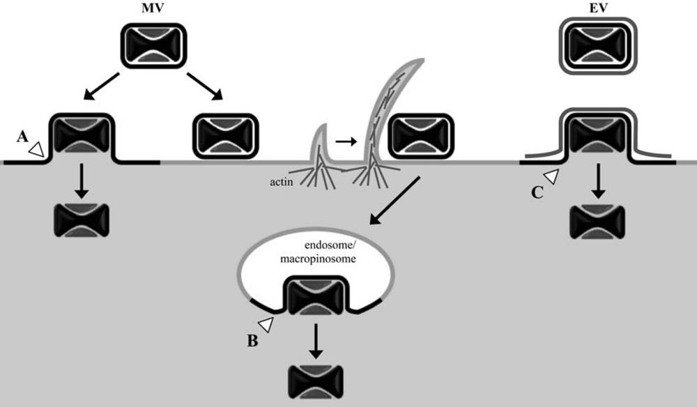
2.1. Roles of membranes and lipids in cell entry</title>
				<sec>
					<title>2.1.1. Entry of MVs</title>
					<p>MVs have been subjected to the great majority of VACV entry studies primarily because they can be highly purified, are stable and are the simplest infectious particle. In addition, the MV is the form used for vaccination. The initial attachment of MVs to cells occurs, in part, through interactions between viral membrane proteins (D8, A27, H3, and A26) and cell surface glycosaminoglycans (specifically, chondroitin sulfate and heparan sulfate) or laminin; however, individually these proteins are not essential [<xref ref-type="bibr" rid="B11">11</xref>,<xref ref-type="bibr" rid="B12">12</xref>,<xref ref-type="bibr" rid="B13">13</xref>,<xref ref-type="bibr" rid="B14">14</xref>]. Viral-cell membrane fusion and entry of the virus core into the cytoplasm can occur at neutral pH at the cell surface, as evidenced by numerous electron micrographs and suggested by biochemical membrane fusion assays [<xref ref-type="bibr" rid="B15">15</xref>,<xref ref-type="bibr" rid="B16">16</xref>,<xref ref-type="bibr" rid="B17">17</xref>,<xref ref-type="bibr" rid="B18">18</xref>,<xref ref-type="bibr" rid="B19">19</xref>]. Electron micrographs have also visualized the uptake of MVs into vesicles [<xref ref-type="bibr" rid="B20">20</xref>]. These observations were reconciled in a recent study showing that both fusion at the plasma membrane and endosomal vesicles can occur even in the same cell [<xref ref-type="bibr" rid="B21">21</xref>] (Figure 1). Further studies showed that the predominant mechanism of entry of the WR strain of VACV is by a low pH-dependent mechanism [<xref ref-type="bibr" rid="B21">21</xref>] following macropinocytosis [<xref ref-type="bibr" rid="B22">22</xref>,<xref ref-type="bibr" rid="B23">23</xref>]. The situation remains complicated, however, as there are VACV strain differences regarding the relative contributions of low and neutral pH entry mechanisms [<xref ref-type="bibr" rid="B24">24</xref>].</p>
					<p>In terms of the number of proteins required, VACV membrane fusion is far more complex than any other studied viral system. At least twelve MV membrane proteins (A16, A21, A28, G3, G9, H2, J5, L5, O3, I2, L1 and F9) – nine or ten of which function in a multi-protein complex termed the entry/fusion complex (EFC) – are individually required [<xref ref-type="bibr" rid="B25">25</xref>,<xref ref-type="bibr" rid="B26">26</xref>,<xref ref-type="bibr" rid="B27">27</xref>,<xref ref-type="bibr" rid="B28">28</xref>,<xref ref-type="bibr" rid="B29">29</xref>,<xref ref-type="bibr" rid="B30">30</xref>,<xref ref-type="bibr" rid="B31">31</xref>,<xref ref-type="bibr" rid="B32">32</xref>,<xref ref-type="bibr" rid="B33">33</xref>,<xref ref-type="bibr" rid="B34">34</xref>,<xref ref-type="bibr" rid="B35">35</xref>]. Two EFC proteins (A16 and G9) and the EFC-associated L1 protein are myristoylated, but the significance of these lipid moieties in virus entry has not been determined. </p>
					<fig id="figure1" position="float">
						<label>Figure 1</label>
						<caption>
							<p>Cell entry pathways of vaccinia virus mature virions (<italic>MV</italic>) and enveloped virions (<italic>EV</italic>) following attachment at the cell plasma membrane. Depending on the VACV strain and target cell, MV cores can enter the cytosol either through direct fusion of the MV membrane with the plasma membrane (<italic>A</italic>) or, following endocytosis/macropinocytosis, through fusion of the MV membrane with vesicles (<italic>B</italic>). EV particles have been observed to shed their additional membrane bilayer prior to direct fusion of the underlying MV membrane with the cell plasma membrane (<italic>C</italic>); entry of EV through an endosomal route might also occur. White arrowheads denote sites of membrane fusion between the MV particle membrane and cellular membranes.</p>
						</caption>
						<graphic xmlns:xlink="http://www.w3.org/1999/xlink" xlink:href="viruses-02-00972-g001.tif" scale="0"/>
					</fig>					
				</sec>
				<sec>
					<title>2.1.2. Lipids of the MV membrane</title>
					<p>Lipids, mostly phospholipids and cholesterol, constitute approximately 5% of the MV mass [<xref ref-type="bibr" rid="B36">36</xref>]. The MV contains several phospholipids with an abundance of phosphatidylcholine and lesser amounts of phosphatidylethanolamine, phosphatidylinositol, and phosphatidylserine (PS) [<xref ref-type="bibr" rid="B37">37</xref>,<xref ref-type="bibr" rid="B38">38</xref>,<xref ref-type="bibr" rid="B39">39</xref>]. An additional phospholipid, semilysobisphosphatidic acid (also known as acylbis(monoacylglycero)phosphate or acylphosphatidylglycerol) has been identified in MVs [<xref ref-type="bibr" rid="B39">39</xref>,<xref ref-type="bibr" rid="B40">40</xref>]. Quantitation of the lipids in the MV by Cluett and Machamer [<xref ref-type="bibr" rid="B39">39</xref>] is shown in <xref ref-type="table" rid="table1">Table 1</xref>. The lipid composition likely reflects the cellular origin of the MV membrane.</p>
					<p>Early studies by Ichihashi and Oie demonstrated the importance of virion-incorporated PS for VACV MV infectivity [<xref ref-type="bibr" rid="B41">41</xref>,<xref ref-type="bibr" rid="B42">42</xref>]. Extraction of virion-associated lipids with Nonidet P-40 detergent rendered MVs non-infectious, but incubation with either crude uninfected cell membrane preparations or PS-containing liposomes partially restored infectivity [<xref ref-type="bibr" rid="B41">41</xref>,<xref ref-type="bibr" rid="B42">42</xref>]. More recently, these results were extended and an apoptotic mimicry model for poxvirus entry, in which virion-associated PS specifically served to flag virions as apoptotic debris for cell uptake by macropinocytosis, was proposed [<xref ref-type="bibr" rid="B23">23</xref>]. However, PS is not unique in its ability to trigger this uptake mechanism as the reconstitution of detergent-extracted MVs with several other anionic phospholipid species could also facilitate virus entry in the absence of PS [<xref ref-type="bibr" rid="B43">43</xref>]. Nonetheless, these studies indicated the importance of the MV membrane lipid composition, specifically the presence of anionic phospholipid species, in cell entry of VACV MVs.</p>
					<table-wrap id="table1" position="float"><object-id pub-id-type="pii">viruses-02-00972_table1</object-id>
						<object-id pub-id-type="pii">viruses-02-00972_table1</object-id>
						<label>Table 1</label>
						<caption>
							<p>Quantitation of phospholipids in vaccinia virus MV and WV(<underline>µg per 10<sup>10</sup> virions</underline>
								<underline>)</underline>
							</p>
						</caption>
						<table>
							<thead>
								<tr>
									<th align="left" valign="middle">Phospholipid</th>
									<th align="left" valign="middle">MV</th>
									<th align="left" valign="middle">WV</th>
									<th align="left" valign="middle">WV-MV</th>
								</tr>
							</thead>
							<tbody>
								<tr>
									<td align="left" valign="middle">
										<italic>N</italic>-acyl phosphatidylethanolamine</td>
									<td align="left" valign="middle">0.38</td>
									<td align="left" valign="middle">0.31</td>
									<td align="left" valign="middle">0</td>
								</tr>
								<tr>
									<td align="left" valign="middle">Semilysobisphosphatidic acid</td>
									<td align="left" valign="middle">1.2</td>
									<td align="left" valign="middle">3.2</td>
									<td align="left" valign="middle">2</td>
								</tr>
								<tr>
									<td align="left" valign="middle">Phosphatidylethanolamine</td>
									<td align="left" valign="middle">1.7</td>
									<td align="left" valign="middle">4.5</td>
									<td align="left" valign="middle">2.8</td>
								</tr>
								<tr>
									<td align="left" valign="middle">Phosphatidylserine</td>
									<td align="left" valign="middle">0.26</td>
									<td align="left" valign="middle">1.3</td>
									<td align="left" valign="middle">1</td>
								</tr>
								<tr>
									<td align="left" valign="middle">Phosphatidylinositol</td>
									<td align="left" valign="middle">1</td>
									<td align="left" valign="middle">2.2</td>
									<td align="left" valign="middle">1.2</td>
								</tr>
								<tr>
									<td align="left" valign="middle">Phosphatidylcholine</td>
									<td align="left" valign="middle">2.8</td>
									<td align="left" valign="middle">7.8</td>
									<td align="left" valign="middle">5</td>
								</tr>
								<tr>
									<td align="left" valign="middle">Sphingomyelin</td>
									<td align="left" valign="middle">0.6</td>
									<td align="left" valign="middle">2.1</td>
									<td align="left" valign="middle">1.5</td>
								</tr>
							</tbody>
						</table>
<table-wrap-foot>
<fn>
					<p>Data from Cluett and Machamer [39].</p>
</fn>
</table-wrap-foot>						
					</table-wrap>
				</sec>
				<sec>
					<title>2.1.3. Entry of EVs and the EV-specific membrane</title>
					<p>The EV contains an additional outer membrane compared to the MV. Relative to MVs, EVs were reported to be enriched in sphingomyelin and PS with lower amounts of phosphatidylinositol [<xref ref-type="bibr" rid="B38">38</xref>]. In a later section, we will discuss the origin and phospholipid composition of the wrapped virion, which contains an additional membrane relative to the EV. MVs and EVs bind differently to cells [<xref ref-type="bibr" rid="B44">44</xref>] but little is known regarding the mechanism or proteins required for binding of the latter particles. Entry of EVs necessitates rupture of the outer membrane to expose the EFC embedded in the MV membrane [<xref ref-type="bibr" rid="B7">7</xref>] (<xref ref-type="fig" rid="figure1">Figure 1</xref>). Electron micrographs reveal a clear dissociation of the EV membrane “wrapper” from individual particles at the cell surface; importantly, no continuity of the EV wrapper with the plasma membrane was observed confirming that it is not fusogenic [<xref ref-type="bibr" rid="B45">45</xref>]. This “ligand-dependent non-fusogenic dissolution” of the outer EV membrane requires the EV membrane glycoproteins A34 and B5 and interactions with cell surface polyanionic molecules, e.g., glycosoaminoglycans, following virus attachment [<xref ref-type="bibr" rid="B45">45</xref>,<xref ref-type="bibr" rid="B46">46</xref>]. The subsequent entry steps are reported to be low pH-dependent [<xref ref-type="bibr" rid="B47">47</xref>].</p>

				</sec>
				<sec>
					<title>2.1.4. Lipids and protein components of the target cell membrane</title>
					<p>Cell surface glycosaminoglycans and laminin are important for the initial attachment of MVs to the cell surface [<xref ref-type="bibr" rid="B11">11</xref>,<xref ref-type="bibr" rid="B13">13</xref>,<xref ref-type="bibr" rid="B14">14</xref>,<xref ref-type="bibr" rid="B48">48</xref>]. The binding of MVs to cells induces the formation of actin-enriched membrane protrusions that engulf the virus particles for entry by macropinocytosis; this cellular response involves actin rearrangements and Rac1 GTPase as well as several serine/threonine, tyrosine, and phosphatidylinositol kinases including Pak1, PKC, and PI(3)K [<xref ref-type="bibr" rid="B23">23</xref>,<xref ref-type="bibr" rid="B49">49</xref>]. In contrast to MVs, entry of EVs does not seem to be signaling dependent [<xref ref-type="bibr" rid="B49">49</xref>]. Specific cell receptors, for either the MV or EV forms of VACV, have not been identified. Although PS-specific receptors for MV have been suggested [<xref ref-type="bibr" rid="B23">23</xref>], this is unlikely since a subsequent study indicated that PS is not uniquely involved in entry [<xref ref-type="bibr" rid="B43">43</xref>]. Depletion of cellular cholesterol levels does not affect cell binding of MVs but significantly inhibits entry of viral cores [<xref ref-type="bibr" rid="B50">50</xref>] indicating the importance of the composition and organization of the plasma membrane and suggesting a role for lipid rafts. Indeed, the block in MV penetration of cholesterol-depleted cells was found to occur at the initial stages of virus-cell membrane fusion (Laliberte and Moss, unpublished). Although the precise role of cellular cholesterol in VACV membrane fusion is not known, it may be indirectly required for triggering specific cell signaling pathways by cell surface-bound MVs. Additionally, reduced levels of cholesterol in the target cell membrane may affect cell surface distribution of an unidentified cell receptor(s) or other factor(s) required for virus entry. </p>
				</sec>
			</sec>
			<sec>
				<title>
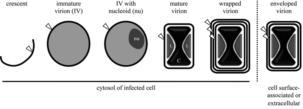
2.2. Role of membranes and lipids during morphogenesis and egress of progeny virions</title>
				<sec>
					<title>Membranes associated with virus factories and immature virions (IVs)</title>
					<p>After virus-cell membrane fusion, viral cores are actively transported on microtubules away from the cell periphery and deeper into the cytoplasm [<xref ref-type="bibr" rid="B51">51</xref>], where early gene transcription occurs. DNA from a single core may serve as a template for genome replication forming a viral factory [<xref ref-type="bibr" rid="B52">52</xref>,<xref ref-type="bibr" rid="B53">53</xref>,<xref ref-type="bibr" rid="B54">54</xref>]. Remarkably, at an early stage, each individual factory is entirely surrounded by ER membranes [<xref ref-type="bibr" rid="B55">55</xref>]. It has been suggested that the latter membranes are associated with replicating DNA. Following viral DNA replication and subsequent intermediate gene expression, the accumulation of late gene products – including viral structural proteins and enzymes/early gene transcription factors destined for virus packaging – allows poxvirus assembly and morphogenesis. The highly complex process of generating progeny virions is orchestrated by more than 80 viral gene products [<xref ref-type="bibr" rid="B5">5</xref>]. </p>
					<p>Viral membranes play a pivotal role in virion assembly. The initial structure observed by electron microscopy is the viral crescent membrane, which expands to form the spherical immature virion (IV) (<xref ref-type="fig" rid="figure2">Figure 2</xref>). The number of lipid bilayers comprising the viral crescent and IV membranes has been the subject of controversy. Electron microscopic images from several different laboratories were consistently interpreted as showing a single bilayer with an external spicule border [<xref ref-type="bibr" rid="B56">56</xref>,<xref ref-type="bibr" rid="B57">57</xref>,<xref ref-type="bibr" rid="B58">58</xref>,<xref ref-type="bibr" rid="B59">59</xref>,<xref ref-type="bibr" rid="B60">60</xref>,<xref ref-type="bibr" rid="B61">61</xref>]. Nevertheless, others interpreted their images as demonstrating two closely apposed bilayers [<xref ref-type="bibr" rid="B38">38</xref>,<xref ref-type="bibr" rid="B62">62</xref>,<xref ref-type="bibr" rid="B63">63</xref>]. The controversy appears to have been settled as no evidence for two bilayers was obtained by freeze-fracture studies [<xref ref-type="bibr" rid="B64">64</xref>] and the initial proponents of the double bilayer now also favor a single bilayer [<xref ref-type="bibr" rid="B65">65</xref>]. The spicule border on the crescent and IV membrane has been shown to be a honeycomb lattice scaffold composed of trimers of the viral D13 protein [<xref ref-type="bibr" rid="B64">64</xref>,<xref ref-type="bibr" rid="B66">66</xref>]; it is possible that the separation of this protein scaffold from the underlying membrane provided images suggesting a double membrane bilayer.</p>
					<p>Neither the crescent nor the IV membrane has been extensively purified and characterized so their lipid compositions remain unknown. The composition is usually inferred to be that of the MV membrane. A number of viral protein components of the crescent and IV membrane have been determined by immunoelectron microscopy and will be discussed below.</p>
					<fig id="figure2" position="float">
						<label>Figure 2</label>
						<caption>
							<p>Enumeration of lipid membrane bilayers (white arrowhead) possessed by vaccinia virus assembly intermediates. Nucleoids (<italic>nu</italic>) are electron-dense, DNA-containing substructures of immature virions. Lateral bodies (<italic>L</italic>) are internal virion structures of heterogenous material surrounding the dumbbell-shaped viral core (<italic>C</italic>). The location of each morphogenic form is indicated below the respective schematic. </p>
						</caption>
						<graphic xmlns:xlink="http://www.w3.org/1999/xlink" xlink:href="viruses-02-00972-g002.tif" scale="0"/>
					</fig>
				</sec>
				<sec>
					<title>Assembly of the IV membrane</title>
					<p>The unusual appearance of the crescent and IV membranes in cytoplasmic virus factories and their separation from cellular organelles led to the idea of their <italic>de novo</italic> synthesis [<xref ref-type="bibr" rid="B56">56</xref>]. However, the unprecedented nature of spontaneous membrane formation encouraged efforts to find a cellular organelle from which the viral membrane might be derived. Electron microscopic images have depicted close association of membranes of the ER-Golgi apparatus intermediate compartment (ERGIC) with IVs [<xref ref-type="bibr" rid="B38">38</xref>,<xref ref-type="bibr" rid="B63">63</xref>,<xref ref-type="bibr" rid="B67">67</xref>,<xref ref-type="bibr" rid="B68">68</xref>] while others have shown a close association with ER membranes [<xref ref-type="bibr" rid="B69">69</xref>]. Nevertheless, direct continuity between cellular organelles and viral membranes has not been demonstrated unambiguously. Although viral membrane proteins have been detected by immunogold labeling in ERGIC and ER, the reverse has not been shown, <italic>i.e.</italic>, cellular proteins in crescent or IV membranes. ERGIC resident proteins have also not been detected in purified MV particles, the membrane of which is presumably derived from the IV [<xref ref-type="bibr" rid="B70">70</xref>]. The lipid profile of purified MV particles was interpreted as being consistent with the MV membrane being of intermediate compartment or <italic>cis</italic>-Golgi network origin [<xref ref-type="bibr" rid="B39">39</xref>]. However, inhibiting transport of proteins from the ER to the Golgi compartment does not prevent the formation of IVs and MVs [<xref ref-type="bibr" rid="B71">71</xref>], implying that IV membranes are not derived from the ERGIC or Golgi compartment. Additional studies have shown that viral proteins can transit directly through the ER to the IV and accumulate in the MV membrane [<xref ref-type="bibr" rid="B69">69</xref>]. </p>
					<p>In addition to the viral D13 scaffold protein, several membrane (A14 and A17) [<xref ref-type="bibr" rid="B72">72</xref>] and non-membrane (A11, F10, G5, and H5) [<xref ref-type="bibr" rid="B73">73</xref>,<xref ref-type="bibr" rid="B74">74</xref>,<xref ref-type="bibr" rid="B75">75</xref>,<xref ref-type="bibr" rid="B76">76</xref>,<xref ref-type="bibr" rid="B77">77</xref>] viral proteins are required for crescent formation, whereas other proteins such as A9 [<xref ref-type="bibr" rid="B78">78</xref>] associate with the crescent membrane but are required at a later stage of morphogenesis. The exact roles of A14 and A17 proteins in crescent formation are uncertain but in the absence of either membrane protein crescent formation is reduced or aberrant and small vesicles accumulate [<xref ref-type="bibr" rid="B79">79</xref>,<xref ref-type="bibr" rid="B80">80</xref>,<xref ref-type="bibr" rid="B81">81</xref>,<xref ref-type="bibr" rid="B82">82</xref>]. </p>
					<p>Elongation and closure of viral crescents around electron dense viroplasm, containing core proteins or their precursors, give rise to the spherical IVs, which are approximately 350 nm in diameter [<xref ref-type="bibr" rid="B5">5</xref>] (<xref ref-type="fig" rid="figure2">Figure 2</xref>). Association of viroplasm with crescents is dependent on the expression of a complex of seven viral core proteins (A15, A30, D2, D3, F10, G7, and J1) [<xref ref-type="bibr" rid="B83">83</xref>]. Following packaging of the viral genome, the IV particle undergoes a gross alteration to become a barrel-shaped particle distinctive of poxvirus MVs. This transition is accompanied by disassembly of the viral D13 scaffold [<xref ref-type="bibr" rid="B84">84</xref>] and the processing of core protein precursors [<xref ref-type="bibr" rid="B85">85</xref>,<xref ref-type="bibr" rid="B86">86</xref>]. Virion assembly has recently been reviewed in detail [<xref ref-type="bibr" rid="B5">5</xref>]. Although crucial for progeny virion infectivity, the EFC as well as the EFC-associated proteins L1 and F9 are dispensable for the MV morphogenesis.</p>
				</sec>
				<sec>
					<title>Membranes of the enveloped particle</title>
					<p>As described earlier, after completion of MV morphogenesis, some particles are actively transported along microtubules away from the viral factory to sites where wrapping [<xref ref-type="bibr" rid="B87">87</xref>,<xref ref-type="bibr" rid="B88">88</xref>] with modified cisternae of endosomal [<xref ref-type="bibr" rid="B89">89</xref>] or trans-Golgi [<xref ref-type="bibr" rid="B90">90</xref>,<xref ref-type="bibr" rid="B91">91</xref>] origin occurs. These wrapped virions (WV; alternate name: intracellular enveloped virion [IEV]) possess an MV membrane and two additional membrane bilayers (<xref ref-type="fig" rid="figure2">Figure 2</xref>). The phospholipids of the WV have been analyzed and the composition of the wrapping membrane inferred by subtracting the values for the MV from the WV [<xref ref-type="bibr" rid="B39">39</xref>] (<xref ref-type="table" rid="table1">Table 1</xref>). The phospholipid components of the WV, with the exception of <italic>N</italic>-acyl phosphatidylethanolamine, are present in 2 to 3 times the molar amount of the MV, consistent with additional lipid bilayers in the WV (<xref ref-type="table" rid="table1">Table 1</xref>). At least seven proteins are associated with the wrapping membrane: A33, A34, A36, A56, B5, F12, F13. Of these, A36 and F12 are located exclusively on the outermost WV membrane. Although the details by which MVs acquire these membranes are unclear, the process requires the expression of several viral proteins, including the MV membrane-associated A27 protein [<xref ref-type="bibr" rid="B88">88</xref>,<xref ref-type="bibr" rid="B92">92</xref>] and the WV membrane-associated B5 [<xref ref-type="bibr" rid="B93">93</xref>,<xref ref-type="bibr" rid="B94">94</xref>] and F13 proteins [<xref ref-type="bibr" rid="B95">95</xref>]; other WV associated viral proteins appear necessary for efficient MV particle wrapping (reviewed in [<xref ref-type="bibr" rid="B9">9</xref>]). Of note, the peripheral F13 membrane protein is palmitylated on a pair of cysteine residues and addition of these 16-carbon fatty acid tails is necessary for membrane association, proper localization, and function of F13 [<xref ref-type="bibr" rid="B90">90</xref>,<xref ref-type="bibr" rid="B96">96</xref>,<xref ref-type="bibr" rid="B97">97</xref>]. The importance of the F13 protein is consistent with the potency both <italic>in vitro</italic> and <italic>in vivo</italic> of the anti-poxvirus therapeutic ST-246, which inhibits F13 protein activity in the infected cell [<xref ref-type="bibr" rid="B98">98</xref>].</p>
					<p>Wrapped virions are transported to the cell periphery in a microtubule-dependent manner relying on an interaction between the motor protein kinesin-1 and the viral A36 protein [<xref ref-type="bibr" rid="B99">99</xref>] and the viral F12 protein [<xref ref-type="bibr" rid="B100">100</xref>]. Once near the plasma membrane, the WV particle traverses the cortical membrane cytoskeleton by enhancing microtubule dynamics near the cell surface and inducing reorganization of the actin cytoskeleton resulting from the inhibition of RhoA signaling by the viral F11 protein [<xref ref-type="bibr" rid="B101">101</xref>,<xref ref-type="bibr" rid="B102">102</xref>]. Following dissociation from microtubules, the outermost membrane bilayer fuses with the plasma membrane resulting in the exocytosis of a doubly-wrapped EV at the cell surface. It is likely that cellular mechanisms are used for this exocytosis step since no viral mutants that have a phenotype in which virions collect under the cell surface have been isolated. Most of the exocytosed particles remain attached to the exterior of the cell and have been referred to as cell-associated EVs (CEV) to distinguish them from the released EVs (alternate name: extracellular enveloped virions [EEV]) [<xref ref-type="bibr" rid="B103">103</xref>]. The cell-associated EVs are largely responsible for cell-to-cell spread at the tips of actin tails.</p>
				</sec>
			</sec>
		</sec>
		<sec sec-type="conclusions">
			<title>3. Conclusions</title>
			<p>Lipoprotein membranes have crucial roles at key stages of the poxvirus life cycle, namely entry, assembly, intracellular transport, exocytosis and spread. The MV membrane is comprised of phospholipids and numerous proteins that together enable attachment and fusion with either the plasma membrane or endocytic vesicles following macropinocytosis. After entry of the core into the cytoplasm, ER membranes surround the early viral factories within which DNA replication occurs. The very first recognizable viral structures in the factories are crescent membranes of yet unknown origin that engulf viroplasm, containing core proteins, to form IVs. The external lattice scaffold responsible for the spherical shape of the IV detaches from the membrane allowing further steps in morphogenesis resulting in the barrel or brick-shaped MV. The latter acquires two additional lipoprotein membranes, derived from trans-Golgi or endosomal cisternae modified by the insertion of viral proteins, that allow high speed transit of the triple-membrane layered virions on microtubules. Upon reaching the periphery of the cell, the outermost viral membrane fuses with the plasma membrane resulting in exocytosis of the double-membrane EV. Upon contacting an uninfected cell, usually at the end of a long actin-containing projection, the outer membrane is disrupted allowing fusion of the MV membrane with the cell and penetration of the viral core into the cytoplasm for continuation of the replicative cycle. </p>
		</sec>
	</body>
	<back>
		<ack>
			<p>The preparation of this review was supported by the Division of Intramural Research, National Institute of Allergy and Infectious Diseases, National Institutes of Health.</p>
		</ack>
		<ref-list><title>References</title>
			<ref id="B1">
				<citation citation-type="book">
					<person-group person-group-type="author">
						<name>
							<surname>Moss</surname>
							<given-names>B.</given-names>
						</name>
					</person-group>
					<article-title>Poxviridae: the viruses and their replication</article-title>
					<source>Fields Virology</source>
					
					<edition>5th ed</edition>
					<person-group person-group-type="editor">
						<name>
							<surname>Knipe</surname>
							<given-names>D.M.</given-names>
						</name>
					</person-group>
					<year>2007</year>
					<publisher-name>Lippincott Williams &amp;amp; Wilkins</publisher-name>
					<publisher-loc>Philadelphia, PA, USA</publisher-loc>
				</citation>
			</ref>
			<ref id="B2">
				<citation citation-type="journal">
					<person-group person-group-type="author">
						<name>
							<surname>Moss</surname>
							<given-names>B.</given-names>
						</name>
					</person-group>
					<article-title>Genetically engineered poxviruses for recombinant gene expression, vaccination, and safety</article-title>
					<source>Proc. Natl. Acad. Sci. USA</source>
					<year>1996</year>
					<volume>93</volume>
					<fpage>11341</fpage>
					<lpage>11348</lpage>
				<pub-id pub-id-type="doi">10.1073/pnas.93.21.11341</pub-id></citation>
			</ref>
			<ref id="B3">
				<citation citation-type="journal">
					<person-group person-group-type="author">
						<name>
							<surname>Jacobs</surname>
							<given-names>B.L.</given-names>
						</name>
						<name>
							<surname>Langland</surname>
							<given-names>J.O.</given-names>
						</name>
						<name>
							<surname>Kibler</surname>
							<given-names>K.V.</given-names>
						</name>
						<name>
							<surname>Denzler</surname>
							<given-names>K.L.</given-names>
						</name>
						<name>
							<surname>White</surname>
							<given-names>S.D.</given-names>
						</name>
						<name>
							<surname>Holechek</surname>
							<given-names>S.A.</given-names>
						</name>
						<name>
							<surname>Wong</surname>
							<given-names>S.</given-names>
						</name>
						<name>
							<surname>Huynh</surname>
							<given-names>T.</given-names>
						</name>
						<name>
							<surname>Baskin</surname>
							<given-names>C.R.</given-names>
						</name>
					</person-group>
					<article-title>Vaccinia virus vaccines: past, present and future</article-title>
					<source>Antiviral Res.</source>
					<year>2009</year>
					<volume>84</volume>
					<fpage>1</fpage>
					<lpage>13</lpage>
				<pub-id pub-id-type="doi">10.1016/j.antiviral.2009.06.006</pub-id></citation>
			</ref>
			<ref id="B4">
				<citation citation-type="book">
					<article-title>Development of the global smallpox eradication programme, 1958-1966</article-title>
<source>Smallpox and Its Eradication</source>
					<person-group person-group-type="editor">
						<name>
							<surname>Fenner</surname>
							<given-names>F.</given-names>
						</name>
						<name>
							<surname>Henderson</surname>
							<given-names>D.A.</given-names>
						</name>
						<name>
							<surname>Arita</surname>
							<given-names>I.</given-names>
						</name>
						<name>
							<surname>Jezek</surname>
							<given-names>Z.</given-names>
						</name>
						<name>
							<surname>Ladnyi</surname>
							<given-names>I.D.</given-names>
						</name>
					</person-group>
					<publisher-name>World Health Organization</publisher-name>
					<publisher-loc>Geneva, Switzerland</publisher-loc>
					<year>1988</year>
					<fpage>365</fpage>
					<lpage>419</lpage>
					
				</citation>
			</ref>
			<ref id="B5">
				<citation citation-type="journal">
					<person-group person-group-type="author">
						<name>
							<surname>Condit</surname>
							<given-names>R.C.</given-names>
						</name>
						<name>
							<surname>Moussatche</surname>
							<given-names>N.</given-names>
						</name>
						<name>
							<surname>Traktman</surname>
							<given-names>P.</given-names>
						</name>
					</person-group>
					<article-title>In a nutshell: structure and assembly of the vaccinia virion</article-title>
					<source>Adv. Virus Res.</source>
					<year>2006</year>
					<volume>66</volume>
					<fpage>31</fpage>
					<lpage>124</lpage>
					<pub-id pub-id-type="pmid">16877059</pub-id>
				<pub-id pub-id-type="doi">10.1016/S0065-3527(06)66002-8</pub-id></citation>
			</ref>
			<ref id="B6">
				<citation citation-type="journal">
					<person-group person-group-type="author">
						<name>
							<surname>Cyrklaff</surname>
							<given-names>M.</given-names>
						</name>
						<name>
							<surname>Risco</surname>
							<given-names>C.</given-names>
						</name>
						<name>
							<surname>Fernandez</surname>
							<given-names>J.J.</given-names>
						</name>
						<name>
							<surname>Jimenez</surname>
							<given-names>M.V.</given-names>
						</name>
						<name>
							<surname>Esteban</surname>
							<given-names>M.</given-names>
						</name>
						<name>
							<surname>Baumeister</surname>
							<given-names>W.</given-names>
						</name>
						<name>
							<surname>Carrascosa</surname>
							<given-names>J.L.</given-names>
						</name>
					</person-group>
					<article-title>Cryo-electron tomography of vaccinia virus</article-title>
					<source>Proc. Natl. Acad. Sci. USA</source>
					<year>2005</year>
					<volume>102</volume>
					<fpage>2772</fpage>
					<lpage>2777</lpage>
				<pub-id pub-id-type="doi">10.1073/pnas.0409825102</pub-id></citation>
			</ref>
			<ref id="B7">
				<citation citation-type="journal">
					<person-group person-group-type="author">
						<name>
							<surname>Moss</surname>
							<given-names>B.</given-names>
						</name>
					</person-group>
					<article-title>Poxvirus entry and membrane fusion</article-title>
					<source>Virology</source>
					<year>2006</year>
					<volume>344</volume>
					<fpage>48</fpage>
					<lpage>54</lpage>
					<pub-id pub-id-type="pmid">16364735</pub-id>
				<pub-id pub-id-type="doi">10.1016/j.virol.2005.09.037</pub-id></citation>
			</ref>
			<ref id="B8">
				<citation citation-type="journal">
					<person-group person-group-type="author">
						<name>
							<surname>Broyles</surname>
							<given-names>S.S.</given-names>
						</name>
					</person-group>
					<article-title>Vaccinia virus transcription</article-title>
					<source>J. Gen. Virol.</source>
					<year>2003</year>
					<volume>84</volume>
					<fpage>2293</fpage>
					<lpage>2303</lpage>
					<pub-id pub-id-type="pmid">12917449</pub-id>
				<pub-id pub-id-type="doi">10.1099/vir.0.18942-0</pub-id></citation>
			</ref>
			<ref id="B9">
				<citation citation-type="journal">
					<person-group person-group-type="author">
						<name>
							<surname>Smith</surname>
							<given-names>G.L.</given-names>
						</name>
						<name>
							<surname>Vanderplasschen</surname>
							<given-names>A.</given-names>
						</name>
						<name>
							<surname>Law</surname>
							<given-names>M.</given-names>
						</name>
					</person-group>
					<article-title>The formation and function of extracellular enveloped vaccinia virus</article-title>
					<source>J. Gen. Virol.</source>
					<year>2002</year>
					<volume>83</volume>
					<fpage>2915</fpage>
					<lpage>2931</lpage>
					<pub-id pub-id-type="pmid">12466468</pub-id>
				</citation>
			</ref>
			<ref id="B10">
				<citation citation-type="journal">
					<person-group person-group-type="author">
						<name>
							<surname>Boulter</surname>
							<given-names>E.A.</given-names>
						</name>
						<name>
							<surname>Appleyard</surname>
							<given-names>G.</given-names>
						</name>
					</person-group>
					<article-title>Differences between extracellular and intracellular forms of poxvirus and their implications</article-title>
					<source>Prog. Med. Virol.</source>
					<year>1973</year>
					<volume>16</volume>
					<fpage>86</fpage>
					<lpage>108</lpage>
					<pub-id pub-id-type="pmid">4356899</pub-id>
				</citation>
			</ref>
			<ref id="B11">
				<citation citation-type="journal">
					<person-group person-group-type="author">
						<name>
							<surname>Chung</surname>
							<given-names>C.S.</given-names>
						</name>
						<name>
							<surname>Hsiao</surname>
							<given-names>J.C.</given-names>
						</name>
						<name>
							<surname>Chang</surname>
							<given-names>Y.S.</given-names>
						</name>
						<name>
							<surname>Chang</surname>
							<given-names>W.</given-names>
						</name>
					</person-group>
					<article-title>A27L protein mediates vaccinia virus interaction with cell surface heparan sulfate</article-title>
					<source>J. Virol.</source>
					<year>1998</year>
					<volume>72</volume>
					<fpage>1577</fpage>
					<lpage>1585</lpage>
					<pub-id pub-id-type="pmid">9445060</pub-id>
				</citation>
			</ref>
			<ref id="B12">
				<citation citation-type="journal">
					<person-group person-group-type="author">
						<name>
							<surname>Hsiao</surname>
							<given-names>J.C.</given-names>
						</name>
						<name>
							<surname>Chung</surname>
							<given-names>C.S.</given-names>
						</name>
						<name>
							<surname>Chang</surname>
							<given-names>W.</given-names>
						</name>
					</person-group>
					<article-title>Cell surface proteoglycans are necessary for A27L protein-mediated cell fusion: identification of the N-terminal region of A27L protein as the glycosaminoglycan-binding domain</article-title>
					<source>J. Virol.</source>
					<year>1998</year>
					<volume>72</volume>
					<fpage>8374</fpage>
					<lpage>8379</lpage>
					<pub-id pub-id-type="pmid">9733888</pub-id>
				</citation>
			</ref>
			<ref id="B13">
				<citation citation-type="journal">
					<person-group person-group-type="author">
						<name>
							<surname>Lin</surname>
							<given-names>C.L.</given-names>
						</name>
						<name>
							<surname>Chung</surname>
							<given-names>C.S.</given-names>
						</name>
						<name>
							<surname>Heine</surname>
							<given-names>H.G.</given-names>
						</name>
						<name>
							<surname>Chang</surname>
							<given-names>W.</given-names>
						</name>
					</person-group>
					<article-title>Vaccinia virus envelope H3L protein binds to cell surface heparan sulfate and is important for intracellular mature virion morphogenesis and virus infection <italic>in vitro</italic> and <italic>in vivo</italic>
					</article-title>
					<source>J. Virol.</source>
					<year>2000</year>
					<volume>74</volume>
					<fpage>3353</fpage>
					<lpage>3365</lpage>
					<pub-id pub-id-type="pmid">10708453</pub-id>
				<pub-id pub-id-type="doi">10.1128/JVI.74.7.3353-3365.2000</pub-id></citation>
			</ref>
			<ref id="B14">
				<citation citation-type="journal">
					<person-group person-group-type="author">
						<name>
							<surname>Chiu</surname>
							<given-names>W.L.</given-names>
						</name>
						<name>
							<surname>Lin</surname>
							<given-names>C.L.</given-names>
						</name>
						<name>
							<surname>Yang</surname>
							<given-names>M.H.</given-names>
						</name>
						<name>
							<surname>Tzou</surname>
							<given-names>D.L.</given-names>
						</name>
						<name>
							<surname>Chang</surname>
							<given-names>W.</given-names>
						</name>
					</person-group>
					<article-title>Vaccinia virus 4c (A26L) protein on intracellular mature virus binds to the extracellular cellular matrix laminin</article-title>
					<source>J. Virol.</source>
					<year>2007</year>
					<volume>81</volume>
					<fpage>2149</fpage>
					<lpage>2157</lpage>
					<pub-id pub-id-type="pmid">17166913</pub-id>
				<pub-id pub-id-type="doi">10.1128/JVI.02302-06</pub-id></citation>
			</ref>
			<ref id="B15">
				<citation citation-type="journal">
					<person-group person-group-type="author">
						<name>
							<surname>Armstrong</surname>
							<given-names>J.A.</given-names>
						</name>
						<name>
							<surname>Metz</surname>
							<given-names>D.H.</given-names>
						</name>
						<name>
							<surname>Young</surname>
							<given-names>M.R.</given-names>
						</name>
					</person-group>
					<article-title>The mode of entry of vaccinia virus into L cells</article-title>
					<source>J. Gen. Virol.</source>
					<year>1973</year>
					<volume>21</volume>
					<fpage>533</fpage>
					<lpage>537</lpage>
					<pub-id pub-id-type="pmid">4128637</pub-id>
				<pub-id pub-id-type="doi">10.1099/0022-1317-21-3-533</pub-id></citation>
			</ref>
			<ref id="B16">
				<citation citation-type="journal">
					<person-group person-group-type="author">
						<name>
							<surname>Carter</surname>
							<given-names>G.C.</given-names>
						</name>
						<name>
							<surname>Law</surname>
							<given-names>M.</given-names>
						</name>
						<name>
							<surname>Hollinshead</surname>
							<given-names>M.</given-names>
						</name>
						<name>
							<surname>Smith</surname>
							<given-names>G.L.</given-names>
						</name>
					</person-group>
					<article-title>Entry of the vaccinia virus intracellular mature virion and its interactions with glycosaminoglycans</article-title>
					<source>J. Gen. Virol.</source>
					<year>2005</year>
					<volume>86</volume>
					<fpage>1279</fpage>
					<lpage>1290</lpage>
					<pub-id pub-id-type="pmid">15831938</pub-id>
				<pub-id pub-id-type="doi">10.1099/vir.0.80831-0</pub-id></citation>
			</ref>
			<ref id="B17">
				<citation citation-type="journal">
					<person-group person-group-type="author">
						<name>
							<surname>Chang</surname>
							<given-names>A.</given-names>
						</name>
						<name>
							<surname>Metz</surname>
							<given-names>D.H.</given-names>
						</name>
					</person-group>
					<article-title>Further investigations on the mode of entry of vaccinia virus into cells</article-title>
					<source>J. Gen. Virol.</source>
					<year>1976</year>
					<volume>32</volume>
					<fpage>275</fpage>
					<lpage>282</lpage>
					<pub-id pub-id-type="pmid">798024</pub-id>
				<pub-id pub-id-type="doi">10.1099/0022-1317-32-2-275</pub-id></citation>
			</ref>
			<ref id="B18">
				<citation citation-type="journal">
					<person-group person-group-type="author">
						<name>
							<surname>Doms</surname>
							<given-names>R.W.</given-names>
						</name>
						<name>
							<surname>Blumenthal</surname>
							<given-names>R.</given-names>
						</name>
						<name>
							<surname>Moss</surname>
							<given-names>B.</given-names>
						</name>
					</person-group>
					<article-title>Fusion of intra- and extracellular forms of vaccinia virus with the cell membrane</article-title>
					<source>J. Virol.</source>
					<year>1990</year>
					<volume>64</volume>
					<fpage>4884</fpage>
					<lpage>4892</lpage>
					<pub-id pub-id-type="pmid">2398531</pub-id>
				</citation>
			</ref>
			<ref id="B19">
				<citation citation-type="journal">
					<person-group person-group-type="author">
						<name>
							<surname>Janeczko</surname>
							<given-names>R.A.</given-names>
						</name>
						<name>
							<surname>Rodriguez</surname>
							<given-names>J.F.</given-names>
						</name>
						<name>
							<surname>Esteban</surname>
							<given-names>M.</given-names>
						</name>
					</person-group>
					<article-title>Studies on the mechanism of entry of vaccinia virus in animal cells</article-title>
					<source>Arch. Virol.</source>
					<year>1987</year>
					<volume>92</volume>
					<fpage>135</fpage>
					<lpage>150</lpage>
					<pub-id pub-id-type="pmid">3800657</pub-id>
				<pub-id pub-id-type="doi">10.1007/BF01310068</pub-id></citation>
			</ref>
			<ref id="B20">
				<citation citation-type="journal">
					<person-group person-group-type="author">
						<name>
							<surname>Dales</surname>
							<given-names>S.</given-names>
						</name>
						<name>
							<surname>Kajioka</surname>
							<given-names>R.</given-names>
						</name>
					</person-group>
					<article-title>The Cycle of Multiplication of Vaccinia Virus in Earle's Strain L Cells</article-title>
					<source>I. Uptake and Penetration. Virology</source>
					<year>1964</year>
					<volume>24</volume>
					<fpage>278</fpage>
					<lpage>294</lpage>
				</citation>
			</ref>
			<ref id="B21">
				<citation citation-type="journal">
					<person-group person-group-type="author">
						<name>
							<surname>Townsley</surname>
							<given-names>A.C.</given-names>
						</name>
						<name>
							<surname>Weisberg</surname>
							<given-names>A.S.</given-names>
						</name>
						<name>
							<surname>Wagenaar</surname>
							<given-names>T.R.</given-names>
						</name>
						<name>
							<surname>Moss</surname>
							<given-names>B.</given-names>
						</name>
					</person-group>
					<article-title>Vaccinia virus entry into cells via a low-pH-dependent endosomal pathway</article-title>
					<source>J. Virol.</source>
					<year>2006</year>
					<volume>80</volume>
					<fpage>8899</fpage>
					<lpage>8908</lpage>
					<pub-id pub-id-type="pmid">16940502</pub-id>
				<pub-id pub-id-type="doi">10.1128/JVI.01053-06</pub-id></citation>
			</ref>
			<ref id="B22">
				<citation citation-type="journal">
					<person-group person-group-type="author">
						<name>
							<surname>Huang</surname>
							<given-names>C.Y.</given-names>
						</name>
						<name>
							<surname>Lu</surname>
							<given-names>T.Y.</given-names>
						</name>
						<name>
							<surname>Bair</surname>
							<given-names>C.H.</given-names>
						</name>
						<name>
							<surname>Chang</surname>
							<given-names>Y.S.</given-names>
						</name>
						<name>
							<surname>Jwo</surname>
							<given-names>J.K.</given-names>
						</name>
						<name>
							<surname>Chang</surname>
							<given-names>W.</given-names>
						</name>
					</person-group>
					<article-title>A novel cellular protein, VPEF, facilitates vaccinia virus penetration into HeLa cells through fluid phase endocytosis</article-title>
					<source>J. Virol.</source>
					<year>2008</year>
					<volume>82</volume>
					<fpage>7988</fpage>
					<lpage>7999</lpage>
					<pub-id pub-id-type="pmid">18550675</pub-id>
				<pub-id pub-id-type="doi">10.1128/JVI.00894-08</pub-id></citation>
			</ref>
			<ref id="B23">
				<citation citation-type="journal">
					<person-group person-group-type="author">
						<name>
							<surname>Mercer</surname>
							<given-names>J.</given-names>
						</name>
						<name>
							<surname>Helenius</surname>
							<given-names>A.</given-names>
						</name>
					</person-group>
					<article-title>Vaccinia virus uses macropinocytosis and apoptotic mimicry to enter host cells</article-title>
					<source>Science</source>
					<year>2008</year>
					<volume>320</volume>
					<fpage>531</fpage>
					<lpage>535</lpage>
					<pub-id pub-id-type="pmid">18436786</pub-id>
				<pub-id pub-id-type="doi">10.1126/science.1155164</pub-id></citation>
			</ref>
			<ref id="B24">
				<citation citation-type="journal">
					<person-group person-group-type="author">
						<name>
							<surname>Bengali</surname>
							<given-names>Z.</given-names>
						</name>
						<name>
							<surname>Townsley</surname>
							<given-names>A.C.</given-names>
						</name>
						<name>
							<surname>Moss</surname>
							<given-names>B.</given-names>
						</name>
					</person-group>
					<article-title>Vaccinia virus strain differences in cell attachment and entry 


</article-title>
					<source>Virology</source>
					<year>2009</year>
					
				</citation>
			</ref>
			<ref id="B25">
				<citation citation-type="journal">
					<person-group person-group-type="author">
						<name>
							<surname>Ojeda</surname>
							<given-names>S.</given-names>
						</name>
						<name>
							<surname>Senkevich</surname>
							<given-names>T.G.</given-names>
						</name>
						<name>
							<surname>Moss</surname>
							<given-names>B.</given-names>
						</name>
					</person-group>
					<article-title>Entry of vaccinia virus and cell-cell fusion require a highly conserved cysteine-rich membrane protein encoded by the A16L gene</article-title>
					<source>J. Virol.</source>
					<year>2006</year>
					<volume>80</volume>
					<fpage>51</fpage>
					<lpage>61</lpage>
					<pub-id pub-id-type="pmid">16352530</pub-id>
				<pub-id pub-id-type="doi">10.1128/JVI.80.1.51-61.2006</pub-id></citation>
			</ref>
			<ref id="B26">
				<citation citation-type="journal">
					<person-group person-group-type="author">
						<name>
							<surname>Brown</surname>
							<given-names>E.</given-names>
						</name>
						<name>
							<surname>Senkevich</surname>
							<given-names>T.G.</given-names>
						</name>
						<name>
							<surname>Moss</surname>
							<given-names>B.</given-names>
						</name>
					</person-group>
					<article-title>Vaccinia virus F9 virion membrane protein is required for entry but not virus assembly, in contrast to the related L1 protein</article-title>
					<source>J. Virol.</source>
					<year>2006</year>
					<volume>80</volume>
					<fpage>9455</fpage>
					<lpage>9464</lpage>
					<pub-id pub-id-type="pmid">16973551</pub-id>
				<pub-id pub-id-type="doi">10.1128/JVI.01149-06</pub-id></citation>
			</ref>
			<ref id="B27">
				<citation citation-type="journal">
					<person-group person-group-type="author">
						<name>
							<surname>Townsley</surname>
							<given-names>A.C.</given-names>
						</name>
						<name>
							<surname>Senkevich</surname>
							<given-names>T.G.</given-names>
						</name>
						<name>
							<surname>Moss</surname>
							<given-names>B.</given-names>
						</name>
					</person-group>
					<article-title>The product of the vaccinia virus L5R gene is a fourth membrane protein encoded by all poxviruses that is required for cell entry and cell-cell fusion</article-title>
					<source>J. Virol.</source>
					<year>2005</year>
					<volume>79</volume>
					<fpage>10988</fpage>
					<lpage>10998</lpage>
					<pub-id pub-id-type="pmid">16103150</pub-id>
				<pub-id pub-id-type="doi">10.1128/JVI.79.17.10988-10998.2005</pub-id></citation>
			</ref>
			<ref id="B28">
				<citation citation-type="journal">
					<person-group person-group-type="author">
						<name>
							<surname>Townsley</surname>
							<given-names>A.C.</given-names>
						</name>
						<name>
							<surname>Senkevich</surname>
							<given-names>T.G.</given-names>
						</name>
						<name>
							<surname>Moss</surname>
							<given-names>B.</given-names>
						</name>
					</person-group>
					<article-title>Vaccinia virus A21 virion membrane protein is required for cell entry and fusion</article-title>
					<source>J. Virol.</source>
					<year>2005</year>
					<volume>79</volume>
					<fpage>9458</fpage>
					<lpage>9469</lpage>
					<pub-id pub-id-type="pmid">16014909</pub-id>
				<pub-id pub-id-type="doi">10.1128/JVI.79.15.9458-9469.2005</pub-id></citation>
			</ref>
			<ref id="B29">
				<citation citation-type="journal">
					<person-group person-group-type="author">
						<name>
							<surname>Senkevich</surname>
							<given-names>T.G.</given-names>
						</name>
						<name>
							<surname>Ojeda</surname>
							<given-names>S.</given-names>
						</name>
						<name>
							<surname>Townsley</surname>
							<given-names>A.</given-names>
						</name>
						<name>
							<surname>Nelson</surname>
							<given-names>G.E.</given-names>
						</name>
						<name>
							<surname>Moss</surname>
							<given-names>B.</given-names>
						</name>
					</person-group>
					<article-title>Poxvirus multiprotein entry-fusion complex</article-title>
					<source>Proc. Natl. Acad. Sci. USA</source>
					<year>2005</year>
					<volume>102</volume>
					<fpage>18572</fpage>
					<lpage>18577</lpage>
				<pub-id pub-id-type="doi">10.1073/pnas.0509239102</pub-id></citation>
			</ref>
			<ref id="B30">
				<citation citation-type="journal">
					<person-group person-group-type="author">
						<name>
							<surname>Senkevich</surname>
							<given-names>T.G.</given-names>
						</name>
						<name>
							<surname>Moss</surname>
							<given-names>B.</given-names>
						</name>
					</person-group>
					<article-title>Vaccinia virus H2 protein is an essential component of a complex involved in virus entry and cell-cell fusion</article-title>
					<source>J. Virol.</source>
					<year>2005</year>
					<volume>79</volume>
					<fpage>4744</fpage>
					<lpage>4754</lpage>
					<pub-id pub-id-type="pmid">15795260</pub-id>
				<pub-id pub-id-type="doi">10.1128/JVI.79.8.4744-4754.2005</pub-id></citation>
			</ref>
			<ref id="B31">
				<citation citation-type="journal">
					<person-group person-group-type="author">
						<name>
							<surname>Senkevich</surname>
							<given-names>T.G.</given-names>
						</name>
						<name>
							<surname>Ward</surname>
							<given-names>B.M.</given-names>
						</name>
						<name>
							<surname>Moss</surname>
							<given-names>B.</given-names>
						</name>
					</person-group>
					<article-title>Vaccinia virus entry into cells is dependent on a virion surface protein encoded by the A28L gene</article-title>
					<source>J. Virol.</source>
					<year>2004</year>
					<volume>78</volume>
					<fpage>2357</fpage>
					<lpage>2366</lpage>
					<pub-id pub-id-type="pmid">14963132</pub-id>
				<pub-id pub-id-type="doi">10.1128/JVI.78.5.2357-2366.2004</pub-id></citation>
			</ref>
			<ref id="B32">
				<citation citation-type="journal">
					<person-group person-group-type="author">
						<name>
							<surname>Bisht</surname>
							<given-names>H.</given-names>
						</name>
						<name>
							<surname>Weisberg</surname>
							<given-names>A.S.</given-names>
						</name>
						<name>
							<surname>Moss</surname>
							<given-names>B.</given-names>
						</name>
					</person-group>
					<article-title>Vaccinia virus l1 protein is required for cell entry and membrane fusion</article-title>
					<source>J. Virol.</source>
					<year>2008</year>
					<volume>82</volume>
					<fpage>8687</fpage>
					<lpage>8694</lpage>
					<pub-id pub-id-type="pmid">18596103</pub-id>
				<pub-id pub-id-type="doi">10.1128/JVI.00852-08</pub-id></citation>
			</ref>
			<ref id="B33">
				<citation citation-type="journal">
					<person-group person-group-type="author">
						<name>
							<surname>Nichols</surname>
							<given-names>R.J.</given-names>
						</name>
						<name>
							<surname>Stanitsa</surname>
							<given-names>E.</given-names>
						</name>
						<name>
							<surname>Unger</surname>
							<given-names>B.</given-names>
						</name>
						<name>
							<surname>Traktman</surname>
							<given-names>P.</given-names>
						</name>
					</person-group>
					<article-title>The vaccinia virus gene I2L encodes a membrane protein with an essential role in virion entry</article-title>
					<source>J. Virol.</source>
					<year>2008</year>
					<volume>82</volume>
					<fpage>10247</fpage>
					<lpage>10261</lpage>
					<pub-id pub-id-type="pmid">18701587</pub-id>
				<pub-id pub-id-type="doi">10.1128/JVI.01035-08</pub-id></citation>
			</ref>
			<ref id="B34">
				<citation citation-type="journal">
					<person-group person-group-type="author">
						<name>
							<surname>Izmailyan</surname>
							<given-names>R.A.</given-names>
						</name>
						<name>
							<surname>Huang</surname>
							<given-names>C.Y.</given-names>
						</name>
						<name>
							<surname>Mohammad</surname>
							<given-names>S.</given-names>
						</name>
						<name>
							<surname>Isaacs</surname>
							<given-names>S.N.</given-names>
						</name>
						<name>
							<surname>Chang</surname>
							<given-names>W.</given-names>
						</name>
					</person-group>
					<article-title>The envelope G3L protein is essential for entry of vaccinia virus into host cells</article-title>
					<source>J. Virol.</source>
					<year>2006</year>
					<volume>80</volume>
					<fpage>8402</fpage>
					<lpage>8410</lpage>
					<pub-id pub-id-type="pmid">16912291</pub-id>
				<pub-id pub-id-type="doi">10.1128/JVI.00624-06</pub-id></citation>
			</ref>
			<ref id="B35">
				<citation citation-type="journal">
					<person-group person-group-type="author">
						<name>
							<surname>Satheshkumar</surname>
							<given-names>P.S.</given-names>
						</name>
						<name>
							<surname>Moss</surname>
							<given-names>B.</given-names>
						</name>
					</person-group>
					<article-title>Characterization of a newly identified 35-amino-acid component of the vaccinia virus entry/fusion complex conserved in all chordopoxviruses</article-title>
					<source>J. Virol.</source>
					<year>2009</year>
					<volume>83</volume>
					<fpage>12822</fpage>
					<lpage>12832</lpage>
					<pub-id pub-id-type="pmid">19812151</pub-id>
				<pub-id pub-id-type="doi">10.1128/JVI.01744-09</pub-id></citation>
			</ref>
			<ref id="B36">
				<citation citation-type="journal">
					<person-group person-group-type="author">
						<name>
							<surname>Zwartouw</surname>
							<given-names>H.T.</given-names>
						</name>
					</person-group>
					<article-title>The Chemical Composition of Vaccinia Virus</article-title>
					<source>J. Gen. Microbiol.</source>
					<year>1964</year>
					<volume>34</volume>
					<fpage>115</fpage>
					<lpage>123</lpage>
					<pub-id pub-id-type="pmid">14121210</pub-id>
				</citation>
			</ref>
			<ref id="B37">
				<citation citation-type="journal">
					<person-group person-group-type="author">
						<name>
							<surname>Stern</surname>
							<given-names>W.</given-names>
						</name>
						<name>
							<surname>Dales</surname>
							<given-names>S.</given-names>
						</name>
					</person-group>
					<article-title>Biogenesis of vaccinia: concerning the origin of the envelope phospholipids</article-title>
					<source>Virology</source>
					<year>1974</year>
					<volume>62</volume>
					<fpage>293</fpage>
					<lpage>306</lpage>
					<pub-id pub-id-type="pmid">4473861</pub-id>
				<pub-id pub-id-type="doi">10.1016/0042-6822(74)90393-6</pub-id></citation>
			</ref>
			<ref id="B38">
				<citation citation-type="journal">
					<person-group person-group-type="author">
						<name>
							<surname>Sodeik</surname>
							<given-names>B.</given-names>
						</name>
						<name>
							<surname>Doms</surname>
							<given-names>R.W.</given-names>
						</name>
						<name>
							<surname>Ericsson</surname>
							<given-names>M.</given-names>
						</name>
						<name>
							<surname>Hiller</surname>
							<given-names>G.</given-names>
						</name>
						<name>
							<surname>Machamer</surname>
							<given-names>C.E.</given-names>
						</name>
						<name>
							<surname>van 't Hof</surname>
							<given-names>W.</given-names>
						</name>
						<name>
							<surname>van Meer</surname>
							<given-names>G.</given-names>
						</name>
						<name>
							<surname>Moss</surname>
							<given-names>B.</given-names>
						</name>
						<name>
							<surname>Griffiths</surname>
							<given-names>G.</given-names>
						</name>
					</person-group>
					<article-title>Assembly of vaccinia virus: role of the intermediate compartment between the endoplasmic reticulum and the Golgi stacks</article-title>
					<source>J. Cell Biol.</source>
					<year>1993</year>
					<volume>121</volume>
					<fpage>521</fpage>
					<lpage>541</lpage>
					<pub-id pub-id-type="pmid">8486734</pub-id>
				<pub-id pub-id-type="doi">10.1083/jcb.121.3.521</pub-id></citation>
			</ref>
			<ref id="B39">
				<citation citation-type="journal">
					<person-group person-group-type="author">
						<name>
							<surname>Cluett</surname>
							<given-names>E.B.</given-names>
						</name>
						<name>
							<surname>Machamer</surname>
							<given-names>C.E.</given-names>
						</name>
					</person-group>
					<article-title>The envelope of vaccinia virus reveals an unusual phospholipid in Golgi complex membranes</article-title>
					<source>J. Cell Sci.</source>
					<year>1996</year>
					<volume>109</volume>
					<fpage>2121</fpage>
					<lpage>2131</lpage>
					<pub-id pub-id-type="pmid">8856508</pub-id>
				</citation>
			</ref>
			<ref id="B40">
				<citation citation-type="journal">
					<person-group person-group-type="author">
						<name>
							<surname>Hiller</surname>
							<given-names>G.</given-names>
						</name>
						<name>
							<surname>Eibl</surname>
							<given-names>H.</given-names>
						</name>
						<name>
							<surname>Weber</surname>
							<given-names>K.</given-names>
						</name>
					</person-group>
					<article-title>Acyl bis(monoacylglycero)phosphate, assumed to be a marker for lysosomes, is a major phospholipid of vaccinia virions</article-title>
					<source>Virology</source>
					<year>1981</year>
					<volume>113</volume>
					<fpage>761</fpage>
					<lpage>764</lpage>
					<pub-id pub-id-type="pmid">7269258</pub-id>
				<pub-id pub-id-type="doi">10.1016/0042-6822(81)90204-X</pub-id></citation>
			</ref>
			<ref id="B41">
				<citation citation-type="journal">
					<person-group person-group-type="author">
						<name>
							<surname>Ichihashi</surname>
							<given-names>Y.</given-names>
						</name>
						<name>
							<surname>Oie</surname>
							<given-names>M.</given-names>
						</name>
					</person-group>
					<article-title>The activation of vaccinia virus infectivity by the transfer of phosphatidylserine from the plasma membrane</article-title>
					<source>Virology</source>
					<year>1983</year>
					<volume>130</volume>
					<fpage>306</fpage>
					<lpage>317</lpage>
					<pub-id pub-id-type="pmid">6649411</pub-id>
				<pub-id pub-id-type="doi">10.1016/0042-6822(83)90085-5</pub-id></citation>
			</ref>
			<ref id="B42">
				<citation citation-type="journal">
					<person-group person-group-type="author">
						<name>
							<surname>Oie</surname>
							<given-names>M.</given-names>
						</name>
					</person-group>
					<article-title>Reversible inactivation and reactivation of vaccinia virus by manipulation of viral lipid composition</article-title>
					<source>Virology</source>
					<year>1985</year>
					<volume>142</volume>
					<fpage>299</fpage>
					<lpage>306</lpage>
					<pub-id pub-id-type="pmid">4060575</pub-id>
				<pub-id pub-id-type="doi">10.1016/0042-6822(85)90338-1</pub-id></citation>
			</ref>
			<ref id="B43">
				<citation citation-type="journal">
					<person-group person-group-type="author">
						<name>
							<surname>Laliberte</surname>
							<given-names>J.P.</given-names>
						</name>
						<name>
							<surname>Moss</surname>
							<given-names>B.</given-names>
						</name>
					</person-group>
					<article-title>Appraising the apoptotic mimicry model and the role of phospholipids for poxvirus entry</article-title>
					<source>Proc. Natl. Acad. Sci. USA</source>
					<year>2009</year>
					<volume>106</volume>
					<fpage>17517</fpage>
					<lpage>17521</lpage>
				<pub-id pub-id-type="doi">10.1073/pnas.0909376106</pub-id></citation>
			</ref>
			<ref id="B44">
				<citation citation-type="journal">
					<person-group person-group-type="author">
						<name>
							<surname>Vanderplasschen</surname>
							<given-names>A.</given-names>
						</name>
						<name>
							<surname>Smith</surname>
							<given-names>G.L.</given-names>
						</name>
					</person-group>
					<article-title>A novel virus binding assay using confocal microscopy: demonstration that the intracellular and extracellular vaccinia virions bind to different cellular receptors</article-title>
					<source>J. Virol.</source>
					<year>1997</year>
					<volume>71</volume>
					<fpage>4032</fpage>
					<lpage>4041</lpage>
					<pub-id pub-id-type="pmid">9094681</pub-id>
				</citation>
			</ref>
			<ref id="B45">
				<citation citation-type="journal">
					<person-group person-group-type="author">
						<name>
							<surname>Law</surname>
							<given-names>M.</given-names>
						</name>
						<name>
							<surname>Carter</surname>
							<given-names>G.C.</given-names>
						</name>
						<name>
							<surname>Roberts</surname>
							<given-names>K.L.</given-names>
						</name>
						<name>
							<surname>Hollinshead</surname>
							<given-names>M.</given-names>
						</name>
						<name>
							<surname>Smith</surname>
							<given-names>G.L.</given-names>
						</name>
					</person-group>
					<article-title>Ligand-induced and nonfusogenic dissolution of a viral membrane</article-title>
					<source>Proc. Natl. Acad. Sci. USA</source>
					<year>2006</year>
					<volume>103</volume>
					<fpage>5989</fpage>
					<lpage>5994</lpage>
				<pub-id pub-id-type="doi">10.1073/pnas.0601025103</pub-id></citation>
			</ref>
			<ref id="B46">
				<citation citation-type="journal">
					<person-group person-group-type="author">
						<name>
							<surname>Roberts</surname>
							<given-names>K.L.</given-names>
						</name>
						<name>
							<surname>Breiman</surname>
							<given-names>A.</given-names>
						</name>
						<name>
							<surname>Carter</surname>
							<given-names>G.C.</given-names>
						</name>
						<name>
							<surname>Ewles</surname>
							<given-names>H.A.</given-names>
						</name>
						<name>
							<surname>Hollinshead</surname>
							<given-names>M.</given-names>
						</name>
						<name>
							<surname>Law</surname>
							<given-names>M.</given-names>
						</name>
						<name>
							<surname>Smith</surname>
							<given-names>G.L.</given-names>
						</name>
					</person-group>
					<article-title>Acidic residues in the membrane-proximal stalk region of vaccinia virus protein B5 are required for glycosaminoglycan-mediated disruption of the extracellular enveloped virus outer membrane</article-title>
					<source>J. Gen. Virol.</source>
					<year>2009</year>
					<volume>90</volume>
					<fpage>1582</fpage>
					<lpage>1591</lpage>
					<pub-id pub-id-type="pmid">19264647</pub-id>
				<pub-id pub-id-type="doi">10.1099/vir.0.009092-0</pub-id></citation>
			</ref>
			<ref id="B47">
				<citation citation-type="journal">
					<person-group person-group-type="author">
						<name>
							<surname>Law</surname>
							<given-names>M.</given-names>
						</name>
						<name>
							<surname>Smith</surname>
							<given-names>G.L.</given-names>
						</name>
					</person-group>
					<article-title>Studying the binding and entry of the intracellular and extracellular enveloped forms of vaccinia virus</article-title>
					<source>Methods Mol. Biol.</source>
					<year>2004</year>
					<volume>269</volume>
					<fpage>187</fpage>
					<lpage>204</lpage>
					<pub-id pub-id-type="pmid">15114017</pub-id>
				</citation>
			</ref>
			<ref id="B48">
				<citation citation-type="journal">
					<person-group person-group-type="author">
						<name>
							<surname>Hsiao</surname>
							<given-names>J.C.</given-names>
						</name>
						<name>
							<surname>Chung</surname>
							<given-names>C.S.</given-names>
						</name>
						<name>
							<surname>Chang</surname>
							<given-names>W.</given-names>
						</name>
					</person-group>
					<article-title>Vaccinia virus envelope D8L protein binds to cell surface chondroitin sulfate and mediates the adsorption of intracellular mature virions to cells</article-title>
					<source>J. Virol.</source>
					<year>1999</year>
					<volume>73</volume>
					<fpage>8750</fpage>
					<lpage>8761</lpage>
					<pub-id pub-id-type="pmid">10482629</pub-id>
				</citation>
			</ref>
			<ref id="B49">
				<citation citation-type="journal">
					<person-group person-group-type="author">
						<name>
							<surname>Locker</surname>
							<given-names>J.K.</given-names>
						</name>
						<name>
							<surname>Kuehn</surname>
							<given-names>A.</given-names>
						</name>
						<name>
							<surname>Schleich</surname>
							<given-names>S.</given-names>
						</name>
						<name>
							<surname>Rutter</surname>
							<given-names>G.</given-names>
						</name>
						<name>
							<surname>Hohenberg</surname>
							<given-names>H.</given-names>
						</name>
						<name>
							<surname>Wepf</surname>
							<given-names>R.</given-names>
						</name>
						<name>
							<surname>Griffiths</surname>
							<given-names>G.</given-names>
						</name>
					</person-group>
					<article-title>Entry of the two infectious forms of vaccinia virus at the plasma membane is signaling-dependent for the IMV but not the EEV</article-title>
					<source>Mol. Biol. Cell</source>
					<year>2000</year>
					<volume>11</volume>
					<fpage>2497</fpage>
					<lpage>2511</lpage>
					<pub-id pub-id-type="pmid">10888684</pub-id>
				</citation>
			</ref>
			<ref id="B50">
				<citation citation-type="journal">
					<person-group person-group-type="author">
						<name>
							<surname>Chung</surname>
							<given-names>C.S.</given-names>
						</name>
						<name>
							<surname>Huang</surname>
							<given-names>C.Y.</given-names>
						</name>
						<name>
							<surname>Chang</surname>
							<given-names>W.</given-names>
						</name>
					</person-group>
					<article-title>Vaccinia virus penetration requires cholesterol and results in specific viral envelope proteins associated with lipid rafts</article-title>
					<source>J. Virol.</source>
					<year>2005</year>
					<volume>79</volume>
					<fpage>1623</fpage>
					<lpage>1634</lpage>
					<pub-id pub-id-type="pmid">15650188</pub-id>
				<pub-id pub-id-type="doi">10.1128/JVI.79.3.1623-1634.2005</pub-id></citation>
			</ref>
			<ref id="B51">
				<citation citation-type="journal">
					<person-group person-group-type="author">
						<name>
							<surname>Carter</surname>
							<given-names>G.C.</given-names>
						</name>
						<name>
							<surname>Rodger</surname>
							<given-names>G.</given-names>
						</name>
						<name>
							<surname>Murphy</surname>
							<given-names>B.J.</given-names>
						</name>
						<name>
							<surname>Law</surname>
							<given-names>M.</given-names>
						</name>
						<name>
							<surname>Krauss</surname>
							<given-names>O.</given-names>
						</name>
						<name>
							<surname>Hollinshead</surname>
							<given-names>M.</given-names>
						</name>
						<name>
							<surname>Smith</surname>
							<given-names>G.L.</given-names>
						</name>
					</person-group>
					<article-title>Vaccinia virus cores are transported on microtubules</article-title>
					<source>J. Gen. Virol.</source>
					<year>2003</year>
					<volume>84</volume>
					<fpage>2443</fpage>
					<lpage>2458</lpage>
					<pub-id pub-id-type="pmid">12917466</pub-id>
				<pub-id pub-id-type="doi">10.1099/vir.0.19271-0</pub-id></citation>
			</ref>
			<ref id="B52">
				<citation citation-type="journal">
					<person-group person-group-type="author">
						<name>
							<surname>Cairns</surname>
							<given-names>J.</given-names>
						</name>
					</person-group>
					<article-title>The initiation of vaccinia infection</article-title>
					<source>Virology</source>
					<year>1960</year>
					<volume>11</volume>
					<fpage>603</fpage>
					<lpage>623</lpage>
					<pub-id pub-id-type="pmid">13806834</pub-id>
				<pub-id pub-id-type="doi">10.1016/0042-6822(60)90103-3</pub-id></citation>
			</ref>
			<ref id="B53">
				<citation citation-type="journal">
					<person-group person-group-type="author">
						<name>
							<surname>Dales</surname>
							<given-names>S.</given-names>
						</name>
						<name>
							<surname>Siminovitch</surname>
							<given-names>L.</given-names>
						</name>
					</person-group>
					<article-title>The development of vaccinia virus in Earle's L strain cells as examined by electron microscopy</article-title>
					<source>J. Biophys. Biochem. Cytol.</source>
					<year>1961</year>
					<volume>10</volume>
					<fpage>475</fpage>
					<lpage>503</lpage>
					<pub-id pub-id-type="pmid">13719413</pub-id>
				<pub-id pub-id-type="doi">10.1083/jcb.10.4.475</pub-id></citation>
			</ref>
			<ref id="B54">
				<citation citation-type="journal">
					<person-group person-group-type="author">
						<name>
							<surname>Katsafanas</surname>
							<given-names>G.C.</given-names>
						</name>
						<name>
							<surname>Moss</surname>
							<given-names>B.</given-names>
						</name>
					</person-group>
					<article-title>Colocalization of transcription and translation within cytoplasmic poxvirus factories coordinates viral expression and subjugates host functions</article-title>
					<source>Cell Host Microbe</source>
					<year>2007</year>
					<volume>2</volume>
					<fpage>221</fpage>
					<lpage>228</lpage>
					<pub-id pub-id-type="pmid">18005740</pub-id>
				<pub-id pub-id-type="doi">10.1016/j.chom.2007.08.005</pub-id></citation>
			</ref>
			<ref id="B55">
				<citation citation-type="journal">
					<person-group person-group-type="author">
						<name>
							<surname>Tolonen</surname>
							<given-names>N.</given-names>
						</name>
						<name>
							<surname>Doglio</surname>
							<given-names>L.</given-names>
						</name>
						<name>
							<surname>Schleich</surname>
							<given-names>S.</given-names>
						</name>
						<name>
							<surname>Krijnse Locker</surname>
							<given-names>J.</given-names>
						</name>
					</person-group>
					<article-title>Vaccinia virus DNA replication occurs in endoplasmic reticulum-enclosed cytoplasmic mini-nuclei</article-title>
					<source>Mol. Biol. Cell</source>
					<year>2001</year>
					<volume>12</volume>
					<fpage>2031</fpage>
					<lpage>2046</lpage>
					<pub-id pub-id-type="pmid">11452001</pub-id>
				</citation>
			</ref>
			<ref id="B56">
				<citation citation-type="journal">
					<person-group person-group-type="author">
						<name>
							<surname>Dales</surname>
							<given-names>S.</given-names>
						</name>
						<name>
							<surname>Mosbach</surname>
							<given-names>E.H.</given-names>
						</name>
					</person-group>
					<article-title>Vaccinia as a model for membrane biogenesis</article-title>
					<source>Virology</source>
					<year>1968</year>
					<volume>35</volume>
					<fpage>564</fpage>
					<lpage>583</lpage>
					<pub-id pub-id-type="pmid">5677800</pub-id>
				<pub-id pub-id-type="doi">10.1016/0042-6822(68)90286-9</pub-id></citation>
			</ref>
			<ref id="B57">
				<citation citation-type="journal">
					<person-group person-group-type="author">
						<name>
							<surname>De Harven</surname>
							<given-names>E.</given-names>
						</name>
						<name>
							<surname>Yohn</surname>
							<given-names>D.S.</given-names>
						</name>
					</person-group>
					<article-title>The fine structure of the Yaba monkey tumor poxvirus</article-title>
					<source>Cancer Res.</source>
					<year>1966</year>
					<volume>26</volume>
					<fpage>995</fpage>
					<lpage>1008</lpage>
					<pub-id pub-id-type="pmid">4286870</pub-id>
				</citation>
			</ref>
			<ref id="B58">
				<citation citation-type="journal">
					<person-group person-group-type="author">
						<name>
							<surname>Grimley</surname>
							<given-names>P.M.</given-names>
						</name>
						<name>
							<surname>Rosenblum</surname>
							<given-names>E.N.</given-names>
						</name>
						<name>
							<surname>Mims</surname>
							<given-names>S.J.</given-names>
						</name>
						<name>
							<surname>Moss</surname>
							<given-names>B.</given-names>
						</name>
					</person-group>
					<article-title>Interruption by Rifampin of an early stage in vaccinia virus morphogenesis: accumulation of membranes which are precursors of virus envelopes</article-title>
					<source>J. Virol.</source>
					<year>1970</year>
					<volume>6</volume>
					<fpage>519</fpage>
					<lpage>533</lpage>
					<pub-id pub-id-type="pmid">5497899</pub-id>
				</citation>
			</ref>
			<ref id="B59">
				<citation citation-type="journal">
					<person-group person-group-type="author">
						<name>
							<surname>Harford</surname>
							<given-names>C.G.</given-names>
						</name>
						<name>
							<surname>Rieders</surname>
							<given-names>E.</given-names>
						</name>
						<name>
							<surname>Osborn</surname>
							<given-names>R.</given-names>
						</name>
					</person-group>
					<article-title>Inhibition of arc-like fragments and immature forms of vaccinia virus by methisazone</article-title>
					<source>Proc. Soc. Exp. Biol. Med.</source>
					<year>1972</year>
					<volume>139</volume>
					<fpage>558</fpage>
					<lpage>561</lpage>
					<pub-id pub-id-type="pmid">5059046</pub-id>
				</citation>
			</ref>
			<ref id="B60">
				<citation citation-type="journal">
					<person-group person-group-type="author">
						<name>
							<surname>Hollinshead</surname>
							<given-names>M.</given-names>
						</name>
						<name>
							<surname>Vanderplasschen</surname>
							<given-names>A.</given-names>
						</name>
						<name>
							<surname>Smith</surname>
							<given-names>G.L.</given-names>
						</name>
						<name>
							<surname>Vaux</surname>
							<given-names>D.J.</given-names>
						</name>
					</person-group>
					<article-title>Vaccinia virus intracellular mature virions contain only one lipid membrane</article-title>
					<source>J. Virol.</source>
					<year>1999</year>
					<volume>73</volume>
					<fpage>1503</fpage>
					<lpage>1517</lpage>
					<pub-id pub-id-type="pmid">9882356</pub-id>
				</citation>
			</ref>
			<ref id="B61">
				<citation citation-type="journal">
					<person-group person-group-type="author">
						<name>
							<surname>Morgan</surname>
							<given-names>C.</given-names>
						</name>
					</person-group>
					<article-title>Vaccinia virus reexamined: development and release</article-title>
					<source>Virology</source>
					<year>1976</year>
					<volume>73</volume>
					<fpage>43</fpage>
					<lpage>58</lpage>
					<pub-id pub-id-type="pmid">960564</pub-id>
				<pub-id pub-id-type="doi">10.1016/0042-6822(76)90059-3</pub-id></citation>
			</ref>
			<ref id="B62">
				<citation citation-type="journal">
					<person-group person-group-type="author">
						<name>
							<surname>Griffiths</surname>
							<given-names>G.</given-names>
						</name>
						<name>
							<surname>Roos</surname>
							<given-names>N.</given-names>
						</name>
						<name>
							<surname>Schleich</surname>
							<given-names>S.</given-names>
						</name>
						<name>
							<surname>Locker</surname>
							<given-names>J.K.</given-names>
						</name>
					</person-group>
					<article-title>Structure and assembly of intracellular mature vaccinia virus: thin-section analyses</article-title>
					<source>J. Virol.</source>
					<year>2001</year>
					<volume>75</volume>
					<fpage>11056</fpage>
					<lpage>11070</lpage>
					<pub-id pub-id-type="pmid">11602745</pub-id>
				<pub-id pub-id-type="doi">10.1128/JVI.75.22.11056-11070.2001</pub-id></citation>
			</ref>
			<ref id="B63">
				<citation citation-type="journal">
					<person-group person-group-type="author">
						<name>
							<surname>Risco</surname>
							<given-names>C.</given-names>
						</name>
						<name>
							<surname>Rodriguez</surname>
							<given-names>J.R.</given-names>
						</name>
						<name>
							<surname>Lopez-Iglesias</surname>
							<given-names>C.</given-names>
						</name>
						<name>
							<surname>Carrascosa</surname>
							<given-names>J.L.</given-names>
						</name>
						<name>
							<surname>Esteban</surname>
							<given-names>M.</given-names>
						</name>
						<name>
							<surname>Rodriguez</surname>
							<given-names>D.</given-names>
						</name>
					</person-group>
					<article-title>Endoplasmic reticulum-Golgi intermediate compartment membranes and vimentin filaments participate in vaccinia virus assembly</article-title>
					<source>J. Virol.</source>
					<year>2002</year>
					<volume>76</volume>
					<fpage>1839</fpage>
					<lpage>1855</lpage>
					<pub-id pub-id-type="pmid">11799179</pub-id>
				<pub-id pub-id-type="doi">10.1128/JVI.76.4.1839-1855.2002</pub-id></citation>
			</ref>
			<ref id="B64">
				<citation citation-type="journal">
					<person-group person-group-type="author">
						<name>
							<surname>Heuser</surname>
							<given-names>J.</given-names>
						</name>
					</person-group>
					<article-title>Deep-etch EM reveals that the early poxvirus envelope is a single membrane bilayer stabilized by a geodetic "honeycomb" surface coat</article-title>
					<source>J. Cell Biol.</source>
					<year>2005</year>
					<volume>169</volume>
					<fpage>269</fpage>
					<lpage>283</lpage>
					<pub-id pub-id-type="pmid">15851517</pub-id>
				<pub-id pub-id-type="doi">10.1083/jcb.200412169</pub-id></citation>
			</ref>
			<ref id="B65">
				<citation citation-type="journal">
					<person-group person-group-type="author">
						<name>
							<surname>Chlanda</surname>
							<given-names>P.</given-names>
						</name>
						<name>
							<surname>Carbajal</surname>
							<given-names>M.A.</given-names>
						</name>
						<name>
							<surname>Cyrklaff</surname>
							<given-names>M.</given-names>
						</name>
						<name>
							<surname>Griffiths</surname>
							<given-names>G.</given-names>
						</name>
						<name>
							<surname>Krijnse-Locker</surname>
							<given-names>J.</given-names>
						</name>
					</person-group>
					<article-title>Membrane rupture generates single open membrane sheets during vaccinia virus assembly</article-title>
					<source>Cell Host Microbe</source>
					<year>2009</year>
					<volume>6</volume>
					<fpage>81</fpage>
					<lpage>90</lpage>
				<pub-id pub-id-type="doi">10.1016/j.chom.2009.05.021</pub-id></citation>
			</ref>
			<ref id="B66">
				<citation citation-type="journal">
					<person-group person-group-type="author">
						<name>
							<surname>Szajner</surname>
							<given-names>P.</given-names>
						</name>
						<name>
							<surname>Weisberg</surname>
							<given-names>A.S.</given-names>
						</name>
						<name>
							<surname>Lebowitz</surname>
							<given-names>J.</given-names>
						</name>
						<name>
							<surname>Heuser</surname>
							<given-names>J.</given-names>
						</name>
						<name>
							<surname>Moss</surname>
							<given-names>B.</given-names>
						</name>
					</person-group>
					<article-title>External scaffold of spherical immature poxvirus particles is made of protein trimers, forming a honeycomb lattice</article-title>
					<source>J. Cell Biol.</source>
					<year>2005</year>
					<volume>170</volume>
					<fpage>971</fpage>
					<lpage>981</lpage>
					<pub-id pub-id-type="pmid">16144903</pub-id>
				<pub-id pub-id-type="doi">10.1083/jcb.200504026</pub-id></citation>
			</ref>
			<ref id="B67">
				<citation citation-type="journal">
					<person-group person-group-type="author">
						<name>
							<surname>Krijnse-Locker</surname>
							<given-names>J.</given-names>
						</name>
						<name>
							<surname>Schleich</surname>
							<given-names>S.</given-names>
						</name>
						<name>
							<surname>Rodriguez</surname>
							<given-names>D.</given-names>
						</name>
						<name>
							<surname>Goud</surname>
							<given-names>B.</given-names>
						</name>
						<name>
							<surname>Snijder</surname>
							<given-names>E.J.</given-names>
						</name>
						<name>
							<surname>Griffiths</surname>
							<given-names>G.</given-names>
						</name>
					</person-group>
					<article-title>The role of a 21-kDa viral membrane protein in the assembly of vaccinia virus from the intermediate compartment</article-title>
					<source>J. Biol. Chem.</source>
					<year>1996</year>
					<volume>271</volume>
					<fpage>14950</fpage>
					<lpage>14958</lpage>
					<pub-id pub-id-type="pmid">8662995</pub-id>
				<pub-id pub-id-type="doi">10.1074/jbc.271.25.14950</pub-id></citation>
			</ref>
			<ref id="B68">
				<citation citation-type="journal">
					<person-group person-group-type="author">
						<name>
							<surname>Salmons</surname>
							<given-names>T.</given-names>
						</name>
						<name>
							<surname>Kuhn</surname>
							<given-names>A.</given-names>
						</name>
						<name>
							<surname>Wylie</surname>
							<given-names>F.</given-names>
						</name>
						<name>
							<surname>Schleich</surname>
							<given-names>S.</given-names>
						</name>
						<name>
							<surname>Rodriguez</surname>
							<given-names>J.R.</given-names>
						</name>
						<name>
							<surname>Rodriguez</surname>
							<given-names>D.</given-names>
						</name>
						<name>
							<surname>Esteban</surname>
							<given-names>M.</given-names>
						</name>
						<name>
							<surname>Griffiths</surname>
							<given-names>G.</given-names>
						</name>
						<name>
							<surname>Locker</surname>
							<given-names>J.K.</given-names>
						</name>
					</person-group>
					<article-title>Vaccinia virus membrane proteins p8 and p16 are cotranslationally inserted into the rough endoplasmic reticulum and retained in the intermediate compartment</article-title>
					<source>J. Virol.</source>
					<year>1997</year>
					<volume>71</volume>
					<fpage>7404</fpage>
					<lpage>7420</lpage>
					<pub-id pub-id-type="pmid">9311819</pub-id>
				</citation>
			</ref>
			<ref id="B69">
				<citation citation-type="journal">
					<person-group person-group-type="author">
						<name>
							<surname>Husain</surname>
							<given-names>M.</given-names>
						</name>
						<name>
							<surname>Weisberg</surname>
							<given-names>A.S.</given-names>
						</name>
						<name>
							<surname>Moss</surname>
							<given-names>B.</given-names>
						</name>
					</person-group>
					<article-title>Existence of an operative pathway from the endoplasmic reticulum to the immature poxvirus membrane</article-title>
					<source>Proc. Natl. Acad. Sci. USA</source>
					<year>2006</year>
					<volume>103</volume>
					<fpage>19506</fpage>
					<lpage>19511</lpage>
				<pub-id pub-id-type="doi">10.1073/pnas.0609406103</pub-id></citation>
			</ref>
			<ref id="B70">
				<citation citation-type="journal">
					<person-group person-group-type="author">
						<name>
							<surname>Krauss</surname>
							<given-names>O.</given-names>
						</name>
						<name>
							<surname>Hollinshead</surname>
							<given-names>R.</given-names>
						</name>
						<name>
							<surname>Hollinshead</surname>
							<given-names>M.</given-names>
						</name>
						<name>
							<surname>Smith</surname>
							<given-names>G.L.</given-names>
						</name>
					</person-group>
					<article-title>An investigation of incorporation of cellular antigens into vaccinia virus particles</article-title>
					<source>J. Gen. Virol.</source>
					<year>2002</year>
					<volume>83</volume>
					<fpage>2347</fpage>
					<lpage>2359</lpage>
					<pub-id pub-id-type="pmid">12237415</pub-id>
				</citation>
			</ref>
			<ref id="B71">
				<citation citation-type="journal">
					<person-group person-group-type="author">
						<name>
							<surname>Husain</surname>
							<given-names>M.</given-names>
						</name>
						<name>
							<surname>Moss</surname>
							<given-names>B.</given-names>
						</name>
					</person-group>
					<article-title>Evidence against an essential role of COPII-mediated cargo transport to the endoplasmic reticulum-Golgi intermediate compartment in the formation of the primary membrane of vaccinia virus</article-title>
					<source>J. Virol.</source>
					<year>2003</year>
					<volume>77</volume>
					<fpage>11754</fpage>
					<lpage>11766</lpage>
					<pub-id pub-id-type="pmid">14557660</pub-id>
				<pub-id pub-id-type="doi">10.1128/JVI.77.21.11754-11766.2003</pub-id></citation>
			</ref>
			<ref id="B72">
				<citation citation-type="journal">
					<person-group person-group-type="author">
						<name>
							<surname>Betakova</surname>
							<given-names>T.</given-names>
						</name>
						<name>
							<surname>Wolffe</surname>
							<given-names>E.J.</given-names>
						</name>
						<name>
							<surname>Moss</surname>
							<given-names>B.</given-names>
						</name>
					</person-group>
					<article-title>Regulation of vaccinia virus morphogenesis: phosphorylation of the A14L and A17L membrane proteins and C-terminal truncation of the A17L protein are dependent on the F10L kinase</article-title>
					<source>J. Virol.</source>
					<year>1999</year>
					<volume>73</volume>
					<fpage>3534</fpage>
					<lpage>3543</lpage>
					<pub-id pub-id-type="pmid">10196242</pub-id>
				</citation>
			</ref>
			<ref id="B73">
				<citation citation-type="journal">
					<person-group person-group-type="author">
						<name>
							<surname>Resch</surname>
							<given-names>W.</given-names>
						</name>
						<name>
							<surname>Weisberg</surname>
							<given-names>A.S.</given-names>
						</name>
						<name>
							<surname>Moss</surname>
							<given-names>B.</given-names>
						</name>
					</person-group>
					<article-title>Vaccinia virus nonstructural protein encoded by the A11R gene is required for formation of the virion membrane</article-title>
					<source>J. Virol.</source>
					<year>2005</year>
					<volume>79</volume>
					<fpage>6598</fpage>
					<lpage>6609</lpage>
					<pub-id pub-id-type="pmid">15890898</pub-id>
				<pub-id pub-id-type="doi">10.1128/JVI.79.11.6598-6609.2005</pub-id></citation>
			</ref>
			<ref id="B74">
				<citation citation-type="journal">
					<person-group person-group-type="author">
						<name>
							<surname>Traktman</surname>
							<given-names>P.</given-names>
						</name>
						<name>
							<surname>Caligiuri</surname>
							<given-names>A.</given-names>
						</name>
						<name>
							<surname>Jesty</surname>
							<given-names>S.A.</given-names>
						</name>
						<name>
							<surname>Liu</surname>
							<given-names>K.</given-names>
						</name>
						<name>
							<surname>Sankar</surname>
							<given-names>U.</given-names>
						</name>
					</person-group>
					<article-title>Temperature-sensitive mutants with lesions in the vaccinia virus F10 kinase undergo arrest at the earliest stage of virion morphogenesis</article-title>
					<source>J. Virol.</source>
					<year>1995</year>
					<volume>69</volume>
					<fpage>6581</fpage>
					<lpage>6587</lpage>
					<pub-id pub-id-type="pmid">7666563</pub-id>
				</citation>
			</ref>
			<ref id="B75">
				<citation citation-type="journal">
					<person-group person-group-type="author">
						<name>
							<surname>Wang</surname>
							<given-names>S.</given-names>
						</name>
						<name>
							<surname>Shuman</surname>
							<given-names>S.</given-names>
						</name>
					</person-group>
					<article-title>Vaccinia virus morphogenesis is blocked by temperature-sensitive mutations in the F10 gene, which encodes protein kinase 2</article-title>
					<source>J. Virol.</source>
					<year>1995</year>
					<volume>69</volume>
					<fpage>6376</fpage>
					<lpage>6388</lpage>
					<pub-id pub-id-type="pmid">7666539</pub-id>
				</citation>
			</ref>
			<ref id="B76">
				<citation citation-type="journal">
					<person-group person-group-type="author">
						<name>
							<surname>da Fonseca</surname>
							<given-names>F.G.</given-names>
						</name>
						<name>
							<surname>Weisberg</surname>
							<given-names>A.S.</given-names>
						</name>
						<name>
							<surname>Caeiro</surname>
							<given-names>M.F.</given-names>
						</name>
						<name>
							<surname>Moss</surname>
							<given-names>B.</given-names>
						</name>
					</person-group>
					<article-title>Vaccinia virus mutants with alanine substitutions in the conserved G5R gene fail to initiate morphogenesis at the nonpermissive temperature</article-title>
					<source>J. Virol.</source>
					<year>2004</year>
					<volume>78</volume>
					<fpage>10238</fpage>
					<lpage>10248</lpage>
					<pub-id pub-id-type="pmid">15367589</pub-id>
				<pub-id pub-id-type="doi">10.1128/JVI.78.19.10238-10248.2004</pub-id></citation>
			</ref>
			<ref id="B77">
				<citation citation-type="journal">
					<person-group person-group-type="author">
						<name>
							<surname>DeMasi</surname>
							<given-names>J.</given-names>
						</name>
						<name>
							<surname>Traktman</surname>
							<given-names>P.</given-names>
						</name>
					</person-group>
					<article-title>Clustered charge-to-alanine mutagenesis of the vaccinia virus H5 gene: isolation of a dominant, temperature-sensitive mutant with a profound defect in morphogenesis</article-title>
					<source>J. Virol.</source>
					<year>2000</year>
					<volume>74</volume>
					<fpage>2393</fpage>
					<lpage>2405</lpage>
					<pub-id pub-id-type="pmid">10666270</pub-id>
				<pub-id pub-id-type="doi">10.1128/JVI.74.5.2393-2405.2000</pub-id></citation>
			</ref>
			<ref id="B78">
				<citation citation-type="journal">
					<person-group person-group-type="author">
						<name>
							<surname>Yeh</surname>
							<given-names>W.W.</given-names>
						</name>
						<name>
							<surname>Moss</surname>
							<given-names>B.</given-names>
						</name>
						<name>
							<surname>Wolffe</surname>
							<given-names>E.J.</given-names>
						</name>
					</person-group>
					<article-title>The vaccinia virus A9L gene encodes a membrane protein required for an early step in virion morphogenesis</article-title>
					<source>J. Virol.</source>
					<year>2000</year>
					<volume>74</volume>
					<fpage>9701</fpage>
					<lpage>9711</lpage>
					<pub-id pub-id-type="pmid">11000242</pub-id>
				<pub-id pub-id-type="doi">10.1128/JVI.74.20.9701-9711.2000</pub-id></citation>
			</ref>
			<ref id="B79">
				<citation citation-type="journal">
					<person-group person-group-type="author">
						<name>
							<surname>Rodriguez</surname>
							<given-names>J.R.</given-names>
						</name>
						<name>
							<surname>Risco</surname>
							<given-names>C.</given-names>
						</name>
						<name>
							<surname>Carrascosa</surname>
							<given-names>J.L.</given-names>
						</name>
						<name>
							<surname>Esteban</surname>
							<given-names>M.</given-names>
						</name>
						<name>
							<surname>Rodriguez</surname>
							<given-names>D.</given-names>
						</name>
					</person-group>
					<article-title>Characterization of early stages in vaccinia virus membrane biogenesis: implications of the 21-kilodalton protein and a newly identified 15-kilodalton envelope protein</article-title>
					<source>J. Virol.</source>
					<year>1997</year>
					<volume>71</volume>
					<fpage>1821</fpage>
					<lpage>1833</lpage>
					<pub-id pub-id-type="pmid">9032312</pub-id>
				</citation>
			</ref>
			<ref id="B80">
				<citation citation-type="journal">
					<person-group person-group-type="author">
						<name>
							<surname>Rodriguez</surname>
							<given-names>J.R.</given-names>
						</name>
						<name>
							<surname>Risco</surname>
							<given-names>C.</given-names>
						</name>
						<name>
							<surname>Carrascosa</surname>
							<given-names>J.L.</given-names>
						</name>
						<name>
							<surname>Esteban</surname>
							<given-names>M.</given-names>
						</name>
						<name>
							<surname>Rodriguez</surname>
							<given-names>D.</given-names>
						</name>
					</person-group>
					<article-title>Vaccinia virus 15-kilodalton (A14L) protein is essential for assembly and attachment of viral crescents to virosomes</article-title>
					<source>J. Virol.</source>
					<year>1998</year>
					<volume>72</volume>
					<fpage>1287</fpage>
					<lpage>1296</lpage>
					<pub-id pub-id-type="pmid">9445029</pub-id>
				</citation>
			</ref>
			<ref id="B81">
				<citation citation-type="journal">
					<person-group person-group-type="author">
						<name>
							<surname>Traktman</surname>
							<given-names>P.</given-names>
						</name>
						<name>
							<surname>Liu</surname>
							<given-names>K.</given-names>
						</name>
						<name>
							<surname>DeMasi</surname>
							<given-names>J.</given-names>
						</name>
						<name>
							<surname>Rollins</surname>
							<given-names>R.</given-names>
						</name>
						<name>
							<surname>Jesty</surname>
							<given-names>S.</given-names>
						</name>
						<name>
							<surname>Unger</surname>
							<given-names>B.</given-names>
						</name>
					</person-group>
					<article-title>Elucidating the essential role of the A14 phosphoprotein in vaccinia virus morphogenesis: construction and characterization of a tetracycline-inducible recombinant</article-title>
					<source>J. Virol.</source>
					<year>2000</year>
					<volume>74</volume>
					<fpage>3682</fpage>
					<lpage>3695</lpage>
					<pub-id pub-id-type="pmid">10729144</pub-id>
				<pub-id pub-id-type="doi">10.1128/JVI.74.8.3682-3695.2000</pub-id></citation>
			</ref>
			<ref id="B82">
				<citation citation-type="journal">
					<person-group person-group-type="author">
						<name>
							<surname>Wolffe</surname>
							<given-names>E.J.</given-names>
						</name>
						<name>
							<surname>Moore</surname>
							<given-names>D.M.</given-names>
						</name>
						<name>
							<surname>Peters</surname>
							<given-names>P.J.</given-names>
						</name>
						<name>
							<surname>Moss</surname>
							<given-names>B.</given-names>
						</name>
					</person-group>
					<article-title>Vaccinia virus A17L open reading frame encodes an essential component of nascent viral membranes that is required to initiate morphogenesis</article-title>
					<source>J. Virol.</source>
					<year>1996</year>
					<volume>70</volume>
					<fpage>2797</fpage>
					<lpage>2808</lpage>
					<pub-id pub-id-type="pmid">8627754</pub-id>
				</citation>
			</ref>
			<ref id="B83">
				<citation citation-type="journal">
					<person-group person-group-type="author">
						<name>
							<surname>Szajner</surname>
							<given-names>P.</given-names>
						</name>
						<name>
							<surname>Jaffe</surname>
							<given-names>H.</given-names>
						</name>
						<name>
							<surname>Weisberg</surname>
							<given-names>A.S.</given-names>
						</name>
						<name>
							<surname>Moss</surname>
							<given-names>B.</given-names>
						</name>
					</person-group>
					<article-title>A complex of seven vaccinia virus proteins conserved in all chordopoxviruses is required for the association of membranes and viroplasm to form immature virions</article-title>
					<source>Virology</source>
					<year>2004</year>
					<volume>330</volume>
					<fpage>447</fpage>
					<lpage>459</lpage>
					<pub-id pub-id-type="pmid">15567438</pub-id>
				<pub-id pub-id-type="doi">10.1016/j.virol.2004.10.008</pub-id></citation>
			</ref>
			<ref id="B84">
				<citation citation-type="journal">
					<person-group person-group-type="author">
						<name>
							<surname>Bisht</surname>
							<given-names>H.</given-names>
						</name>
						<name>
							<surname>Weisberg</surname>
							<given-names>A.S.</given-names>
						</name>
						<name>
							<surname>Szajner</surname>
							<given-names>P.</given-names>
						</name>
						<name>
							<surname>Moss</surname>
							<given-names>B.</given-names>
						</name>
					</person-group>
					<article-title>Assembly and disassembly of the capsid-like external scaffold of immature virions during vaccinia virus morphogenesis</article-title>
					<source>J. Virol.</source>
					<year>2009</year>
					<volume>83</volume>
					<fpage>9140</fpage>
					<lpage>9150</lpage>
					<pub-id pub-id-type="pmid">19570860</pub-id>
				<pub-id pub-id-type="doi">10.1128/JVI.00875-09</pub-id></citation>
			</ref>
			<ref id="B85">
				<citation citation-type="journal">
					<person-group person-group-type="author">
						<name>
							<surname>Ansarah-Sobrinho</surname>
							<given-names>C.</given-names>
						</name>
						<name>
							<surname>Moss</surname>
							<given-names>B.</given-names>
						</name>
					</person-group>
					<article-title>Role of the I7 protein in proteolytic processing of vaccinia virus membrane and core components</article-title>
					<source>J. Virol.</source>
					<year>2004</year>
					<volume>78</volume>
					<fpage>6335</fpage>
					<lpage>6343</lpage>
					<pub-id pub-id-type="pmid">15163727</pub-id>
				<pub-id pub-id-type="doi">10.1128/JVI.78.12.6335-6343.2004</pub-id></citation>
			</ref>
			<ref id="B86">
				<citation citation-type="journal">
					<person-group person-group-type="author">
						<name>
							<surname>Byrd</surname>
							<given-names>C.M.</given-names>
						</name>
						<name>
							<surname>Hruby</surname>
							<given-names>D.E.</given-names>
						</name>
					</person-group>
					<article-title>Vaccinia virus proteolysis--a review</article-title>
					<source>Rev Med Virol</source>
					<year>2006</year>
					<volume>16</volume>
					<fpage>187</fpage>
					<lpage>202</lpage>
					<pub-id pub-id-type="pmid">16710840</pub-id>
				<pub-id pub-id-type="doi">10.1002/rmv.499</pub-id></citation>
			</ref>
			<ref id="B87">
				<citation citation-type="journal">
					<person-group person-group-type="author">
						<name>
							<surname>Sanderson</surname>
							<given-names>C.M.</given-names>
						</name>
						<name>
							<surname>Hollinshead</surname>
							<given-names>M.</given-names>
						</name>
						<name>
							<surname>Smith</surname>
							<given-names>G.L.</given-names>
						</name>
					</person-group>
					<article-title>The vaccinia virus A27L protein is needed for the microtubule-dependent transport of intracellular mature virus particles</article-title>
					<source>J. Gen. Virol.</source>
					<year>2000</year>
					<volume>81</volume>
					<fpage>47</fpage>
					<lpage>58</lpage>
					<pub-id pub-id-type="pmid">10640541</pub-id>
				</citation>
			</ref>
			<ref id="B88">
				<citation citation-type="journal">
					<person-group person-group-type="author">
						<name>
							<surname>Ward</surname>
							<given-names>B.M.</given-names>
						</name>
					</person-group>
					<article-title>Visualization and characterization of the intracellular movement of vaccinia virus intracellular mature virions</article-title>
					<source>J. Virol.</source>
					<year>2005</year>
					<volume>79</volume>
					<fpage>4755</fpage>
					<lpage>4763</lpage>
					<pub-id pub-id-type="pmid">15795261</pub-id>
				<pub-id pub-id-type="doi">10.1128/JVI.79.8.4755-4763.2005</pub-id></citation>
			</ref>
			<ref id="B89">
				<citation citation-type="journal">
					<person-group person-group-type="author">
						<name>
							<surname>Tooze</surname>
							<given-names>J.</given-names>
						</name>
						<name>
							<surname>Hollinshead</surname>
							<given-names>M.</given-names>
						</name>
						<name>
							<surname>Reis</surname>
							<given-names>B.</given-names>
						</name>
						<name>
							<surname>Radsak</surname>
							<given-names>K.</given-names>
						</name>
						<name>
							<surname>Kern</surname>
							<given-names>H.</given-names>
						</name>
					</person-group>
					<article-title>Progeny vaccinia and human cytomegalovirus particles utilize early endosomal cisternae for their envelopes</article-title>
					<source>Eur. J. Cell Biol.</source>
					<year>1993</year>
					<volume>60</volume>
					<fpage>163</fpage>
					<lpage>178</lpage>
					<pub-id pub-id-type="pmid">8385018</pub-id>
				</citation>
			</ref>
			<ref id="B90">
				<citation citation-type="journal">
					<person-group person-group-type="author">
						<name>
							<surname>Hiller</surname>
							<given-names>G.</given-names>
						</name>
						<name>
							<surname>Weber</surname>
							<given-names>K.</given-names>
						</name>
					</person-group>
					<article-title>Golgi-derived membranes that contain an acylated viral polypeptide are used for vaccinia virus envelopment</article-title>
					<source>J. Virol.</source>
					<year>1985</year>
					<volume>55</volume>
					<fpage>651</fpage>
					<lpage>659</lpage>
					<pub-id pub-id-type="pmid">4020961</pub-id>
				</citation>
			</ref>
			<ref id="B91">
				<citation citation-type="journal">
					<person-group person-group-type="author">
						<name>
							<surname>Schmelz</surname>
							<given-names>M.</given-names>
						</name>
						<name>
							<surname>Sodeik</surname>
							<given-names>B.</given-names>
						</name>
						<name>
							<surname>Ericsson</surname>
							<given-names>M.</given-names>
						</name>
						<name>
							<surname>Wolffe</surname>
							<given-names>E.J.</given-names>
						</name>
						<name>
							<surname>Shida</surname>
							<given-names>H.</given-names>
						</name>
						<name>
							<surname>Hiller</surname>
							<given-names>G.</given-names>
						</name>
						<name>
							<surname>Griffiths</surname>
							<given-names>G.</given-names>
						</name>
					</person-group>
					<article-title>Assembly of vaccinia virus: the second wrapping cisterna is derived from the trans Golgi network</article-title>
					<source>J. Virol.</source>
					<year>1994</year>
					<volume>68</volume>
					<fpage>130</fpage>
					<lpage>147</lpage>
					<pub-id pub-id-type="pmid">8254722</pub-id>
				</citation>
			</ref>
			<ref id="B92">
				<citation citation-type="journal">
					<person-group person-group-type="author">
						<name>
							<surname>Rodriguez</surname>
							<given-names>J.F.</given-names>
						</name>
						<name>
							<surname>Smith</surname>
							<given-names>G.L.</given-names>
						</name>
					</person-group>
					<article-title>IPTG-dependent vaccinia virus: identification of a virus protein enabling virion envelopment by Golgi membrane and egress</article-title>
					<source>Nucleic Acids Res.</source>
					<year>1990</year>
					<volume>18</volume>
					<fpage>5347</fpage>
					<lpage>5351</lpage>
					<pub-id pub-id-type="pmid">2216706</pub-id>
				<pub-id pub-id-type="doi">10.1093/nar/18.18.5347</pub-id></citation>
			</ref>
			<ref id="B93">
				<citation citation-type="journal">
					<person-group person-group-type="author">
						<name>
							<surname>Engelstad</surname>
							<given-names>M.</given-names>
						</name>
						<name>
							<surname>Smith</surname>
							<given-names>G.L.</given-names>
						</name>
					</person-group>
					<article-title>The vaccinia virus 42-kDa envelope protein is required for the envelopment and egress of extracellular virus and for virus virulence</article-title>
					<source>Virology</source>
					<year>1993</year>
					<volume>194</volume>
					<fpage>627</fpage>
					<lpage>637</lpage>
					<pub-id pub-id-type="pmid">8503178</pub-id>
				<pub-id pub-id-type="doi">10.1006/viro.1993.1302</pub-id></citation>
			</ref>
			<ref id="B94">
				<citation citation-type="journal">
					<person-group person-group-type="author">
						<name>
							<surname>Wolffe</surname>
							<given-names>E.J.</given-names>
						</name>
						<name>
							<surname>Isaacs</surname>
							<given-names>S.N.</given-names>
						</name>
						<name>
							<surname>Moss</surname>
							<given-names>B.</given-names>
						</name>
					</person-group>
					<article-title>Deletion of the vaccinia virus B5R gene encoding a 42-kilodalton membrane glycoprotein inhibits extracellular virus envelope formation and dissemination</article-title>
					<source>J. Virol.</source>
					<year>1993</year>
					<volume>67</volume>
					<fpage>4732</fpage>
					<lpage>4741</lpage>
					<pub-id pub-id-type="pmid">8331727</pub-id>
				</citation>
			</ref>
			<ref id="B95">
				<citation citation-type="journal">
					<person-group person-group-type="author">
						<name>
							<surname>Blasco</surname>
							<given-names>R.</given-names>
						</name>
						<name>
							<surname>Moss</surname>
							<given-names>B.</given-names>
						</name>
					</person-group>
					<article-title>Extracellular vaccinia virus formation and cell-to-cell virus transmission are prevented by deletion of the gene encoding the 37,000-Dalton outer envelope protein</article-title>
					<source>J. Virol.</source>
					<year>1991</year>
					<volume>65</volume>
					<fpage>5910</fpage>
					<lpage>5920</lpage>
					<pub-id pub-id-type="pmid">1920620</pub-id>
				</citation>
			</ref>
			<ref id="B96">
				<citation citation-type="journal">
					<person-group person-group-type="author">
						<name>
							<surname>Child</surname>
							<given-names>S.J.</given-names>
						</name>
						<name>
							<surname>Hruby</surname>
							<given-names>D.E.</given-names>
						</name>
					</person-group>
					<article-title>Evidence for multiple species of vaccinia virus-encoded palmitylated proteins</article-title>
					<source>Virology</source>
					<year>1992</year>
					<volume>191</volume>
					<fpage>262</fpage>
					<lpage>271</lpage>
					<pub-id pub-id-type="pmid">1413506</pub-id>
				<pub-id pub-id-type="doi">10.1016/0042-6822(92)90188-U</pub-id></citation>
			</ref>
			<ref id="B97">
				<citation citation-type="journal">
					<person-group person-group-type="author">
						<name>
							<surname>Grosenbach</surname>
							<given-names>D.W.</given-names>
						</name>
						<name>
							<surname>Ulaeto</surname>
							<given-names>D.O.</given-names>
						</name>
						<name>
							<surname>Hruby</surname>
							<given-names>D.E.</given-names>
						</name>
					</person-group>
					<article-title>Palmitylation of the vaccinia virus 37-kDa major envelope antigen. Identification of a conserved acceptor motif and biological relevance</article-title>
					<source>J. Biol. Chem.</source>
					<year>1997</year>
					<volume>272</volume>
					<fpage>1956</fpage>
					<lpage>1964</lpage>
					<pub-id pub-id-type="pmid">8999886</pub-id>
				<pub-id pub-id-type="doi">10.1074/jbc.272.3.1956</pub-id></citation>
			</ref>
			<ref id="B98">
				<citation citation-type="journal">
					<person-group person-group-type="author">
						<name>
							<surname>Yang</surname>
							<given-names>G.</given-names>
						</name>
						<name>
							<surname>Pevear</surname>
							<given-names>D.C.</given-names>
						</name>
						<name>
							<surname>Davies</surname>
							<given-names>M.H.</given-names>
						</name>
						<name>
							<surname>Collett</surname>
							<given-names>M.S.</given-names>
						</name>
						<name>
							<surname>Bailey</surname>
							<given-names>T.</given-names>
						</name>
						<name>
							<surname>Rippen</surname>
							<given-names>S.</given-names>
						</name>
						<name>
							<surname>Barone</surname>
							<given-names>L.</given-names>
						</name>
						<name>
							<surname>Burns</surname>
							<given-names>C.</given-names>
						</name>
						<name>
							<surname>Rhodes</surname>
							<given-names>G.</given-names>
						</name>
						<name>
							<surname>Tohan</surname>
							<given-names>S.</given-names>
						</name>
						<name>
							<surname>Huggins</surname>
							<given-names>J.W.</given-names>
						</name>
						<name>
							<surname>Baker</surname>
							<given-names>R.O.</given-names>
						</name>
						<name>
							<surname>Buller</surname>
							<given-names>R.L.</given-names>
						</name>
						<name>
							<surname>Touchette</surname>
							<given-names>E.</given-names>
						</name>
						<name>
							<surname>Waller</surname>
							<given-names>K.</given-names>
						</name>
						<name>
							<surname>Schriewer</surname>
							<given-names>J.</given-names>
						</name>
						<name>
							<surname>Neyts</surname>
							<given-names>J.</given-names>
						</name>
						<name>
							<surname>DeClercq</surname>
							<given-names>E.</given-names>
						</name>
						<name>
							<surname>Jones</surname>
							<given-names>K.</given-names>
						</name>
						<name>
							<surname>Hruby</surname>
							<given-names>D.</given-names>
						</name>
						<name>
							<surname>Jordan</surname>
							<given-names>R.</given-names>
						</name>
					</person-group>
					<article-title>An orally bioavailable antipoxvirus compound (ST-246) inhibits extracellular virus formation and protects mice from lethal orthopoxvirus Challenge</article-title>
					<source>J. Virol.</source>
					<year>2005</year>
					<volume>79</volume>
					<fpage>13139</fpage>
					<lpage>13149</lpage>
					<pub-id pub-id-type="pmid">16189015</pub-id>
				<pub-id pub-id-type="doi">10.1128/JVI.79.20.13139-13149.2005</pub-id></citation>
			</ref>
			<ref id="B99">
				<citation citation-type="journal">
					<person-group person-group-type="author">
						<name>
							<surname>Ward</surname>
							<given-names>B.M.</given-names>
						</name>
						<name>
							<surname>Moss</surname>
							<given-names>B.</given-names>
						</name>
					</person-group>
					<article-title>Vaccinia virus A36R membrane protein provides a direct link between intracellular enveloped virions and the microtubule motor kinesin</article-title>
					<source>J. Virol.</source>
					<year>2004</year>
					<volume>78</volume>
					<fpage>2486</fpage>
					<lpage>2493</lpage>
					<pub-id pub-id-type="pmid">14963148</pub-id>
				<pub-id pub-id-type="doi">10.1128/JVI.78.5.2486-2493.2004</pub-id></citation>
			</ref>
			<ref id="B100">
				<citation citation-type="journal">
					<person-group person-group-type="author">
						<name>
							<surname>van Eijl</surname>
							<given-names>H.</given-names>
						</name>
						<name>
							<surname>Hollinshead</surname>
							<given-names>M.</given-names>
						</name>
						<name>
							<surname>Rodger</surname>
							<given-names>G.</given-names>
						</name>
						<name>
							<surname>Zhang</surname>
							<given-names>W.H.</given-names>
						</name>
						<name>
							<surname>Smith</surname>
							<given-names>G.L.</given-names>
						</name>
					</person-group>
					<article-title>The vaccinia virus F12L protein is associated with intracellular enveloped virus particles and is required for their egress to the cell surface</article-title>
					<source>J. Gen. Virol.</source>
					<year>2002</year>
					<volume>83</volume>
					<fpage>195</fpage>
					<lpage>207</lpage>
					<pub-id pub-id-type="pmid">11752717</pub-id>
				</citation>
			</ref>
			<ref id="B101">
				<citation citation-type="journal">
					<person-group person-group-type="author">
						<name>
							<surname>Arakawa</surname>
							<given-names>Y.</given-names>
						</name>
						<name>
							<surname>Cordeiro</surname>
							<given-names>J.V.</given-names>
						</name>
						<name>
							<surname>Way</surname>
							<given-names>M.</given-names>
						</name>
					</person-group>
					<article-title>F11L-mediated inhibition of RhoA-mDia signaling stimulates microtubule dynamics during vaccinia virus infection</article-title>
					<source>Cell Host Microbe</source>
					<year>2007</year>
					<volume>1</volume>
					<fpage>213</fpage>
					<lpage>226</lpage>
				<pub-id pub-id-type="doi">10.1016/j.chom.2007.04.007</pub-id></citation>
			</ref>
			<ref id="B102">
				<citation citation-type="journal">
					<person-group person-group-type="author">
						<name>
							<surname>Arakawa</surname>
							<given-names>Y.</given-names>
						</name>
						<name>
							<surname>Cordeiro</surname>
							<given-names>J.V.</given-names>
						</name>
						<name>
							<surname>Schleich</surname>
							<given-names>S.</given-names>
						</name>
						<name>
							<surname>Newsome</surname>
							<given-names>T.P.</given-names>
						</name>
						<name>
							<surname>Way</surname>
							<given-names>M.</given-names>
						</name>
					</person-group>
					<article-title>The release of vaccinia virus from infected cells requires RhoA-mDia modulation of cortical actin</article-title>
					<source>Cell Host Microbe</source>
					<year>2007</year>
					<volume>1</volume>
					<fpage>227</fpage>
					<lpage>240</lpage>
				<pub-id pub-id-type="doi">10.1016/j.chom.2007.04.006</pub-id></citation>
			</ref>
			<ref id="B103">
				<citation citation-type="journal">
					<person-group person-group-type="author">
						<name>
							<surname>Blasco</surname>
							<given-names>R.</given-names>
						</name>
						<name>
							<surname>Moss</surname>
							<given-names>B.</given-names>
						</name>
					</person-group>
					<article-title>Role of cell-associated enveloped vaccinia virus in cell-to-cell spread</article-title>
					<source>J. Virol.</source>
					<year>1992</year>
					<volume>66</volume>
					<fpage>4170</fpage>
					<lpage>4179</lpage>
					<pub-id pub-id-type="pmid">1602540</pub-id>
				</citation>
			</ref>
		</ref-list>
	</back>
</article>
