The Efficacy of the Quorum Sensing Inhibitor FS8 and Tigecycline in Preventing Prosthesis Biofilm in an Animal Model of Staphylococcal Infection

Abstract
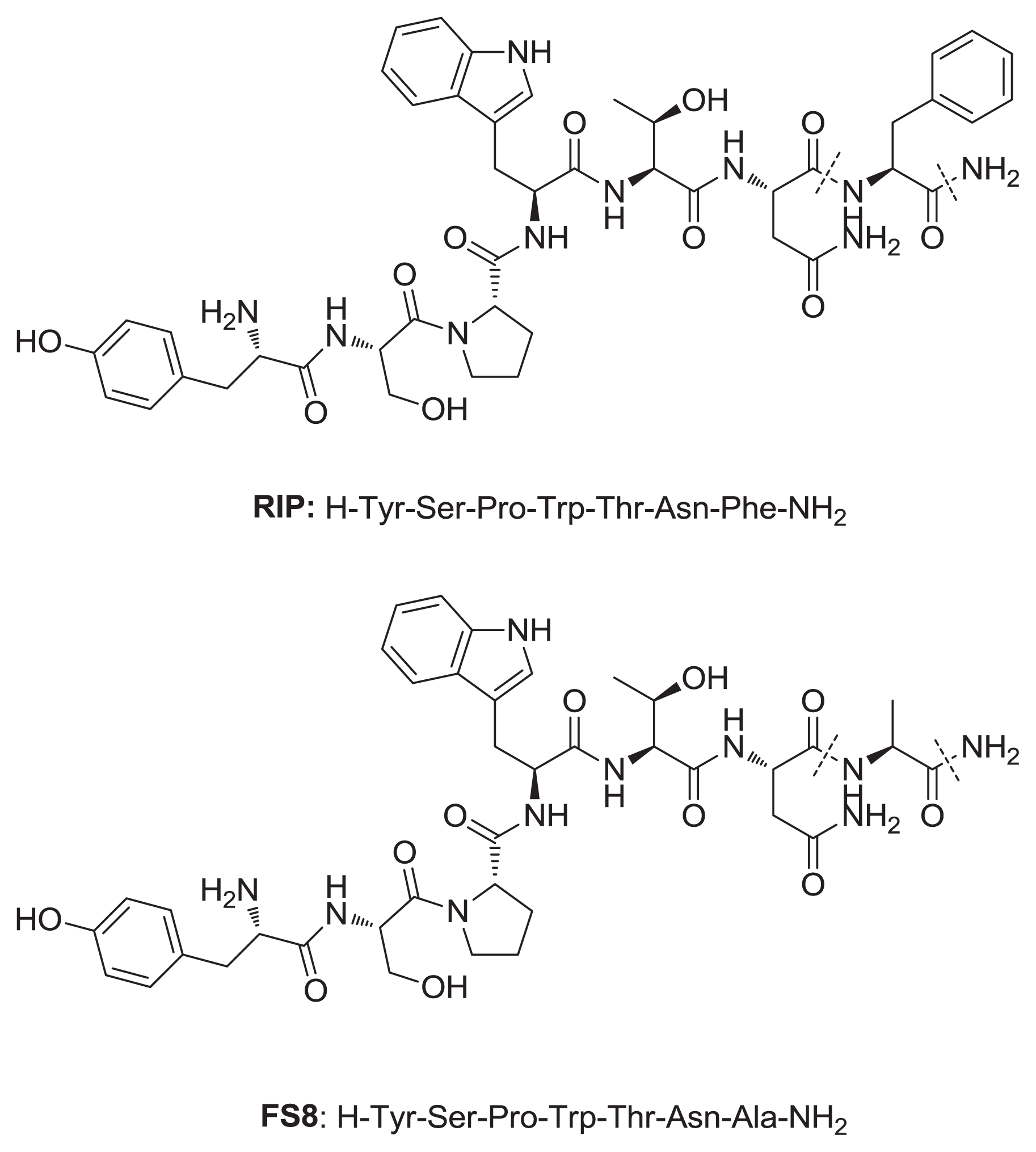
:1. Introduction
2. Results
2.1. In Vitro Studies
2.2. In Vivo Studies
2.3. Peptide Binding to Dacron Graft
3. Discussion
4. Materials and Methods
4.1. Organisms
4.2. Antimicrobial Agents
4.3. Adherent Biofilm Formation for Susceptibility Testing
4.4. Susceptibility Testing with Adherent Cells
4.5. Susceptibility Testing with Planktonic Bacteria
4.6. Peptide Binding to Dacron Graft
4.7. Assessment of the Infection
4.8. Statistical Analysis
5. Conclusions
Acknowledgements
Conflicts of Interest
References
- Reilly, J.; Stewart, S.; Allardice, G.A.; Noone, A.; Robertson, C.; Walker, A.; Coubrough, S. Results from the Scottish national HAI prevalence survey. J. Hosp. Infect 2008, 69, 62–68. [Google Scholar]
- Agarwal, A.; Singh, K.P.; Jain, A. Medical significance and management of staphylococcal biofilm. FEMS Immunol. Med. Microbiol 2010, 58, 147–160. [Google Scholar]
- Otto, M. Looking toward basic science for potential drug discovery targets against community-associated MRSA. Med. Res. Rev 2010, 30, 1–22. [Google Scholar]
- Costerton, J.W.; Stewart, P.S.; Greenberg, E.P. Bacterial biofilm: A common cause of persistent infections. Science 1999, 284, 1318–1322. [Google Scholar]
- Owens, C.D.; Stoessel, K. Surgical site infections: Epidemiology, microbiology and prevention. J. Hosp. Infect 2008, 70, 3–10. [Google Scholar]
- Kaila, V.C. Quorum sensing inhibitors: An overview. Biotechnol. Adv 2013, 31, 224–245. [Google Scholar]
- Gray, K.M. Intercellular communication and group behavior in bacteria. Trends Microbiol 1997, 5, 184–188. [Google Scholar]
- Langsrud, S.; Sidhu, M.S.; Heir, E.; Holck, A.L. Bacterial disinfectant resistance—A challenge for the food industry. Int. Biodeterior. Biodegrad 2003, 51, 283–290. [Google Scholar]
- Olson, M.E.; Ceri, H.; Morck, D.W.; Buret, A.G.; Read, R.R. Biofilm bacteria: Formation and comparative susceptibility to antibiotics. Can. J. Vet. Res 2002, 66, 86–92. [Google Scholar]
- Baldassarre, L.; Pinnen, F.; Cornacchia, C.; Fornasari, E.; Cellini, L.; Baffoni, M.; Cacciatore, I. Synthesis of short cationic antimicrobial peptidomimetics containing arginine analogues. J. Pept. Sci 2012, 18, 567–578. [Google Scholar]
- Cirioni, O.; Giacometti, A.; Ghiselli, R.; Kamysz, W.; Orlando, F.; Mocchegiani, F.; Silvestri, C.; Licci, A.; Chiodi, L.; Lukasiak, J.; et al. Citropin 1.1-treated central venous catheters improve the efficacy of hydrophobic antibiotics in the treatment of experimental staphylococcal catheter-related infection. Peptides 2006, 27, 1210–1216. [Google Scholar]
- Cirioni, O.; Giacometti, A.; Ghiselli, R.; Dell’Acqua, G.; Orlando, F.; Mocchegiani, F.; Silvestri, C.; Licci, A.; Saba, V.; Scalise, G.; et al. RNAIII inhibiting peptide significantly reduces bacterial load and enhances the effect of antibiotics in the treatment of central venous catheter-associated S. aureus infections. J. Infect. Dis 2006, 193, 180–186. [Google Scholar]
- Bergamini, T.M.; Corpus, R.A., Jr; McCurry, T.M.; Peyton, J.C.; Brittian, K.R.; Cheadle, W.G. Immunosuppression augments growth of graft-adherent Staphylococcus epidermidis. Arch. Surg. 1995, 130, 1345–1350. [Google Scholar]
- Giacometti, A.; Ghiselli, R.; Cirioni, O.; Mocchegiani, F.; Orlando, F.; del Prete, M.S.; D’Amato, G.; Silvestri, C.; Saba, V.; Scalise, G. Prophylactic efficacy of linezolid alone or combined with levofloxacin and vancomycin in a rat subcutaneous pouch model of graft infection caused by Staphylococcus epidermidis with intermediate resistance to glycopeptides. J. Antimicrob. Chemother 2003, 52, 724–726. [Google Scholar]
- Raad, I.; Alrahwan, A.; Rolston, K. Staphylococcus epidermidis: Emerging resistance and need for alternative agents. Clin. Infect. Dis 1998, 26, 1182–1187. [Google Scholar]
- Livermore, D.M. Has the era of untreatable infections arrived? J. Antimicrob. Chemother 2009, 64, i29–i36. [Google Scholar]
- Gales, A.C.; Jones, R.N. Antimicrobial activity and spectrum of the new glycylcycline, GAR-936 tested against 1203 recent clinical bacterial isolates. Diagn. Microbiol. Infect. Dis 2000, 36, 19–36. [Google Scholar]
- Peterson, L.R. A review of tigecycline—The first glycylcycline. Int. J. Antimicrob. Agents 2008, 32, S215–S222. [Google Scholar]
- Simonetti, O.; Cirioni, O.; Lucarini, G.; Orlando, F.; Ghiselli, R.; Silvestri, C.; Brescini, L.; Rocchi, M.; Provinciali, M.; Guerrieri, M.; et al. Tigecycline accelerates staphylococcal-infected burn wound healing through matrix metalloproteinase-9 modulation. J. Antimicrob. Chemother 2012, 67, 191–201. [Google Scholar]
- Gov, Y.; Bitler, A.; Dell’Acqua, G.; Torres, J.V.; Balaban, N. RNAIII inhibiting peptide (RIP), a global inhibitor of Staphylococcus aureus pathogenesis: Structure and function analysis. Peptides 2001, 22, 1609–1620. [Google Scholar]
- Davies, D.G.; Parsek, M.R.; Pearson, J.P.; Iglewski, B.H.; Costerton, J.W.; Greenberg, E.P. The involvement of cell-to-cell signals in the development of a bacterial biofilm. Science 1998, 280, 295–298. [Google Scholar]
- Kiran, M.D.; Akiyoshi, D.E.; Giacometti, A.; Cirioni, O.; Scalise, G.; Balaban, N. OpuC—An ABC transporter that is associated with Staphylococcus aureus pathogenesis. Int. J. Artif. Organs 2009, 32, 600–610. [Google Scholar]
- Kiran, M.D.; Akidesavan, N.V.; Cirioni, O.; Giacometti, A.; Silvestri, C.; Scalise, G.; Ghiselli, R.; Saba, V.; Orlando, F.; Shoham, M.; et al. Discovery of a quorum sensing inhibitor of drug resistant staphylococcal infections by structure-based virtual screening. Mol. Pharmacol 2008, 73, 1578–1586. [Google Scholar]
- Balaban, N.; Cirioni, O.; Giacometti, A.; Ghiselli, R.; Braunstein, J.B.; Silvestri, C.; Mocchegiani, F.; Saba, V.; Scalise, G. Treatment of Staphylococcus aureus biofilm infection by the quorum-sensing inhibitor RIP. Antimicrob. Agents Chemother 2007, 51, 2226–2229. [Google Scholar]
- Simonetti, O.; Cirioni, O.; Ghiselli, R.; Goteri, G.; Scalise, A.; Orlando, F.; Silvestri, C.; Riva, A.; Saba, V.; Madanahally, K.D.; et al. RIP enhances healing of-wounds infected with methicillin resistant Staphylococcus aureus. Antimicrob. Agents Chemother 2008, 52, 2205–2211. [Google Scholar]
- Giacometti, A.; Cirioni, G.; Ghiselli, R.; Dell’Acqua, G.; Orlando, F.; D’Amato, G.; Mocchegiani, F.; Silvestri, S.; del Prete, M.S.; Rocchi, M.; et al. RNAIII-inhibiting peptide improves efficacy of clinically used antibiotics in a murine model of staphylococcal sepsis. Peptides 2005, 26, 169–175. [Google Scholar]
- Jamieson, A.G.; Boutard, N.; Sabatino, D.; Lubell, W.D. Peptide scanning for studying structure-activity relationships in drug discovery. Chem. Biol. Drug Des 2013, 81, 148–165. [Google Scholar]
- Cirioni, O.; Mocchegiani, F.; Cacciatore, I.; Vecchiet, J.; Silvestri, C.; Baldassarre, L.; Ucciferri, C.; Orsetti, E.; Castelli, P.; Provinciali, M.; et al. Quorum sensing inhibitor FS3-coated vascular graft enhances daptomycin efficacy in a rat model of staphylococcal infection. Peptides 2013, 40, 77–81. [Google Scholar]
- Barie, P.S. Antibiotic-resistant gram-positive cocci: Implications for surgical practice. World J. Surg 1998, 22, 118–126. [Google Scholar]
- Henke, P.K.; Bergamini, T.M.; Rose, S.M.; Richardson, J.D. Current options in prosthetic vascular graft infection. Am. Surg 1998, 64, 39–45. [Google Scholar]
- Donlan, R.M.; Costerton, J.W. Biofilms: Survival mechanisms of clinically relevant-microorganisms. Clin. Microbiol. Rev 2002, 15, 167–193. [Google Scholar]
- Sardelic, F.; Ao, P.Y.; Taylor, D.A.; Fletcher, J.P. Prophylaxis against Staphylococcus epidermidis vascular graft infection with rifampicin-soaked, gelatin-sealed Dacron. Cardiovasc. Surg 1996, 4, 389–392. [Google Scholar]
- Gordon, R.J.; Lowy, F.D. Pathogenesis of methicillin-resistant Staphylococcus aureus infections. Clin. Infect. Dis 2008, 46, S350–S359. [Google Scholar]
- Novick, R.P. Autoinduction and signal transduction in the regulation of staphylococcal virulence. Mol. Microbiol 2003, 48, 1429–1449. [Google Scholar]
- Bronner, S.; Monteil, H.; Prévost, G. Regulation of virulence determinants in Staphylococcus aureus: Complexity and applications. FEMS Microbiol. Rev 2004, 28, 183–200. [Google Scholar]
- Cacciatore, I.; Fornasari, E.; Cornacchia, C.; Baldassarre, L.; di Stefano, A.; Sozio, P.; Cerasa, L.S.; Fulle, S.; di Filippo, E.S.; La Rovere, R.M.L.; et al. (R)-alpha-Lipoyl-Glycyl-L-Prolyl-L-Glutamyl dimethyl ester codrug as multifunctional agent with potential neuroprotective activities. Chem. Med. Chem. 2012, 7, 2021–2029. [Google Scholar]
- Christensen, G.D.; Simpson, W.A.; Younger, J.J.; Baddour, L.M.; Barrett, F.F.; Melton, D.M.; Beachey, E.H. Adherence of coagulase-negative staphylococci to plastic tissue culture plates: A quantitative model for the adherence of staphylococci to medical devices. J. Clin. Microbiol 1985, 22, 996–1006. [Google Scholar]
- Stepanovic, S.; Vukovic, D.; Dakic, I.; Savic, B.; Svabic-Vlahovic, M. A modified microtiter-plate test for quantification of staphylococcal biofilm formation. J. Microbiol. Methods 2000, 40, 175–179. [Google Scholar]
- Clinical and Laboratory Standards Institute (CLSI), Methods for Dilution Antimicrobial Susceptibility Tests for Bacteria That Grow Aerobically: Approved Standard; CLSI: Wayne, PA, USA, 2003.


| Biofilm | ||
|---|---|---|
| Agent | MIC (μg/mL) | MBC (μg/mL) |
| Tigecycline | 8.00 | 16.00 |
| Tigecycline * | 2.00 | 4.00 |
| Group a | Graft-bonded drug b | Intraperitoneal preoperative drug c | Quantitative graft culture (CFU/mL) d | p-value (comparison with untreated control group) | p-value (comparison with double-treated group) |
|---|---|---|---|---|---|
| Uncontaminated control | - | <10 | |||
| Untreated control | - | 7.5 × 10⁶ ± 2.1 × 10⁶ | |||
| Group 1 | - | Tigecycline | 4.4 × 10⁴ ± 1.2 × 10⁴ | <0.001 | <0.001 |
| Group 2 | FS8 | - | 6.8 × 10⁴ ± 2.0 × 10⁴ | <0.001 | <0.001 |
| Group 3 | FS8 | Tigecycline | 3.7 × 101 ± 0.7 × 101 | <0.001 | <0.001 |
| RP-HPLC retention time (min) | Purity (%) | Mass
| Binding to the grafts (μg/cm2) | |
|---|---|---|---|---|
| Calculated | Found | |||
| 14.43 | 97.85 | 836.393 | 836.388 | 0.67 ± 0.04 |
© 2013 by the authors; licensee MDPI, Basel, Switzerland This article is an open access article distributed under the terms and conditions of the Creative Commons Attribution license (http://creativecommons.org/licenses/by/3.0/).
Share and Cite
Simonetti, O.; Cirioni, O.; Mocchegiani, F.; Cacciatore, I.; Silvestri, C.; Baldassarre, L.; Orlando, F.; Castelli, P.; Provinciali, M.; Vivarelli, M.; et al. The Efficacy of the Quorum Sensing Inhibitor FS8 and Tigecycline in Preventing Prosthesis Biofilm in an Animal Model of Staphylococcal Infection. Int. J. Mol. Sci. 2013, 14, 16321-16332. https://doi.org/10.3390/ijms140816321
Simonetti O, Cirioni O, Mocchegiani F, Cacciatore I, Silvestri C, Baldassarre L, Orlando F, Castelli P, Provinciali M, Vivarelli M, et al. The Efficacy of the Quorum Sensing Inhibitor FS8 and Tigecycline in Preventing Prosthesis Biofilm in an Animal Model of Staphylococcal Infection. International Journal of Molecular Sciences. 2013; 14(8):16321-16332. https://doi.org/10.3390/ijms140816321
Chicago/Turabian StyleSimonetti, Oriana, Oscar Cirioni, Federico Mocchegiani, Ivana Cacciatore, Carmela Silvestri, Leonardo Baldassarre, Fiorenza Orlando, Pamela Castelli, Mauro Provinciali, Marco Vivarelli, and et al. 2013. "The Efficacy of the Quorum Sensing Inhibitor FS8 and Tigecycline in Preventing Prosthesis Biofilm in an Animal Model of Staphylococcal Infection" International Journal of Molecular Sciences 14, no. 8: 16321-16332. https://doi.org/10.3390/ijms140816321