Review

Phytochemicals: Target-Based Therapeutic Strategies for Diabetic Retinopathy

1
Department of Pharmacognosy, College of Pharmacy, Government College University Faisalabad, Faisalabad 3800, Pakistan
2
College of Pharmacy, Gachon University, Hambakmoero, Yeonsu-gu, Incheon 406-799, Korea
3
Gachon Institute of Pharmaceutical Science, Gachon University, Hambakmoe-ro, Yeonsu-gu, Incheon 406-799, Korea
*
Author to whom correspondence should be addressed.
Molecules 2018, 23(7), 1519; https://doi.org/10.3390/molecules23071519
Submission received: 7 June 2018 / Revised: 18 June 2018 / Accepted: 19 June 2018 / Published: 23 June 2018
(This article belongs to the Special Issue Medicinal Plants and Diabetes)

Abstract

:
Background: A variety of causative factors are involved in the initiation of diabetic retinopathy (DR). Current antidiabetic therapies are expensive and not easily accessible by the public. Furthermore, the use of multiple synthetic drugs leads to severe side effects, which worsen the diabetic patient’s condition. Medicinal plants and their derived phytochemicals are considered safe and effective treatment and their consumption can reduce the DR risk. In this article, we discuss a variety of medicinal plants, and their noteworthy bio-active constituents, that will be utilized as target based therapeutic strategies for DR. Methods: A broad-spectrum study was conducted using published English works in various electronic databases including Science Direct, PubMed, Scopus, and Google Scholar. Results: Targeting the multiple pathological factors including ROS, AGEs formation, hexosamine flux, PARP, PKC, and MAPK activation through variety of bioactive constituents in medicinal plants, diabetes progression can be delayed with improved loss of vision. Conclusions: Data reveals that traditional herbs and their prominent bioactive components control and normalize pathological cellular factors involved in DR progression. Therefore, studies should be carried out to explore the protective retinopathy effects of medicinal plants using experimental animal and humans models.

Graphical Abstract

1. Introduction

Diabetes mellitus (DM) occurs due to an abnormality in blood glucose metabolism in which production or activity of insulin is adversely affected. The global prevalence of diabetes is rising rapidly and has serious socio-economic implications as it primarily affects the working-age population. According to a World Health Organization (WHO) report, the number of diabetic patients will have doubled from 171 million in 2000 to 366 million in 2030. In addition, the global prevalence of diabetes among adults older than 18 years has risen from 4.7% to 8.5% in the period from 1980 to 2014 [1]. This diabetic epidemic is of concern in that it does not only detriment patients’ health, but also is an economic burden on society as well as the patient. Increased risk of development of macro- and microvascular diseases including diabetic nephropathy, retinopathy, neuropathy, and of conditions affecting the cardiovascular system, is considered as the foremost causes of mortality among diabetic patients. Among the various diabetic complications, diabetic retinopathy (DR) is not only considered a serious microvascular complication of diabetes, and the main cause of decreased vision and blindness worldwide, but also has an additional neurodegenerative aspect. With the increasing prevalence of diabetes, the number of people affected by DR is expected to rise to 191 million by 2030 [2].
In diabetic retinopathy development, the microvasculature of the retina is damaged and its penetrability increased, which potentially results in loss of vision. Experimental evidence indicates that thinned retinas isolated from animal models exhibit a reduced number of retinal ganglion cells. Increased numbers of apoptotic cells within those retinas result in more rapid apoptosis before the appearance of histopathological manifestations [3].
Diabetes stimulates several factors and metabolites within the retina of diabetic animals that cause damage to retinal cells and affect disease progression. Oxidative stress (OS), which interacts with various signaling pathways, is considered the main factor for the initiation of the complex mechanism of diabetes [4]. DR is a complex mechanism interlinked with a number of dysregulated metabolic functions including inflammation, oxidative stress, increased formation of advanced glycation end-products (AGE), activation of protein kinases C (PKC), and stimulation of the polyol and hexosamine pathways [5]. Dysregulation of various metabolites within the diabetic retina causes a change in the production pattern of various mediators including growth factors, cytokines, chemokines, neurotrophic factors, and adhesion molecules. This results in increased oxidative stress, which exacerbates the retinal cells, triggering diabetic retinopathy [6].
Natural products offer a wide variety of therapeutically active compounds, which are considered less toxic, safer, and cheaper than synthetic compounds. These naturally derived compounds including alkaloids, tannins, polyphenols, terpenoids, flavonoids, and steroids exhibit different pharmacological effects by simultaneously targeting multiple metabolic pathways [7,8,9]. In addition, these naturally derived chemicals also contribute to improved intercellular drug delivery, which is considered an important factor in attaining good therapeutic effects [10,11]. Using modern natural research techniques, many potentially active compounds have been discovered and utilized to ameliorate the DR. This review highlights recently-studied agents derived from natural products with respect to their role in the treatment of diabetes-related complications, and which may be a source of alternate therapies against retinal damage caused by diabetes.

2. Results

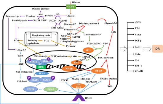
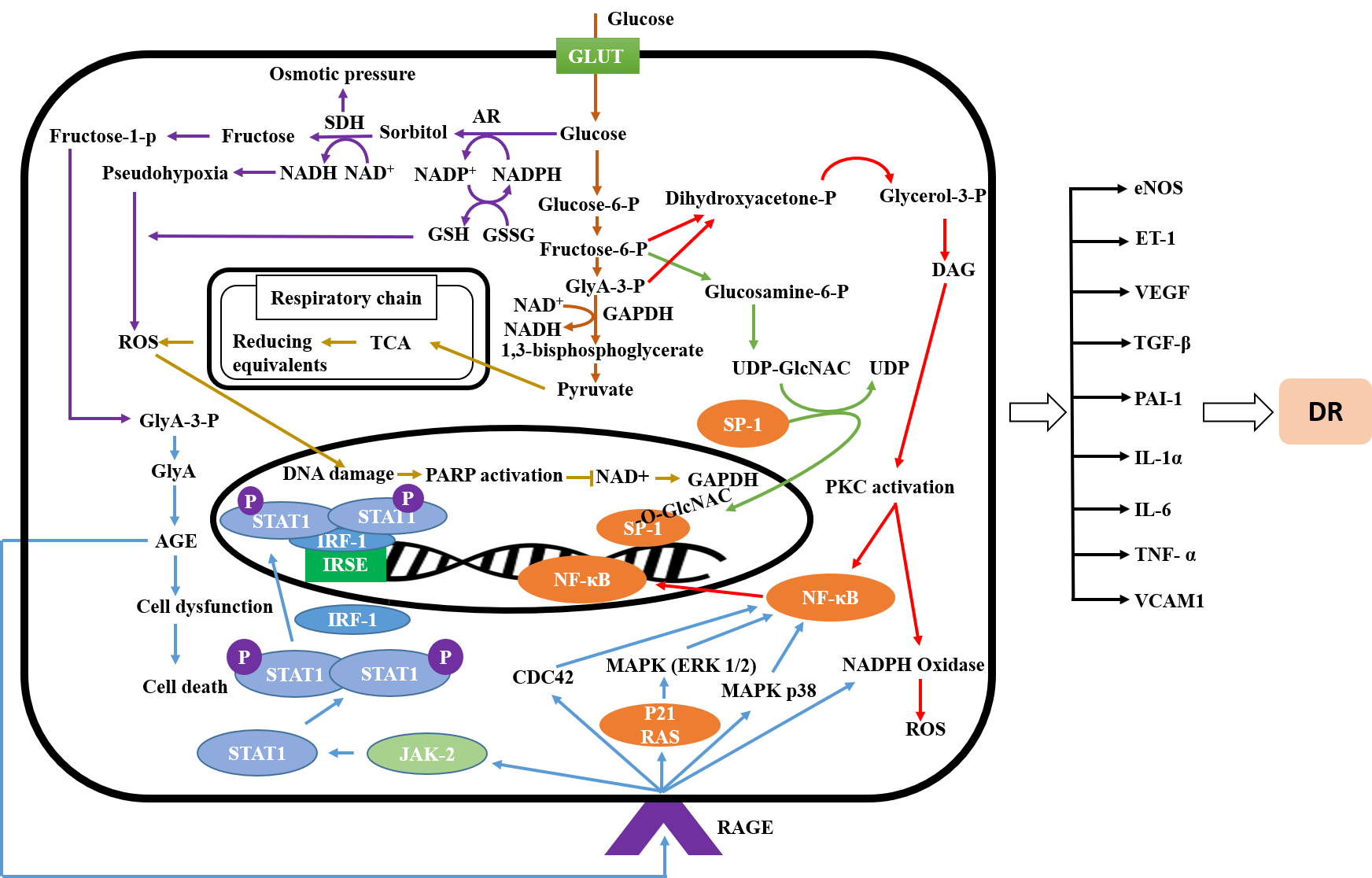
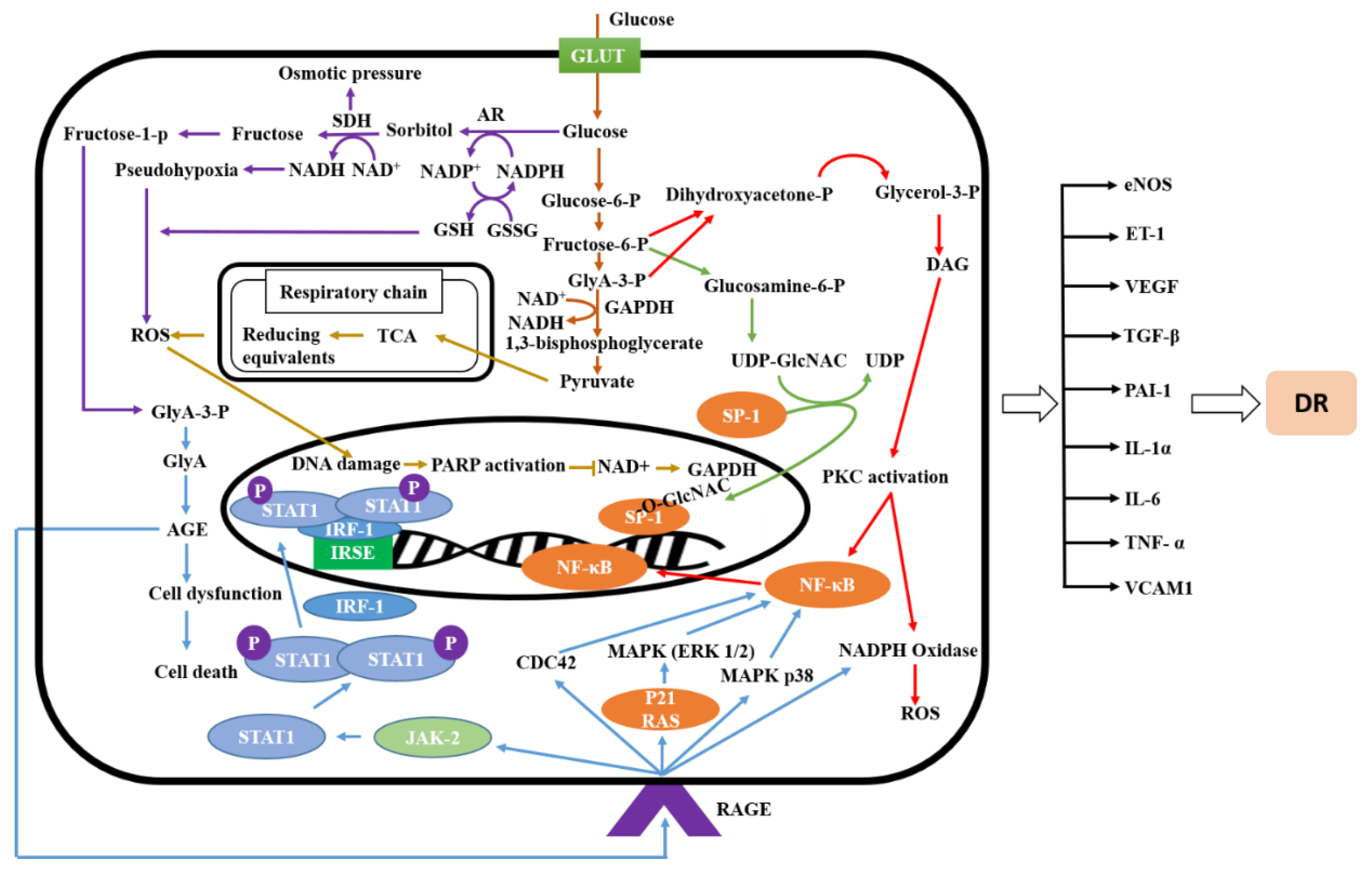
2.1. Signaling Pathway of Diabetic Retinopathy

In diabetic retinopathy, a multifactorial disease, retinal cells produce a variety of cytokines, growth factors, and enzymes, which are interlinked and are believed to initiate the cellular mechanisms underlying the condition [12]. The development of DR requires a variety of mechanisms including oxidative stress [6], PKC activation, the formation of AGEs, hexosamine flux, poly (ADP-ribose) polymerase (PARP) activation, and mitogen-activated protein kinase (MAPK) activation, which cause faster apoptosis of retinal cells in diabetes [5] and ultimately lead to the development of DR.
Various substances interact with each other to drive Janus kinase/signal transducers and activate transcription of the JAK/STAT pathway, which plays a significant role in retinal diseases. Receptor tyrosine kinase 2 (TYK2), JAK1, JAK2, and JAK3 belong to the JAK class of tyrosine kinases while STAT-1, -2, -3, -4, -5a, 5b, and 6b are members of the signal transducer and activator of transcription (STAT) family [13]. Interferon-gamma (INF-γ) triggers JAK1 and JAK2 phosphorylation through receptor binding, and activates STAT-1 through its tyrosine residue, followed by dimerization of STAT1 molecules and, together with interferon regulatory factor 1 (IRF-1), translocate into the nucleus and forms a complex. This complex activates interferon-stimulated response element (ISRE), which is followed by cytokines production [14]. Cytokines production can activate ROS production and vice versa, suggesting a bidirectional mechanism. Increased ROS results in retinal cell injury through interaction with different cellular components, and ultimately increases the incidence of DR [4].
The interaction between AGE and its receptors (AGE-RAGE) results in activation of various signaling pathways including those involving nicotinamide adenine dinucleotide phosphate (NADPH) oxidase and reactive oxygen species (ROS), MAP kinase, p38 MAPK, RAC, cell division control protein 42 (CDC42), and p21 RAS expression. This causes nuclear factor kappa b (NF-κB) activation followed by elevation of transcriptional protein, the inflammatory response including apoptosis, pro-inflammatory gene expression, and migration, ultimately aggravating DR [5,15].
Intracellular hyperglycemia elevates de novo synthesis of diacylglycerol (DAG), increased levels of which, in turn, activate several kinase isoforms from the PKC family, and control abnormalities in retinal and renal blood flow in diabetic mice. Activation of PKC induces vascular endothelial growth factor (VEGF) and transforming growth factor beta 1 (TGF-β1) expression [5], followed by accumulation of fibronectin and type IV collagen in glomeruli of diabetic rats. All of these changes ultimately contributed to changes in the microvascular protein matrix [16]. Furthermore, PKC is also involved in controlling these changes by inhibiting the nitric oxide production. In addition, PKC activation contributes to NF-κB activation, plasminogen activator-1 (PAI-1) overexpression, and to stimulation of NADPH dependent oxidases. Therefore, treating diabetic mice with PKC inhibitors can reduce PKC activity and ultimately control hyperglycemia, providing protection against abnormalities related to DR [5,17].
Several diabetic complications may result from the shunting of excess intracellular glucose into the hexosamine pathway. Elevated levels of substances involved in the hexosamine signaling pathway have been found in retinal tissues from rats and humans with diabetes. During glycolysis, fructose-6-phosphate triggers glucosamine-mediated activation of the PAI-1 promotor via serine protease 1 (SP1) sites in glomerular mesangial cells. Modification of SP1 through N-acetylglucosamine may explain the relationship between activation of the hexosamine pathway and hyperglycemia. Additionally, the hexosamine pathway regulates the PAI-1 gene promoter, as well as SP1 transcriptional gene activation via PKC-β1 and -γ and other transcription factors which play an important role in the pathogenesis of diabetic complications [5,18].
The polyol pathway, a key component in glucose metabolism, plays a pivotal role in the pathogenesis of cataract formation, refractive changes, and diabetic retinopathy in diabetic individuals. High expression of aldose reductase, a key rate-limiting enzyme in polyol pathway, reduces the conversion of glucose and galactose into sorbitol and galactitol. Excess sorbitol lowers available NADPH levels, which results in elevated oxidative stress, ultimately resulting in tissue retinal damage, aggravating DR [19,20]. Therefore, DR can be ameliorated through mediation and control of the polyol pathway.
PARP, a nuclear enzyme believed to be active in the retina of diabetic animals, causes DNA damage and exacerbates nitrosative and oxidative stress, potentially activating NF-κB and its dependent genes intercellular adhesion molecule (ICAM), monocyte chemoattractant protein-1 (MCP-1), and tumor necrosis factor alpha (TNF-α). Inhibition of glyceraldehyde-3-phosphate dehydrogenase activity by PARP activates PKC, the hexosamine pathway, and AGE formation, triggering ROS production, thereby playing an important role in the pathogenesis of DR [21]. Therefore, by blocking PARP activity, PKC activation, the hexosamine pathway, and AGE formation associated with DR can be blocked or attenuated.
In diabetes, metalloproteinase-9 (MMP-9) is activated. MMP-9 regulation is controlled by various transcriptional factors, including NF-κB and activator protein 1 (AP-1). PARP-1, a well-characterized member of PARP family, triggers and regulates the formation of complexes between these transcriptional factors with MMP-9. All these changes lead to increase the expression of MMP-9 causing mitochondrial damage and capillary cell apoptosis. Therefore, through inhibition of PARP-1 activity, retinopathy can be prevented in diabetic individuals [22].
ROS, PKC activation, AGEs production, hexosamine flux, PARP activation, and MAPK activation are interlinked to each other, and are the main causative agents in the pathogenesis of DR. Inhibition of the mechanisms described above can prevent the apoptosis of retinal cells in diabetes, and ultimately delay and ameliorate DR as shown in Figure 1.

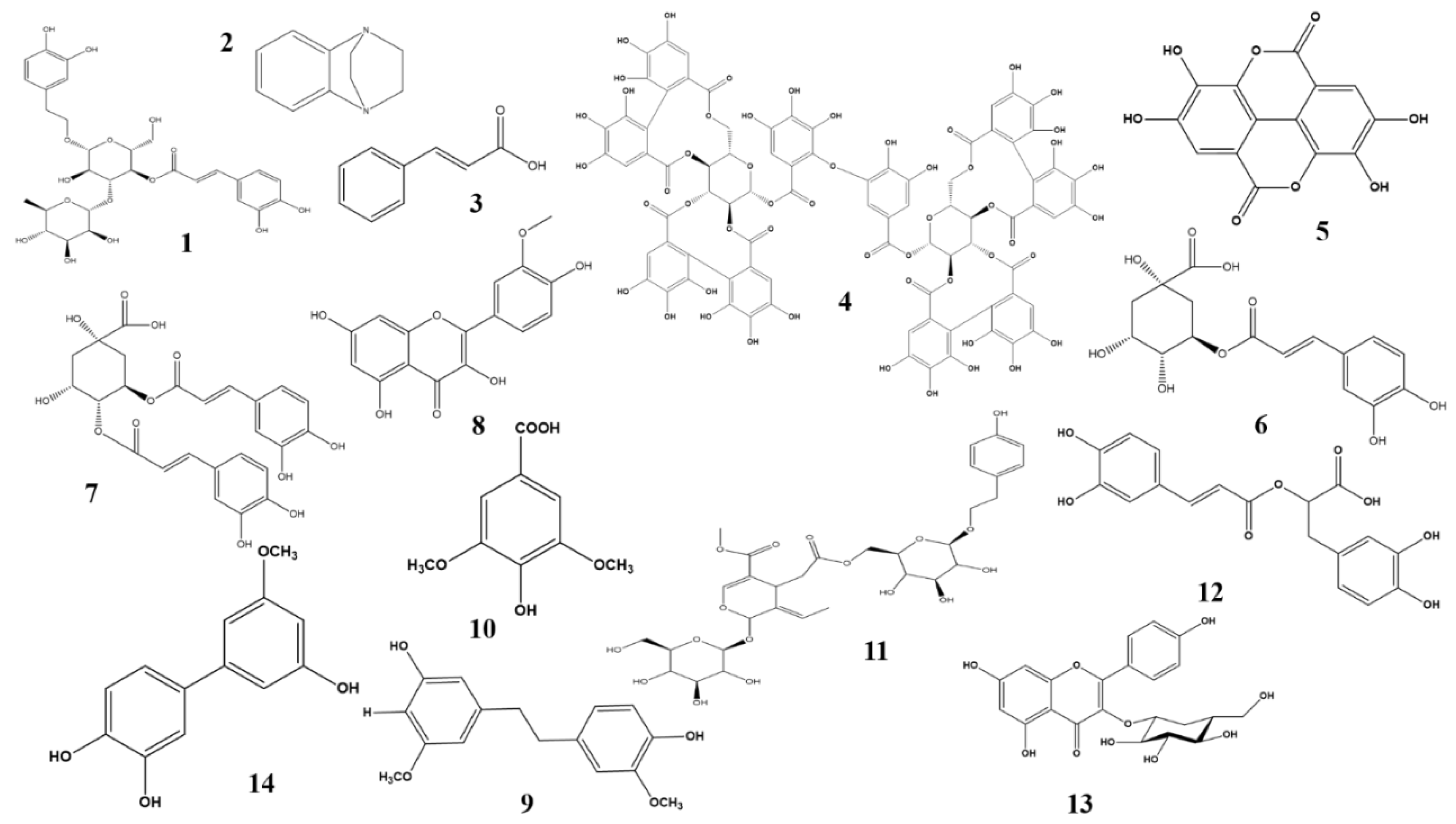
2.2. Anti-diabetic Effects of Phenolic and Flavonoid Phytochemicals

2.2.1. Abeliophyllum distichum

A. distichum, also called Korean abeliophyllum or white forsythia, is a monotypic genus of flowering plants. A. distichum Nakai is endemic to the southern and central regions of Korea. Major isolated compounds are quinol glycosides, acteoside, and halleridone. These have a potent inhibitory effect on aldose reductase. Acetoside (Figure 2), the most potent ingredient, has a five times greater efficacy against aldose reductase than quercetin [23].

2.2.2. Aegle marmelos

A. marmelos, commonly called Bael, is distributed across several regions of India and South East Asia. Evidence from in vitro, ex vivo, and in vivo experiments suggest that it has the potential to inhibit aldose reductase in rat lens, which is associated with lens opacification. Greater chaperone activity was reported upon treatment with α-crystalline, a water-soluble protein isolated from rat’s lens, with ethyl acetate extract. Phytochemical profiling showed the presence of three major compounds, namely benzo[b])-1,4-diazabicyclo[2.2.2]octane (Figure 2), cinnamic acid (Figure 2), and 3,4-dimethoxybenzoic anhydride. These were isolated from an ethyl acetate extract but, while these compounds may be responsible for ameliorating DR, activity assays involving these compounds have not yet been performed [24].

2.2.3. Agrimonia pilosa Ledeb

A. pilosa, is well known in traditional Chinese medicine. The butanol fraction of a methanol extract of this plant was found to be a rich source of flavonoids and contained bioactive compounds including agrimoniin (Figure 2), quercetin, afzelin, luteolin, and luteolin-7-glucoside. These exhibited significant inhibitory activity against aldose reductase [25]. A triterpenoid compound isolated from this plant helps in preadipocyte-differentiation via peroxisome proliferator-activated receptor gamma (PPAR-γ) activation. In addition, it can potentially activate the mRNA expression of adiponectin and glucose transporter type 4 (GLUT4) [26].

2.2.4. Aster koraiensis

A. koraiensis, a valuable Korean native plant, is employed in traditional medicine and as a food in Korea. Kim et al. demonstrated in 2016 that A. koraiensis extract attenuates retinal pericyte apoptosis by suppressing AGE formation and NF-κB activation. Chlorogenic acid (Figure 2) and 3,5-dicaffeoylquinic acid (Figure 2) are the two major bioactive constituents responsible for this activity in this plant [27]. Further studies have indicated that this plant can also prevent retinal vascular dysfunction, block blood-retinal barrier (BRB) breakage, and inhibit tight junction protein expression. This highlights the potential benefits of A. koraiensis as a dietary supplement for attenuating DR.

2.2.5. Camellia nitidissima Chi

C. nitidissima Chi, an edible plant, is used as a tea in China. Usually, it grows in a narrow region of Northern Vietnam and Southern China. This plant is a good source of flavonoids and glycosides and exhibits beneficial properties including anti-AGE formation activity and methylglyoxal scavenging. This suggests that it can reduce diabetic complications, including DR, via anti-AGE formation and anti-oxidant mechanisms [28]. However, additional studies need to be carried out to characterize its bioactive phytochemicals and to elucidate their anti-AGE formation mechanism and preventive role in DR.

2.2.6. Carpobrotus edulis

C. edulis is widely distributed in the Eastern Cape of South Africa. This plant contains ferulic acid and ellagic acid (Figure 2), which exhibit the attenuative properties against DR via inhibition of AGE formation, and of OS [29]. Recently, a study conducted to note the potency of ellagic acid revealed that it is involved in the prevention of retinal abnormalities through inhibition of AGE formation. A detailed mechanism indicates that ellagic acid inhibits the expression of GAP, VEGF, Bax, and hypoxia-inducible factor 1-alpha (HIF-1α), and apoptosis in the retina [30].

2.2.7. Cochlospermum religiosum

C. religiosum, a small or medium-sized deciduous tree, is commonly known as “Golden silk cotton”. Isorhamnetin (Figure 2), a flavonoid glycoside, present in this plant exhibited an anti-oxidant effect by suppressing lipoprotein diene formation induced by Cu2+, and provided a protective effect against cataracts induced by selenite in an in vitro cell culture model. The anti-oxidant effect of this compound makes this plant valuable in the treatment of DR [31,32]. In vitro and in vivo studies have revealed that myricetin, a bioactive flavonoid, is active against DR through inhibition of RAGE-Src-ERK1/2-FAK-1 paxillin signaling pathway, followed by prevention of retinal pericyte migration [28].

2.2.8. Dendrobium chrysotoxum L.

In 2014, Wang et al. described the potency of an ethanolic extract of D. chrysotoxum against DR and its mechanism. The experimental results indicated that the extract decreases elevated retinal vessels in diabetic rats [33]. In addition, it lowered the increased expression of VEGF and VEGFR2 mRNA, as well as serum VEGF levels. In treated diabetic rats, the levels of retinal MMP2/9, serum MMP 2/9, platelet-derived growth factor (PDGF)A/B, basic fibroblast growth factor, insulin-like growth factor, IL-1β, IL-6, p65 phosphorylation, and ICAM-1 were suppressed. These findings demonstrate the potency of D. chrysotoxum in reversing DR [34]. Further experiments on the potency of D. chrysotoxum showed that it attenuates the breakdown of the blood-retinal barrier. It also reverses the decreased expression of retinal mRNA for the junction proteins occludin and claudin-1 in diabetic rats. Furthermore, it reduces ICAM-1 expression, TNFα, IL-6, IL-12, IL-3, IL-2, IL-10, IκB, and IκB kinases. Together, this reveals that this plant reduces DR by preventing retinal inflammation and reducing the amount of tight junction protein in diabetic rats. It can, therefore, be recommended as a supplementary treatment for diabetic patients [35]. Further studies on its bioactive constituents demonstrated that gigantol (Figure 2), an important bioactive constituent, has a synergistic effect with syringic acid (Figure 2), and plays an important role in preventing diabetic cataracts by inhibiting aldose reductase activity through downregulation of its expression via reduced transcription, and by controlling sorbitol levels. The synergistic effect is thus mainly due to disruption of AR activity and the polyol pathway [36].

2.2.9. Ginkgo biloba

G. biloba is commonly known as maiden hair. A combined extract of red berry, white willow bark, and G. biloba was evaluated against DR, with the experimental results indicating lowered TNF-α and VEGF expression, thereby attenuating elevated plasma lipid peroxidation and retinal inflammation in diabetic rats [37]. Extracts of this plant have been found to notably improve retinal capillary blood flow rate in patients with diabetic retinopathy [38]. In addition, clinical trials on the effectiveness of G. biloba extract (GBE) in patients with DR showed that GBE prevents and suppresses microvascular alterations in the retinas of diabetic patients, and ultimately improves DR [39]. Recently, a study demonstrated that GBE significantly decreases the number of retinal microaneurysms and areas of retinal hemorrhage, and also decreases in total cholesterol and low-density lipoprotein. In addition, decreases in the rate of platelet aggregation and adhesion in diabetic patients upon treatment with GBE proves its efficacy against DR [40]. Several clinical trials have now demonstrated that GBE may be used as a first-line therapy against DR.

2.2.10. Glycyrrhiza uralensi

G. uralensi is used as a natural sweetener. Many studies have suggested a potential role of G. uralensi in improving glucose tolerance through the PPAR-γ pathway, due to the presence of bioactive compounds such as glycycoumarin, glycyrin, and dihydroglyasperin C and D. Additionally, semilicoisoflavone B, liquiritigenin, and isoliquiritigenin have, through their effects on DR and sorbitol formation, indicated the plant’s potential therapeutic importance against diabetes and diabetes-related complications [41].

2.2.11. Juglans regia L.

J. regia L. has hypoglycemic effects of J. regia L. that indicates the value of this plant in significantly lowering the blood glucose during fasting and HbA1c. Moreover, leaf extracts from this plant attenuate lipid peroxidation, anti-oxidant status, S100B, PARP, cyclooxygenase 2 (COX-2), and caspase-3 expression in the retina of diabetic rats [42]. J. regia L. is a rich source of flavonoids and phenolic compounds that might be related to its potency against DR.

2.2.12. Litchi chinensis

L. chinensis is commonly known as lychee. Its natural range includes Vietnam, China, and the Malay Peninsula. The plant is a rich source of anthocyanins, phenolics, and flavonoids which demonstrate diverse pharmacological activities including anti-cancer, anti-oxidant, anti-coagulant, immunomodulatory, and liver-protective effects. Extracts of the fruit pericarp, which are under consideration as additives in the food and pharmaceutical industries, have inhibitory effects against glycoxidation and AGE formation in serum and lens homogenate. L. chinenesis also inhibits changes in fundus structure and protects retinal cells in streptozotocin (STZ)-induced diabetic rats. These findings demonstrate that L. chinenesis delays the progression and development of DR by inhibiting oxidative stress, glycoxidation and AGE formation due to its high polyphenol and tannin content [43].

2.2.13. Ligustrum lucidum Ait

L. lucidum is a well-known traditional Chinese medicine possessing a wide range of pharmacological functions. Specnuezhenide (Figure 2), a constituent of this plant, is valued for its effectiveness against DR through suppression of the HIF-1α/VEGF signaling pathway. Specnuezhenide suppresses VEGF expression, downregulating mRNA expression of VEGFA, prolyl-hydroxylase 2, and HIF-1α. In addition, it prevents hypoxia-induced retinal neovascularization in a rat model and prevents DR. This indicates that specnuezhenide can potentially be used to treat DR [44].

2.2.14. Lonicerae japonicae Flos

L. japonicae Flos decreases retinal angiogenesis in diabetic mice and reduces VEGF-induced tube formation owing to the presence of chlorogenic acid, caffeic acid, and luteolin [45]. Chlorogenic acid is considered to be the main compound involved in suppression of retinal angiogenesis and works by decreasing VEGF expression, which is followed by a decreased breakdown of BRB, and of vascular leakage. This provides a protective effect against DR production in diabetic rats [46]. Further studies have indicated that it lowered VEGF expression and VEGF-mediated retinal neoangiogenesis in STZ-induced hyperglycemic mice. Collectively, these results highlight the potency of chlorogenic acid contained in L. japonicae against DR [47].

2.2.15. Melissa officinalis

M. officinalis anti-oxidant property has been studied extensively, and found to be principally responsible for lowering hyperglycemia and AGE formation. M. officinalis extracts have the ability to elude the cross-β structure pathway during glycation. Research involving alloxan-induced diabetic rats revealed that M. officinalis significantly suppresses diabetes by modulating blood sugar, lipoprotein, and serum lipid levels. In addition, it has a beneficial effect on high-density lipoprotein (HDL) levels [48]. The plant, therefore, has potential therapeutic uses in the treatment and prevention of diabetes and related disorders [49]. Rosmarinic acid (Figure 2), a major phenolic bioactive compound isolated from this plant, has a potential role in suppressing DR. It inhibits the proliferation of retinal endothelial cells and inhibits angiogenic tube formation. According to a study using a mouse model, rosmarinic acid’s suppression of retinal neovascularization is linked to cell cycle arrest followed by an increase in p21 (WAF1) expression. Interestingly, rosmarinic acid does not exhibit any retinal toxicity [50]. It is clear that M. officinalis-derived rosmarinic acid acts as a potent inhibitor of retinal neovascularization. However, the details of the DR-linked signaling mechanism still need to be explored.

2.2.16. Moringa oleifera Lam

M. oleifera Lam is mainly consumed in Pakistan, India, Hawaii, and across Africa. Polyphenolic compounds from M. oleifera are responsible for lowering blood glucose by stimulating glucose uptake. In the diabetic retina, an increased expression of TNF-α, IL-1β, VEGF, PKC-β was observed. On treating the diabetic retinae with M. olefera, a marked inhibition in these expressions was noted. In addition, M. oleifera-treated retinae exhibited intact retinal vasculature and reduced thickening of the capillary basement membrane. This was evidence of M. oleifera’s efficacy against diabetic retinopathy via anti-oxidant, anti-inflammatory, and anti-angiogenic mechanisms [51]. Astragalin (Figure 2), a major bioactive compound present in this plant, has been evaluated as an anti-diabetic, and was found to both lower VEGF expression and to inhibit the negative effects of high glucose levels, effectively preventing DR [52].

2.2.17. Morus alba

M. alba is commonly known as white mulberry and is broadly cultivated in subtropical, tropical, and moderate regions. Owing to the presence of flavonoids, coumarins, and phenols, the leaves of this plant possess a variety of anti-hypotensive, anti-hyperglycemic, and neuroprotective properties. Recently, it has been demonstrated that oral administration of M. albla potentially reduces glucose levels and suppresses the production of sorbitol, PKC, OS, and proinflammatory cytokines in retinas of diabetic rats. Moreover, it also downregulates caspase-3 and Bax and upregulates B-cell lymphoma 2 (Bcl-2). In addition, it also inhibits the VEGF expression in the retina of diabetic rats. Collectively, these findings suggest that M. albla exerts its protective effect against DR by inhibiting hyperglycemia-induced apoptosis, inflammation, polyol pathway activation, oxidative stress, and VEGF expression [53].

2.2.18. Osteomeles schwerinae C.K. Schneid

O. schwerinae C.K. Schneid ethanolic extract contains significant quantities of quercitrin and hyperoside, which exhibit inhibitory properties against rat aldose reductase. An additional potentially-bioactive constituent is 5′-methoxybiphenyl-3,4,3′-triol (Figure 2), which also exhibited inhibitory activity against aldose reductase. Further study revealed that it interrupts AGE formation, reduces VEGF expression, causes breakdown of the blood-retinal barrier, and stabilizes the junction protein occludin. It also downregulates the various angiogenic factors including fibroblast growth factor (FGF-2), insulin-like growth factor-binding proteins (IGFBPs), and PAI-1. All these unveils the potency of 5′-methoxybiphenyl-3,4,3′-triol against DR pathogenesis via an anti-VEGF mechanism and the hexosamine pathway [33]. Further study, exploring the potency of this compound against DR via different signaling pathways, is warranted. In addition, in order to improve sample quality, standardization of O. schwerinae extractions using 5′-methoxybiphenyl-3,4,3′-triol as a marker bioactive compound should be undertaken.

2.2.19. Perilla frutescens

P. frutescens, commonly known as perilla or Korean perilla. It is an annual short-day plant. Rosmarinic acid, a potent bioactive compound present in ethyl acetate fractions of methanol extracts of P. frutescens, exhibited aldose reductase inhibitory activity. [54]. In a prior study, it is indicated that rosmarinic acid suppresses the proliferation of retinal endothelial cells and prevents retinal neovascularization through cell cycle arrest by increasing expression of p21WAF1. [50].

2.2.20. Platycodon grandiflorum

The potency of P. grandiflorum against diabetes is due to the presence of luteolin (Figure 3), luteolin glucoside, luteolin acetyl glucoside, and chlorogenic acid methyl ester, which, isolated from this plant, proved to be potent inhibitors of AGE formation. Luteolin glucoside exhibited a potent inhibitory effect against rat lens aldose reductase (RLAR) activity [55]. Recently, the clinical effect of a herbal decoction containing different medicinal plants, including P. grandiflorum, was studied in patients with stage II DR, with the finding that it effectively prevented and treated DR. Clinical reports has proven that supplementation of DR patients with P. grandiflorum could be an effective therapy for DR [56].

2.2.21. Polygonatum odoratum

P. odoratum contain three homoisolflavones, namely 3-(4′-hydroxybenzyl)-5,7-dihydroxy-6-methyl-8-methoxychroman-4-one, 3-(4′-methoxybenzyl)-5,7-dihydroxy-6-methyl-8-methoxychroman-4-one, and 3-(4′-hydroxybenzyl)-5,7-dihydroxy-6,8-dimethylchroman-4-one, are isolated from the chloroform fraction of the ethanol extract of this plant’s rhizome. These compounds exhibit inhibitory activity against AGE formation in both in-vitro and in-vivo studies [57].

2.2.22. Polygonum cuspidatum

P. cuspidatum is used in folk medicines from China, Japan, and Korea, as it possesses a variety of biological activities against allergies, diabetes, and as an anti-viral agent. Kim et al. (2012) demonstrated that P. cuspidatum can ameliorate HMGB1, RAGE, and NF-κB activities in the retina. In addition, it decreases retinal vascular permeability and the loosening of tight junctions in diabetic rats [35]. This plant can, therefore, be recommended as an effective supplement for the treatment of DR. Additional research into the effectiveness of P. cuspidatum indicated that it controls RAGE-mediated activation of NF-κB, followed by inhibition of upregulation of HMGB1 and prevention of diabetes-induced retinal vascular hyperpermeability [58]. P. cuspidatum is known to contain four major active compounds: polydatin, resveratrol (Figure 3), emodin glucopyranoside and emodin. It has been shown that resveratrol provides protection against ADR via inhibition of the AKT/NF-κB signaling pathway as well as through suppression of high morbidity group box 1 (HMBG1), RAGE, TNF-α, and IL-4 in skin inflammation induced by atopic dermatitis. Emodin also suppresses HMGB1, NF-κB, and gene expression of cell surface adhesion proteins. Emodin glucopyranoside, on the other hand, has the capacity to function as an antioxidant [58]. Recently, it has been demonstrated that polydatin provides it hepatoprotective effect through the peroxisome proliferator-activated receptor alpha/beta (PPAR-α/-β) signaling pathway in STZ induced diabetic mice [59]. Collectively, these bioactive compounds allow P. cuspidatum to exert its effects against DR.

2.2.23. Polygonum multiflorum

P. multiflorum is still popular in both China and Japan, but is now also popular in North America. Tetrahydroxystilbine glucoside, a major bioactive compound in this plant, inhibits AGE formation by trapping reactive methylglyoxal under physiological conditions in a dose-dependent manner [60] (Lv, Shao et al. 2010). These results indicate that it has the capability to delay DR.

2.2.24. Prunella vulgaris

P. vulgaris, a self-healing herb, is broadly distributed across Asia, Europe, North America, and Northwest Africa. It contains several bioactive compounds, including oleanolic acid, ursolic acid, botulinic acid, rosmarinic acid, caffeic acid ethyl ester, 2-hydroxy cinnamic acid, and prunellin. Caffeic acid ethyl ester, p-hydroxy cinnamic acid, and prunellin are the main active compounds responsible for inhibition of RLAR activity. Furthermore, caffeic acid ethyl ester ameliorates diabetic retinopathy via an anti-oxidant, anti-AGE formation mechanism, and has been shown to possess stronger activity than the commonly-prescribed drug aminoguanidine. These findings suggest that Prunella vulgaris is a potential source of active components capable of ameliorating DR [61]. However, as is the case with many other medicinal plants, additional studies using animal models are needed to unveil the mechanisms underlying its anti-AGE formation activities.

2.2.25. Pueraria lobata

P. lobate contains bioactive isoflavonoid puerarin (Figure 3), which is isolated from its dried root. Puerarin prevents retinal and neuronal cells from undergoing apoptosis via an anti-oxidant mechanism, and possesses a number of additional activities. It reduces the mRNA expression of inducible nitric oxide synthase (iNOS), increases SOD activity, and inhibits radical formation. It reduces oxidative stress by downregulating RAGE expression and prevents assembly of the receptor complex. It also suppresses NF-κB and NADPH oxidase activation via suppression of p47phox and Rac1 signaling and prevents apoptosis in retinal pericytes. Additionally, it exhibits anti-angiogenic properties by suppressing the expression of HIF-1α and VEGF mRNA. Finally, it exhibits its anti-inflammatory effect by attenuating IL-1β, ICAM, VCAM-1, cell apoptosis, Bax, and Caspase-3 while simultaneously upregulating Bcl-2 expression in mitochondria, and blocking BRB breakdown, ultimately preventing DR. Genistein, a bioactive flavonoid, attenuates diabetic retinopathy by inhibiting tyrosine kinase, proinflammatory cytokine production, TNF-α activation, and microglial activation. It prevents leukocytes/endothelial interaction and vascular dysfunction and inhibits extracellular-signal-regulated kinase (ERK) and p38 MAPK phosphorylation, ultimately preventing retinal edema. An isoflavone also present in this plant, daidzein, exerts its anti-inflammatory effect by acting on the PPAR receptor, providing protection against diabetic retinopathy [62]. Puerarin, P. lobata’s principal isoflavone glycoside, has been isolated from the plant and, upon evaluation, has been found to modulate VEGF expression by suppressing hypoxia-inducible factor-1 mRNA, ultimately exerting a protective effect against DR in experimental animals [63]. Additionally, it has been found to inhibit AGE formation. NADPH oxidase consists of membrane-integrated cytochrome b 558, which comprises several regulatory subunits that are required for the activation of AGE-BSA-induced NADPH oxidase and ROS production. Puerarin blocks the activation of AGE-BSA-induced phosphorylation of p47phox and Rac1, thus achieving the inhibitory effect. AGE-BSA also stimulates the translocation of NF-κB, also causing hyperglycemia. Therefore, treatment with puerarin inhibits the AGE-BSA-induced NF-κB activity, and ultimately provides protection from diabetes and diabetes-related disorders [64]. These factors combined indicate that P. lobata is a potential source of flavonoid which highly-effective against DR. Animal trials indicate that this plant may form the basis for promising, effective medicines for use in first-line DR treatment therapies.

2.2.26. Salvia miltiorrhiza Bge

S. miltiorrhiza Bge contains several active compounds which exhibit anti-diabetic effects, including isosalvianolic acid C methyl ester, tanshinone IIA, rosmarinic acid, salvianolic acid, and lithospermic acid dimethyl ester are present in this plant. Injection of S. miltiorrhiza extracts into the retinal hypoxic-ischemic tissues of DR mice promotes blood oxygen transport and retinal hemangioma absorption, while also controlling blood sugar levels and improving microcirculation. S. miltiorrhiza may prove an effective treatment for DR by way of the blood-ocular barrier, as it protects cells from damage and contributes to visual improvements [65]. Controlled clinical studies have been conducted to evaluate the effectiveness and safety of S. miltiorrhiza and have proven its effectiveness intreating DR [66,67]. Further studies indicate that salvianolic acid decreases the morbidity rate of cataract and improves the retinal pathological changes by preventing the chronic inflammation, inhibiting lipid peroxidation, and reducing the lipoprotein-related phospholipase A2 (Lp-PLA2) through down-regulating the IL-6, IL-1, oxidized low-density lipoprotein, and (Lp-PLA2) level [68].

2.2.27. Stauntonia hexaphylla

S. hexaphylla (Thunb) Decne is broadly distributed in the lowlands and foothills of mostly warmer areas of Korea, Japan, and China, where it occurs as dense groups of bushes. The known chemical constituents of S. hexaphylla include glucosides, flavonoids, triterpenoids, phenolic glucosides, phenylpropanoids and chlorogenic acid analogs. Chlorogenic acid, quercetin-3-O-𝛽-d-glucopyranoside, luteolin-7-O-𝛽-d glucopyranoside, and calceolarioside B exhibit activity against RLAR. Additionally, luteolin-7-O-𝛽-d glucopyranoside, 3-(3-(3,4-dihydroxyphenyl)-propionic acid, and neochlorogenic acid showed anti-AGE formation activity. These results suggest that S. hexaphylla extracts have potential application in the treatment of cataracts caused by hyperglycemia and DR [69].

2.2.28. Tephrosia purpurea

T. purpurea is widely cultivated for its anti-diabetic properties, which are contained within a flavonoid-rich alcoholic fraction of extractions. The mechanism underlying this indicates that it is involved in increasing the level of soluble proteins that prevent the cross-linking, aggregation and distribution of the soluble protein. It also possesses antioxidant properties that prevent insolubilization of proteins, delaying development of lens opacity. In addition to this, results from docking experiments have suggested that most of the known constituents including rutin, quercetin, lupeol, fidarestat, tephrosin degurelin rotenone, and elliptone have a common binding mode in the vicinity of the active site of subunit A of aldose reductase, lying between the catalytic amino acid residues, Trp20, Tyr48, Trp111, Phe122, and His110. T. purpurea possesses significant anti-hyperglycemic activity, as well as antioxidant activity in diabetic rats and has shown significantly in vitro AR inhibiting activity. Together with the ability to reduce oxidative stress and inhibition of AR, T. purpurea might be beneficial not only in preventing hyperglycemia but also in delaying the onset of diabetes-induced complications resulting from hyperglycemia-induced oxidative and osmotic stress [70].

2.2.29. Terminalia catappa

T. catappa grows in the warmer regions of India. T. catappa is a rich source of tannin, which is linked to its anti-diabetic effect. The bark extract of T. catappa induced improvement in body weight and lipid profile along with regeneration of pancreatic β-cells in STZ-induced diabetic rats. Research into the effectiveness of T. catappa has indicated that it is rich in phytochemicals which have the capacity to reverse a number of pathologies induced by hyperglycemia. These include suppression of mitochondrial dysfunction, inhibition of superoxide generation, downregulation of inflammatory and angiogenic factors, restoration of balance between low-density lipoprotein (LDL) and high-density lipoprotein (HDL) cholesterol, and suppression of enzymatic-activity-related glucose absorption. T. catappa is thus valuable as an effective natural therapy to inhibit the progression of DR and has potential application in first-line therapies [71].

2.2.30. Vitex negundo

V. negundo, commonly known as the five-leafed chaste tree, is famous for its diverse pharmacological properties. It is widely used in traditional medicine in South Asia. These are attributable to V. negundo’s rich variety of flavonoids and flavonoid glycosides, which include luteolin-7-glucoside (Figure 3), a compound that exhibits anti-oxidant activity and prevents selenite toxicity and cataractogenesis [31,72]. However, further study is required in order to fully elucidate its efficacy against DR.

2.2.31. Zea mays L.

Z. mays L. (purple waxy corn) is easy to find anywhere in the world. Z. mays is an important source of anthocyanins, and of phenolic acids including p-coumaric acid (Figure 3), vanillic acid, protocatechuic acid, and flavonoids like quercetin and hesperetin. Traditionally, Native Americans have used it to treat a wide range of ailments. It makes an effective poultice, which can be used to treat bruises, swellings, sores, boils, and similar conditions. Chickasaw Indians treated itching skin by burning old corncobs and holding the affected part over the smoke. Recently, a study conducted to explore the combined anti-cataract and anti-retinopathy activities of Z. mays and ginger revealed suppression of both aldose reductase’s inhibitory activity, and of OS. These changes effectively delayed cataractogenesis in diabetes, and increased the number of neurons in the ganglionic cell layer was present. Additionally, the thickness of total retina and of the retinal nuclear layer were found to be increased in diabetic rats. These studies have proven the capability of this two-functional food against DR [73].

2.3. Anti-diabetic Effects of Terpenoid and Steroid Phytochemicals

2.3.1. Alpinia zerumbet

A. zerumbet is widely distributed in subtropical and tropical regions. The secondary metabolites found in its roots inhibit AGE formation. Labdadiene (Figure 3), a triterpenoid bioactive compound, inhibits the formation of α-dicarbonyl compounds and fructosamine, both of which are important causative factors in AGE formation, and thereby reduces the occurrence of diabetic retinopathy [74]. While the potency of labdadiene against DR is clear, there remains a need to conduct further trials using in vitro and in vivo animal models.

2.3.2. Andrographis paniculata Nees

A. paniculata nees is widely distributed and cultivated in Southern Asia, China, and Europe. Andrographolide (Figure 3), a major bioactive compound isolated from this plant possesses anti-inflammatory activity, which makes it useful for treating inflammatory bowel disease, pulmonary fibrosis, and cigarette smoke-induced lung injury. It also possesses anti-angiogenic effects and inhibits DR. In STZ-induced diabetic mice, it reduced the retinal vessel numbers and decreased elevated VEGF levels via binding KDR or FLT1 receptors. It also lowered DR via an anti-inflammatory mechanism, as retinal inflammation is critically involved in the progression of DR. It has been shown to inhibit the expression of inflammatory cytokines such as TNF-α, IL-1β, and IL-6; reduce leukostasis; and to decrease BRB breakage during DR development. In addition, it inhibits the activation of an NF-κB signaling pathway by decreasing the increased nuclear translocation and phosphorylation of NF-κB, p65, kappa-B, and kappa-B kinase. Furthermore, it inhibits the nuclear translocation of Egr1 along with related downstream genes such as serpentine 1, PAI-1, and a transmembrane glycoprotein, which is involved in the progression of DR. Ultimately, andrographolide is a promising compound for the treatment of DR, which functions by inhibiting activated VEGF, NF-κB, and early growth response 1 (EGR1) signaling pathways [75]. However, its toxicity is not well understood, and clinical trials in experimental animal models are required.

2.3.3. Astragalus membranaceous

A. membranaceous possesses astragaloside IV, a novel saponin, is the major active compound isolated from this plant. It ameliorates DR via inhibition of AGE formation, hepatic glycogen phosphorylase, and glucose-6 phosphate activities, oxidative stress, and activation of NF-κB in STZ-induced diabetic mice. Its mechanism of action includes inhibition of ERK1/2 phosphorylation, NF-κB activation, RGCs dysfunction, which provides protection against retinal neurodegeneration. It also inhibits the activation of AR and downregulates the expression of VEGF, TNF-α, IL-1, IL-6, and MMPs. Astragaloside IV (Figure 3) is a good potential source of antidiabetic agents further investigated of its clinical efficacy may prove fruitful for the pharmaceutical industry [76]. Astragalus polysaccharides (APS), bioactive components of this plant lower the IL-1, 6, -12, and IL-1β, iNOS, and INF-gamma and increase the IL-4, -5, -10, and TGF-β. All this reveals that bioactive components of A. membranaceous have a potential antidiabetic effect and its related complications.

2.3.4. Origanum majorana L.

O. majorana, an herbaceous and perennial plant, is native to Southern Europe and the Mediterranean. It was considered a symbol of love or happiness by the ancient Greeks. O. majorana extracts exhibit anti-AGE formation, antiglycation, and anti-oxidant activities. It also promotes the major mechanism underlying inhibition of AGE formation, inhibiting of conversion of dicarbonyl intermediates, reducing sugars, and amadori adducts to AGE [77]. The antiglycative effects of triterpenoids ursolic acid (UA) (Figure 3) and oleanolic acid (OA) (Figure 3), which are major bioactive compounds present in this plant, have been studied in terms of their ability to attenuate DR. Both potentially reduce renal sorbitol dehydrogenase activity, with OL promoting renal glyoxalase I activity, increasing renal GLI mRNA expression, and reducing renal methylglyoxal levels [78]. Furthermore, experimental studies in animals on the protective effect and mechanism of UA have revealed that UA suppresses the vascularization, reduces the VEGF, COX-2, and MMP-2 expression in renal tissue, and promotes recovery from DR [79]. All this indicate that supplementation with UA or OA may help in alleviating the DR.

2.3.5. Panax quinquefolius

P. quinquefolius is commonly known as ginseng and is valued for its bioactive ginsenosides, which are protopanaxatriol derivatives. North American ginseng’s effectiveness in amelioration of DR and cardiopathy is accomplished through its antioxidant and antihyperglycemic properties, and it has gained recognition as an inexpensive means of treating diabetic complications [80]. Ginsenoside-Rb1 (Figure 3), a bioactive terpenoid glycoside, is isolated from the Panax ginseng root, and has a long history of medicinal use. It suppresses VEGF release from retinal pigment epithelial cells and prevents the progression of DR [81]. Ginsenoside-Rg3 is another Panax-derived bioactive triterpenoid saponin which has been evaluated in retinal cells of diabetic rats, Where it was found to downregulate VEGF and TNF-α expression, ultimately ameliorating DR [82].

2.3.6. Zingiber zerumbet

Z. zerumbet is well distributed throughout tropical and subtropical areas of South East Asia. Zerumbone (Figure 3), a sesquiterpene isolated from the rhizome of several species of genus Zingiber, is mainly used as food flavoring agent but also possesses a range of anti-tumor, anti-inflammatory, anti-oxidant, and anti-proliferative properties. It can also reduce the effects of nutritional steatohepatitis by regulating the causative genes involved in oxidative, inflammatory and fibrogenesis processes. It reduced high sugar levels and ameliorated diabetes by inhibiting pro-inflammatory cytokines, chemokines, and regulatory genes in the kidneys of diabetic mice. Additionally, it blocks AGE formation and provides protection against retinal damage induced by hyperglycemia. A further mode of action involves suppression of retinal permeability, which in turn decreases vascular permeability and inhibits BRB breakdown. BRB breakdown is an important causative factor in DR progression. It can further ameliorate DR through its anti-inflammatory activity by inhibiting the NF-κB activation signaling pathway and blocking degradation and phosphorylation of kappa-B. Finally, it blocks the p38 MAPK activation and AGE/RAGE signaling pathways in the retina of diabetic mice, providing an additional protective effect. Owing to its ability to hit multiple DR-related targets, zerumbone plays an important role in the recovery of DR and can be utilized as an adjunct therapy for treating diabetic microvascular complications [83].

2.4. Anti-diabetic Effects of Alkaloid Phytochemicals

Cnidium officinale

C. officinale contain butylidenephthalide (BP), an alkaloid phthalide, is isolated from its volatile oil and possesses antiangiogenic and other pharmacological properties that play a role in p38 and ERK1/2 signaling pathway activation, exhibiting anti-platelet, anti-inflammatory, anti-anginal, and anti-atherosclerotic activities. Extracts from C. officinale and BP inhibit AGE, RAGE, VEGF, and DLL4 expression and exert antiangiogenic effects on retinal neovascularization in-vitro and in-vivo experimental models, with commensurate inhibition of DR [84].

2.5. Anti-diabetic Effect of Other Phytochemicals

2.5.1. Lycium barbarum

This plant is commonly known as Goji- or wolf berry. This plant is famous in Chinese medicine and has a variety of pharmacological effects due to the presence of valuable phytochemicals including zeaxanthin, betaine, carotene, β-sitosterol, polysaccharide, and several vitamins. L. barbarum’s anti-apoptotic properties make it a potential source of therapeutics for the treatment of DR. In retinal cells, plant-derived polysaccharides prevent oxidative stress-induced cell apoptosis by up-regulation of anti-apoptotic gene Bcl-2 and downregulation the pro-apoptotic gene BAX. Taurine, a non-essential free amino acid isolated from L. barbarum, reverses diabetes and cell apoptosis via multiple mechanisms; it inhibits caspase-3 activity, PPAR receptor activation, and downregulation of VEGF mRNA expression. Neuronal and retinal pigment epithelium cells are an important part of the blood-brain barrier that prevents apoptosis and helps in maintaining the integrity of BRB. This ameliorates inflammation and angiogenesis, subsequently preventing retinal tissue damage and loss of vision. These findings show that L. barbarum is effective against diabetic retinopathy by means of an anti-inflammatory, antiapoptotic, and anti-angiogenic mechanism [62,85]. Experiments in the diabetic retina of rats indicate that chronic turaine supplementation lowers the level of glutamate, γ-aminobutyric acid, intermediate filament glial fibrillary acidic protein, and N-methyl-d-aspartate receptor subunit 1, and elevates glutamate transporter expression, which is followed by amelioration of DR. This mechanism is known as anti-excitotoxicity of glutamate in rats [86]. Additional studies on the protective effect of taurine indicate that it possesses an anti-diabetic effect, prevents loss of body weight, and minimizes electrophysiological changes in the retina of diabetic animals [87].

2.5.2. Paenonia lactiflora

P. lactiflora is widely distributed throughout China. It is mainly used for the ornamental purposes. Peoniflorin, a monoterpene glucoside, is the principal bioactive component isolated from the root of this plant and effective against rheumatoid arthritis, mesenteric hyperplastic nephritis, systemic lupus erythematosus, and hepatitis. It attenuates diabetic retinopathy by alleviating retinal inflammation through upregulation of SOCS3 expression, suppression of toll-like receptor 4 (TLR4) signaling, and reduction of MMP-9 production, and of inflammatory factors including TLR4/p38/NF-κB signaling pathways in BV2 cells [88]. Aminoguanidine (Figure 3) is a nucleophilic hydrazine compound that suppresses glycation and prevents AGE formation with commensurate inhibition of DR [89].

2.5.3. Scutellaria barbata

In 2017, Zhoou determined that treatment of STZ-induced diabetic mice with S. barbata reversed BRB breakdown and decreased expression of tight junction proteins including claudin-1, and -19. Moreover, it diminished mRNA expression of TNF-α, IL-1β, ICAM-1, and NF-κB [90]. These findings indicated that S. barbata reverses DR by preventing retinal inflammation and lowering expression of tight junction proteins. As with other medicinal plants, there remains a need to explore in detail the mechanism(s) by which S. barbata phytochemicals ameliorate DR.

3. Discussion

Natural products are a rich source of bioactive compounds with potential activity against several diseases and metabolic ailments, including oxidative stress-induced diseases, gastrointestinal tract infections, inflammatory diseases, diabetes, cancers, and drug-induced nephro- and hepatotoxicity (Table 1). DM is associated with several complications, including DR, in which cellular damage occurs to the retina, causing impaired vision or total loss of vision. Importantly, a variety of population-based studies have revealed that diabetic complications such as DR, diabetic nephropathy, and diabetic cardiomyopathy are interlinked, and share common pathways [91,92]. Current therapies for these conditions include anti-VEGF injections [93], laser photocoagulation, vitreoretinal surgery, and administration of steroid agents [94]. Although, these therapies are largely effective but have certain limitations. For example, majority of world’s population have severely limited access to affordable drugs and specialized care [93]. In addition to this, serious adverse effects to existing treatments have been reported. For example, anti-VEGF injections have a short duration of action, and may trigger retinal detachment in patients with pre-existing preretinal fibrosis [95].
Laser photocoagulation can destroy and burn part of retina, resulting in permanent loss of vision. Difluprednate, a corticosteroid used in eye drops, reduces inflammation and pain in patients who have undergone eye surgery, but may temporarily cause blurred vision, and prolonged use of this medication may result in fungal infection [96]. Furthermore, multiple factors are involved in DR progression, including VEGF, IL-6, IL-α, TNF-α, AGE, NF-κB, ROS, and many others are involved in signaling pathway regulation. Considering these factors, there is a need to find multitargeted and cost-effective therapies, which can easily be made available to the majority of world’s population. Medicinal plants contain a large variety of biologically active ingredients that have the ability to act on multiple pharmaceutical targets at a time. By hitting multiple targets, recovery from DR is faster, which potentially saves lives, time, and money. In addition, the use of phytomedicine in combination with laser or surgical treatments can dramatically lower prognosis, improving vision and reversing the effects of diabetes. Therefore, medicinal plants are recommended as an innovative approach and best-choice of treatment for diabetes-related complications including DR.
In spite of various beneficial properties associated with the use of medicinal plants, several factors need to be considered with regard to their safe use and quality standards. Standardized procedures should be taken into consideration when producing natural products, and also when using them in preclinical and clinical experiments. While the majority of herbal medicines are easy to acquire, as there is no requirement for a prescription from an authorized health care practitioner or physician. In addition, consistent quality, effectiveness, and safety are not guaranteed as these medicines are not formally analyzed for toxicity or quality. Therefore, standardization and quality control should be a high priority for improving their efficacy and consistency. In addition, while some natural products exert powerful in vitro effects, they may not be effective in vivo, for a variety of reasons including poor pharmacokinetic properties such as poor absorption and low bioavailability. Therefore, there is an additional need to develop advanced procedures for improving both their pharmacokinetic and pharmacodynamic characteristics. All these considerations recommend that relevant checkpoints and supervisory authorities should be established to improve public health with suitable standards and safety criteria where herbal medicines are concerned.
Medicinal plants have emerged as an excellent source of therapeutically-active compounds, which have been shown to have a positive effect against diabetes and diabetes-related complications. A variety of non-nutrient secondary metabolites, including phenolics, alkaloids, and terpenoids, are isolated from different parts of plants that can modulate the cell signaling pathway involved in the pathogenesis of DR . Evidence from numerous studies suggests that phytochemicals exhibit their effects after crossing through the blood-retinal barrier and ultimately provide protection against the development and progression of diabetic retinopathy as shown in Table 1.
On the basis of above literature review, a variety of recommendations may be put forth. Several plants have been evaluated in terms of their efficacy against aldose reductase activity only. These include, for example, A. distichum, A. marmelos, P. frutescens, S. hexaphylla, and T. purpurea [23,24,54,70]. Therefore, detailed studies should be conducted to explore multiply-targeted signaling pathways affected by these medicinal plants and their bioactive compounds using a variety of in vitro and in vivo experimental models. Few medicinal plants have been studied in terms of their antioxidant activity-related effectiveness against DR. Those that have include J. regia and V. negundo [31,42,72]. However, these plants and their phytoactive extracts still require detailed investigation into the mechanisms by which they affect signaling pathways. Inhibition of AGE formation is the most common means by which DR may be prevented, treated and cured, and much research has been undertaken in order to identify medicinal plants, and their phytochemicals, which have activity as AGE formation inhibitors. A number of species have been identified, including A. koraiensis, P. multiflorum, P. vulgaris and A. zerumbet. However, signaling pathways for many medicinal plants and their phytochemicals still need to be investigated, for example, L. japonica, M. officinalis, M. oleifera, M. albla, 5′-methoxybiphenyl-3,4,3′-triol from O. schwerinae, S. miltiorrhiza, S. hexaphylla, Z. mays L, A. paniculata, P. quinquefolius, C. officinale, and P. lactiflora. Various plants extracts have been studied, and their potency against DR demonstrated, for example those from A. pilosa, G. uralensi, J. regia, M. albla, C. officinale, L. barbarum, and S. barbata, but as yet no one bioactive ingredient has been pointed out as a specific blocking agent for DR. Therefore, there is an urgent need to unveil the main bioactive compound, or combination of compounds, responsible for inhibiting DR. Many plants exhibit potential pharmacological activities and antidiabetic effects, but thus far no standardization and toxicological experiments have been carried out using the active ingredient as a biomarker. For example, while C. religiosum is commonly employed to treat jaundice, syphilis, and gonorrhea, and is a source of two bioactive constituents, namely isorhamnetin and myricetin (Stefek [28]), and D. chrysotoxum is a rich source potential agents such as gigantol and syringic acid, having antihyperglycemic, antioxidant, immunomodulatory, and anti-tumor effects [36], no standardization study aimed at reliably getting high-quality extracts from these plants has been performed to date. Fruit pericarp L. Chinensis, usually wasted after eating the fruit, is a rich source of polyphenols and tannins, having potential ethnopharmacological anti-tussive, antipyretic, analgesic, and diuretic properties, could be utilized as a source of antidiabetic agents in different pharmaceutical industries [43]. Therefore, attention should also be given to standardization of suitable techniques for getting quality extracts from L. chinenesis. Additionally, L. lucidum, L. japonicae, M. officinalis, C. edulis, and M. albla extraction methods also require standardization. Some plants, including A. distichum, A. marmelos, A. pilosa, and Z. zerumbet, have demonstrated activity against DR in in vitro studies. However, to date no study has been conducted to establish their potency in in vivo. Therefore, such a study should be undertaken within the near future. Various medicinal plants and their phytochemicals demonstrate potential ethnopharmacological effects and their ability to block DR has been studied in detail. These species include C. religiosum, D. chrysotoxum, G. biloba, P. grandiflorum, P. cuspidatum, P. lobate, S. miltiorrhiza, T. catappa, and A. membranaceous. Some of these have been proven to be effective in the treatment of DR in clinical trials. P. grandiflorum, for example, demonstrated anti-obesity, anticancer, antiallergy, and neuroprotective effects, with decoctions proving effective in the treatment of patients with DR [56]. S. miltiorrhiza, similarly, is anti-inflammatory, provides vascular endothelial protection, and has anti-pulmonary fibrosis effects. Clinical trials have revealed that this plant contributes to improved vision in DR patients [66]. G. biloba has been used for many years due to its pharmacological activities against a variety of diseases and has anticancer, anti-platelet activation, neuroprotective, and antioxidant properties in addition to its antidiabetic effects. Successful clinical trials have been conducted in which G. biloba was used as a supplement to cure treat DR patients [39,40].
A variety of edible medicinal plants i.e., A. marmelos, A. paniculate, A. koraiensis, C. nitidissima, C. edulis, D. chrysotoxum, G. uralensi, L. chinensis, L. japonia, L. barbarum, M. officinalis, M. oleifera, M. alba, O. majorana, O. schwerinae, P. frutescens, P. grandiflorum, P. odoratum, P. cuspidatum, P. vulgaris, S. miltiorrhiza, S. hexaphylla, Z. mays, and Z. zerumbet are routinely consumed across different regions of the world [30,33,97,98,99]. These edible plants are a source of food as well as medicine. Therefore, regular consumption as part of a daily diet may form part of a first-line therapy in the treatment of DR.
Above literature review reveals that no study has yet been conducted specifically targeting the JAK/STAT signaling pathway, an important mechanism involved in DR. Consequently, research should be carried out to explore the active compounds targeting this pathway. In addition, the hexosamine pathway is another important signaling pathway but few studies investigating its role in the progression of DR have been conducted in recent years.
Phytochemicals controlling DR though several signaling pathways provide hope for the development of safe and effective therapies to reverse diabetes-related vision impairment (Figure 4). In addition, controlling and deactivation of PKC, hexosamine, polyol, PARP, and OS signaling pathway can block the progression of several diabetes-related complications. It has been suggested that it is good to slow down or block the DR progression pharmacologically for development of new disease treatments. In DR, AGE formation and OS contribute to retinal tissue damage and loss of vision. We propose that nutritional phytochemicals, which act through several signaling pathways, should be deeply explored in clinical trials and preclinical animal models. In addition, based on their chemical structure, derivatives of potent natural drugs may by synthesized using a structure/activity modelling procedure.

4. Materials and Methods

A broad-spectrum study was conducted using published English works in recent five years in various electronic databases including Science Direct, PubMed, Scopus, Google Scholar, and Embase. We investigated for medicinal plants along with their bioactive phytochemicals exhibiting their role in attenuating DR focused on the target based mechanism.

5. Conclusions

The scientific works discussed here provide evidence of medicinal plant use, either as the source of individual extracted compounds, or as a mixture of various bioactive components, which demonstrate remarkable suppression of cellular damage to the retina, or the improvement of vision in general. Use of synthetic retinoprotective agents or therapies is limited due to their cost and availability. Therefore, natural products, which contain diverse and potent active agents, and are inexpensive and easily-available, should be prioritized as a means for restoring human health. Furthermore, the addition of medicinal herbs in daily diets can reduce the risk of diabetes and diabetes-related complications, including DR, and significantly improve human health.

Author Contributions

Amna Parveen conceived the original idea and wrote the manuscript with support from Jin Hyun Kim and Byeong Gyu Oh. Lalita Subedi and Zahra Khan contributed to the final version of the manuscript. Sun Yeou Kim supervised the project.

Funding

This study was carried out with the support of “High value-added Food Technology Development Program (Project No. 114006-4) provided by Ministry of Agriculture, Food, and Rural affairs”. Also, this research was partially supported by a grant from the National Institute of Biological Resources (NIBR), funded by the Ministry of Environment (MOE) of the Republic of Korea (NIBR201828201).

Conflicts of Interest

There is no conflict of interest among authors.

References

  1. Roglic, G. WHO Global report on diabetes: A summary. Int. J. Noncommun. Dis. 2016, 1, 3–8. [Google Scholar] [CrossRef]
  2. Ting, D.S.W.; Cheung, G.C.M.; Wong, T.Y. Diabetic retinopathy: Global prevalence, major risk factors, screening practices and public health challenges: A review. Clin. Exp. Ophthalmol. 2016, 44, 260–277. [Google Scholar] [CrossRef] [PubMed]
  3. Feenstra, D.J.; Yego, E.C.; Mohr, S. Modes of retinal cell death in diabetic retinopathy. J. Clin. Exp. Ophthalmol. 2013, 4, 298. [Google Scholar] [PubMed]
  4. Calderon, G.D.; Juarez, O.H.; Hernandez, G.E.; Punzo, S.M.; De la Cruz, Z.D. Oxidative stress and diabetic retinopathy: Development and treatment. Eye 2017, 31, 1122–1130. [Google Scholar] [CrossRef] [PubMed]
  5. Safi, S.Z.; Qvist, R.; Kumar, S.; Batumalaie, K.; Ismail, I.S.B. Molecular mechanisms of diabetic retinopathy, general preventive strategies, and novel therapeutic targets. BioMed res. Int. 2014, 2014, 801269. [Google Scholar] [CrossRef] [PubMed]
  6. Li, C.; Miao, X.; Li, F.; Wang, S.; Liu, Q.; Wang, Y.; Sun, J. Oxidative Stress-Related Mechanisms and Antioxidant Therapy in Diabetic Retinopathy. Oxidative Med. Cell. Longev. 2017, 2017, 9702820. [Google Scholar] [CrossRef] [PubMed]
  7. Efferth, T.; Koch, E. Complex interactions between phytochemicals. The multi-target therapeutic concept of phytotherapy. Curr. Drug Targets 2011, 12, 122–132. [Google Scholar] [CrossRef] [PubMed]
  8. Dal, S.; Sigrist, S. The protective effect of antioxidants consumption on diabetes and vascular complications. Diseases 2016, 4, 24. [Google Scholar] [CrossRef] [PubMed]
  9. Parveen, A.; Jin, M.; Kim, S.Y. Bioactive phytochemicals that regulate the cellular processes involved in Diabetic nephropathy. Phytomedicine 2018, 39, 146–159. [Google Scholar] [CrossRef] [PubMed]
  10. Jeetah, R.; Bhaw-Luximon, A.; Jhurry, D. Nanopharmaceutics: Phytochemical-based controlled or sustained drug-delivery systems for cancer treatment. J. Biomed. Nanotechnol. 2014, 10, 1810–1840. [Google Scholar] [CrossRef] [PubMed]
  11. Dewan, N.; Dasgupta, D.; Pandit, S.; Ahmed, P. Review on-Herbosomes, A new arena for drug delivery. J. Pharmacogn. Phytochem. 2016, 5, 104. [Google Scholar]
  12. Mishra, B.; Swaroop, A.; Kandpal, R.P. Genetic components in diabetic retinopathy. Indian J. Ophthalmol. 2016, 64, 55–61. [Google Scholar] [PubMed]
  13. Chuang, P.Y.; He, J.C. JAK/STAT signaling in renal diseases. Kidney Int. 2010, 78, 231–234. [Google Scholar] [CrossRef] [PubMed]
  14. Marrero, M.B.; Banes-Berceli, A.K.; Stern, D.M.; Eaton, D.C. Role of the JAK/STAT signaling pathway in diabetic nephropathy. Am. J. Physiol.-Renal Physiol. 2006, 290, F762–F768. [Google Scholar] [CrossRef] [PubMed]
  15. Zong, H.; Ward, M.; Stitt, A.W. AGEs, RAGE, and diabetic retinopathy. Curr. Diabetes Rep. 2011, 11, 244–252. [Google Scholar] [CrossRef] [PubMed]
  16. Geraldes, P.; King, G.L. Activation of protein kinase C isoforms and its impact on diabetic complications. Circ. Res. 2010, 106, 1319–1331. [Google Scholar] [CrossRef] [PubMed]
  17. Koya, D.; Haneda, M.; Nakagawa, H.; Isshiki, K.; Sato, H.; Maeda, S.; Sugimoto, T.; Yasuda, H.; Kashiwagi, A.; Ways, D.K. Amelioration of accelerated diabetic mesangial expansion by treatment with a PKC β inhibitor in diabetic db/db mice, a rodent model for type 2 diabetes. FASEB J. 2000, 14, 439–447. [Google Scholar] [CrossRef] [PubMed] [Green Version]
  18. Goldberg, H.J.; Whiteside, C.I.; Fantus, I.G. The hexosamine pathway regulates the plasminogen activator inhibitor-1 gene promoter and Sp1 transcriptional activation through protein kinase C-βI and-δ. J. Biol. Chem. 2002, 277, 33833–33841. [Google Scholar] [CrossRef] [PubMed]
  19. Lorenzi, M. The polyol pathway as a mechanism for diabetic retinopathy: Attractive, elusive, and resilient. J. Diabetes Res. 2007, 2007, 61038. [Google Scholar] [CrossRef] [PubMed]
  20. Mathebula, S.D. Polyol pathway: A possible mechanism of diabetes complications in the eye. Afr. Vis. Eye Health 2015, 74. [Google Scholar] [CrossRef] [Green Version]
  21. Luo, D.-W.; Zheng, Z.; Wang, H.; Fan, Y.; Chen, F.; Sun, Y.; Wang, W.-J.; Sun, T.; Xu, X. UPP mediated Diabetic Retinopathy via ROS/PARP and NF-κB inflammatory factor pathways. Curr. Mol. Med. 2015, 15, 790–799. [Google Scholar] [CrossRef] [PubMed]
  22. Mishra, M.; Kowluru, R.A. Role of PARP-1 as a novel transcriptional regulator of MMP-9 in diabetic retinopathy. Biochim. Biophys. Acta (BBA)-Mol. Basis Dis. 2017, 1863, 1761–1769. [Google Scholar] [CrossRef] [PubMed]
  23. Li, H.M.; Kim, J.K.; Jang, J.M.; Cui, C.B.; Lim, S.S. Analysis of the inhibitory activity of Abeliophyllum distichum leaf constituents against aldose reductase by using high-speed counter current chromatography. Arch. Pharm. Res. 2013, 36, 1104–1112. [Google Scholar] [CrossRef] [PubMed]
  24. Sankeshi, V.; Kumar, P.A.; Naik, R.R.; Sridhar, G.; Kumar, M.P.; Gopal, V.V.; Raju, T.N. Inhibition of aldose reductase by Aegle marmelos and its protective role in diabetic cataract. J. Ethnopharmacol. 2013, 149, 215–221. [Google Scholar] [CrossRef] [PubMed]
  25. Kim, S.B.; Hwang, S.H.; Suh, H.W.; Lim, S.S. Phytochemical Analysis of Agrimonia pilosa Ledeb, Its Antioxidant Activity and Aldose Reductase Inhibitory Potential. Int. J. Mol. Sci. 2017, 18, 379. [Google Scholar] [CrossRef] [PubMed]
  26. Guo, T.; Zhu, L.; Tan, J.; Zhou, X.; Xiao, L.; Liu, X.; Wang, B. Promoting effect of triterpenoid compound from Agrimonia pilosa Ledeb on preadipocytes differentiation via up-regulation of PPARγ expression. Pharmacogn. Mag. 2015, 11, 219–225. [Google Scholar] [PubMed]
  27. Kim, J.; Jo, K.; Lee, I.-S.; Kim, C.-S.; Kim, J.S. The extract of aster koraiensis prevents retinal pericyte apoptosis in diabetic rats and its active compound, chlorogenic acid inhibits AGE formation and AGE/RAGE interaction. Nutrients 2016, 8, 585. [Google Scholar] [CrossRef] [PubMed]
  28. Kim, Y.S.; Kim, J.; Kim, K.M.; Jung, D.H.; Choi, S.; Kim, C.-S.; Kim, J.S. Myricetin inhibits advanced glycation end product (AGE)-induced migration of retinal pericytes through phosphorylation of ERK1/2, FAK-1, and paxillin in vitro and in vivo. Biochem. Pharmacol. 2015, 93, 496–505. [Google Scholar] [CrossRef] [PubMed]
  29. Hafsa, J.; Hammi, K.M.; Khedher, M.R.B.; Smach, M.A.; Charfeddine, B.; Limem, K.; Majdoub, H. Inhibition of protein glycation, antioxidant and antiproliferative activities of Carpobrotus edulis extracts. Biomed. Pharmacother. 2016, 84, 1496–1503. [Google Scholar] [CrossRef] [PubMed]
  30. Raghu, G.; Akileshwari, C.; Reddy, V.S.; Reddy, G.B. Attenuation of diabetic retinopathy in rats by ellagic acid through inhibition of AGE formation. J. Food Sci. Technol. 2017, 54, 2411–2421. [Google Scholar] [CrossRef] [PubMed]
  31. Stefek, M. Natural flavonoids as potential multifunctional agents in prevention of diabetic cataract. Interdiscip. Toxicol. 2011, 4, 69–77. [Google Scholar] [CrossRef] [PubMed]
  32. Devi, V.G.; Rooban, B.N.; Sasikala, V.; Sahasranamam, V.; Abraham, A. Isorhamnetin-3-glucoside alleviates oxidative stress and opacification in selenite cataract in vitro. Toxicol In Vitro 2010, 24, 1662–1669. [Google Scholar] [CrossRef] [PubMed]
  33. Lee, Y.M.; Kim, J.; Kim, C.-S.; Jo, K.; Yoo, N.H.; Sohn, E.; Kim, J.S. Anti-glycation and anti-angiogenic activities of 5′-methoxybiphenyl-3,4,3′-triol, a novel phytochemical component of Osteomeles schwerinae. Eur. J. Pharmacol. 2015, 760, 172–178. [Google Scholar] [CrossRef] [PubMed]
  34. Gong, C.-Y.; Yu, Z.-Y.; Lu, B.; Yang, L.; Sheng, Y.-C.; Fan, Y.-M.; Ji, L.-L.; Wang, Z.-T. Ethanol extract of Dendrobium chrysotoxum Lindl ameliorates diabetic retinopathy and its mechanism. Vasc. Pharmacol. 2014, 62, 134–142. [Google Scholar] [CrossRef] [PubMed]
  35. Yu, Z.; Gong, C.; Lu, B.; Yang, L.; Sheng, Y.; Ji, L.; Wang, Z. Dendrobium chrysotoxum Lindl. alleviates diabetic retinopathy by preventing retinal inflammation and tight junction protein decrease. J. Diabetes Res. 2015, 2015, 518317. [Google Scholar] [CrossRef] [PubMed]
  36. Chen, C.O.; Chen, D.; Fang, H.; Wu, J.; Wei, X.; Gao, X.; Li, X.; Long, Y.; Yi, Y. Investigation of synergistic mechanism and identification of interaction site of aldose reductase with the combination of gigantol and syringic acid for prevention of diabetic cataract. BMC Complement. Altern. Med. 2016, 16, 286. [Google Scholar]
  37. Bucolo, C.; Marrazzo, G.; Platania, C.B.M.; Drago, F.; Leggio, G.M.; Salomone, S. Fortified extract of red berry, Ginkgo biloba, and white willow bark in experimental early diabetic retinopathy. J. Diabetes Res. 2013, 2013, 432695. [Google Scholar] [CrossRef] [PubMed]
  38. Huang, S.-Y.; Jeng, C.; Kao, S.-C.; Yu, J.J.-H.; Liu, D.-Z. Improved haemorrheological properties by Ginkgo biloba extract (Egb 761) in type 2 diabetes mellitus complicated with retinopathy. Clin. Nutr. 2004, 23, 615–621. [Google Scholar] [CrossRef] [PubMed]
  39. Spadiene, A.; Savickiene, N.; Jurgeviciene, N.; Zalinkevicius, R.; Norkus, A.; Ostrauskas, R.; Skesters, A.; Silova, A.; Rodovicius, H.; Francaite-Daugeliene, M. Effect of ginkgo extract on eye microcirculation in patients with diabetes. Cent. Eur. J. Med. 2013, 8, 736–741. [Google Scholar] [CrossRef] [Green Version]
  40. Zhu, C.-Y.; Yi, Q.; Ma, J.-L.; Wei, Q.-P. Clinical evaluation of Ginkgo biloba extract for diabetic retinopathy. Guoji Yanke Zazhi 2016, 16, 361–364. [Google Scholar]
  41. Lee, Y.S.; Kim, S.H.; Jung, S.H.; Kim, J.K.; Pan, C.-H.; Lim, S.S. Aldose reductase inhibitory compounds from Glycyrrhiza uralensis. Biol. Pharm. Bull. 2010, 33, 917–921. [Google Scholar] [CrossRef] [PubMed]
  42. Nasiry, D.; Khalatbary, A.R.; Ahmadvand, H. Therapeutic potential of Juglans regia L. leaf extract against diabetic retinopathy in rat. Iran. J. Basic Med. Sci. 2017, 20, 1275–1281. [Google Scholar] [PubMed]
  43. Kilari, E.K.; Putta, S. Delayed progression of diabetic cataractogenesis and retinopathy by Litchi chinensis in STZ-induced diabetic rats. Cutan. Ocul. Toxicol. 2017, 36, 52–59. [Google Scholar] [CrossRef] [PubMed]
  44. Wu, J.; Ke, X.; Fu, W.; Gao, X.; Zhang, H.; Wang, W.; Ma, N.; Zhao, M.; Hao, X.; Zhang, Z. Inhibition of Hypoxia-Induced Retinal Angiogenesis by Specnuezhenide, an Effective Constituent of Ligustrum lucidum Ait., through Suppression of the HIF-1α/VEGF Signaling Pathway. Molecules 2016, 21, 1756. [Google Scholar] [CrossRef] [PubMed]
  45. Zhou, L.; Zhang, T.; Lu, B.; Yu, Z.; Mei, X.; Abulizi, P.; Ji, L. Lonicerae Japonicae Flos attenuates diabetic retinopathy by inhibiting retinal angiogenesis. J. Ethnopharmacol. 2016, 189, 117–125. [Google Scholar] [CrossRef] [PubMed]
  46. Shin, J.Y.; Sohn, J.; Park, K.H. Chlorogenic acid decreases retinal vascular hyperpermeability in diabetic rat model. J. Korean Med. Sci. 2013, 28, 608–613. [Google Scholar] [CrossRef] [PubMed]
  47. Mei, X.; Zhou, L.; Zhang, T.; Lu, B.; Sheng, Y.; Ji, L. Chlorogenic acid attenuates diabetic retinopathy by reducing VEGF expression and inhibiting VEGF-mediated retinal neoangiogenesis. Vasc. Pharmacol. 2017, 101, 29–37. [Google Scholar] [CrossRef] [PubMed]
  48. Khodsooz, S.; Moshtaghian, J.; Eivani, M. Antihyperglycemic and antihyperlipidemic effects of hydroalcoholic extract of Melissa officinalis (Lemon Balm) in alloxan-induced diabetic rats. Physiol. Pharmacol. 2016, 20, 24–30. [Google Scholar]
  49. Miroliaei, M.; Khazaei, S.; Moshkelgosha, S.; Shirvani, M. Inhibitory effects of Lemon balm (Melissa officinalis, L.) extract on the formation of advanced glycation end products. Food Chem. 2011, 129, 267–271. [Google Scholar] [CrossRef]
  50. Kim, J.H.; Lee, B.J.; Kim, J.H.; Yu, Y.S.; Kim, M.Y.; Kim, K.-W. Rosmarinic acid suppresses retinal neovascularization via cell cycle arrest with increase of p21 WAF1 expression. Eur. J. Pharmacol. 2009, 615, 150–154. [Google Scholar] [CrossRef] [PubMed]
  51. Kumar Gupta, S.; Kumar, B.; Srinivasan, B.; Nag, T.C.; Srivastava, S.; Saxena, R.; Aggarwal, A. Retinoprotective effects of Moringa oleifera via antioxidant, anti-inflammatory, and anti-angiogenic mechanisms in streptozotocin-induced diabetic rats. J. Ocul. Pharmacol. Ther. 2013, 29, 419–426. [Google Scholar] [CrossRef] [PubMed]
  52. Ke, M.; Hu, X.-Q.; Ouyang, J.; Dai, B.; Xu, Y. The effect of astragalin on the VEGF production of cultured Müller cells under high glucose conditions. Bio-med. Mater. Eng. 2012, 22, 113–119. [Google Scholar]
  53. Mahmoud, A.M.; El-Twab, S.M.A.; Abdel-Reheim, E.S. Consumption of polyphenol-rich Morus alba leaves extract attenuates early diabetic retinopathy: The underlying mechanism. Eur. J. Nutr. 2017, 56, 1671–1684. [Google Scholar] [CrossRef] [PubMed]
  54. Paek, J.H.; Shin, K.H.; Kang, Y.H.; Lee, J.Y.; Lim, S.S. Rapid identification of aldose reductase inhibitory compounds from Perilla frutescens. Biomed. Res. Int. 2013, 2013, 679463. [Google Scholar] [CrossRef] [PubMed]
  55. Jang, D.S.; Lee, Y.M.; Jeong, I.H.; Kim, J.S. Constituents of the flowers of Platycodon grandiflorum with inhibitory activity on advanced glycation end products and rat lens aldose reductase in vitro. Arch. Pharm. Res. 2010, 33, 875–880. [Google Scholar] [CrossRef] [PubMed]
  56. Ojha, S.; Balaji, V.; Sadek, B.; Rajesh, M. Beneficial effects of phytochemicals in diabetic retinopathy: Experimental and clinical evidence. Eur. Rev. Med. Pharmacol. Sci. 2017, 21, 2769–2783. [Google Scholar] [PubMed]
  57. Dong, W.; Shi, H.B.; Ma, H.; Miao, Y.B.; Liu, T.J.; Wang, W. Homoisoflavanones from Polygonatum odoratum rhizomes inhibit advanced glycation end product formation. Arch. Pharm. Res. 2010, 33, 669–674. [Google Scholar] [CrossRef] [PubMed]
  58. Sohn, E.; Kim, J.; Kim, C.-S.; Lee, Y.M.; Kim, J.S. Extract of Polygonum cuspidatum Attenuates Diabetic Retinopathy by Inhibiting the High-Mobility Group Box-1 (HMGB1) Signaling Pathway in Streptozotocin-Induced Diabetic Rats. Nutrients 2016, 8, 140. [Google Scholar] [CrossRef] [PubMed]
  59. Xue, L.; Wu, K.; Qiu, H.; Huang, B.; Chen, R.; Xie, W.; Jiang, Q. Polydatin exhibits the hepatoprotective effects through PPAR-α/-β signaling pathway in Streptozocin-induced diabetic mice. J. Funct. Foods 2017, 36, 341–347. [Google Scholar] [CrossRef]
  60. Lv, L.; Shao, X.; Wang, L.; Huang, D.; Ho, C.-T.; Sang, S. Stilbene glucoside from Polygonum multiflorum Thunb.: A novel natural inhibitor of advanced glycation end product formation by trapping of methylglyoxal. J. Agric. Food Chem. 2010, 58, 2239–2245. [Google Scholar] [CrossRef] [PubMed]
  61. Li, H.M.; Kim, J.K.; Jang, J.M.; Kwon, S.O.; Cui, C.B.; Lim, S.S. The inhibitory effect of Prunella vulgaris L. on aldose reductase and protein glycation. BioMed Res. Int. 2012, 2012, 928159. [Google Scholar]
  62. Behl, T.; Kotwani, A. Chinese herbal drugs for the treatment of diabetic retinopathy. J. Pharm. Pharmacol. 2017, 69, 223–235. [Google Scholar] [CrossRef] [PubMed] [Green Version]
  63. Teng, Y.; Cui, H.; Yang, M.; Song, H.; Zhang, Q.; Su, Y.; Zheng, J. Protective effect of puerarin on diabetic retinopathy in rats. Mol. Biol. Rep. 2009, 36, 1129. [Google Scholar] [CrossRef] [PubMed]
  64. Kim, J.; Kim, K.M.; Kim, C.S.; Sohn, E.; Lee, Y.M.; Jo, K.; Kim, J.S. Puerarin inhibits the retinal pericyte apoptosis induced by advanced glycation end products in vitro and in vivo by inhibiting NADPH oxidase-related oxidative stress. Free Radic. Biol. Med. 2012, 53, 357–365. [Google Scholar] [CrossRef] [PubMed]
  65. Zhang, L.; Dai, S.-Z.; Nie, X.-D.; Zhu, L.; Xing, F.; Wang, L.-Y. Effect of Salvia miltiorrhiza on retinopathy. Asian Pac. J. Trop. Med. 2013, 6, 145–149. [Google Scholar] [CrossRef]
  66. Lian, F.; Wu, L.; Tian, J.; Jin, M.; Zhou, S.; Zhao, M.; Wei, L.; Zheng, Y.; Wang, Y.; Zhang, M. The effectiveness and safety of a danshen-containing Chinese herbal medicine for diabetic retinopathy: A randomized, double-blind, placebo-controlled multicenter clinical trial. J. Ethnopharmacol. 2015, 164, 71–77. [Google Scholar] [CrossRef] [PubMed]
  67. Ma, H.Y.; Gao, H.Y.; Sun, L.; Huang, J.; Xu, X.M.; Wu, L.J. Constituents with alpha-glucosidase and advanced glycation end-product formation inhibitory activities from Salvia miltiorrhiza Bge. J. Nat. Med. 2011, 65, 37–42. [Google Scholar] [CrossRef] [PubMed]
  68. Qiang, G.; Yang, X.; Shi, L.; Zhang, H.; Chen, B.; Zhao, Y.; Zu, M.; Zhou, D.; Guo, J.; Yang, H. Antidiabetic effect of salvianolic acid A on diabetic animal models via AMPK activation and mitochondrial regulation. Cell. Physiol. Biochem. 2015, 36, 395–408. [Google Scholar] [CrossRef] [PubMed]
  69. Hwang, S.H.; Kwon, S.H.; Kim, S.B.; Lim, S.S. Inhibitory Activities of Stauntonia hexaphylla Leaf Constituents on Rat Lens Aldose Reductase and Formation of Advanced Glycation End Products and Antioxidant. BioMed Res. Int. 2017, 2017, 4273257. [Google Scholar] [CrossRef] [PubMed]
  70. Bhadada, S.V.; Vyas, V.K.; Goyal, R.K. Protective effect of Tephrosia purpurea in diabetic cataract through aldose reductase inhibitory activity. Biomed. Pharmacother. 2016, 83, 221–228. [Google Scholar] [CrossRef] [PubMed]
  71. Behl, T.; Kotwani, A. Proposed mechanisms of Terminalia catappa in hyperglycaemia and associated diabetic complications. J. Pharmacy Pharmacol. 2016. [Google Scholar] [CrossRef]
  72. Huang, M.; Zhang, Y.; Xu, S.; Xu, W.; Chu, K.; Xu, W.; Zhao, H.; Lu, J. Identification and quantification of phenolic compounds in Vitex negundo L. var. cannabifolia (Siebold et Zucc.) Hand.-Mazz. using liquid chromatography combined with quadrupole time-of-flight and triple quadrupole mass spectrometers. J. Pharm. Biomed. Anal. 2015, 108, 11–20. [Google Scholar] [CrossRef] [PubMed]
  73. Thiraphatthanavong, P.; Wattanathorn, J.; Muchimapura, S.; Wipawee, T.M.; Wannanon, P.; Terdthai, T.U.; Suriharn, B.; Lertrat, K. Preventive effect of Zea mays L. (purple waxy corn) on experimental diabetic cataract. Biomed. Res. Int. 2014, 2014, 507435. [Google Scholar] [CrossRef] [PubMed]
  74. Chompoo, J.; Upadhyay, A.; Kishimoto, W.; Makise, T.; Tawata, S. Advanced glycation end products inhibitors from Alpinia zerumbet rhizomes. Food Chem. 2011, 129, 709–715. [Google Scholar] [CrossRef] [PubMed]
  75. Yu, Z.; Lu, B.; Sheng, Y.; Zhou, L.; Ji, L.; Wang, Z. Andrographolide ameliorates diabetic retinopathy by inhibiting retinal angiogenesis and inflammation. Biochim. Biophys. Acta 2015, 1850, 824–831. [Google Scholar] [CrossRef] [PubMed]
  76. Ding, Y.; Yuan, S.; Liu, X.; Mao, P.; Zhao, C.; Huang, Q.; Zhang, R.; Fang, Y.; Song, Q.; Yuan, D.; et al. Protective effects of astragaloside IV on db/db mice with diabetic retinopathy. PLoS ONE 2014, 9, e112207. [Google Scholar] [CrossRef] [PubMed]
  77. Perez Gutierrez, R.M. Inhibition of Advanced Glycation End-Product Formation by Origanum majorana L. In Vitro and in Streptozotocin-Induced Diabetic Rats. Evid. Based Complement. Altern. Med. 2012, 2012, 598638. [Google Scholar] [CrossRef] [PubMed]
  78. Wang, Z.-H.; Hsu, C.-C.; Huang, C.-N.; Yin, M.-C. Anti-glycative effects of oleanolic acid and ursolic acid in kidney of diabetic mice. Eur. J. Pharmacol. 2010, 628, 255–260. [Google Scholar] [CrossRef] [PubMed]
  79. Sun, Y.; Sun, X.; Wang, F.; Liu, S. Inhibitory effects of ursolic acid on diabetic retinopathy in mice. Zhonghua Yi Xue Za Zhi 2015, 95, 2589–2593. [Google Scholar] [PubMed]
  80. Sen, S.; Chen, S.; Wu, Y.; Feng, B.; Lui, E.K.; Chakrabarti, S. Preventive effects of North American ginseng (Panax quinquefolius) on diabetic retinopathy and cardiomyopathy. Phytother. Res. 2013, 27, 290–298. [Google Scholar] [CrossRef] [PubMed]
  81. Godoy, D.L.; Betts-Obregon, B.S.; Tsin, A.T. Ginsenoside-Rb1 inhibition of VEGF release–structure and activity relations (sar) perspective. Med. Hypothesis Discov. Innov. Ophthalmol. 2014, 3, 38–39. [Google Scholar] [PubMed]
  82. Sun, H.-Q.; Zhou, Z.-Y. Effect of ginsenoside-Rg3 on the expression of VEGF and TNF-α in retina with diabetic rats. Int. J. Ophthalmol. 2010, 3, 220–223. [Google Scholar] [PubMed]
  83. Liu, W.Y.; Tzeng, T.F.; Liu, I.M. Zerumbone, a Bioactive Sesquiterpene, Ameliorates Diabetes-Induced Retinal Microvascular Damage through Inhibition of Phospho-p38 Mitogen-Activated Protein Kinase and Nuclear Factor-kappaB Pathways. Molecules 2016, 21, 1708. [Google Scholar] [CrossRef] [PubMed]
  84. Kim, C.-S.; Sohn, E.; Kim, J.S.; Kim, J.; Jo, K.; Lee, Y.-R.; Lee, Y.M. Cnidium officinale extract and butylidenephthalide inhibits retinal neovascularization in vitro and in vivo. BMC Complement. Altern. Med. 2016, 16, 231. [Google Scholar]
  85. Song, M.; Salam, N.K.; Roufogalis, B.D.; Huang, T.H.-W. Lycium barbarum (Goji Berry) extracts and its taurine component inhibit PPAR-γ-dependent gene transcription in human retinal pigment epithelial cells: Possible implications for diabetic retinopathy treatment. Biochem. Pharmacol. 2011, 82, 1209–1218. [Google Scholar] [CrossRef] [PubMed]
  86. Yu, X.; Xu, Z.; Mi, M.; Xu, H.; Zhu, J.; Wei, N.; Chen, K.; Zhang, Q.; Zeng, K.; Wang, J. Dietary taurine supplementation ameliorates diabetic retinopathy via anti-excitotoxicity of glutamate in streptozotocin-induced Sprague-Dawley rats. Neurochem. Res. 2008, 33, 500–507. [Google Scholar] [CrossRef] [PubMed]
  87. Chiang, S.T.-H.; Yeh, S.-M.; Chen, Y.-C.; Lin, S.-L.; Tseng, J.-K. Investigation of the protective effects of taurine against alloxan-induced diabetic retinal changes via electroretinogram and retinal histology with New Zealand white rabbits. Int. J. Endocrinol. 2014, 2014, 631549. [Google Scholar] [CrossRef] [PubMed]
  88. Zhu, S.H.; Liu, B.Q.; Hao, M.J.; Fan, Y.X.; Qian, C.; Teng, P.; Zhou, X.W.; Hu, L.; Liu, W.T.; Yuan, Z.L.; et al. Paeoniflorin Suppressed High Glucose-Induced Retinal Microglia MMP-9 Expression and Inflammatory Response via Inhibition of TLR4/NF-κB Pathway through Upregulation of SOCS3 in Diabetic Retinopathy. Inflammation 2017, 40, 1475–1486. [Google Scholar] [CrossRef] [PubMed]
  89. Kim, J.; Kim, C.-S.; Kim, Y.S.; Lee, I.S.; Kim, J.S. Jakyakgamcho-tang and Its Major Component, Paeonia Lactiflora, Exhibit Potent Anti-glycation Properties. J. Exerc. Nutr. Biochem. 2016, 20, 60–64. [Google Scholar] [CrossRef] [PubMed]
  90. Mei, X.-Y.; Zhou, L.-Y.; Zhang, T.-Y.; Lu, B.; Ji, L.-L. Scutellaria barbata attenuates diabetic retinopathy by preventing retinal inflammation and the decreased expression of tight junction protein. Int. J. Ophthalmol. 2017, 10, 870–877. [Google Scholar] [PubMed]
  91. Pedro, R.-A.; Ramon, S.-A.; Marc, B.-B.; Juan, F.-B.; Isabel, M.-M. Prevalence and relationship between diabetic retinopathy and nephropathy, and its risk factors in the North-East of Spain, a population-based study. Ophthalmic Epidemiol. 2010, 17, 251–265. [Google Scholar] [CrossRef] [PubMed]
  92. Lee, W.J.; Sobrin, L.; Lee, M.J.; Kang, M.H.; Seong, M.; Cho, H. The relationship between diabetic retinopathy and diabetic nephropathy in a population-based study in Korea (KNHANES V-2, 3). Investig. Ophthalmol. Vis. Sci. 2014, 55, 6547–6553. [Google Scholar] [CrossRef] [PubMed]
  93. Stewart, M.W. Treatment of diabetic retinopathy: Recent advances and unresolved challenges. World J. Diabetes 2016, 7, 333–341. [Google Scholar] [CrossRef] [PubMed]
  94. Duh, E.J.; Sun, J.K.; Stitt, A.W. Diabetic retinopathy: Current understanding, mechanisms, and treatment strategies. JCI Insight 2017, 2, e93751. [Google Scholar] [CrossRef] [PubMed]
  95. Osaadon, P.; Fagan, X.; Lifshitz, T.; Levy, J. A review of anti-VEGF agents for proliferative diabetic retinopathy. Eye 2014, 28, 510–520. [Google Scholar] [CrossRef] [PubMed] [Green Version]
  96. Mulki, L.; Foster, C. Difluprednate for inflammatory eye disorders. Drugs Today (Barc.) 2011, 47, 327–333. [Google Scholar] [CrossRef] [PubMed]
  97. Song, M.-J.; Kim, H.; Brian, H.; Choi, K.H.; Lee, B.-Y. Traditional Knowledge of Wild Edible Plants on Jeju Island, Korea; NISCAIR-CSIR: New Delhi, India, 2013. [Google Scholar]
  98. Ahmad, I.; Aqil, F.; Owais, M. Modern Phytomedicine: Turning Medicinal Plants into Drugs; John Wiley & Sons: Hoboken, NJ, USA, 2006. [Google Scholar]
  99. Dogan, Y.; Baslar, S.; Ay, G.; Mert, H.H. The use of wild edible plants in western and central Anatolia (Turkey). Econ. Bot. 2004, 58, 684–690. [Google Scholar] [CrossRef]
Figure 1. Signaling pathways involved in DR. eNOS, Endothelial nitric oxide synthase; ET-1, Endothelin-1; VEGF, Vascular endothelial growth factor; TGF-β1, Transforming growth factor beta; PAI-1, Plasminogen activator inhibitor-1; IL- Interleukin; TNF-α, Tumor necrosis factor alpha; VACM-1, Vascular cell adhesion molecules-1; DAG, Diacylglycerol; PKC, Protein kinase C; NF-κB, Nuclear factor kappa; ROS, Reactive oxygen species; MAPK, Mitogen-activated protein kinase; RAGE; Receptor for AGE; CDC42, Cell division control protein 42; UDP-GlcNAC, Uridine diphosphate N-acetylglucosamine; AR, Aldose reductase; TCA, Tricarboxylic acid; STAT1, Signal transducer and activator of transcription 1; IRF-1; Interferon regulated factor 1; PARP; Poly (ADP-ribose) polymerase; GLUT, Glucose transporter; RAS, Renin-angiotensin system.
Molecules 23 01519 g001
Figure 2. 1. Acetoside—Abeliophyllum distichum; 2. benzo[b]-1,4-diazabicyclo[2.2.2]octane—Aegle marmelos; 3. Cinnamic acid—Aegle marmelos; 4. Agrimoniin—Agrimonia pilosa; 5. Ellagic acid—Carpobrotus edulis; 6. Chlorogenic acid—Aster Koraiensis; 7. 3,5-dicaffeoylquinic acid—Aster koraiensis; 8. Isorhamnetin—Cochlospermum religiosum; 9. Gigantol—Dendrobium chrysotoxum; 10. Syringic acid—Dendrobium chrysotoxum; 11. Specnuezhenide—Ligustrum lucidum; 12. Rosmarinic acid—Melissa officinalis; 13. Astragalin—Moringa oleifera; 14. 5′-methoxybiphenyl-3,4,3′-triol—Osteomeles schwerinae.
Molecules 23 01519 g002
Figure 3. 15. Luteolin—Platycodon grandiflorum; 16. Resveratrol—Polygonum cuspidatum; 17. Puerarin—Puerariae lobate; 18. luteolin-7-glucoside—Vitex negundo; 19. Coumaric acid—Zea mays; 20. Labdadiene—Alpinia zerumbet; 21. Andrographolide—Andrographis paniculata; 22. Astragaloside IV—Astragalus membranaceous; 23. Ursolic acid—Origanum majorana L.; 24. Oleanolic acid—Origanum majorana L.; 25. Ginsenoside-Rb1—Panax quinquefolius; 26. Zerumbone—Zingiber zerumbet; 27. Aminoguanidine—Paenonia lactiflora.
Molecules 23 01519 g003
Figure 4. 1. Abeliophyllum distichum; 2. Aegle marmelos; 3. Agrimonia pilosa ledeb; 4. Aster Koraiensis; 5. Camellia nitidissima Chi; 6. Carpobrotus edulis; 7. Cochlospermum religiosum; 8. Dendrobium chrysotoxum; 9. Ginkgo biloba; 10. Glycyrrhiza uralensi; 11. Juglans regia L.; 12. Litchi chinenesis; 13. Ligustrum lucidum Ait; 14. Lonicerae japonicae Flos; 15. Melissa officinalis; 16. Morniga oleifera Lam; 17. Morus alba; 18. Osteomeles schwerinae C.K. Schneid; 19. Perilla frutescens; 20. Platycodon grandiflorum; 21. Polygonatum odoratum; 22. Polygonum cuspidatum; 23. Polygonum multiflorum; 24. Prunella vulgaris; 25. Pueraria lobata; 26. Salvia miltiorrhiza Bge; 27. Stauntonia hexaphylla; 28. Tephrosia purpurea; 29. Terminalia catappa; 30. Vitex negundo; 31. Zea mays L.; 32. Alpinia zerumbet; 33. Andrographis paniculata Nees; 34. Astragalus membranaceous; 35. Origanum majorana L.; 36. Panax quinquefolius; 37. Zingiber zerumbet; 38. Cnidium Officinale; 39. Lycium barbarum; 40. Paenonia lactiflora.
Molecules 23 01519 g004
Table 1. Phytochemicals in diabetic retinopathy.
Sr. NoPlant NameActive ConstituentTraditional UseFamilyClassPharmacological TargetPharmacological ActivityReference
1Abeliophyllum distichumActeoside OleaceaePhenolicAldose reductaseAnti-hypertensive Anti-diabetic
Anti-inflammatory
Anti-cancer
[23]
2Aegle marmelosCinnamic acidDiabetes mellitus
Ulcer
Beriberi
Cholera
Ophthalmia
RutaceaePhenolic acidAldose reductaseAnti-inflammatory
Analgesic
Anti-pyretic
Anti-hyperglycemic
Anti-diarrheal
Anti-microfilarial
[24]
3Agrimonia pilosa ledebAgrimoniinAbdominal pain
Heat stroke
Headache
Sore throat
RosaceaePhenolicAldose reductase
PPAR-γ
Glucose transporter type 4
Anti-cancer
Anti-oxidant
Anti-allergy
Anti-inflammatory
Anti-nociception
[26]
4Alpinia zerumbetLabdadieneAnti-inflammatory
Ati-fungal
Anti-bacterial
ZingiberaceaeTriterpenoidAGEAnti-hypertensive
Anti-ulcerogenic
Diuretic
Sedative
[74]
5Andrographis paniculataAndrographolideCooling effect
Detoxification
Sore throat
Respiratory tract infection
AcanthaceaeDiterpenoidTNF-α
IL-1β
IL-6
NF-κB
PAI-1
EGR1
Hepatoprotective
Anti-oxidant
Anti-hyperglycemic
Anti-cancer
Anti-platelet aggregation
[75]
6Aster KoraiensisChlorogenic acid
3,5,di-caffeoylquinic acid
Diabetes
Pertussis
Chronic bronchitis
Pneumonia
AsteraceaePhenolic acidAGE
NF-κB
Anti-diabetic
Anti-apoptosis
[27]
7Astragalus membranaceousAstragaloside IVStomach ulcer
Diabetes
Diuretics Tonic
Fever
LeguminosaeTriterpenoidNF-κB
VEGF
TNF-α
IL-1
IL-4
IL-5
IL-10
IL-6
MMPs
IL-1β
iNOS
TGF-β
Immunomodulatory
Anti-apoptosis
Anti-inflammatory
[76]
8Camellia nitidissima Cancer
Diarrhea
Sore throat
High blood pressure
Irregular menstruation
TheaceaeFlavonoidsAGE
OS
Anti-oxidant[33]
9Carpobrotus edulisEllagic acidThroat infection
Tuberculosis
Diarrhea
Dysentery
Mouth ulcer
Stomach ailments
AizoaceaePhenolic acidAGE
OS
GAP
VEGF
Bax
HIF-1α
Anti-bacterial
Anti-oxidant
Anti-proteus
Anti-microbial
[30]
10Cnidium officinaleButylidenephthalideInflammation
High blood pressure
Menstrual pain
UmbelliferaeAlkaloidERK1/2
AGE/RAGE
VEGF
DLL4
Larvicidal
Acaricidal
Anti-hyperglycemic
Anti-angiogenic
Anti-inflammatory
[84]
11Cochlospermum religiosumIsorhamnetinSedative
Jaundice
Gonorrhea
Syphilis
Stomach ailments
CochlospermaceaeFlavonoidRAGE-Src-ERK1/2-FAK-1 paxillin signaling pathwayAnti-oxidant
Anti-microbial
Immunomodulatory
[28]
12Dendrobium chrysotoxumGigantol
Syringic acid
Moisten and nourish skin
Longevity
Tuberculosis
Anorexia
Eye sight
OrichidaceaePhenolicVEGF
MMP2/9
IL-1β, IL-3, IL-6, IL-10, IL-12
IκB
ICAM-1
Anti-angiogenesis
Anti-inflammatory
Anti-oxidant
Anti-hyperglycemic
Immunomodulatory
[36]
13Ginkgo biloba Blood disordersGinkgoaceaeFlavonoidTNF-α
VEGF
Anti-angiogenesis
Anti-oxidant
Anti-inflammatory
Neuroprotective
Hepatoprotective
Anti-stress
[40]
14Glycyrrhiza uralensi Hepatitis C
Peptic ulcer
Diabetes
Skin diseases
Pulmonary diseases
FabaceaeFlavonoidPPAR-γAnti-depressant
Anti-oxidant
Immunomodulatory
Hepatoprotective
Anti-viral
Anti-inflammatory
Hepatoprotective
Anti-cancer
[41]
15Juglans regia Diabetes
Inflammation
Infection
Flavonoid
Phenolic
PARP
OS
COX-2
Caspase-3
Anti-oxidant
Anti-microbial
Sedative
Anti-hyperglycemic
[42]
16Litchi chinenesis Diabetes
Obesity
Epigastric pain
Herniae-like conditions
Neuralgic apin
SapindaceaePolyphenolAGE
OS
Anti-inflammatory
Ant-ioxidant
Hepatoprotective
[43]
17Ligustrum lucidumSpecnuezhenideEyesight
Dizziness
Fever
Insomnia
Cancer
OleaceaePhenolicHIF-1α
VEGF
Anti-angiogenesis
Hepatoprotective
Anti-diabetic
[44]
18Lonicerae japonicaeChlorogenic acid Inflammation
Headache
Acute fever
Eye sight
Heat stroke
CaprifoliaceaePhenolicVEGFanti-angiogenesis
anti-nociceptive
anti-inflammatory
analgesic
anti-bacterial
[47]
19Lycium barbarumTaurine Blurry vision
Abdominal pain
Infertility
Dry cough
Fatigue
Dizziness
Headache
SolanceaeAmino acidBcl-2
Bax
Caspase-3
PPAR
VEGF
Anti-apoptosis
Anti-angiogenesis
Immunomodulator
Anti-aging
Neuroprotective
[87]
20Melissa officinalisRosmarinic acidIndigestion
Cardiac failure
Anemia
LamiaceaePhenolic acidAGEAnti-angiogenesis
Anti-cancer
Anti-oxidant
Neurotropic
Anti-microbial
Anti-bacterial
[48]
21Moringa oleifera Culinary use
Malnutrition
MornigaceaePolyphenolicTNF-α
IL-1β
VEGF
PKC-β
Hypolipidemic
Anti-atherosclerosis
Hypocholesterolemic
Anti-angiogenesis
Anti-inflammatory
Anti-oxidant
[51]
22Morus albla Feedstock for silkworms
Constipation
Diabetes
MoraceaePhenolicPKC
OS
Caspase-3
VEGF
Bax
Bcl-2
Anti-apoptosis
Anti-angiogenesis
Antioxidant
Anti-microbial
Hypoglycemic
hepatoprotective
Anti-inflammatory
[53]
23Origanum majorana L.Ursolic acid
Oleanolic acid
Disinfectant
Headache
Indigestion
Rheumatism
Insomnia
Diabetes
Asthma
Cataract
Nervousness
Lamiaceae TriterpenoidAGE
COX-2
MMP-2
OS
Anti-oxidant
Anti-hyperglycemic
Anti-microbial
Anti-proliferative
Anti-cholinesterase
[79]
24Osteomeles schwerinae5′-methoxybiphenyl-3,4,3′-triolDiarrhea
Sore throat
Arthritis
Dysentery
RosaceaePhenolicAldose reductase VEGF
FGF-2
IGFBPS
PAI-1
Anti-diabetic
Anti-angiogenesis
Anti-oxidant
[33]
25Paenonia lactifloraAminoguanidine Rheumatoid arthritis
Systemic lupus erythematous
Fever
Spasm
Muscles cramping
Dysmenorrhea
Paeoniaceae TLR4
MMP-9
NF-κB
Anti-inflammatory
Anti-oxidant
Anti-thrombosis
[88]
26Panax quinquefoliusGinsenoside-Rb1Aphrodisiac
Restorative
Nootropic
Antiaging
Tonic
AraliaceaeSteroid glycosideVEGF
TNF-α
Antioxidant
Anti-angiogenesis
Antidiabetic
Anti-coagulant
[81]
27Perilla frutescensRosmarinic acidCough
Bacterial infection
Fungal infection
Allergy
Tumor
Intestinal disorder
LamiaceaePhenolic acidp21WAF1Anti-angiogenesis
Anticancer
Anti-inflammatory
Anti-allergy
Anti-depressant
Anti-allergy
[54]
28Platycodon grandiflorumLuteolinCough
Inflammation
Fever
CmpanulaceaeFlavonoidAldose reductaseAnti-oxidant
Anti-cancer
Hepatoprotective
Anti-hyperlipidemia
[56]
29Polygonatum odoratum DiabetesLiliaceaeFlavonoidAGEAntihyperglycemic
Antioxidant
[57]
30Polygonum cuspidatumPolydatin
Resveratrol
Emodin glucopyranoside
Emodin
Allergy
Diabetes
PolygonaceaePhenolHMGB1
AGE
NF-κB
IL-4
PPAR-α/-β
Anti-AGE formation
Anti-inflammatory
Anti-oxidant
Anti-bacterial
Anti-apoptosis
[57]
31Polygonum multiflorum Anti-aging
Tonic
PolygonaceaePhenolic
Flavonoid
AGENeuroprotective
Anti-oxidant
Myocardial protective
Anti-inflammatory
[60]
32Prunella vulgaris Headache
Goiter
Cancer
High blood pressure
Lymphatic system disorder
Lamiaceae Phenolic
Flavonoid
Aldose reductase
AGE
OS
Anti-oxidant
Immunostimulatory
Anti-HIV
Anti-allergy
Anti-inflammatory
[61]
33Pueraria lobataPuerarinNeuro-protective
Hepato-protective
Analgesic inflammation
Fever
FabaceaeFlavoneiNOS
IL-1β
ICAM
HIF-1α
VEGF
Bax
Caspase-3
TNF-α
ERK
p38 MAPK
Anti-apoptosis
Anti-angiogenesis
Antioxidant
Vasodilatory
Neuroprotective
Hepatoprotective
Anti-pyretic
Analgesic
Anti-inflammatory
[64]
34Salvia miltiorrhiza Coronary heart disease
Cerebrovascular
LabiataePhenolicLp-PLA2
IL-1
IL-6
Antihyperglycemic
Anti-arrhythmic
Anti-pulmonary fibrosis
Anti-inflammatory
[68]
35Scutellaria barbata Toxicity
Heat relief
Blood circulation promoter
Pain and swelling
Lamiaceae TNF-α
IL-1β
ICAM
NF-κB
Anti-cancer
Anti-leukemic
[90]
36Stauntonia hexaphylla Sedative
Analgesic
Diuretic
LardizabalaceaeFlavonoid and phenolicAldose reductaseAnti-inflammatory
Anti-HIV
[69]
37Tephrosia purpurea Ulcer
Asthma
Leprosy
Cancer
FabaceaeFlavonoid PhenolicAldose reductaseAnti-ulcer
Anticarcinogenic
Anti-lipidperoxidative
Immunomodulator
Anti-cancer
[70]
38Terminalia catappa Dermatitis
Hepatitis
Pyresis
Diarrhea
CombretaceaeTanninLDL
HDL
Anti-oxidant
Anti-angiogenesis
Anti-inflammatory
Hepatoprotective
Anti-diabetic
Anti-bacterial
Analgesic
[71]
39Vitex negundoLuteolin-7-glucosideEczema
Ringworm
Liver disorder
Rheumatic pain
Gout
vermicide
VerbenaceaeFlavonoidOSAnti-oxidant
Anti-inflammatory
Analgesic
Anti-microfilarial
[72]
40Zea maysQuercetin
Coumaric acid
Diuretic
Dysuria vasodilator Menorrhagia
Nose bleeds
PoaceaeFlavonoid
Phenolic
Aldose reductase
OS
Anti-oxidant[73]
41Zingiber zerumbetZerumboneInflammation
Toothache
Fever
Ingestion
Diarrhea
ZingiberaceaeSesquiterpeneNF-κB AGE/RAGE
p38 MAPK
Anti-microbial
Anti-nociceptive
Anti-hyperglycemic Anti-inflammatory Anti-cancer
Anti-allergy
[83]

